Article

The Effect of MicroRNA bantam on Baculovirus AcMNPV Infection in Vitro and in Vivo

State Key Laboratory of Genetic Engineering, Department of Microbiology and Microbial Engineering, School of Life Sciences, Fudan University, Shanghai 200438, China
*
Author to whom correspondence should be addressed.
Viruses 2016, 8(5), 136; https://doi.org/10.3390/v8050136
Submission received: 3 March 2016 / Revised: 24 April 2016 / Accepted: 10 May 2016 / Published: 16 May 2016
(This article belongs to the Section Insect Viruses)

Abstract

:
The role of microRNA bantam, one of the most abundant microRNAs in Sf9 cells, was studied for its role in baculovirus infection in vitro and in vivo. The expression level of bantam was increased after AcMNPV infection in Sf9 cells and in Spodoptera litura larvae. In Sf9 cells, application of bantam inhibitor or mimic altered the expression of many virus genes, the most affected gene being lef8, gp41 and p10, the expression level of which was increased by 8, 10 and 40 times, respectively, in the presence of bantam inhibitor. Virus DNA replication was decreased in the presence of bantam mimic and increased in the presence of bantam inhibitor in a dose dependent manner. However, the production of budded virus did not change significantly. Feeding the larvae of S. litura and Spodoptera exigua with bantam antagomiR, a more stable form of the inhibitor, resulted in an abnormal larval growth and a decreased pupation rate. In S. litura, larvae died 3.5 days sooner than the control when bantam antagomiR was applied, together with AcMNPV. In infected S. exigua, larval mortality increased from 47% without antagomiR to 80% with it. The results suggest that microRNA bantam plays an important role in insect growth, as well as in baculovirus-insect interaction.

Graphical Abstract

1. Introduction

MicroRNA is a kind of highly conserved endogenous small RNA that regulates gene expression at the post-transcriptional stage. They have major roles in cell proliferation, apoptosis, development, metabolism, and many other biological processes in almost all eukaryotic organisms [1]. MicroRNA is also known to play important roles in virus–host cell interactions [2,3]. Changes in cellular microRNA pattern in virus-infected cells have been reported for different viruses [4,5,6]. Some host cell microRNAs interfere with virus infection [7,8,9], while some others are exploited by viruses to facilitate their own replication [10]. Viruses may also encode their own microRNA to interfere with host defense mechanism, or to regulate cellular and virus gene expression [11,12,13].
Baculovirus is a group of invertebrate virus, widely used in the production of recombinant proteins in insect cells. It is also a potential biological pesticide. Mehrabadi et al. [14] studied the microRNAome of Sf9 cells before and after the infection of baculovirus Autographa californica multicapsid nucleopolyhedrovirus (AcMNPV) by a combination of deep sequencing, bioinformatics, and experimental approaches. They identified 90 novel and conserved microRNAs, with 76 identical to known Bombyx mori microRNAs. Similar study by our group also found a few dozen potentially novel microRNA in Sf9 cells. Both studies showed that microRNA bantam was one of the most abundant in the cells.
Previous studies showed that microRNA bantam was a versatile player in many biological processes in Drosophila. As an essential component in Hippo, Notch, and epidermal growth factor receptor (EGFR) pathways [15,16,17], it controls cell proliferation and apoptosis in many cell types [18,19,20] and fly development [21,22], possibly by targeting the proapoptotic gene hid [23]. It also regulates the production of the molting hormone ecdysone [24] and even directly targeting Clk gene, which is responsible for the maintenance of circadian rhythms [25]. However, the role of microRNA bantam in lepidopteran insects has not been well studied yet.
Here, we studied the role of microRNAs bantam in AcMNPV infection. We found that bantam level was increased after virus infection both in Sf9 cells and in Spodoptera larvae, and altering bantam levels by mimic and inhibitors affected the pattern of virus gene expression, viral DNA replication in Sf9 cells, and affected virus infectivity in the larvae.

2. Materials and Methods

2.1. Cells, Viruses, MicroRNA Mimic, Inhibitor and AntagomiR

Insect Sf9 cells were maintained at 27 °C using TNM-FH medium (Sigma-Aldrich, St. Louis, MO, USA) supplemented with 10% FBS. Cell viability was determined by CCK8 reagent (TOYOBO, Osaka, Japan) following manufacturer’s instructions. Recombinant baculovirus AcEGFP, which was constructed by inserting egfp gene in the polyhedrin locus of AcMNPV [26], was used for in vitro infection of Sf9 cells. Wild-type AcMNPV (strain 1A) was used for in vivo infection of Spodoptera litura (leafworm) and Spodoptera exigua (beet armyworm) larvae.
bantam mimic, inhibitor and antagomiR were chemically synthesized and modified by GenePharma (Shanghai). bantam mimic was unmodified RNA oligo with the same sequence as bantam (5′-UGAGAUCAUUGUGAAAGCUGAU-3′). bantam inhibitor and antagomiR were complementary to bantam (5′-AUCAGCUUUCACAAUGAUCUCA-3′) with modifications. Both of them had 2′-O-methyl modification in the sugar, while antagomiR had additional modifications of phosphorothioate backbone for first two ribonucleotides at both the 5′ and 3′ ends, and a cholesterol conjugation [27]. As a more stable form of inhibitor, antagomiR was used for in vivo experiments. Control RNA oligos for mimic, inhibitor and antagomiR were provided by the manufacturer.

2.2. Transfection and Infection

MicroRNA mimic and inhibitor were used for in vitro experiments. RNA oligos were dissolved in RNase-free water to the concentration of 20 μM, and used to transfect the overnight culture of Sf9 cells in a 24-well plate using Turbofect for siRNA reagent (Fermentas, Waltham, MA, USA), following manufacturer’s instructions. The culture medium was replaced by fresh medium, and, when necessary, virus was added to the required MOI (multiplicity of infection) for infection. The cells were transfected again to maintain the effective level of mimic or inhibitor if the culturing time was over 3 days.

2.3. Real-Time PCR

Total small RNAs (<200 nt) were harvested from Sf9 cells or insect tissues (grinded in liquid nitrogen) using miRVana microRNA isolation kit (Ambion, Life Technologies, Carlsbad, CA, USA). RT-qPCR detection of microRNAs was performed using miScript (Qiagen, Venlo, The Netherlands) according to instructions. To determine the expression level of virus genes, total RNA was extracted from AcEGFP-infected Sf9 cells (MOI = 0.5 pfu/cell) at different time points for early, late, and very late genes, by TRIzol, and mRNA was reverse-transcribed to cDNA using Primescript RT Master Mix (TaKaRa, Shiga, Japan). Virus DNA in infected cells was extracted by Cell Total DNA Extraction Kit (Tiangen, Beijing, China). Real-time PCR for virus DNA and cDNA were carried out using iTaq mix (Bio-Rad, Hercules, CA, USA). Primers for real-time PCR are listed in Table 1. Stratagene MX3000p was employed for quantitative PCR and data processing was done with software MxPro. The 2−ΔΔt method was used to calculate the relative level of microRNA or mRNA by comparing to the internal controls, which were U6 RNA or tubulin mRNA for microRNA and virus mRNA, respectively. All experiments were repeated three or more times, and the representative results were shown.

2.4. Insect Experiments
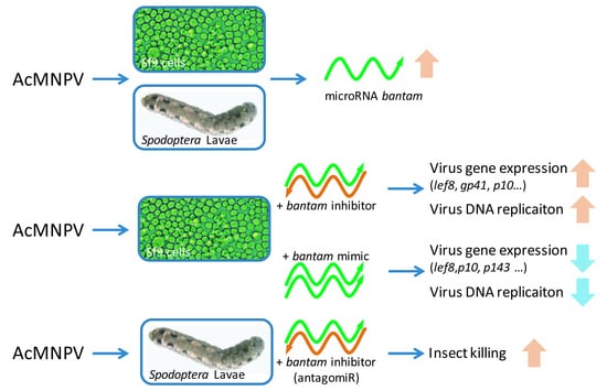
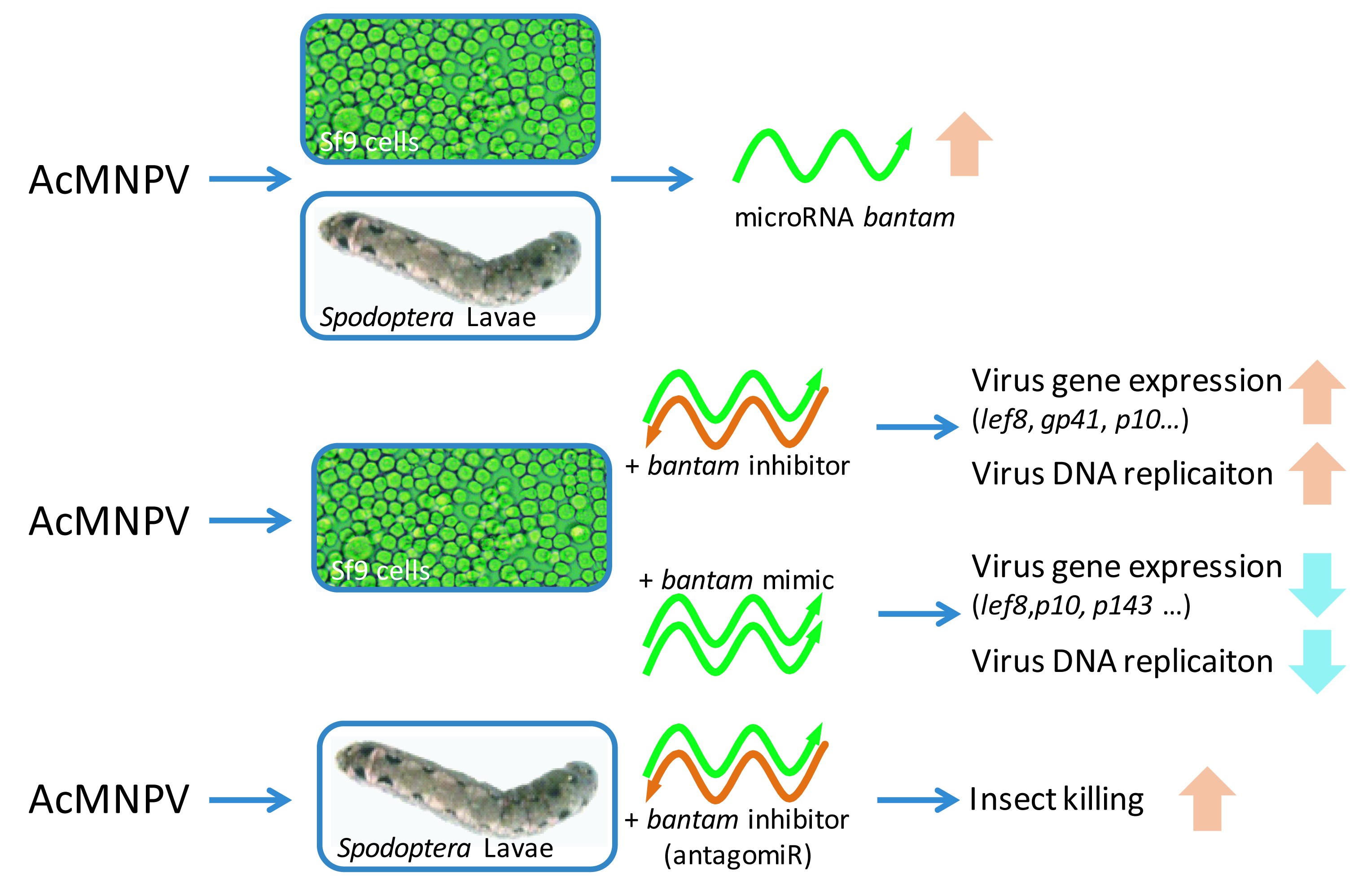
Lavae of S. litura and S. exigua were reared individually in polymer cups on artificial diet (Keyun Biocontrol, Jiyuan, China), under the condition of 28 °C, 70%–90% humidity, and photoperiod of 15:9. Larvae of the same hatching time were used for experiments.
AntagomirR was used to silence bantam in larvae. The bantam antagomirR and control antagomiR were dissolved in RNase free water to the concentration of 75 nM. They were added to the surface of a small piece of diet every day so that they could be consumed completely. MicroRNA antagomiRs were administrated on a daily basis from early 2nd instar until the pre-pupal stage. Wild type AcMNPV inclusion bodies (2 × 10⁷ polyhedra/mL) was inoculated per os by surface contamination of the diet, one day after antagomiR administration for the first time, and once again 2 days later. There were no less than 15 larvae in each group of treatment. Mortality and pupation were recorded daily.

3. Results

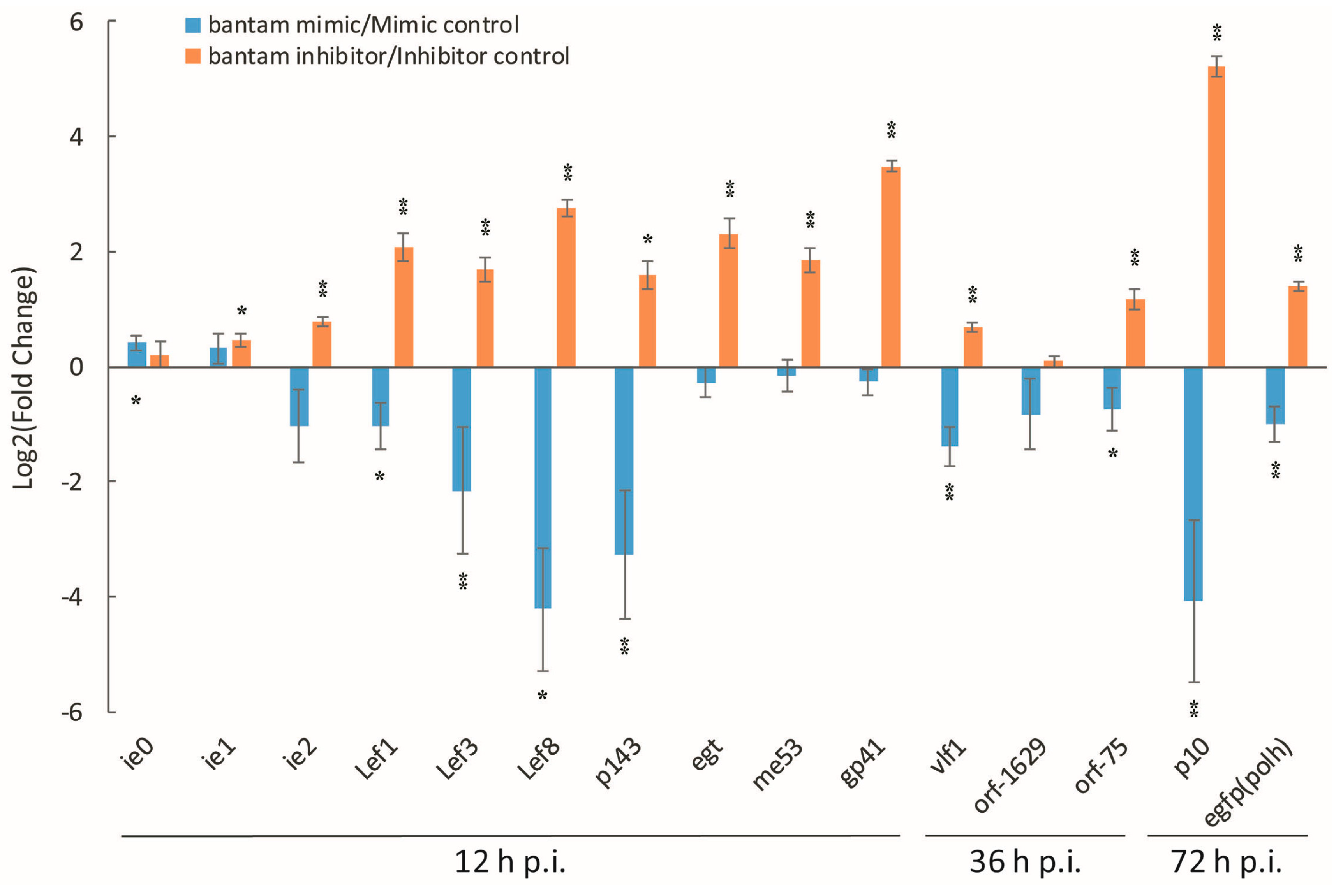
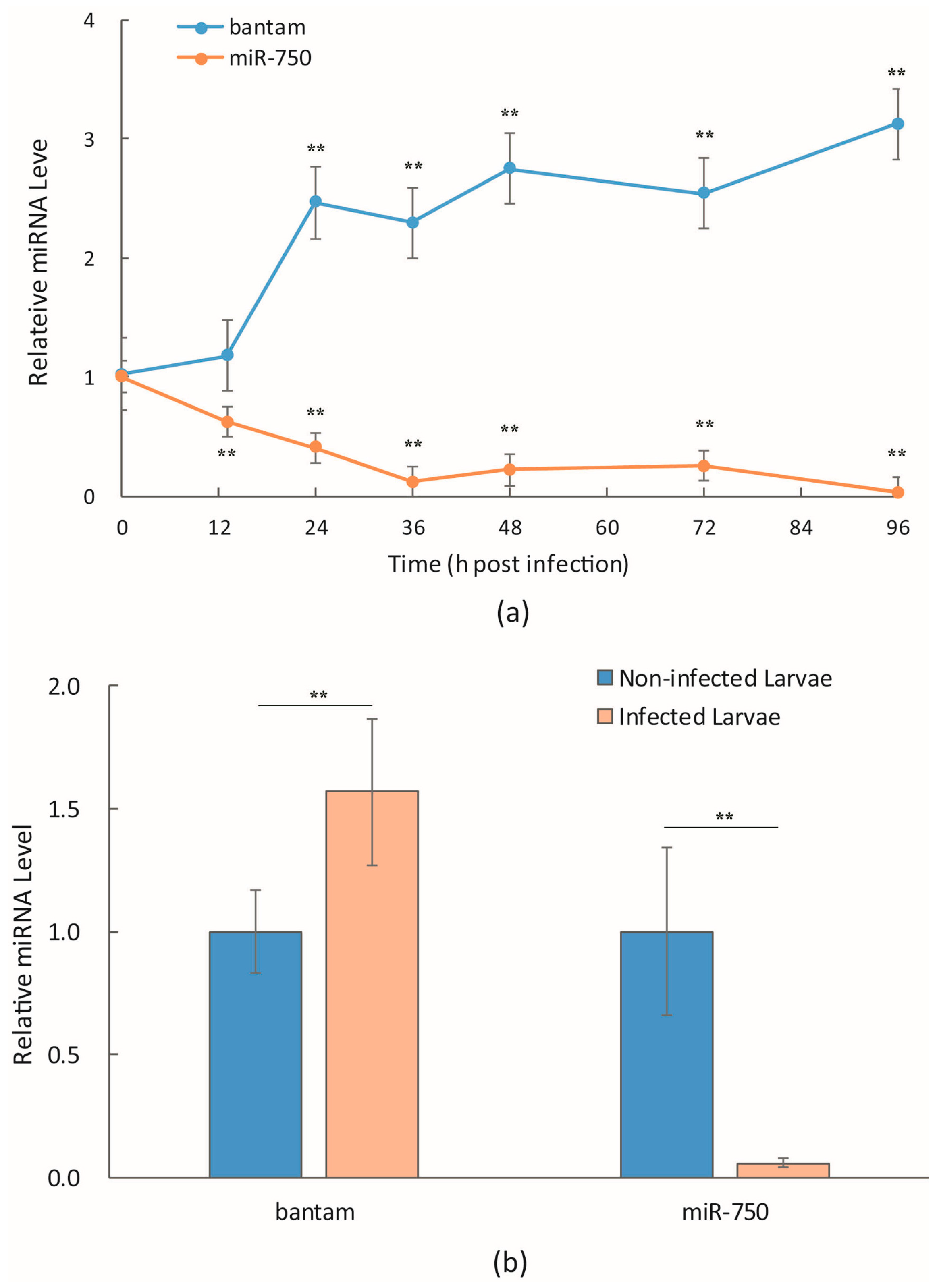
3.1. bantam Level after Infection

Mehrabadi et al. [14] and our own unpublished data on microRNAome of Sf9 cells showed that bantam was one of the most abundant microRNAs, which accounted for more than 5% of total microRNA. Our sequence data also suggested that the level of bantam increased late in AcMNPV infection. This was confirmed with real-time quantitative PCR in infected Sf9 cells and S. litura larvae (Figure 1). Unlike most microRNAs that decreased in the expression level after infection, as represented by miR-750 in Figure 1, the level of bantam increased to about 2.5-fold of uninfected cells 24 h p.i., and remained at a high level late in the infection (Figure 1a). The level of bantam was also about 1.5-fold higher than the control in AcMNPV-infected S. litura larvae (Figure 1b).

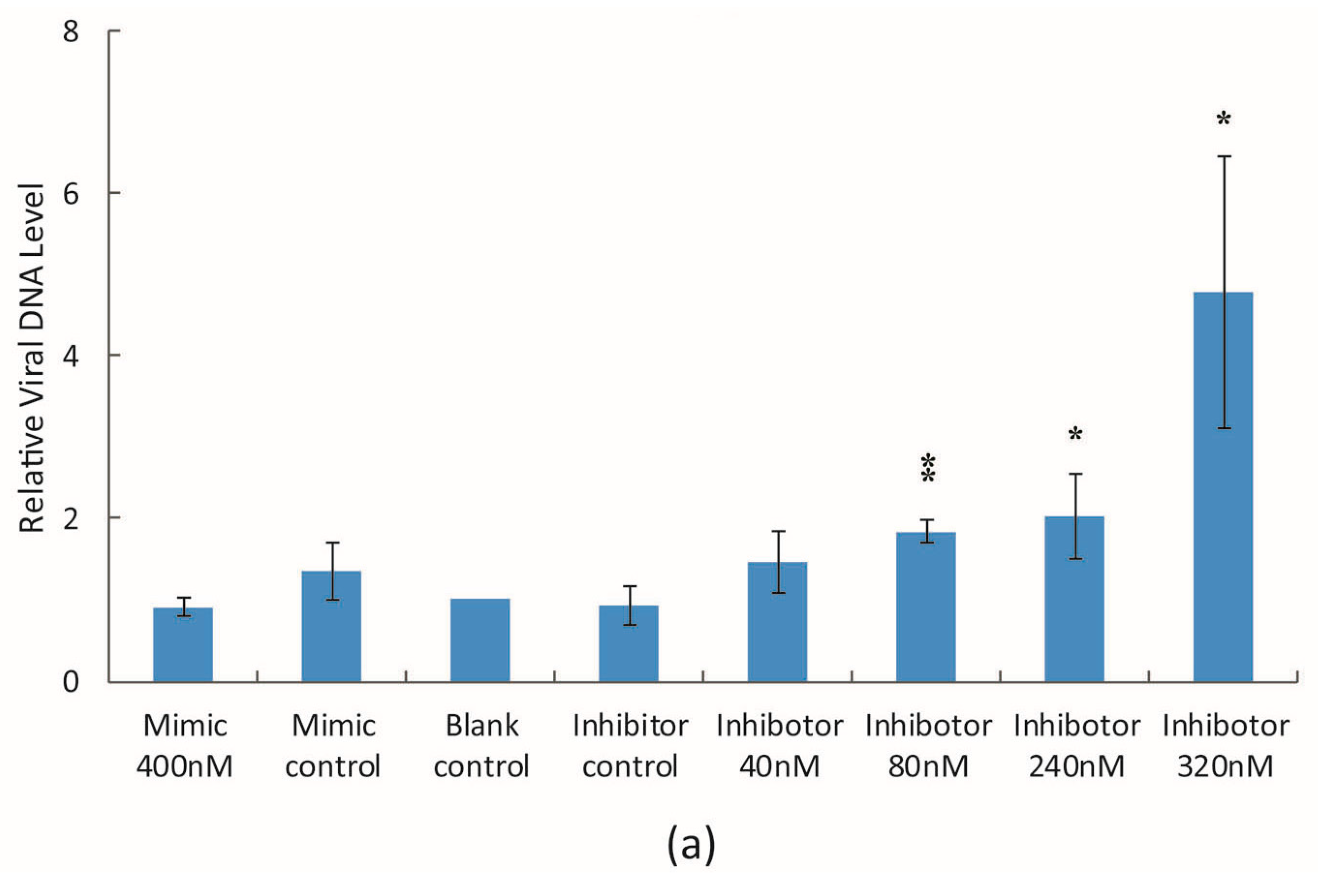
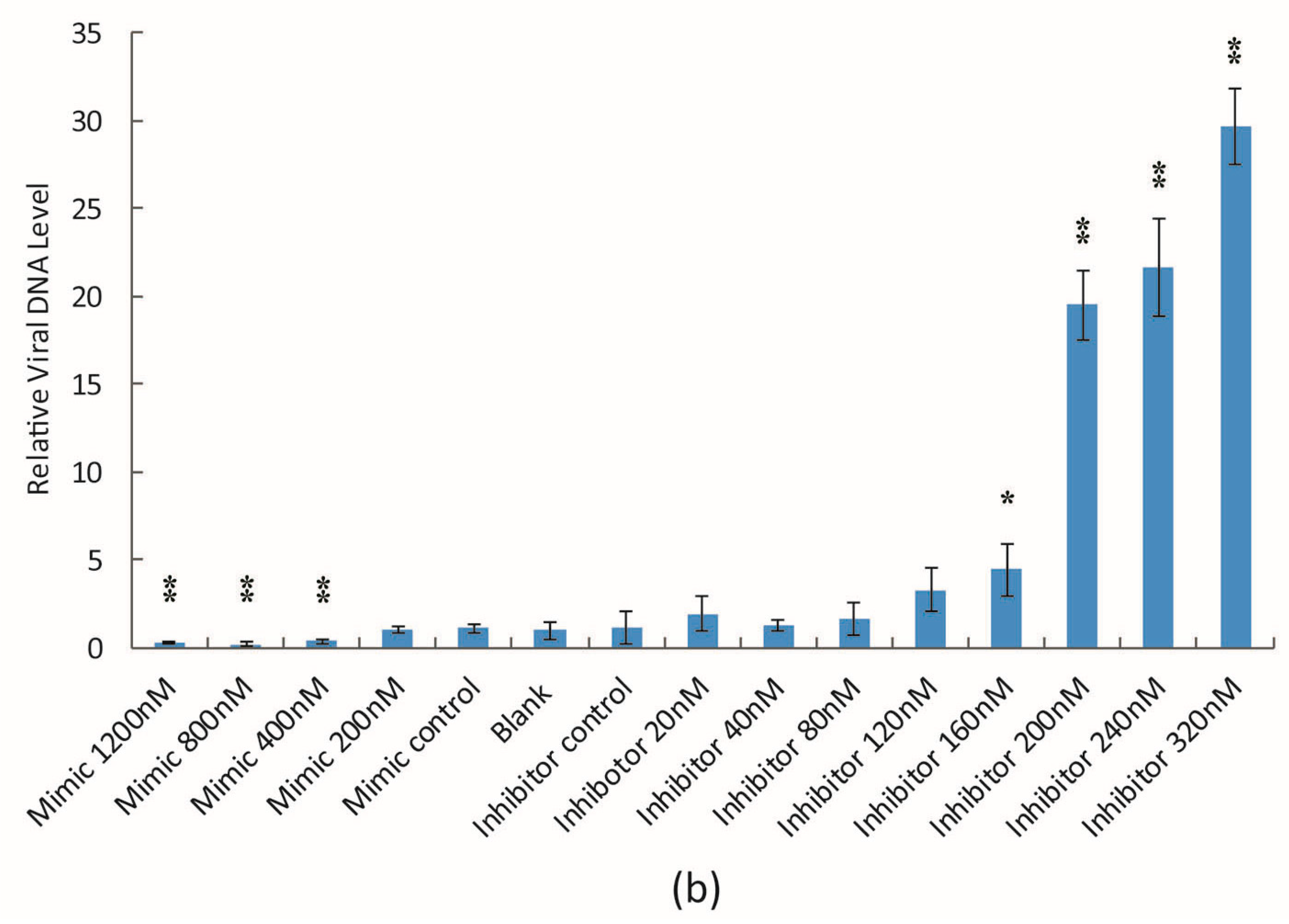
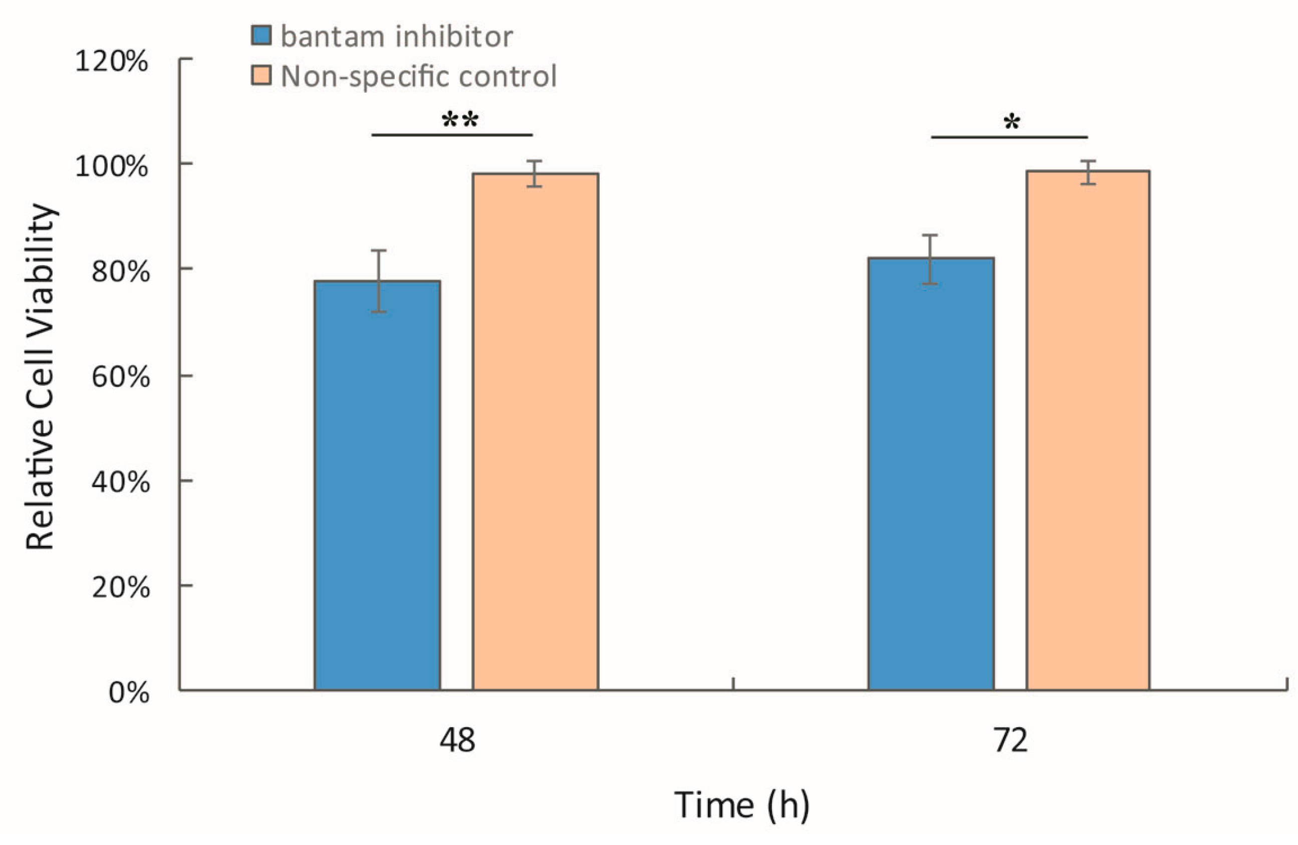
3.2. The Effect of bantam Mimic and Inhibitor on AcMNPV Replication in Sf9 Cells

The bantam mimic and inhibitor were used to modify the bantam level in Sf9 cells in order to study the effect of bantam in AcMNPV infection. In the presence of bantam inhibitor (200 nM), the viability of Sf9 cells was about 20% lower than the blank control (Figure 2). Treatment with non-specific inhibitor control had no effect on cell viability.
AcEGFP, a recombinant AcMNPV with egfp gene in the polyhedrin locus, was used to infect Sf9 cells in the presence of bantam mimic or inhibitor, and the expression of a series of virus genes was determined with real-time RT-PCR. These genes included virus immediate early and delayed early genes ie0, ie1, ie2, egt, p143, me53, lef1, lef3, lef8, and vlf1, late genes gp41, orf1629 and orf75, and very late genes p10, polh (represented by egfp). Different time points were used for genes of different categories, which were 12 h, 36 h and 72 h p.i. for immediate early and delayed early, late, and very late genes, respectively. The results showed that bantam mimic and inhibitor significantly affected the transcription of many of the tested genes (Figure 3). In most cases, the expression level was higher than the control in the presence of bantam inhibitor (the logarithm of fold change was positive), and lower than the control in the presence of bantam mimic (the logarithm of fold change was negative). Among all genes, the three most affected genes were lef8, gp41 and p10, the expression level of which increased by about 8, 10 and 40 times, respectively, in the presence of bantam inhibitor. The expression of immediate early genes ie0 and ie1 remained stable in the presence of either bantam mimic or inhibitor.
The viral DNA level in infected cells was also determined 48 h p.i. when cells were infected at MOI of 2 pfu/cell, or 72 h p.i. when the cells were infected at MOI of 0.05 pfu/cell, in the presence of different concentrations of bantam mimic and inhibitor. A dose-dependent effect was observed in both cases (Figure 4). The more bantam inhibitor that was added, the higher the level of virus genome that was detected; the more bantam mimic that was added, the lower the level of virus genome that was detected. The effect was more significant at a low MOI. However, bantam mimic or inhibitor had no major effect on the titer of budded virus in the medium 48 h p.i. and 72 h p.i..

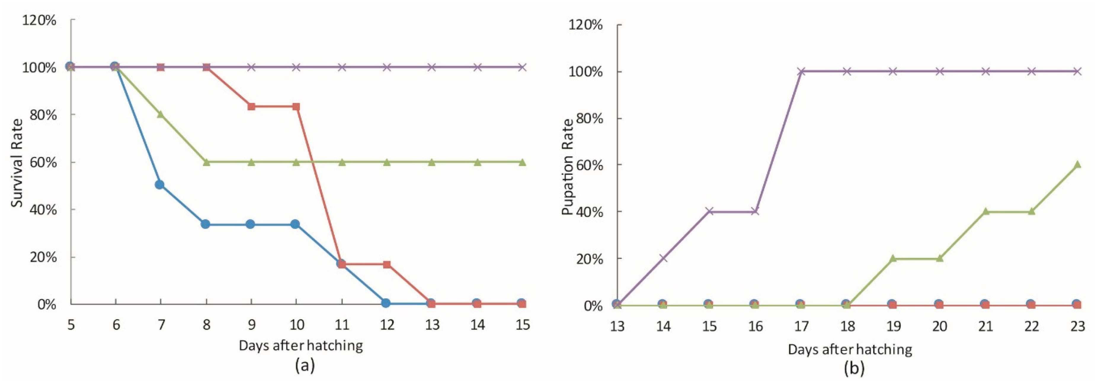
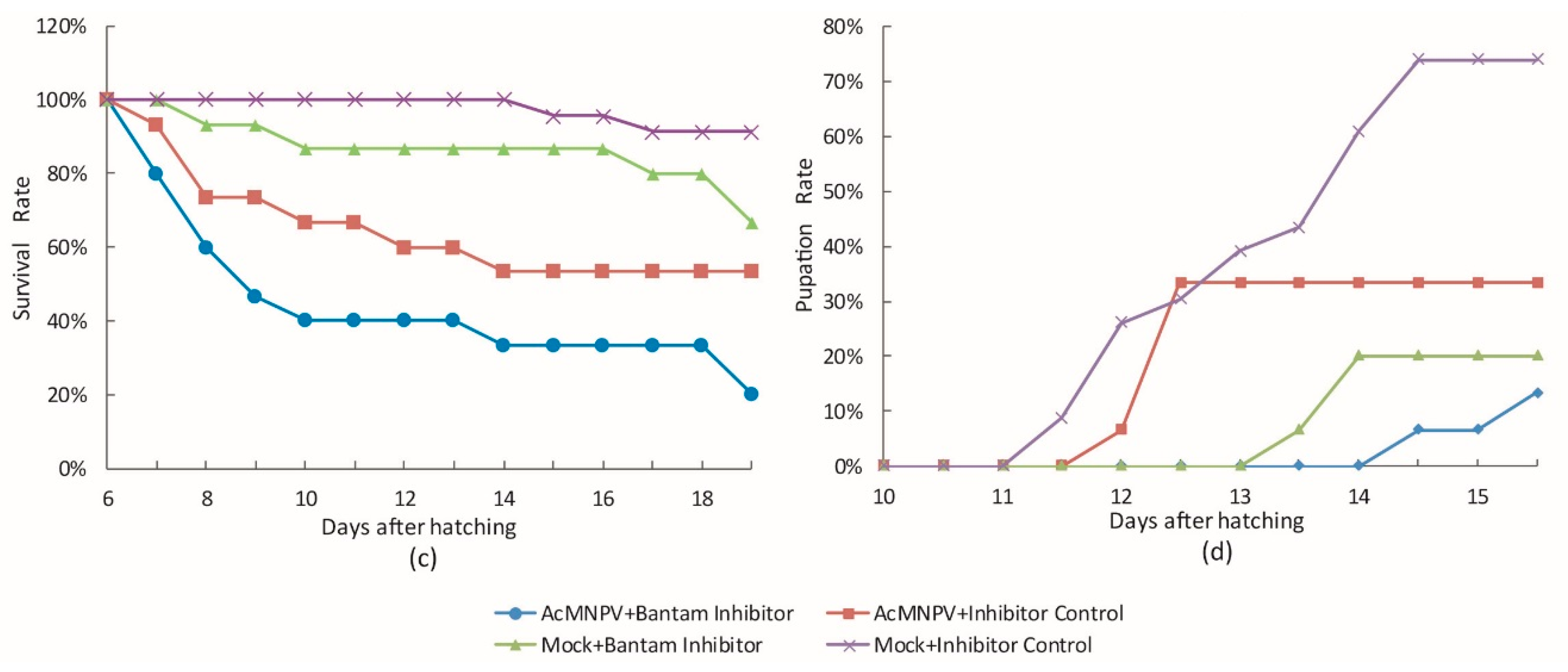
3.3. The Effect of bantam antagomiR in AcMNPV Infection in Spodoptera larvae

bantam antagomiR was used to study the effect of bantam on AcMNPV infection in vivo. Second instar larvae of S. litura and S. exigua were treated with bantam antagomiR before being inoculated with occlusion bodies of wild type AcMNPV per os. As shown in Figure 5, treating larvae with bantam antagomiR resulted in a 20% or 35% of larval death in S. litura and S. exigua, respectively, and a delay in the pupation of survived larvae.
Meanwhile, bantam antagomiR also displayed the ability to improve killing by AcMNPV in these insects (Figure 5). In S. litura, although 100% mortality was reached either with or without antagomiR treatment, the larvae died sooner with antagomiR treatment than without it. The antagomiR treatment group reached 50% mortality about 3.5 days earlier than the control group. In S. exigua, the mortality increased from 47% in the control to 80% in the antagomiR treatment group, and the average time to death was more than 2 days shorter with antagomiR treatment than without it.

4. Discussion

RNA interference pathway is thought to be important in virus-host interactions in insect. Exogenous viral RNA could be processed by this pathway to generate antiviral activity [28,29,30], whereas host-encoded microRNA is also produced by this pathway. As a kind of endogenous functional small RNA, host microRNA has important roles in many physiological processes as well as in the defense against infection [31]. Published work [14] and our early data showed that microRNA bantam was among the few most abundant microRNAs in Sf9 cells. bantam is an insect-specific microRNA identified in Drosophila, B. mori [32], and some other insects [33,34]. It has also been reported in blood fluke Schistosoma japonica [35,36]. Functional study in Drosophila [18,19,20,21,22,23,24,25] and Schistosoma [36] showed that it plays an important role in development and cell proliferation. In silkworm B. mori, bantam was found to be constantly expressed in the body wall, silk glands and midgut, but much less abundant in the fat body [32]. However, little is known of its role in lepidopteran insects. The results in the current work suggested that bantam also played a similarly important role in Spodoptera insects. Inhibition of the bantam activity resulted in decreased cell viability in Sf9 cells, and abnormal larvae growth in S. litura and S. exigua.
Most of the host microRNAs decreased in the expression level after AcMNPV infection. bantam was among the few microRNAs of which the expression level increased after infection both in Sf9 cells and in S. litura larvae. Similar to the expression of most proteins, the biogenesis of microRNA is under fine regulation. Therefore, the changed bantam expression level upon virus infection implied a possible role in virus–host interaction. The expression regulation of bantam has not been well studied yet. Since bantam is an effector in Hippo, Notch and EGFR/mitogen-activated protein kinase (MAPK) pathways, it is possible that baculovirus infection activated some elements in these pathways, and indirectly increased the expression level of bantam.
It is not clear how important the increase in bantam level was for the cellular defense against virus infection, since the cells and insects were susceptible to AcMNPV infection, and the production of progeny virus in infected Sf9 cells did not change much in the presence of bantam inhibitor. However, altering bantam level using a mimic or inhibitor did affect the expression of some important virus genes and the replication of virus DNA. Meanwhile, some other essential genes, such as ie0 and ie1, were less affected. The mechanism of the difference is not clear. MicroRNA tends to have multiple target sites in the genome. Bioinformatic analysis did not find any potential target of bantam in these genes. The only target gene known for bantam was the proapoptotic gene head involution defect (hid) in Drosophila [23,37]. Inhibition of hid by bantam will relieve the suppression on caspase activation, and resulted in apoptosis. Whether this is connected to the change in virus gene expression and DNA replication needs further study.
Inhibiting bantam activity had more significant effect on AcMNPV infection in S. litura and S. exigua larvae. bantam antagomiR treatment resulted in increased and accelerated killing in AcMNPV-infected larvae. This might relate to the growth retardation effect of bantam antagomiR on larvae. bantam is known to have a major effect on cell proliferation and apoptosis, the level molting hormone ecdysone [24], and even the circadian rhythms [25] in insects. Treatment of insects with bantam antagomiR alone also resulted in abnormal growth, low pupation rate, and even larval death. It is likely that such treatments made the larvae more vulnerable to infection, and enhanced the killing by virus. Preliminary results in our laboratory showed that administration of juvenile hormone partially compensated the effect of bantam antagomiR, which is consistent with the reported role of bantam in repressing the release of ecdysone.
In summary, we found that the level of microRNA bantam increased after AcMNPV infection in vitro and in vivo. Interfering bantam resulted in the change in virus gene expression, virus DNA replication in Sf9 cells, and increased insect killing in Spodoptera larvae. Although further studies are needed to elucidate the mechanisms under these observations; these findings provided a new approach to improve baculovirus as a biological pesticide.

5. Conclusions

Inhibiting host microRNA bantam resulted in the change in virus gene expression and virus genomic DNA synthesis in AcMNPV-infected Sf9 cells in vitro. It also improved AcMNPV infection of S. litura and S. exigua larvae, most likely through regulating the growth and development of the larvae. These findings may have applications in biological control of insect pests.

Acknowledgments

The work is supported by National Natural Science Foundation of China (31370183).

Author Contributions

Xiaojie Shi and Jiang Zhong conceived and designed the experiments; Xiaojie Shi, Zihan Ran, Sisi Li and Juan Yin performed the experiments; Jiang Zhong and Xiaojie Shi wrote the paper.

Conflicts of Interest

The authors declare no conflict of interest. The funding sponsors had no role in the design of the study; in the collection, analyses, or interpretation of data; in the writing of the manuscript, and in the decision to publish the results.

Abbreviations

The following abbreviations are used in this manuscript:
MOImultiplicity of infection
AcMNPVAutographa californica multicapsid nucleopolyhedrovirus

References

  1. Ambros, V. The functions of animal microRNAs. Nature 2004, 431, 350–355. [Google Scholar] [CrossRef] [PubMed]
  2. Cullen, B.R. MicroRNAs as mediators of viral evasion of the immune system. Nat. Immunol. 2013, 14, 205–210. [Google Scholar] [CrossRef] [PubMed]
  3. Guo, Y.E.; Steitz, J.A. Virus meets host microRNA: The destroyer, the booster, the hijacker. Mol. Cell. Biol. 2014, 34, 3780–3787. [Google Scholar] [CrossRef] [PubMed]
  4. Buggele, W.A.; Johnson, K.E.; Horvath, C.M. Influenza A virus infection of human respiratory cells induces primary microRNA expression. J. Biol. Chem. 2012, 287, 31027–31040. [Google Scholar] [CrossRef] [PubMed]
  5. Thornburg, N.J.; Hayward, S.L.; Crowe, J.E., Jr. Respiratory syncytial virus regulates human microRNAs by using mechanisms involving beta interferon and NF-κB. MBio 2012, 3. [Google Scholar] [CrossRef] [PubMed]
  6. Backes, S.; Shapiro, J.S.; Sabin, L.R.; Pham, A.M.; Reyes, I.; Moss, B.; Cherry, S.; tenOever, B.R. Degradation of host microRNAs by poxvirus poly(A) polymerase reveals terminal RNA methylation as a protective antiviral mechanism. Cell Host Microbe 2012, 12, 200–210. [Google Scholar] [CrossRef] [PubMed]
  7. Jeang, K.-T. RNAi in the regulation of mammalian viral infections. BMC Biol. 2012, 10. [Google Scholar] [CrossRef] [PubMed]
  8. Sanghvi, V.R.; Steel, L.F. RNA silencing as a cellular defense against HIV-1 infection: Progress and issues. FASEB J. 2012, 26, 3937–3945. [Google Scholar] [CrossRef] [PubMed]
  9. Nathans, R.; Chu, C.-Y.; Serquina, A.K.; Lu, C.-C.; Cao, H.; Rana, T.M. Cellular microRNA and P bodies modulate host-HIV-1 interactions. Mol. Cell 2009, 34, 696–709. [Google Scholar] [CrossRef] [PubMed]
  10. Jopling, C.L.; Yi, M.K.; Lancaster, A.M.; Lemon, S.M.; Sarnow, P. Modulation of Hepatitis C virus RNA abundance by a liver-specific microRNA. Science 2005, 309, 1577–1581. [Google Scholar] [CrossRef] [PubMed]
  11. Seto, E.; Moosmann, A.; Groemminger, S.; Walz, N.; Grundhoff, A.; Hammerschmidt, W. MicroRNAs of Epstein-Barr virus promote cell cycle progression and prevent apoptosis of primary human B cells. PLoS Pathog. 2010, 6, e1001063. [Google Scholar] [CrossRef] [PubMed] [Green Version]
  12. Feederle, R.; Linnstaedt, S.D.; Bannert, H.; Lips, H.; Bencun, M.; Cullen, B.R.; Delecluse, H.-J. A viral microRNA cluster strongly potentiates the transforming properties of a human herpesvirus. PLoS Pathog. 2011, 7, e1001294. [Google Scholar] [CrossRef] [PubMed]
  13. Zhao, Y.; Xu, H.; Yao, Y.; Smith, L.P.; Kgosana, L.; Green, J.; Petherbridge, L.; Baigent, S.J.; Nair, V. Critical role of the virus-encoded microRNA-155 ortholog in the induction of Marek’s disease lymphomas. PLoS Pathog. 2011, 7, e1001305. [Google Scholar] [CrossRef] [PubMed]
  14. Mehrabadi, M.; Hussain, M.; Asgari, S. MicroRNAome of Spodoptera frugiperda cells (Sf9) and its alteration following baculovirus infection. J. Gen. Virol. 2013, 94, 1385–1397. [Google Scholar] [CrossRef] [PubMed]
  15. Thompson, B.J.; Cohen, S.M. The Hippo pathway regulates the bantam microRNA to control cell proliferation and apoptosis in Drosophila. Cell 2006, 126, 767–774. [Google Scholar] [CrossRef] [PubMed]
  16. Becam, I.; Rafel, N.; Hong, X.; Cohen, S.M.; Milan, M. Notch-mediated repression of bantam miRNA contributes to boundary formation in the Drosophila wing. Development 2011, 138, 3781–3789. [Google Scholar] [CrossRef] [PubMed]
  17. Herranz, H.; Hong, X.; Cohen, S.M. Mutual repression by bantam miRNA and capicua links the EGFR/MAPK and Hippo pathways in growth control. Curr. Biol. 2012, 22, 651–657. [Google Scholar] [CrossRef] [PubMed]
  18. Harti, M.; Kadow, I.C.G. New roles for “old” microRNAs in nervous system function and disease. Front. Mol. Neurosci. 2013, 6. [Google Scholar] [CrossRef]
  19. Dong, L.; Li, J.; Huang, H.; Yin, M.-X.; Xu, J.; Li, P.; Lu, Y.; Wu, W.; Yang, H.; Zhao, Y.; et al. Growth suppressor lingerer regulates bantam microRNA to restrict organ size. J. Mol. Cell Biol. 2015, 7, 415–428. [Google Scholar] [CrossRef] [PubMed]
  20. Bras-Pereira, C.; Casares, F.; Janody, F. The retinal determination gene dachshund restricts cell proliferation by limiting the activity of the homothorax-yorkie complex. Development 2015, 142, 1470–1479. [Google Scholar] [CrossRef] [PubMed]
  21. Zhang, X.; Luo, D.; Pflugfelder, G.O.; Shen, J. Dpp signaling inhibits proliferation in the Drosophila wing by Omb-dependent regional control of bantam. Development 2013, 140, 2917–2922. [Google Scholar] [CrossRef] [PubMed]
  22. Li, Y.; Padgett, R.W. bantam Is required for optic lobe development and glial cell proliferation. PLoS ONE 2012, 7, e32910. [Google Scholar] [CrossRef] [PubMed]
  23. Brennecke, J.; Hipfner, D.R.; Stark, A.; Russell, R.B.; Cohen, S.M. bantam encodes a developmentally regulated microRNA that controls cell proliferation and regulates the proapoptotic gene hid in Drosophila. Cell 2003, 113, 25–36. [Google Scholar] [CrossRef]
  24. Boulan, L.; Martin, D.; Milan, M. bantam miRNA promotes systemic growth by connecting insulin signaling and ecdysone production. Curr. Biol. 2013, 23, 473–478. [Google Scholar] [CrossRef] [PubMed]
  25. Lerner, I.; Bartok, O.; Wolfson, V.; Menet, J.S.; Weissbein, U.; Afik, S.; Haimovich, D.; Gafni, C.; Friedman, N.; Rosbash, M.; et al. Clk post-transcriptional control denoises circadian transcription both temporally and spatially. Nat. Commun. 2015, 6. [Google Scholar] [CrossRef] [PubMed]
  26. Wang, X.; Li, L.; Ding, S.; Huang, X.; Zhang, J.; Yin, J.; Zhong, J. Chicken HS4 insulator significantly improves baculovirus-mediated foreign gene expression in insect cells by modifying the structure of neighbouring chromatin in virus minichromosome. J. Biotechnol. 2009, 142, 193–199. [Google Scholar] [CrossRef] [PubMed]
  27. Krutzfeldt, J.; Rajewsky, N.; Braich, R.; Rajeev, K.G.; Tuschl, T.; Manoharan, M.; Stoffel, M. Silencing of microRNAs in vivo with ‘antagomirs’. Nature 2005, 438, 685–689. [Google Scholar] [CrossRef] [PubMed]
  28. Kingsolver, M.B.; Huang, Z.; Hardy, R.W. Insect antiviral innate immunity: Pathways, effectors, and connections. J. Mol. Biol. 2013, 425, 4921–4936. [Google Scholar] [CrossRef] [PubMed]
  29. Zografidis, A.; van Nieuwerburgh, F.; Kolliopoulou, A.; Apostolou-Karampelis, K.; Head, S.R.; Deforce, D.; Smagghe, G.; Swevers, L. Viral small-RNA analysis of Bombyx mori larval midgut during persistent and pathogenic cytoplasmic polyhedrosis virus infection. J. Virol. 2015, 89, 11473–11486. [Google Scholar] [CrossRef] [PubMed]
  30. Niu, J.; Smagghe, G.; de Coninck, D.I.; van Nieuwerburgh, F.; Deforce, D.; Meeus, I. In vivo study of Dicer-2-mediated immune response of the small interfering RNA pathway upon systemic infections of virulent and avirulent viruses in Bombus terrestris. Insect Biochem. Mol. Biol. 2016, 70, 127–137. [Google Scholar] [CrossRef] [PubMed]
  31. Hussain, M.; Asgari, S. MicroRNAs as mediators of insect host-pathogen interactions and immunity. J. Insect Physiol. 2014, 70, 151–158. [Google Scholar] [CrossRef] [PubMed] [Green Version]
  32. Liu, S.; Gao, S.; Zhang, D.; Yin, J.; Xiang, Z.; Xia, Q. MicroRNAs show diverse and dynamic expression patterns in multiple tissues of Bombyx mori. BMC Genom. 2010, 11. [Google Scholar] [CrossRef] [PubMed]
  33. Gao, L.; Zuo, H.; Liu, K.; Li, H.; Zhong, G. A new strategy for identification of highly conserved microRNAs in non-model insect, Spodoptera litura. Int. J. Mol. Sci. 2012, 13, 612–627. [Google Scholar] [CrossRef] [PubMed]
  34. Etebari, K.; Hussain, M.; Asgari, S. Identification of microRNAs from Plutella xylostella larvae associated with parasitization by Diadegma semiclausum. Insect Biochem. Mol. Biol. 2013, 43, 309–318. [Google Scholar] [CrossRef] [PubMed]
  35. Xue, X.; Sun, J.; Zhang, Q.; Wang, Z.; Huang, Y.; Pan, W. Identification and characterization of novel microRNAs from Schistosoma japonicum. PLoS ONE 2008, 3, e4034. [Google Scholar] [CrossRef] [PubMed]
  36. Zhu, L.; Zhao, J.; Wang, J.; Hu, C.; Peng, J.; Luo, R.; Zhou, C.; Liu, J.; Lin, J.; Jin, Y.; et al. MicroRNAs are involved in the regulation of ovary development in the pathogenic blood fluke Schistosoma japonicum. PLoS Pathog. 2016, 12, e1005423. [Google Scholar] [CrossRef] [PubMed]
  37. Nahvi, A.; Shoemaker, C.J.; Green, R. An expanded seed sequence definition accounts for full regulation of the hid 3′ UTR by bantam miRNA. RNA 2009, 15, 814–822. [Google Scholar] [CrossRef] [PubMed]
Figure 1. Expression level of microRNA bantam in AcMNPV-infected Sf9 cells and Spodoptera litura larvae. Total small RNA samples were prepared from infected Sf9 cells at different time points (a), or infected S. litura larvae (4 d p.i.) (b). The levels of microRNA bantam and miR-750 were determined with real-time RT-PCR using U6 RNA as the internal control. The fold change comparing to the non-infected control was shown. The means of three or more replicates were shown, with the error bars indicating the standard deviation. The significance of difference between certain time point and 0 h (a), or between infected and non-infected larvae (b), was also shown (*: p < 0.05; **: p < 0.01).
Viruses 08 00136 g001
Figure 2. The effect of bantam inhibitor on Sf9 cells. Sf9 cells were transfected with bantam inhibitor (200 nM), and cell viability was determined 48 h and 72 h post treatment. The cell viability in blank control was set as 100%. The means of three replicates was shown, with the error bars indicating the standard deviation. The significance of difference between bantam inhibitor and non-specific treatment was also shown (*: p < 0.05; **: p < 0.01).
Viruses 08 00136 g002
Figure 3. The effect of bantam mimic and inhibitor on the transcription of virus genes in Sf9 cells. Total mRNA was prepared from AcEGFP-infected Sf9 cells in the presence of bantam inhibitor (200 nM) or mimic (400 nM), and the transcription level of representative virus genes was determined with real-time RT-PCR. Cellular tubulin gene was used as the internal control. Sampling time was shown under the gene names. The data was normalized with either inhibitor control or mimic control. The mean of three or more replicates was shown, with the error bars indicating the standard deviation. The significance of difference between bantam mimic and mimic control, or between bantam inhibitor and inhibitor control was also shown (*: p < 0.05; **: p < 0.01).
Viruses 08 00136 g003
Figure 4. The effect of bantam mimic and inhibitor on viral DNA replication in Sf9 cells. Total DNA was prepared from AcEGFP-infected Sf9 cells in the in the presence of bantam inhibitor or mimic, and the relative level of viral DNA was determined 48 h p.i. (MOI = 2, (a)) or 72 h p.i. (MOI = 0.05, (b)) with real-time PCR. egfp gene in AcEGFP genome was used as the amplification target, and cellular tubulin gene was used as the control. The mean of three or more replicates was shown, with the error bars indicating the standard deviation. The significance of difference between certain concentration of bantam mimic or inhibitor with mimic control or inhibitor control, respectively, was also shown (*: p < 0.05; **: p < 0.01).
Viruses 08 00136 g004aViruses 08 00136 g004b
Figure 5. The effect of bantam-antagomiR on AcMNPV infection in larvae. Second instar larvae of S. litura (a,b) and S. exigua (c,d) were administrated with bantam-antagomiR or control on a daily basis. They were then inoculated with AcMNPV polyhedra per os, or mock infected twice, on Day 2 and 4 after first antagomirR administration. Larval death (a,c) and pupation (b,d) were recorded. Each group includes 15 or more larvae.
Viruses 08 00136 g005aViruses 08 00136 g005b
Table 1. Sequences of real-time PCR primers.
TargetTypeSequence (5′–3′)
bantamprimer assayCTTTCTGAGATCATTGTGAAAG
miR-750primer assayCAGTTGCCAGATCTATCTTTC
U6 (B. mori)primer assayAGAGACGATTAGCATGGCCC
ie-0upper primerCATATTCGTGCGAGGCAACG
lower primerCGAGTTGACGCTTGCCAAAA
ie-1upper primerTTTTAACGCGTCGTACACCA
lower primerGTTGACGCTTGCCAAAAAGT
ie2upper primerAGCGAAGAAAACGTGCAGAT
lower primerCTCCGACGCAATGTTATCCT
lef-1upper primerTTCCGCATATGCAAGATTCA
lower primerACCACATCCACCAGTTCCAT
lef-3upper primerAACCCCCATGAGCGACTATTTG
lower primerACCGCTGTTGATTTTCTTGTA
lef-8upper primerAATGGCTACTCTGTGGCGGTAA
lower primerTTACCGCCACAGAGTAGCCATT
egtupper primerACCGTTTCCAGCGATCAACT
lower primerTAGAGGCGGAAACGTTGCTT
me53upper primerCGCATCTCTCCGCCTAAACA
lower primerGCGGGCAAACCTTCAAACTT
p143upper primerAAGTATTTGCCCGAGGAC
lower primerATTTTGGCGATGTGATAAGT
p35upper primerACGACACGGGACTTTACGAG
lower primerGTTTTTCGACGCTTCGTTGT
vlf-1upper primerCGTGCCAATTCCAACGGTTT
lower primerCAACTCAGCGTGGACGATCT
gp41upper primerAGAGTTGGGACAGAGCAACG
lower primerGCGCCACCGTTGTAAAACTT
orf-1629upper primerGTTAGGCACGGGAGAA
lower primerCGAAGCAGACGACCTT
p10upper primerGACGCCGTTACGGAAACTAA
lower primerGTCTGGAAGATCCGGAACAA
egfpupper primerCGTAAACGGCCACAAGTTCAT
lower primerGGGTCAGCTTGCCGTAGGT
orf-75upper primerGCTGGCAGTTGGTATGCTTC
lower primerACTCAGTAGGCGACAGGTTG
β-tublin (B. mori)upper primerTTGCATTGGTACACTGGCGA
lower primerACACCAGGTCGTTCATGTTGC

