Article

Isolation and Genome Characterization of the Virulent Staphylococcus aureus Bacteriophage SA97

1
Department of Food and Animal Biotechnology, Department of Agricultural Biotechnology, Research Institute of Agriculture and Life Sciences, and Center for Food and Bioconvergence, Seoul National University, Seoul 151-921, Korea
2
Department of Food Science and Biotechnology, Kyung Hee University, Yongin 446-701, Korea
3
Department of Dermatology, College of Medicine, the Catholic University of Korea, Seoul 137-701, Korea
4
Department of Microbiology, College of Medicine, the Catholic University of Korea, Seoul 137-701, Korea
*
Author to whom correspondence should be addressed.
Viruses 2015, 7(10), 5225-5242; https://doi.org/10.3390/v7102870
Submission received: 21 July 2015 / Revised: 31 August 2015 / Accepted: 22 September 2015 / Published: 1 October 2015
(This article belongs to the Section Bacterial Viruses)

Abstract

:
A novel bacteriophage that infects S. aureus, SA97, was isolated and characterized. The phage SA97 belongs to the Siphoviridae family, and the cell wall teichoic acid (WTA) was found to be a host receptor of the phage SA97. Genome analysis revealed that SA97 contains 40,592 bp of DNA encoding 54 predicted open reading frames (ORFs), and none of these genes were related to virulence or drug resistance. Although a few genes associated with lysogen formation were detected in the phage SA97 genome, the phage SA97 produced neither lysogen nor transductant in S. aureus. These results suggest that the phage SA97 may be a promising candidate for controlling S. aureus.

1. Introduction

Bacteriophages are natural antibacterial agents that lyse a specific bacterial host strain [1]. Based on their bacterial host specificity and bacteriolytic activity, the use of bacteriophages has been suggested as a potential biocontrol strategy for various pathogenic bacteria. Staphylococcus aureus is a major foodborne and clinical pathogen that causes food poisoning, endocarditis, staphylococcal scalded skin syndrome and toxic shock syndrome [2]. Furthermore, the emergence of antibiotic-resistant S. aureus, including methicillin-resistant S. aureus (MRSA) [3], has complicated the treatment of S. aureus infections [4]. Bacteriophage treatment could be an alternative method for controlling S. aureus. Various bacteriophages have been used as alternative antibacterial therapies for human patients in eastern Europe for several decades [5]. In addition, the US Food and Drug Association (FDA) has approved the application of bacteriophages as a safe food preservative [6].
Virulent phages are generally preferred as biocontrol agents as they cannot integrate their genome into the bacterial chromosome to form lysogens and will always lyse and kill infected target bacterial cells [7]. Staphylococcal lytic phages have demonstrated their potential as biocontrol agents of food [1] and phage therapy of human or animal infections [8,9,10]. For example, Myovirus phages S25-3 [11], K [9] and the Podoviruses S13’ [11], S24-1 [11] are virulent phages that have been applied to food samples or mice. However, most reported S. aureus phages are siphoviruses and known as temperate phages that contain lysogeny modules in their genomes [12,13]. Enterobacteria phage lambda [14,15,16] and S. aureus phage phi11 [14] are well-known temperate phages that form lysogens. These phages all contain typical lysogen modules including cro-like repressor, antirepressor, integrase, excisionase, cI-like repressor, and lambda contains additional cII and cIII regulatory proteins. Integrase mediates site-specific recombination between two DNA recognition sequences in phage and host bacteria, while excisionase is involved in excisional recombination by excising the phage genome from the bacterial chromosome. CI-like repressor represses lytic functions; Cro-like repressor, which is a repressor of cI, leads the phage to the lytic infection cycle, while the antirepressor blocks phage repressors. Among the lysogen formation-related genes, cI-like repressor gene is the key regulator that maintains the phage in the lysogenic life cycle [14,15,16].
Here, the phage SA97, which specifically infects S. aureus without lysogen formation, was isolated and characterized. An analysis of the whole genome of the phage SA97 revealed part of the genes encoding a lysogeny module but no genes related to virulence or drug resistance. An analysis of lysogen formation or transduction by SA97 showed that SA97 produced neither lysogen nor transductant, suggesting that the phage SA97 may be applicable as a biocontrol agent against S. aureus.

2. Materials and Methods

2.1. Bacterial Strains, Media, and Growth Conditions

The bacterial strains used in this study are summarized in Table 1. Food isolates 1 to 4 were from vegetable, pork, beef and Korean meal kimbab (rice with laver, ham, egg, cucumber, pickled radish and crab meat), respectively. They were isolated and purified by Baird-Parker agar medium (Difco, Detroit, MI, USA) and identified by VITEK® 2 Compact (bioMerieux, Inc., Hazelwood, MO, USA). These strains were all grown in tryptic soy broth (TSB) medium (Difco) at 37 °C with agitation. Tryptic soy agar (Difco) containing 1.5% (w/v) agar was used to count the bacteria.

2.2. Bacteriophage Isolation and Propagation

Bacteriophage SA97 was isolated from the skin using the strain S. aureus RN4220 as a bacterial host strain. To isolate the phage, 0.5 cm2 of skin was rubbed with a cotton swab that was subsequently homogenized with 1 mL of sodium chloride and magnesium sulfate (SM) buffer (100 mM NaCl, 10 mM MgSO4, and 50 mM Tris-HCl, pH 7.5). The sample was then mixed with TSB broth supplemented with 10 mM CaCl2, sub-cultured with S. aureus RN4220 and incubated at 37 °C for 12 h with shaking. After incubation, the sample was centrifuged at 8000× g for 10 min and filtered to remove bacterial cells and obtain the supernatant that contained the bacteriophage. For phage propagation, TSB broth containing 10 mM CaCl2 was first sub-inoculated with S. aureus RN4220, and the culture was incubated at 37 °C for 1.5 h. Subsequently, the phage was added at a multiplicity of infection (MOI) of 1, and the culture was incubated for 3 h at the same temperature with shaking. To prepare a high-titer phage, the phages were precipitated with polyethylene glycol (PEG) 6000 and concentrated using CsCl density gradient ultracentrifugation [17]. Finally, to confirm the phage plaque formation, the supernatant was overlaid on 0.4% TSA soft agar containing 10 mM CaCl2 and S. aureus RN4220. Plaques were evident after incubation in at 37 °C for 12 h.

2.3. Transmission Electron Microscopy (TEM)

The phage SA97 was analyzed using transmission electron microscopy (TEM). The phage suspension was placed on a carbon-coated copper grid and negatively stained with 2% uranyl-acetate (pH 4.0). The sample was examined under an energy-filtering transmission electron microscope at an operating voltage of 120 kV [18]. Phage SA97 was identified and classified according to the guidelines of the International Committee on Taxonomy of Viruses [19].

2.4. Bacteriophage Host Range

The bacterial strains listed in Table 1 were incubated overnight at 37 °C. Each bacterial culture was added to 5 mL of the 0.4% TSA soft agar and overlaid on TSA agar plates. Subsequently, the phage SA97 containing diluted lysates was spotted onto the prepared plate and incubated at 37 °C for at least 6 h to obtain single plaques. After incubation, we could determine the infectivity based on the clarity of the spots, and efficiency of plating (EOP) was calculated by dividing the phage titer on the test strain by the phage titer on the reference strain. All the experiments were performed in triplicate.

2.5. Bacterial Challenge Assay

Fifty milliliters of TSB broth containing 10 mM CaCl2 was sub-inoculated with S. aureus RN4220, and the culture was incubated at 37 °C until it reached the early exponential growth phase (3.5 × 10⁸ ± 0.2 × 10⁸). The culture was then infected with the phage at a MOI of 1 or 10. The OD600 was measured each hour after phage infection for 15 h [20]. An un-infected culture was used as a control. All the experiments were performed in triplicate.

2.6. Lysogen Formation

To evaluate the ability of the phage SA97 to form lysogens, the phage-resistant bacteria were isolated 10 h after phage infection, and the presence of SA97 gene fragments in the fifty of SA97-resistant colonies was analyzed by PCR using primers specific to the transcriptional regulator gene (SA97_044) or antirepressor gene (SA97_045). In addition, the phage-resistant strains grown to exponential phase were induced with mitomycin C at a final concentration of 0.5 μg/mL and centrifuged for 5 min at 10,000 × g after incubation at 37 °C. The supernatants were filtered using 0.22-μm filter and spotted onto S. aureus overlay.

2.7. One-Step Growth Curve Assay

One-step growth curve analysis was performed as previously described [21]. Briefly, phage was mixed with S. aureus RN4220 in its early exponential growth phase at MOI of 0.001 in the presence of 10 mM CaCl2 and allowed to adsorb for 10 min at room temperature, then it was centrifuged at 6000 × g for 10 min. The pellet containing infected cells was suspended in 50 mL of fresh TSB liquid medium and incubated at 37 °C with shaking. Two sets of samples were taken every 5 min up to 1 h. To release the intracellular phages, CHCl3 was added to one of them, and the eclipse period was determined. Subsequently, each sample was immediately titered by the double-layer agar plate method and latent period, eclipse period and burst size were calculated. All the experiments were performed in triplicate.

2.8. Receptor Analysis

S. aureus RN4220, a strain free of prophages, restriction mechanisms and capsules [22,23], was used in this study (Table 1). To identify the phage receptor, we obtained the ΔtagO/RN4220 mutant, which is deficient in the peptidoglycan-anchored wall teichoic acid (WTA) [24], and its complemented strain using plasmid pRB474-tagO, which was constructed by sub-cloning the tagO gene into an Escherichia coli-S. aureus shuttle expression vector [25]. We carried out a spotting assay with the wild type RN4220, ΔtagO/RN4220 mutant and the tagO complemented strains.

2.9. Adsorption Assay

The phage adsorption ability was assayed as previously described [26]. Overnight cultures of the bacterial strains were diluted 1:100 in TSB and incubated until the OD600 reached 2.0. One milliliter of the strain cultures was diluted 10-fold in fresh TSB, and the phage was added to the each diluted culture at an MOI of 0.01. The cultures were incubated at 37 °C for 10 min. A sample was collected immediately after reaction and 10 min after incubation for each culture and immediately centrifuged at 14,000 × g and 4 °C for 1 min. The samples were then filtered using 0.22-μm filters. Subsequently, the supernatants were serially diluted and overlaid on TSA plates to determine the titers of unabsorbed phages. All the experiments were performed in triplicate.

2.10. Determination of the Frequency of the Bacteriophage-Insensitive Mutants (BIMs)

The frequency of the BIMs was determined as previously described [27]. Phage was mixed with S. aureus (109 CFU/mL) cell suspension containing 10 mM of CaCl2 with a MOI of 100. After incubation at 37 °C for 10 min, serial dilutions of the mixtures were plated. The BIMs frequency was determined by dividing remaining viable counts by the initial viable counts. All the experiments were performed in triplicate.

2.11. Transduction Assay

A transduction assay was performed to determine the ability of phage SA97 to induce horizontal gene transfer between the host bacteria cells and bacteriophages. Donor phage lysate was prepared by infecting SA97 phage to the host bacteria containing the erythromycin antibiotic-resistant cassette (ΔypfP::erm, the glycolipid-deficient mutant) [28]. After infection, the cultures were incubated in TSB for 8 h at 37 °C. After incubation, the cultures were treated with chloroform, and the supernatant lysate was harvested. To determine the ability of the phage to transduce host bacteria, supernatant phage lysate was added to the recipient host bacteria culture (S. aureus RN4220, 1010 CFU/mL) at an MOI of 10 and the culture was incubated for 15 min at 37 °C. TSB containing ethylene glycol tetraacetic acid (EGTA) was added to the culture (final concentration of 20 mM) to stop the reaction. After incubation for 20 min under the same conditions, the phage-infected bacterial cells were harvested, spread on TSA agar media containing erythromycin and incubated at 37 °C. All the experiments were performed in triplicate, and the transduction frequencies were calculated as the quotient of the number of transductants divided by the number of input PFU per plate [29].

2.12. Bacteriophage Genomic DNA Purification

Bacteriophage genomic DNA was purified as previously described [30]. Prior to purification, the phage lysates were treated with DNase and RNaseA at 37 °C for 1 h to remove bacterial DNA and RNA contamination, respectively. The phage lysate was then treated with lysis buffer containing 0.5 M of EDTA, 10 mg/mL of proteinase K and 1% of sodium dodecyl sulfate (SDS) for 15 min at 65 °C. Finally, ethanol precipitation was performed, followed by phenol-chloroform DNA purification.

2.13. Full-Genome Sequencing of Bacteriophage SA97 and Bioinformatics Analysis

The extracted SA97 phage genomic DNA was sequenced with a Genome Sequencer FLX titanium sequencer (Roche, Mannheim, Germany) and assembled with the GS de novo assembler software (Roche) at Macrogen Inc., South Korea. Open reading frames (ORFs) were predicted using the FGENESB (http://www.softberry.com), Glimmer v3.02 [31] and GeneMark.hmm [32] software packages. The ORFs were annotated using the InterProScan [33] and BLASTP [34] programs. Comparative genomic analysis of SA97 with other phietalikevirus Subgroup2 phages and visualization were conducted with BLASTN [35] and ACT12 [36]. The complete genome sequence of S. aureus phage SA97 was deposited in GenBank under accession number KJ716334.

3. Results and Discussion

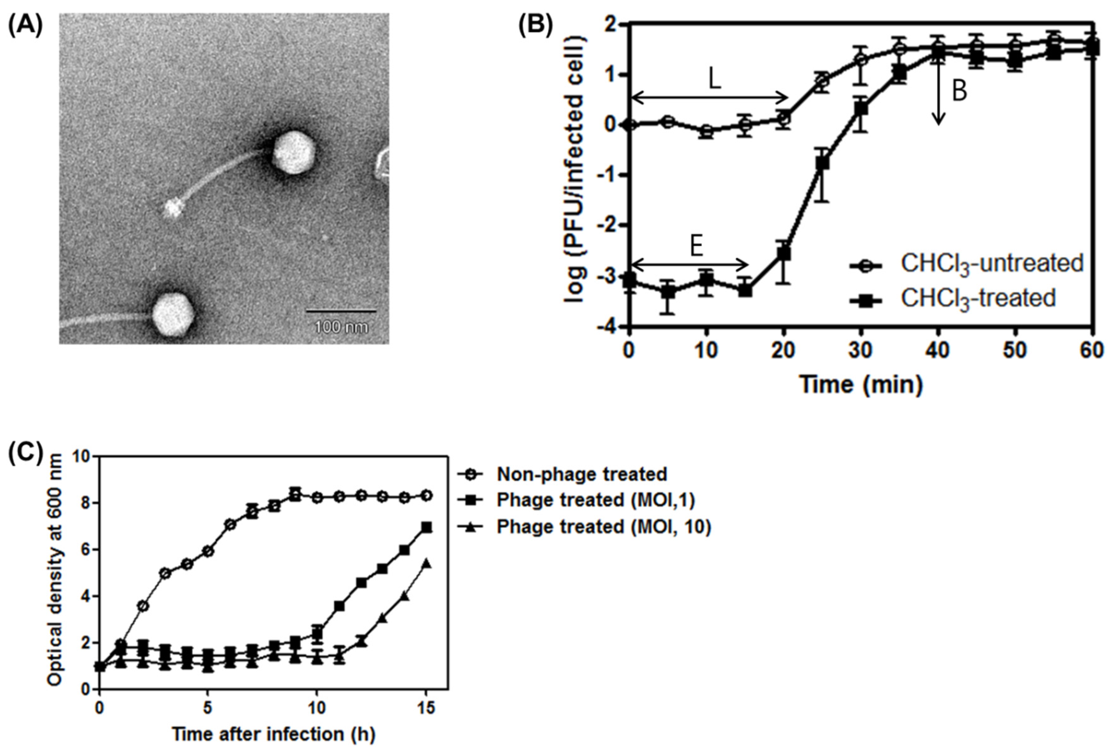
3.1. Phage SA97 Isolation and Its Morphological Analysis

The S. aureus infecting bacteriophage SA97 was newly isolated. This phage formed clear plaques against the S. aureus RN4220 bacterial host strain. Transmission electron microscopy (TEM) revealed an icosahedral capsid (63 ± 2 nm, n = 5) with a long, flexible and non-contractile tail (171 ± 6 nm, n = 5) for phage SA97, indicating that this phage belongs to the Siphoviridae family (Figure 1A). The baseplate structure of the phage SA97 consists of multiple disc baseplates and is similar to those of phages StB12, StB27 and StB20 [37]. However, the tail of phage SA97 is shorter than those of the above-mentioned phages (206 nm for StB12, 213 nm for StB27, and 250 nm for StB20) [37]. According to Xia and Wolz [38], phage SA97 belongs to serogroup B, which contains phages with a tail length of less than 200 nm.
Figure 1. General characterizations of bacteriophage SA97. (A) Electron micrograph of phage SA97. The phage belongs to the Siphoviridae family. The scale bar represents 100 nm; (B) One-step growth curve analysis of phage SA97 on exponential culture of S. aureus RN4220 incubated in tryptic soy broth (TSB) medium at 37 °C under agitation. Cells were chloroform treated (○) or untreated (■). E, eclipse period; L, latent period; B, burst size; (C) Inhibition assays of S. aureus RN4220 with phage SA97 at 37 °C. ○, control group without phage; ■, ▲, experimental groups with phage SA97 (MOI of 1 and 10, respectively). The data shown are the mean values from three independent measurements and the error bars represent the standard deviations.
Viruses 07 02870 g001

3.2. One-Step Growth Curve of SA97and Bacterial Challenge Assay

The one-step growth kinetics of SA97 showed a latent period time of 20 min, an eclipse period time of 15 min and a burst size of 32 PFU/infected cell when infecting S. aureus RN4220 (Figure 1B).
To determine the bacteriolytic activity of the phage SA97, a bacterial growth inhibition assay was performed in the presence of SA97 (Figure 1C). The growth inhibition of S. aureus by SA97 persisted for up to 10 or 11 h after infection with MOI of 1 or 10, respectively. SA97 showed a longer inhibitory effect than most other S. aureus phages such as CS1, DW2 [10] and SAH-1 [39], all of which showed only 3 h of growth inhibition. These results demonstrate that SA97 possesses strong bacteriolytic activity, which is essential in phage therapy. Re-growth of the culture 10 h after infection with SA97 suggested the possibility of development of lysogen or resistant mutant. However, PCR analysis of the 50 S. aureus colonies resistant to SA97 obtained 10 h after phage infection using primers specific to the transcriptional regulator gene (SA97_044) or antirepressor gene (SA97_045) detected neither gene. In addition, no plaques were observed after induction of the resistant strains with mitomycin C, indicating that the resistant strains were not SA97 lysogens but resistant mutants.

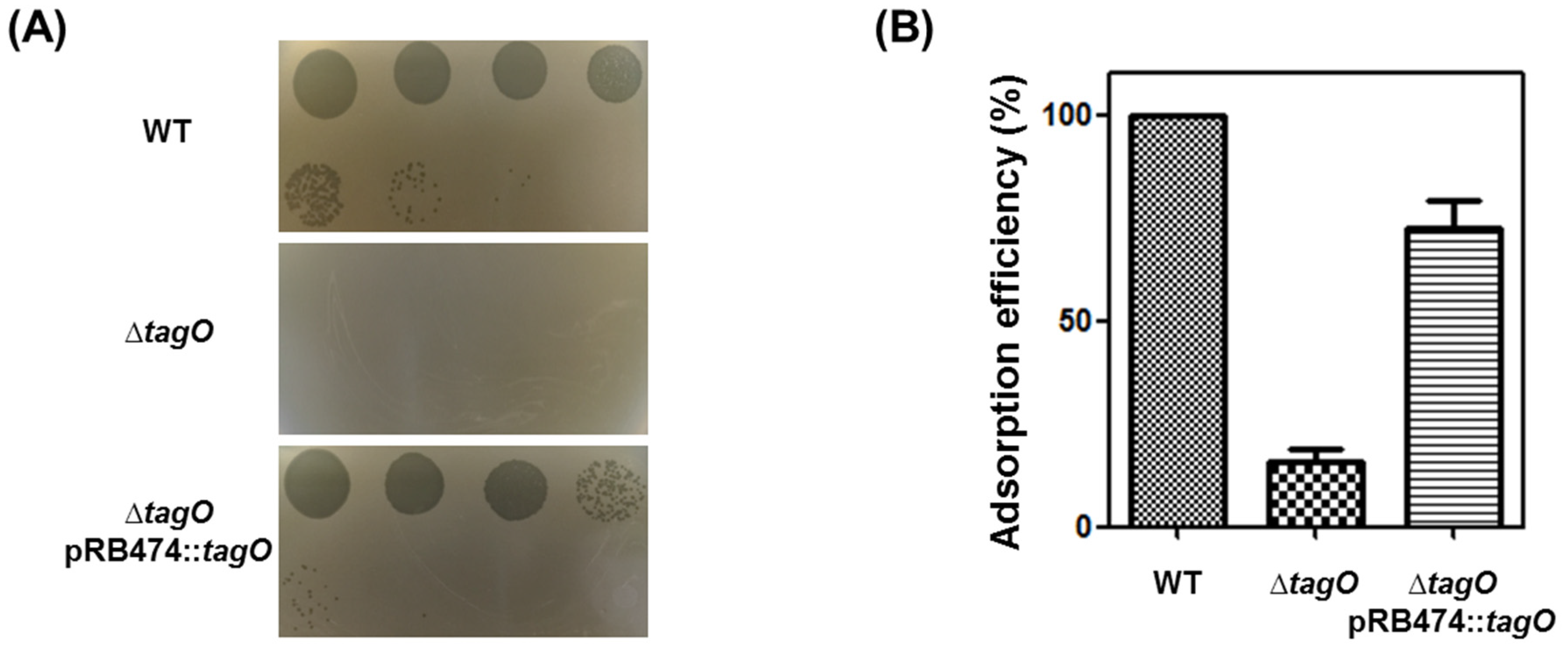
3.3. Receptor Analysis

The only known host receptor of S. aureus phages is peptidoglycan-anchored wall teichoic acid (WTA). Xia et al. [40] demonstrated that WTA is required for siphovirus and myovirus infection of S. aureus; thus, a ΔtagO mutant of S. aureus (RN4220ΔtagO) [41] was tested (Figure 2A). The RN4220ΔtagO strain was resistant to SA97, and the phage sensitivity was recovered by complementing the strain with tagO [42]. The phage adsorption assay also showed that phage adsorption was severely reduced in the RN4220ΔtagO strain (Figure 2B), indicating that the host receptor for phage SA97 is peptidoglycan-anchored WTA.
Figure 2. (A) Wall teichoic acid (WTA)-dependent infection of S. aureus phage SA97. The phage SA97 lysate was spotted onto lawns of wild-type RN4220 (WT), ΔtagO mutant (ΔtagO), and tagO-complemented (ΔtagO pBR474::tagO) strains. Plaque formation indicates successful adsorption and infection by the phage SA97. EOP relative to WT strain was 7.5 × 10⁻² ± 0.2 × 10⁻² on ΔtagO pBR474::tagO strain; (B) WTA-dependent adsorption ability of S. aureus phage SA97. The adsorption efficiency relative to the adsorption to wild-type strain RN4220, which was set to 100%, is indicated. The data shown are the mean values from three independent measurements and the error bars represent the standard deviations.
Viruses 07 02870 g002

3.4. Host Range of Bacteriophage SA97

Plaque formation efficiency of the phage SA97 was determined for 19 S. aureus strains and several other strains of both Gram-positive and Gram-negative bacteria as shown on Table 1. SA97 phage could infect only S. aureus strains. Six out of 13 S. aureus type strains and all six S. aureus local isolates tested were sensitive to SA97. It is interesting to note that nine of these 12 sensitive strains were isolated in Korea, indicating that SA97 phage may be more effective for controlling S. aureus strains isolated in Korea.

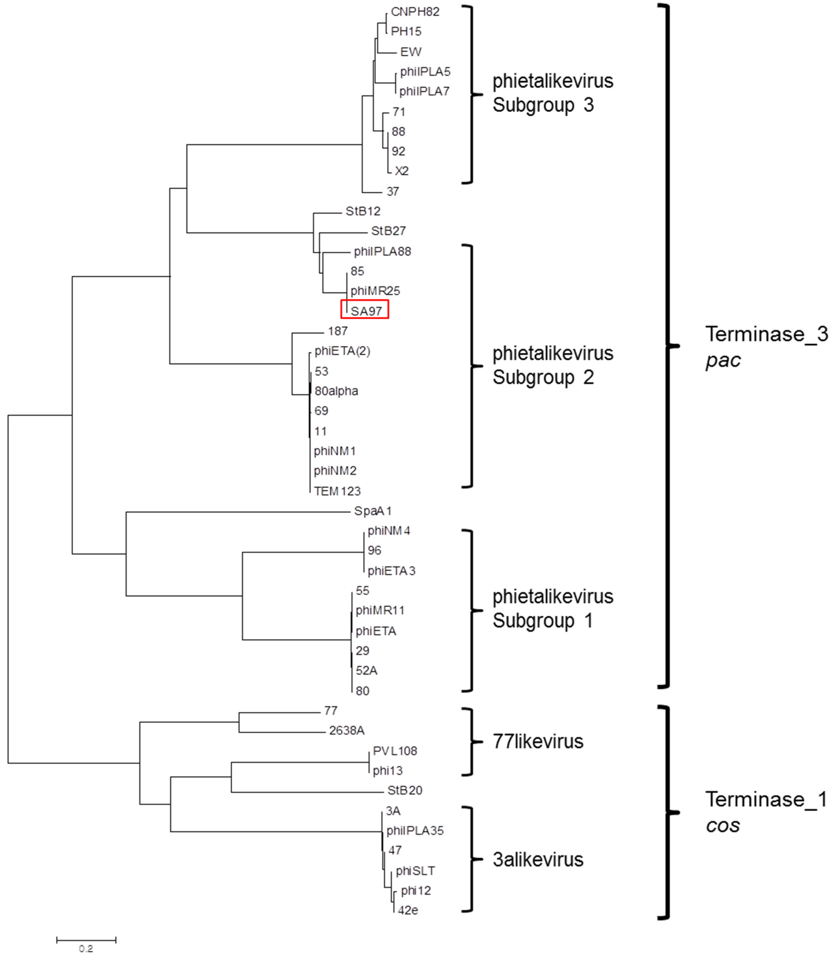
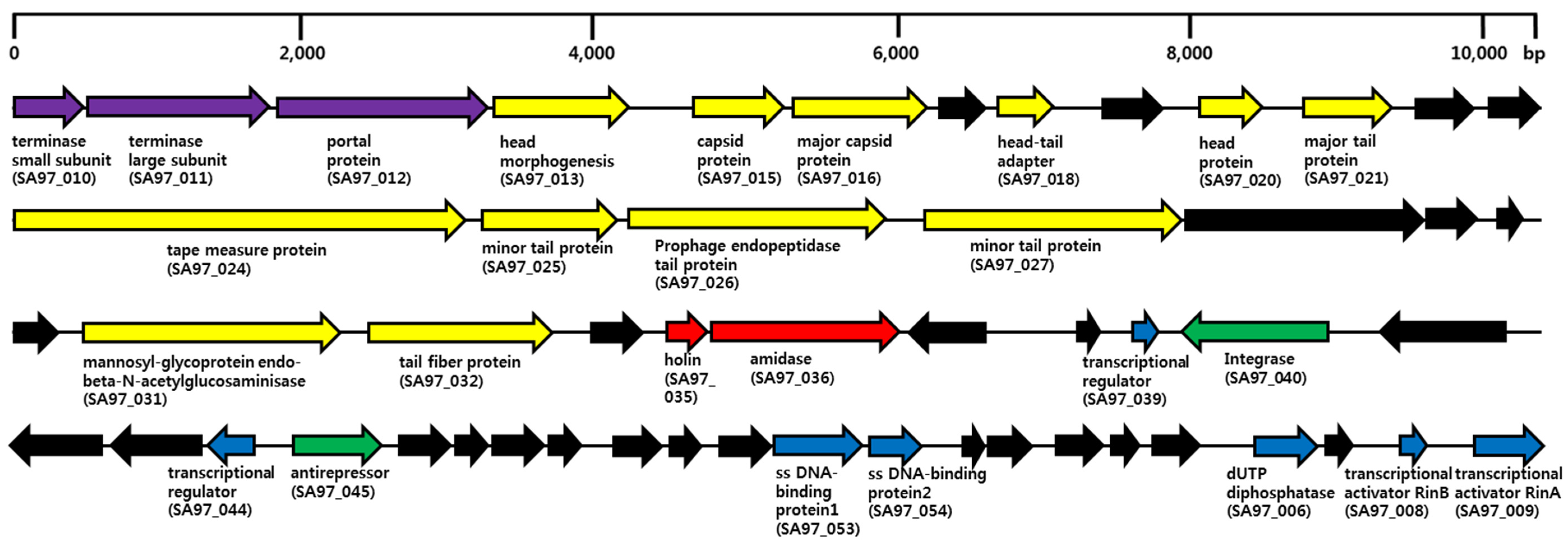
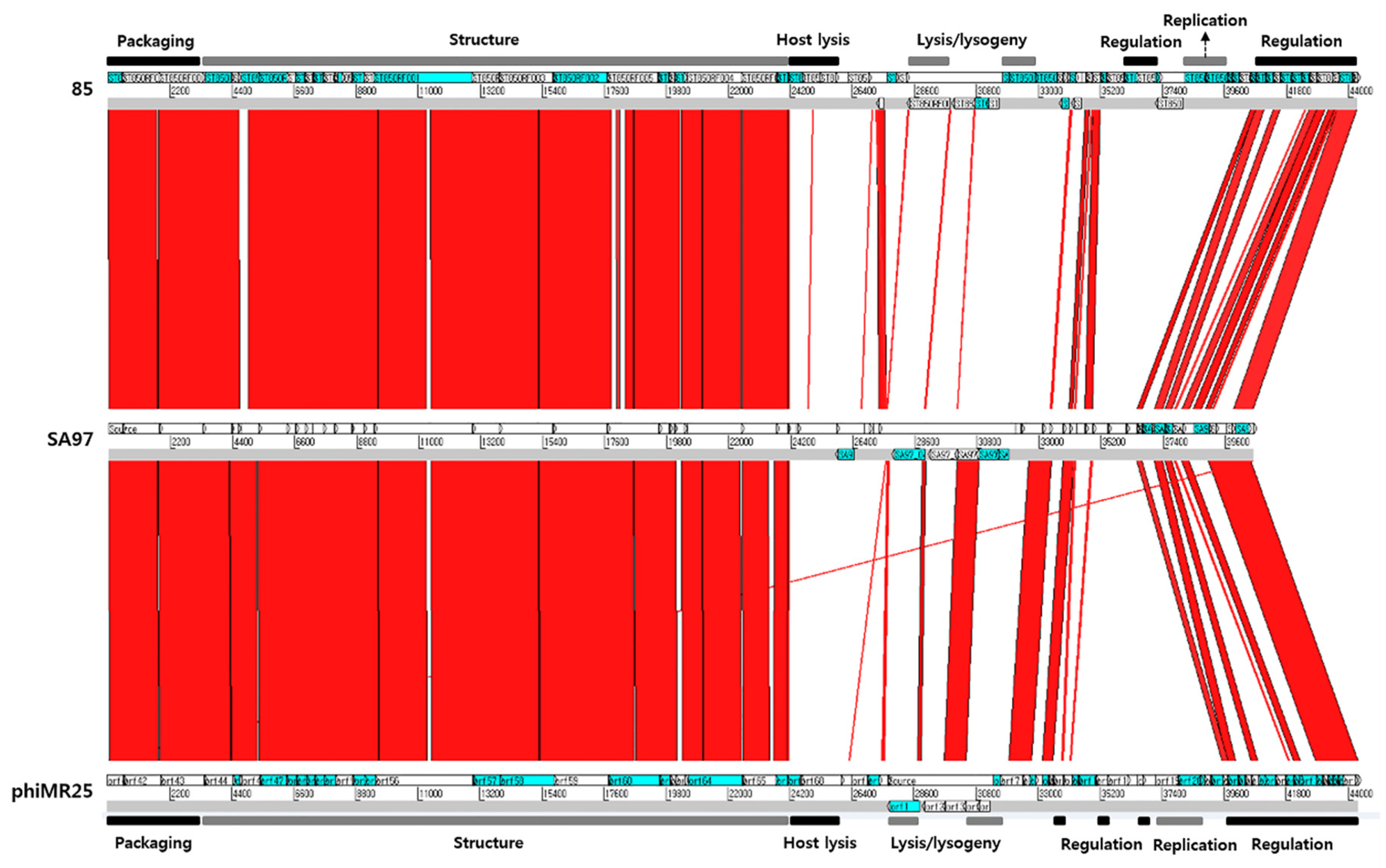
3.5. Bacteriophage SA97 Genomic Analysis

The phage SA97 genome was sequenced and analyzed. The complete genome sequence of the phage SA97 revealed that the genome consists of 40,592 bp with a GC content of 34.25% encoding 54 predicted open reading frames (ORFs) and no tRNA genes (Figure 3).
The functional analysis of the predicted ORFs revealed that they can be classified into five functional groups: phage structure (major capsid protein, head morphogenesis protein, head-tail connector protein, major and minor tail proteins, tape measure protein, minor structure proteins, tail fiber protein and endo-β-N-acetylglucosaminidase), packaging (portal protein and terminase small/large subunits), host lysis (holin and endolysin), DNA regulation (single-stranded DNA-binding protein, dUTP diphosphatase, transcriptional activators RinAB and transcriptional regulators) and lysis/lysogeny module (integrase and antirepressor) (Table 2).
Table 1. The antimicrobial spectrum of bacteriophage SA97
BacteriaPlaque Formation of SA97aE.O.P bReference or Source c
Staphylococcus strains
Staphylococcus aureus RN4220C1.0 × 100 ± 0.0 × 100[42]
Staphylococcus aureus KCTC 1916C1.1 × 100 ± 1.8 × 10⁻¹KCTC
Staphylococcus aureus ATCC 29213C1.7 × 10−4 ± 2.1 × 10⁻⁵ATCC
Staphylococcus aureus ATCC 33593C1.2 × 100 ± 8.7 × 10⁻²ATCC
Methicillin resistant Staphylococcus aureus CCARM 3089C1.0 × 100 ± 8.1 × 10⁻²CCARM
Methicillin resistant Staphylococcus aureus CCARM 3090C7.4 × 10−2 ± 3.1 × 10⁻²CCARM
Staphylococcus aureus food isolate 1 (from vegetable)C4.9 × 10−2 ± 1.6 × 10⁻²This study
Staphylococcus aureus food isolate 2 (from pork)C1.2 × 100 ± 1.0 × 10⁻¹This study
Staphylococcus aureus food isolate 3 (from beef)C1.2 × 100 ± 2.8 × 10⁻¹This study
Staphylococcus aureus food isolate 4 (from Kimbab)C1.0 × 100 ± 4.0 × 10⁻¹This study
Staphylococcus aureus clinical isolate 1C2.0 × 10−4 ± 5.0 × 10⁻⁵This study
Staphylococcus aureus clinical isolate 0055C1.1 × 10−3 ± 5.5 × 10⁻⁵GNUH-NCCP
Staphylococcus aureus Newman--[43]
Staphylococcus aureus ATCC 6538--ATCC
Staphylococcus aureus ATCC 23235--ATCC
Staphylococcus aureus ATCC 25923--ATCC
Staphylococcus aureus ATCC 12600--ATCC
Staphylococcus aureus ATCC 13301--ATCC
Staphylococcus aureus ATCC 27664--ATCC
Staphylococcus xylosis ATCC 29971--ATCC
Staphylococcus cohnii ATCC 29974--ATCC
Staphylococcus warneri ATCC 10209--ATCC
Staphylococcus intermedius ATCC 29663--ATCC
Other Gram positive bacteria
Enterococcus faecalis ATCC 29212--ATCC
Bacillus cereus ATCC 14579--ATCC
Bacillus subtilis ATCC 23857--ATCC
Listeria monocytogenes ATCC 19114--ATCC
Gram negative bacteria
Salmonella enterica serovar Typhimurium SL1344--ATCC
Escherichia coli MG1655 ATCC 47076--ATCC
Escherichia coli O157:H7 ATCC 35150--ATCC
Cronobacter sakazakii ATCC 29544--ATCC
Pseudomonas aeruginosa ATCC 27853--ATCC
a C, clear plaques; -, no plaque. b The EOP is relative to an average result obtained for RN4220. c ATCC, American Type Culture Collection; KCTC, Korean Collection for Type Cultures; GNUH-NCCP, Gyeongsang National University Hospital Branch of National Culture Collection for Pathogens; CCARM, Culture Collection of Antimicrobial Resistant Microbes.
Interestingly, three genes in the category of host lysis may be responsible for the host lysis activity of phage SA97. Holin (SA97_035) is generally known to create a hole in the host inner membrane to expose the host peptidoglycan layer to endolysin. Endolysin (SA97_036) may then lyse the host peptidoglycan to allow phage burst-out. In general, Gram positive endolysins form in a modular structure, consisting of enzymatically active domains (EADs) and a cell wall binding domain (CBD) [44] which leads the enzyme specifically to the substrate bacterial peptidoglycan. The BLASTP analysis of endolysin revealed two major protein domains containing CHAP (PF05257) and amidase_3 (PF01520), which may be involved in host lysis via the activities of endopeptidase and amidase. However, a host cell wall-binding domain (e.g., SH3 domain) was not detected, suggesting that this phage endolysin may contain a new type of CBD. Endo-β-N-acetylglucosaminidase (SA97_032), which is known as a tail-associated cell wall hydrolase, may degrade the peptidoglycan of the bacterial cell wall during infection [45].
A few putative genes associated with lysogen formation [antirepressor (SA97_045) and integrase (SA97_040)] were detected in the category of DNA manipulation and regulation; however, the major components of the lytic/lysogenic decision gene cluster, cI- or cro-like repressors, were missing.
A phylogenetic analysis of the terminase large subunits in the staphylococcal bacteriophages, including phage SA97, was performed (Figure 4). According to a previous study [46], Siphovirus staphylococcal phages cluster into three groups: “3alikevirus”, “77likevirus” and “phietalikevirus”. “phietalikevirus” includes 31 staphylococcal phages, which are theoretically divided again into three subgroups based on DNA similarity. The terminase large subunit of phage SA97 is similar to those of phage 85 [47] and phiMR25 [48], so that SA97 could be classified into the phietalikevirus subgroup2 as shown in Figure 4. Terminase large subunit of SA97 (SA97_011) has a terminase_3 domain like B. subtilis phage SPP1, which is known to use the pac site for DNA packaging [49], suggesting the possibility that SA97 would also use a pac mechanism for packaging DNA. Moreover, the tail morphogenesis module of SA97 consists of the SGNH hydrolase superfamily (SA97_026), tail protein (SA97_027), virion-associated peptidoglycan hydrolase (SA97_032) and tail fiber protein (SA97_033). The composition is the same as that of phages involved in “phietalikevirus subgroup2” as analyzed by Gutierrez et al [46]. Additional comparative genomic analysis of phage SA97 with phietalikevirus subgroup2 phages such as phage 85 and phage phiMR25 (Figure 5) revealed that DNA packaging and structure (heads and tails) gene clusters are highly conserved but the lysis/lysogeny control regions (Integrase, Repressor, Antirepressor and Cro-like protein) are not, suggesting that phage SA97 may have distinctive lytic/lysogenic decision mechanism even though phage SA97 is grouped in the phietalikevirus subgroup2 with phage 85 and phage phiMR25. The host cell lysis mechanism of SA97 is also expected to be different from those of phage 85 and phiMR25 because the endolysin and holin genes of SA97 differ from those of phage 85 and phiMR25.
Table 2. Functional categories of predicted open reading frames (ORFs) in bacteriophage SA97.
GroupSubgroupLocus_tag aFunctionBLASTP Best MatchIdentity (%) bAccession no. c
packaging SA97_010terminase small subunitphage terminase small subunit [Staphylococcus aureus subsp. aureus JH1]164/164 (100)YP_001316047.1
packaging SA97_011terminase large subunitPBSX family phage terminase large subunit [Staphylococcus aureus subsp. aureus JH9]431/431 (100)YP_001246264.1
packaging SA97_012portal proteinSPP1 family phage portal protein [Staphylococcus aureus subsp. aureus JH9]512/512 (100)YP_001246265.1
structureheadSA97_013head morphogenesis proteinphage head morphogenesis protein [Staphylococcus aureus subsp. aureus JH9]331/331 (100)YP_001246266.1
hypothetical SA97_014hypothetical proteinhypothetical protein phiETA2_gp45 [Staphylococcus phage phiETA2]56/56 (100)YP_001004305.1
structureheadSA97_015capsid proteinphage capsid protein [Staphylococcus aureus]206/206 (100)WP_000392146.1
structureheadSA97_016major capsid proteinPhage major capsid protein [Staphylococcus aureus subsp. aureus DSM 20231]324/324 (100)WP_001836233.1
hypothetical SA97_017hypothetical proteinphage protein [Staphylococcus aureus subsp. aureus DSM 20231]95/95 (100)ELP28728.1
structurehead-tailSA97_018head-tail adapter proteinhead-tail connector protein [Staphylococcus phage SA12]110/110 (100)YP_008241892.1
hypothetical SA97_019hypothetical proteinhypothetical protein NWMN_1023 [Staphylococcus aureus subsp. aureus str. Newman]115/115 (100)YP_001332057.1
structureheadSA97_020head proteinhypothetical protein SAV0897 [Staphylococcus aureus subsp. aureus Mu50]127/127 (100)NP_371421.1
structuretailSA97_021major tail proteinphage major tail protein [Staphylococcus phage phiNM]193/193 (100)YP_873998.1
hypothetical SA97_022hypothetical proteinhypothetical protein SaurJH9_0899 [Staphylococcus aureus subsp. aureus JH9]121/121 (100)YP_001246276.1
hypothetical SA97_023hypothetical proteinhypothetical protein T874_02808, partial [Staphylococcus aureus SJOS6126]114/114 (100)EVG26589.1
structuretailSA97_024tape measure proteinputative tape measure protein [Staphylococcus phage phiSauS-IPLA88]1130/1154 (98)YP_002332525.1
structuretailSA97_025putative minor tail proteinputative minor tail protein [Staphylococcus phage TEM123]314/315 (99)YP_ 006382275.1
structuretailSA97_026prophage endopeptidase tail proteinSGNH hydrolase superfamily [Staphylococcus aureus subsp. aureus 21283]632/632 (100)EHO98287.1
structuretailSA97_027putative minor tail proteinminor structural protein [Staphylococcus aureus]635/636 (99)WP_000369023.1
hypothetical SA97_028hypothetical proteinhypothetical protein NWMN_0306 [Staphylococcus aureus subsp. aureus str. Newman]607/607 (100)YP_001331340.1
hypothetical SA97_029hypothetical proteinSLT orf 129-like protein [Staphylococcus aureus subsp. aureus JH1]125/125 (100)YP_001316067.1
hypothetical SA97_030hypothetical proteinhypothetical protein SAP26_gp20 [Staphylococcus phage SAP-26]57/57 (100)YP_003857088.1
hypothetical SA97_031hypothetical proteinhypothetical protein phiETA2_gp63 [Staphylococcus phage phiETA2]99/99 (100)YP_001004323.1
structuretailSA97_032mannosyl-glycoprotein endo-beta-N-acetylglucosminidasemannosyl-glycoprotein endo-beta-N-acetylglucosaminidase [Staphylococcus aureus subsp. aureus DSM 20231]632/632 (100)WP_001836236.1
structuretailSA97_033putative tail fiber proteintail fiber [Staphylococcus aureus subsp. aureus JH1]412/412 (100)YP_001316071.1
hypothetical SA97_034hypothetical proteinhypothetical protein SASA_03740 [Staphylococcus aureus subsp. aureus DSM 20231]131/131 (100)ELP28745.1
host lysis SA97_035holinholin, SPP1 family [Staphylococcus aureus subsp. aureus 21267]91/91 (100)EZH90125.1
host lysis SA97_036amidaseCHAP domain-containing protein [Staphylococcus aureus subsp. aureus JH9]470/470 (100)YP_001246290.1
hypothetical SA97_037hypothetical proteinhypothetical protein SaurJH1_0932 [Staphylococcus aureus subsp. aureus JH1]185/185 (100)YP_001316075.1
hypothetical SA97_038hypothetical proteinhypothetical protein SAOUHSC_02018 [Staphylococcus aureus subsp. aureus NCTC 8325]50/50 (100)YP_500515.1
regulation SA97_039putative transcriptional regulatory proteinXRE family transcriptional regulator [Staphylococcus aureus]61/61 (100)WP_000382163.1
lysis/lysogeny SA97_040putative integraseintegrase [Staphylococcus aureus subsp. aureus DSM 20231]349/349 (100)WP_001145725.1
hypothetical SA97_041hypothetical proteinhypothetical protein SAOUHSC_02087 [Staphylococcus aureus subsp. aureus NCTC 8325]301/301 (100)YP_500579.1
hypothetical SA97_042hypothetical proteinhypothetical protein phiETA2_gp04 [Staphylococcus phage phiETA2]241/241 (100)YP_001004264.1
hypothetical SA97_043hypothetical proteinhypothetical protein SaurJH1_0868 [Staphylococcus aureus subsp. aureus JH1]224/224 (100)YP_001316011.1
regulation SA97_044putative transcriptional regulatorXRE family transcriptional regulator [Staphylococcus aureus]98/110 (89)WP_001055143.1
lysis/lysogeny SA97_045anti-repressoranti-repressor protein [Staphylococcus phage phiETA2]255/255 (100)YP_001004268.1
hypothetical SA97_046hypothetical proteinhypothetical protein WWS_01005 [Staphylococcus aureus M1510]154/155 (99)ENN21654.1|
hypothetical SA97_047hypothetical proteinhypothetical protein SAV0856 [Staphylococcus aureus subsp. aureus Mu50]74/74 (100)NP_371380.1
hypothetical SA97_048hypothetical proteinhypothetical protein phiETA2_gp10 [Staphylococcus phage phiETA2]148/149 (99)YP_001004270.1
hypothetical SA97_049hypothetical proteinhypothetical protein phiETA3_gp11 [Staphylococcus phage phiETA3]73/73 (100)YP_001004340.1
hypothetical SA97_050hypothetical proteinhypothetical protein SaurJH1_0876 [Staphylococcus aureus subsp. aureus JH1]100/100 (100)YP_001316019.1
hypothetical SA97_051hypothetical proteinhypothetical protein SASA_03250 [Staphylococcus aureus subsp. aureus DSM 20231]86/86 (100)ELP28696.1
hypothetical SA97_052hypothetical proteinhypothetical protein phiSLTp16 [Staphylococcus phage phiSLT]177/178 (99)NP_075479.1
regulation SA97_053single-stranded DNA-binding protein 1ssDNA-binding protein St aphylococcus aureus SA_ST125_MupR]216/216 (100)EPR23947.1
regulation SA97_054single-stranded DNA-binding protein 2ssDNA-binding protein [Staphylococcus phage phiETA3]104/105 (99)YP_001004346.1
hypothetical SA97_001hypothetical proteinhypothetical protein U622_02779, partial [Staphylococcus aureus F53399]61/61 (100)EWV07862.1
hypothetical SA97_002hypothetical proteinhypothetical protein SaurJH9_0873 [Staphylococcus aureus subsp. aureus JH9]101/101 (100)YP_001246250.1
hypothetical SA97_003hypothetical proteinhypothetical protein phiETA3_gp28 [Staphylococcus phage phiETA3]118/118 (100)YP_001004357.1
hypothetical SA97_004hypothetical proteinhypothetical protein SASA_03390 [Staphylococcus aureus subsp. aureus DSM 20231]80/80 (100)ELP28710.1
hypothetical SA97_005hypothetical proteinphage protein [Staphylococcus aureus M0157]110/110 (100)EUU37240.1
regulation SA97_006dUTP diphosphatasedUTP diphosphatase [Staphylococcus aureus A9765]175/176 (99)WP_001797468.1
hypothetical SA97_007hypothetical proteinhypothetical protein [Staphylococcus phage phiMR25]67/68 (99)YP_001949833.1
regulation SA97_008transcriptional activator RinBtranscriptional activator RinB [Staphylococcus aureus subsp. aureus CM05]57/57 (100)WP_001657250.1
regulation SA97_009transcriptional activator RinAtranscriptional activator RinA [Staphylococcus phage phiETA3]140/140 (100)YP_001004368.1
a, Each predicted ORFs of SA97 phage was labeled with locus_tag. b, Identities were calculated between amino acid sequences. c, Genbank accession numbers.
Figure 3. Genome map of phage SA97. Representation of the ORF (ORF 1 to 54) organization of phage SA97. The predicted genes are indicated as arrows. Blue arrows, DNA regulation module; purple arrows, packaging module; yellow arrows, phage structural proteins; red arrows, host lysis proteins; green arrows, lysis/lysogeny module; black arrows, hypothetical proteins.
Viruses 07 02870 g003
Figure 4. Phylogenetic analysis of terminase large subunits in the staphylococcal bacteriophages. The terminase large subunits were compared using the ClustalW program, and the phylogenetic tree was generated with the neighbor-joining method with P distance values using the MEGA5 program.
Viruses 07 02870 g004

3.6. Determination of the Frequency of BIMs and Horizontal Gene Transfer by SA97

The frequencies of BIMs were calculated with SA97 and SA12 phages. S. aureus temperate phage SA12 [17] was compared because the temperate phage is generally known to show a higher frequency of BIMs compared to the lytic phages [27]. The frequency of BIMs of S. aureus RN4220 by SA97 was 4.4 × 10⁻⁶ ± 2.9 × 10⁻⁶, which was much lower than 2.8 × 10⁻³ ± 2.79 × 10⁻³ observed by SA12 phage. These results indicate that the possibility of lysogen formation by SA97 is very low compared with SA12.
Figure 5. Comparative genomic analysis of phietalikevirus subgroup2 phages (SA97, 85 and phiMR25) with BLASTN and ACT12. Black and gray bars indicate the functions of gene clusters in the genomes.
Viruses 07 02870 g005
To evaluate the ability of the SA97 to transduce, frequencies of erythromycin-resistant gene transduction by SA97 were calculated with S. aureus RN4220 and four isolates (Table 3). SA12 phage was used as a positive control. No erythromycin-resistant colonies were found from any strain tested after transduction with SA97, suggesting that the horizontal gene transfer by SA97 may have hardly occurred. However, the transduction was possible with SA12 with transduction frequencies ranging from 1.7 × 10⁻⁶ ± 1.2 × 10⁻⁸ to 6.0 × 10⁻⁷ ± 1.6 × 10⁻⁷. We do not understand the cause of this difference because most pac-type phages such as SA12 and SA97 are known to be able to transduce bacterial DNA, but it is speculated that SA12 may transduce better than SA97 because SA12 can produce lysogen while SA97 cannot. Genome analysis revealed that SA12 has intact lysogeny-related genes [recombinase (SA12_ORF07), cI-like repressor (SA12_ORF54), cro-like repressor (SA12_ORF55) and antirepressor (SA12_ORF57)] but that cI-like repressor, a key gene of the lysogeny module, is missing in the phage SA97 genome.
Table 3. Transduction frequencies with phages SA97 and SA12.
Recipient StrainTransductionFrequency
SA97SA12
S. aureus RN42200.0 ± 0.01.7 × 10−8± 1.2 × 10⁻⁸
S. aureus food isolate 10.0 ± 0.02.6 × 10−7± 1.5 × 10⁻⁷
S. aureus food isolate 20.0 ± 0.06.0 × 10−7± 1.6 × 10⁻⁷
S. aureus food isolate 30.0 ± 0.05.1 × 10−8± 1.2 × 10⁻⁸
S. aureus clinical isolate 10.0 ± 0.01.3 × 10−7± 0.5 × 10⁻⁷
In conclusion, SA97 could not cause horizontal gene transfer despite containing genes encoding a lysogeny module, likely because the phage lacks a key gene (cI) important for lysogen formation. Our results suggest that even phages containing a lysogen module should be examined for their ability to transduce or lysogenize because they can be used as biocontrol agents if they can form neither lysogen nor transductant.

Acknowledgments

We thank Bok Luel Lee at Pusan National University for sharing the S. aureus strains (Newman, RN4220, ΔtagO::erm/RN4220 mutant and tagO-complemented (ΔtagO pBR474::tagO)). This research was supported by the Agriculture, Food and Rural Affairs Research Center Support Program, the Ministry of Agriculture, Food and Rural Affairs, Republic of Korea and the “Cooperative Research Program for Agriculture Science & Technology Development (Project No. PJ009842)”, Rural Development Administration, Republic of Korea.

Author Contributions

Y.C. carried out all the experiments; she participated in the phage characterization, genome analysis and study design and drafted the manuscript. H.S. and J.L. participated in the phage genome analysis, and C.P. and S.P. participated in the phage isolation. S.R. conceived of the study, participated in its design and wrote the manuscript. All authors read and approved the final manuscript.

Conflicts of Interest

The authors declare no conflict of interest.

References

  1. Mahony, J.; McAuliffe, O.; Ross, R.P.; van Sinderen, D. Bacteriophages as biocontrol agents of food pathogens. Curr. Opin. Biotech. 2011, 22, 157–163. [Google Scholar] [CrossRef]
  2. Lowy, F.D. Staphylococcus aureus infections. N. Engl. J. Med. 1998, 339, 520–532. [Google Scholar]
  3. Bal, A.M.; Gould, I.M. Antibiotic resistance in Staphylococcus aureus and its relevance in therapy. Expert Opin. Pharmacother. 2005, 6, 2257–2269. [Google Scholar] [CrossRef] [PubMed]
  4. Appelbaum, P.C. The emergence of vancomycin-intermediate and vancomycin-resistant Staphylococcus aureus. Clin. Microbiol. Infect. 2006, 12, 16–23. [Google Scholar] [CrossRef] [PubMed]
  5. Sulakvelidze, A.; Alavidze, Z.; Morris, J.G., Jr. Bacteriophage therapy. Antimicrob. Agents Chemother. 2001, 45, 649–659. [Google Scholar] [CrossRef] [PubMed]
  6. Lang, L.H. FDA approves use of bacteriophages to be added to meat and poultry products. Gastroenterology 2006, 131, 1370. [Google Scholar] [CrossRef] [PubMed]
  7. Guenther, S.; Huwyler, D.; Richard, S.; Loessner, M.J. Virulent bacteriophage for efficient biocontrol of Listeria monocytogenes in ready-to-eat foods. Appl. Environ. Microb. 2009, 75, 93–100. [Google Scholar] [CrossRef] [PubMed]
  8. Matsuzaki, S.; Yasuda, M.; Nishikawa, H.; Kuroda, M.; Ujihara, T.; Shuin, T.; Shen, Y.; Jin, Z.; Fujimoto, S.; Nasimuzzaman, M.D.; et al. Experimental protection of mice against lethal Staphylococcus aureus infection by novel bacteriophage phiMR11. J. Infect. Dis. 2003, 187, 613–624. [Google Scholar] [CrossRef] [PubMed]
  9. O’Flaherty, S.; Ross, R.P.; Meaney, W.; Fitzgerald, G.F.; Elbreki, M.F.; Coffey, A. Potential of the polyvalent anti-Staphylococcus bacteriophage K for control of antibiotic-resistant staphylococci from hospitals. Appl. Environ. Microbiol. 2005, 71, 1836–1842. [Google Scholar] [CrossRef] [PubMed]
  10. O'Flaherty, S.; Ross, R.P.; Flynn, J.; Meaney, W.J.; Fitzgerald, G.F.; Coffey, A. Isolation and characterization of two anti-staphylococcal bacteriophages specific for pathogenic Staphylococcus aureus associated with bovine infections. Lett. Appl. Microbiol. 2005, 41, 482–486. [Google Scholar] [CrossRef]
  11. Takemura-Uchiyama, I.; Uchiyama, J.; Kato, S.; Inoue, T.; Ujihara, T.; Ohara, N.; Daibata, M.; Matsuzaki, S. Evaluating efficacy of bacteriophage therapy against Staphylococcus aureus infections using a silkworm larval infection model. FEMS Microbiol. Lett. 2013, 347, 52–60. [Google Scholar] [CrossRef] [PubMed]
  12. Goerke, C.; Pantucek, R.; Holtfreter, S.; Schulte, B.; Zink, M.; Grumann, D.; Broker, B.M.; Doskar, J.; Wolz, C. Diversity of prophages in dominant Staphylococcus aureus clonal lineages. J. Bacteriology 2009, 191, 3462–3468. [Google Scholar] [CrossRef]
  13. Blair, J.E.; Carr, M. Lysogeny in staphylococci. J. Bacteriol. 1961, 82, 984–993. [Google Scholar]
  14. Simatake, H.; Rosenberg, M. Purified lambda regulatory protein CII positively activates promoters for lysogenic development. Nature 1981, 292, 128–132. [Google Scholar] [CrossRef] [PubMed]
  15. Ptashne, M.; Hopkins, N. The operators controlled by the lambda phage repressor. Proc. Natl. Acad. Sci. USA 1968, 60, 1282–1287. [Google Scholar] [CrossRef] [PubMed]
  16. Groth, A.C.; Calos, M.P. Phage integrases: Biology and applications. J. Mol. Biol. 2004, 335, 667–678. [Google Scholar] [CrossRef] [PubMed]
  17. Chang, Y.; Lee, J.H.; Shin, H.; Heu, S.; Ryu, S. Characterization and complete genome sequence analysis of Staphylococcus aureus bacteriophage SA12. Virus Genes 2013, 47, 389–393. [Google Scholar] [CrossRef] [PubMed]
  18. Kwiatek, M.; Parasion, S.; Mizak, L.; Gryko, R.; Bartoszcze, M.; Kocik, J. Characterization of a bacteriophage, isolated from a cow with mastitis, that is lytic against Staphylococcus aureus strains. Arch. Virol. 2012, 157, 225–234. [Google Scholar] [CrossRef] [PubMed]
  19. Rodhain, F. Classification and Nomenclature of Viruses—5th Report of the International Committee for Virus Taxonomy, 5th ed.; Francki, R.I.B., Fauquet, C.M., Knudson, D.I., Brown, F., Eds.; Springer: Verlag Wien, Austria, 1995; p. 95. [Google Scholar]
  20. Park, M.; Lee, J.H.; Shin, H.; Kim, M.; Choi, J.; Kang, D.H.; Heu, S.; Ryu, S. Characterization and comparative genomic analysis of a novel bacteriophage, SFP10, simultaneously inhibiting both Salmonella enterica and Escherichia coli O157:H7. Appl. Environ. Microb. 2012, 78, 58–69. [Google Scholar] [CrossRef] [PubMed]
  21. Lu, Z.; Breidt, F., Jr.; Fleming, H.P.; Altermann, E.; Klaenhammer, T.R. Isolation and characterization of a Lactobacillus plantarum bacteriophage, phiJL-1, from a cucumber fermentation. Int. J. Food Microbiol. 2003, 84, 225–235. [Google Scholar] [CrossRef]
  22. Kreiswirth, B.N.; Lofdahl, S.; Betley, M.J.; O’Reilly, M.; Schlievert, P.M.; Bergdoll, M.S.; Novick, R.P. The toxic shock syndrome exotoxin structural gene is not detectably transmitted by a prophage. Nature 1983, 305, 709–712. [Google Scholar] [CrossRef] [PubMed]
  23. Wann, E.R.; Dassy, B.; Fournier, J.M.; Foster, T.J. Genetic analysis of the cap5 locus of Staphylococcus aureus. FEMS Microbiol. Lett. 1999, 170, 97–103. [Google Scholar] [CrossRef] [PubMed]
  24. Swoboda, J.G.; Campbell, J.; Meredith, T.C.; Walker, S. Wall teichoic acid function, biosynthesis, and inhibition. Chembiochem 2010, 11, 35–45. [Google Scholar] [CrossRef] [PubMed]
  25. Bruckner, R. A series of shuttle vectors for Bacillus subtilis and Escherichia coli. Gene 1992, 122, 187–192. [Google Scholar] [CrossRef]
  26. Yoon, H.; Yun, J.; Lim, J.A.; Roh, E.; Jung, K.S.; Chang, Y.; Ryu, S.; Heu, S. Characterization and genomic analysis of two Staphylococcus aureus bacteriophages isolated from poultry/livestock farms. J. Gen. Virol. 2013, 94, 2569–2576. [Google Scholar] [CrossRef]
  27. Garcia, P.; Madera, C.; Martinez, B.; Rodriguez, A. Biocontrol of Staphylococcus aureus in curd manufacturing processes using bacteriophages. Int. Dairy J. 2007, 17, 1232–1239. [Google Scholar] [CrossRef]
  28. Kiriukhin, M.Y.; Debabov, D.V.; Shinabarger, D.L.; Neuhaus, F.C. Biosynthesis of the glycolipid anchor in lipoteichoic acid of Staphylococcus aureus RN4220: Role of YpfP, the diglucosyldiacylglycerol synthase. J. Bacteriol. 2001, 183, 3506–3514. [Google Scholar] [CrossRef] [PubMed]
  29. Welker, N.E. Transduction in Bacillus stearothermophilus. J. Bacteriol. 1988, 170, 3761–3764. [Google Scholar]
  30. Wilcox, S.A.; Toder, R.; Foster, J.W. Rapid isolation of recombinant lambda phage DNA for use in fluorescence in situ hybridization. Chromosome Res. 1996, 4, 397–398. [Google Scholar] [CrossRef] [PubMed]
  31. Delcher, A.L.; Bratke, K.A.; Powers, E.C.; Salzberg, S.L. Identifying bacterial genes and endosymbiont DNA with Glimmer. Bioinformatics 2007, 23, 673–679. [Google Scholar] [CrossRef] [PubMed]
  32. Lukashin, A.V.; Borodovsky, M. GeneMark.hmm: New solutions for gene finding. Nucleic Acids Res. 1998, 26, 1107–1115. [Google Scholar] [CrossRef] [PubMed]
  33. Ziedaite, G.; Daugelavicius, R.; Bamford, J.K.; Bamford, D.H. The Holin protein of bacteriophage PRD1 forms a pore for small-molecule and endolysin translocation. J. Bacteriol. 2005, 187, 5397–5405. [Google Scholar] [CrossRef] [PubMed]
  34. Altschul, S.F.; Madden, T.L.; Schaffer, A.A.; Zhang, J.; Zhang, Z.; Miller, W.; Lipman, D.J. Gapped BLAST and PSI-BLAST: A new generation of protein database search programs. Nucleic Acids Res. 1997, 25, 3389–3402. [Google Scholar] [CrossRef]
  35. Altschul, S.F.; Gish, W.; Miller, W.; Myers, E.W.; Lipman, D.J. Basic local alignment search tool. J. Mol. Biol. 1990, 215, 403–410. [Google Scholar] [CrossRef]
  36. Carver, T.; Berriman, M.; Tivey, A.; Patel, C.; Bohme, U.; Barrell, B.G.; Parkhill, J.; Rahandream, M.A. Artemis and ACT: Viewing, annotating and comparing sequences stored in a relational database. Bioinformatics 2008, 24, 2672–2676. [Google Scholar] [CrossRef] [PubMed]
  37. Deghorain, M.; Bobay, L.M.; Smeesters, P.R.; Bousbata, S.; Vermeersch, M.; Perez-Morga, D.; Dreze, P.A.; Rocha, E.P.; Touchon, M.; van Melderen, L. Characterization of novel phages isolated in coagulase-negative staphylococci reveals evolutionary relationships with Staphylococcus aureus phages. J. Bacteriol. 2012, 194, 5829–5839. [Google Scholar] [CrossRef] [PubMed]
  38. Guoqing, X.; Christiane, W. Phages of Staphylococcus aureus and their impact on host evolution. Infect. Genet. Evol. 2014, 21, 593–601. [Google Scholar]
  39. Han, J.E.; Kim, J.H.; Hwang, S.Y.; Choresca, C.H., Jr.; Shin, S.P.; Jun, J.W.; Chai, J.Y.; Park, Y.H.; Park, S.C. Isolation and characterization of a Myoviridae bacteriophage against Staphylococcus aureus isolated from dairy cows with mastitis. Res. Vet. Sci. 2013, 95, 758–763. [Google Scholar] [CrossRef] [PubMed]
  40. Xia, G.; Corrigan, R.M.; Winstel, V.; Goerke, C.; Grundling, A.; Peschel, A. Wall teichoic Acid-dependent adsorption of staphylococcal siphovirus and myovirus. J. Bacteriol. 2011, 193, 4006–4009. [Google Scholar] [CrossRef]
  41. Oku, Y.; Kurokawa, K.; Matsuo, M.; Yamada, S.; Lee, B.L.; Sekimizu, K. Pleiotropic roles of polyglycerolphosphate synthase of lipoteichoic acid in growth of Staphylococcus aureus cells. J. Bacteriol. 2009, 191, 141–151. [Google Scholar] [CrossRef] [PubMed]
  42. Park, K.H.; Kurokawa, K.; Zheng, L.; Jung, D.J.; Tateishi, K.; Jin, J.O.; Ha, N.C.; Kang, H.J.; Matsushita, M.; Kwak, J.Y.; et al. Human serum mannose-binding lectin senses wall teichoic acid Glycopolymer of Staphylococcus aureus, which is restricted in infancy. J. Biol. Chem. 2010, 285, 27167–27175. [Google Scholar] [CrossRef] [PubMed]
  43. Baba, T.; Bae, T.; Schneewind, O.; Takeuchi, F.; Hiramatsu, K. Genome sequence of Staphylococcus aureus strain Newman and comparative analysis of staphylococcal genomes: Polymorphism and evolution of two major pathogenicity islands. J. Bacteriol. 2008, 190, 300–310. [Google Scholar] [CrossRef] [PubMed]
  44. Loessner, M.J. Bacteriophage endolysins-current state of research and applications. Curr. Opin. Microbiol. 2005, 8, 480–487. [Google Scholar] [CrossRef] [PubMed]
  45. Rodriguez-Rubio, L.; Martinez, B.; Donovan, D.M.; Rodriguez, A.; Garcia, P. Bacteriophage virion-associated peptidoglycan hydrolases: Potential new enzybiotics. Crc. Cr. Rev. Microbiol. 2013, 39, 427–434. [Google Scholar] [CrossRef] [PubMed]
  46. Gutierrez, D.; Adriaenssens, E.M.; Martinez, B.; Rodriguez, A.; Lavigne, R.; Kropinski, A.M.; Garcia, P. Three proposed new bacteriophage genera of staphylococcal phages: “3alikevirus”, “77likevirus”, “Phietalikevirus”. Arch. Virol. 2014, 159, 389–398. [Google Scholar] [CrossRef] [PubMed] [Green Version]
  47. Kwan, T.; Liu, J.; DuBow, M.; Gros, P.; Pelletier, J. The complete genomes and proteomes of 27 Staphylococcus aureus bacteriophages. Proc. Natl. Acad. Sci. USA 2005, 102, 5174–5179. [Google Scholar] [CrossRef] [PubMed]
  48. Hoshiba, H.; Uchiyama, J.; Kato, S.; Ujihara, T.; Muraoka, A.; Daibata, M.; Wakiguchi, H.; Matsuzaki, S. Isolation and characterization of a novel Staphylococcus aureus bacteriophage, phiMR25, and its therapeutic potential. Arch. Virol. 2010, 155, 545–552. [Google Scholar]
  49. Chai, S.; Bravo, A.; Luder, G.; Nedlin, A.; Trautner, T.A.; Alonso, J.C. Molecular analysis of the Bacillus subtilis bacteriophage SPP1 region encompassing genes 1 to 6. J. Mol. Biol. 1992, 224, 87–102. [Google Scholar] [CrossRef]

