Porcine Bocavirus: Achievements in the Past Five Years

1
Department of Clinical Medicine, Huashan Hospital, Fudan University, Shanghai 200032, China
2
Department of Medical Microbiology and Parasitology, School of Basic Medical Sciences, Fudan University, Shanghai 200032, China
*
Author to whom correspondence should be addressed.
†
These authors contributed equally to this work.
Viruses 2014, 6(12), 4946-4960; https://doi.org/10.3390/v6124946
Submission received: 18 August 2014
/
Revised: 1 December 2014
/
Accepted: 2 December 2014
/
Published: 10 December 2014
Abstract
:Porcine bocavirus is a recently discovered virus that infects pigs and is classified within the Bocavirus genus (family Parvoviridae, subfamily Parvovirinae). The viral genome constitutes linear single-stranded DNA and has three open reading frames that encode four proteins: NS1, NP1, VP1, and VP2. There have been more than seven genotypes discovered to date. These genotypes have been classified into three groups based on VP1 sequence. Porcine bocavirus is much more prevalent in piglets that are co-infected with other pathogens than in healthy piglets. The virus can be detected using PCR, loop-mediated isothermal amplification, cell cultures, indirect immunofluorescence, and other molecular virology techniques. Porcine bocavirus has been detected in various samples, including stool, serum, lymph nodes, and tonsils. Because this virus was discovered only five years ago, there are still many unanswered questions that require further research. This review summarizes the current state of knowledge and primary research achievements regarding porcine bocavirus.
1. Introduction
Bocaviruses have been recognized in veterinary medicine since the early 1960s. Bocaviruses can be detected in humans [1,2,3,4,5,6], cattle [7,8,9], canines [10,11], gorillas [12,13,14], cats [15,16], sea lions [17], and possibly other species. The most recently discovered bocaviruses are human bocavirus (HBoV) and porcine bocavirus (PBoV), which have been classified within the genus Bocavirus and family Parvoviridae [18].
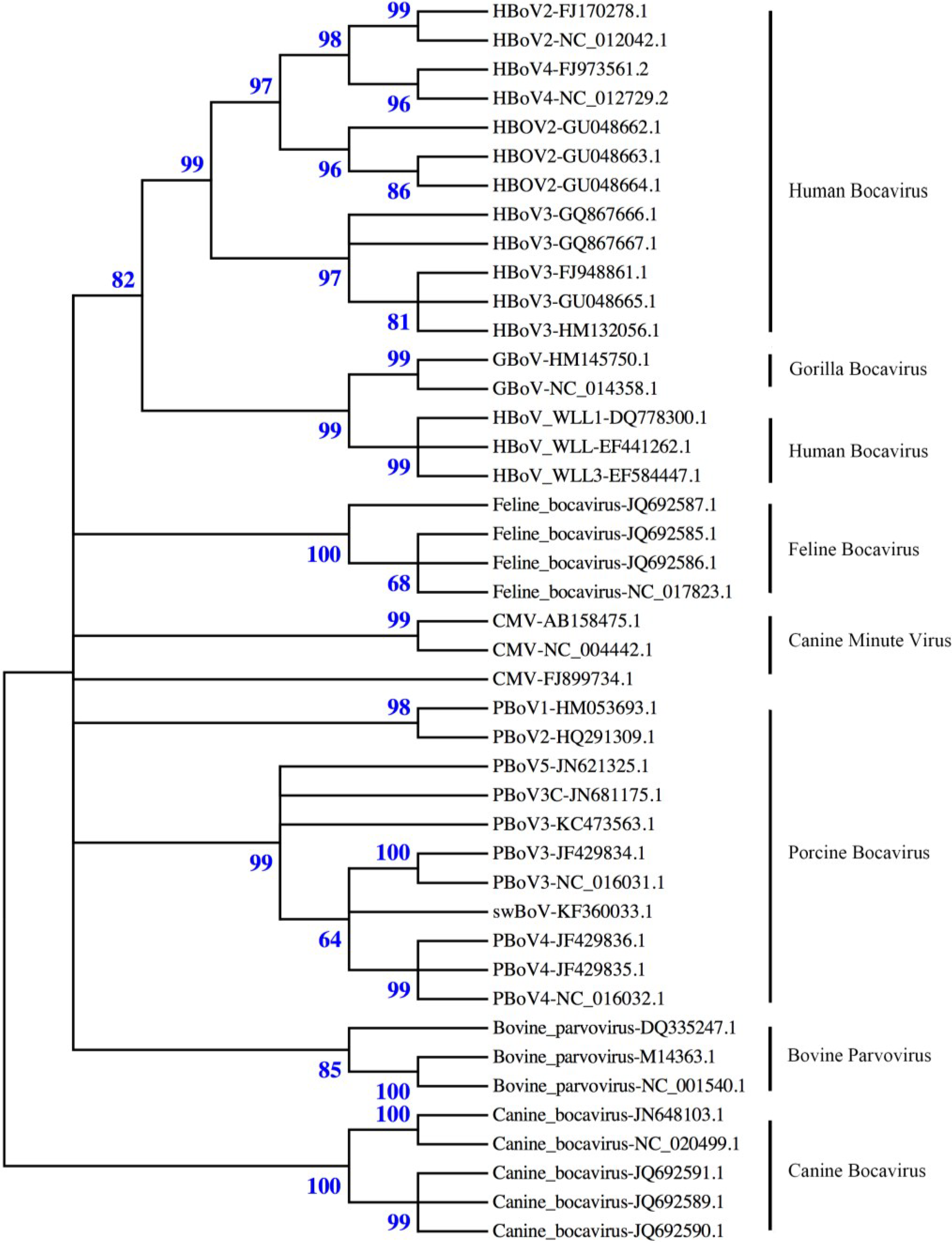
In 2009, an 1879-bp sequence of porcine boca-like virus (PBo-likeV) was found in Sweden in lymph nodes from swine with post-weaning multisystemic wasting syndrome (PMWS) [19]. Since this discovery, sequences of bocaviruses have been identified throughout the world. In 2010, the nearly complete genomes of PBoV1/2-CHN were characterized from fecal samples of swine in China, and the partial genomes of two additional boca-like viruses (named 6V and 7V) were also described [20]. In 2011, PBoV3/4-UK was isolated from a primary pig kidney cell culture that was inoculated with a homogenate of small intestine (PBoV3-UK) and the fecal suspension (PBoV4-UK), respectively, of pigs with PMWS from farms in Northern Ireland [21]. Later that year, PBoV3/4-HK was also detected from samples that were obtained in China during the period of 2005-2007 [22]. In 2012, PBoV3C was found with a high detection rate in fecal samples from healthy piglets [23]. In the same year, PBoV5 was discovered in stool from piglets that had clinical diarrhea on a farm in China [24]. In 2013, a novel PBoV strain swBoV CH437 was detected from clinical samples in Northwest China [25]. The phylogenetic relationship between PBoV and the other bocaviruses is shown in Figure 1.
Since its discovery in 2009, PBoV has been detected globally (Table 1). To date, eleven countries, including Sweden, China, the USA, Canada, Mexico, Romania, Hungary, Uganda, Korea, Cameroon, and the UK, have reported infections of PBoV, although the frequency of the reported infections has varied. In the past five years, with developing methods and techniques, the research on PBoV has expanded rapidly. Nevertheless, some questions remain unresolved. This article summarizes research results from the past five years to present a brief review of porcine bocavirus.
2. Virus Structure
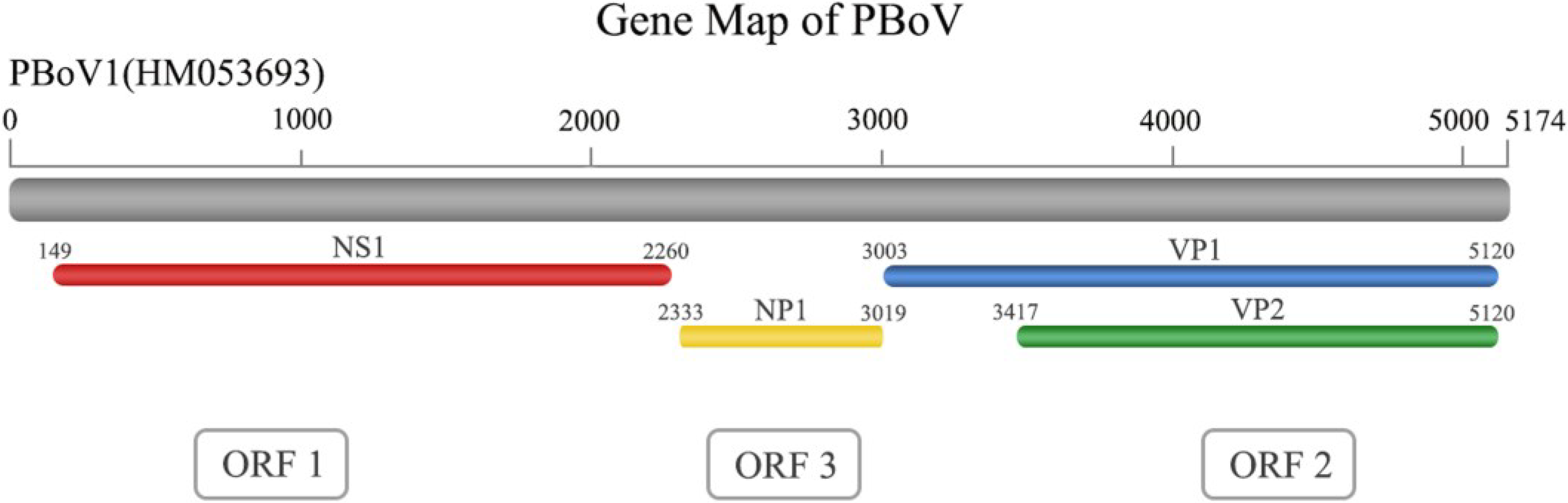
Porcine bocaviruses (PBoVs), new members of the Bocavirus genus, belong to the family Parvoviridae and subfamily Parvovirinae. PBoVs are non-lipid enveloped viruses that exhibit icosahedral symmetry and are 25–30 nm in diameter. Their linear single-stranded genome is only ~5 kb in length and contains terminal palindromic sequences [26]. The genome contains three primary open reading frames (ORFs), which are named ORF1, ORF2, and ORF3. ORF1 codes a non-structural protein (NS1 or replicase) and is located at the 5’-end of the genome. ORF1 has been demonstrated to contain conserved motifs that are associated with rolling-circle replication and helicase and ATPase activities [22]. At the amino acid sequence level, the predicted NS1 gene of PBoV exhibited sequence identities with the canine minute virus (CMV) of 42.0%–48.5%, bovine parvovirus (BPV) of 30.0%–49.1%, gorilla bocavirus (GBoV) of 40.0%–49.0% and human bocavirus (HBoV) of 38.0%–54.7% [22,27]. NS1 has been demonstrated to be essential for viral DNA replication in CMV [28,29], which indicates a similar function in PBoV. ORF2 codes the capsid proteins VP1 and VP2, which overlap in the genome (Figure 2). A conserved “YXGXF” motif domain was found in the unique VP1 protein (VP1u) [23]. This domain indicates a secretory phospholipase A2 (sPLA2) activity that is critical for parvovirus infectivity [20]. Finally, ORF3 codes the NP protein that is located between ORF1 and ORF2 (Figure 2), which is a characteristic genetic feature of the members of the Bocavirus genus. Although the function of NP1 in PBoV has not yet been determined, NP1 is essential for CMV DNA replication [28]. A recent study has demonstrated that in HBoV, NP1 blocks IFN production, which suggests a potential immune-evasion mechanism [30].
Figure 1.
Phylogenetic analysis of bocaviruses. The phylogenetic tree was constructed using nearly full-length nucleotide sequences from bocaviruses in GenBank using the MEGA 5.2 software package [31] (neighbor-joining method with 1000 bootstrap replicates). The relationship among the seven PBoV genotypes (clade Porcine bocavirus) and other bocaviruses can be observed from the tree. PBoV has a close relationship with other bocaviruses, such as feline bocavirus and bovine bocavirus.

| Published Year | First Author | Country | Name | Accession No. |
|---|---|---|---|---|
| 2009 | Blomstrom, A.L. [19] | Sweden | PBo-likeV | FJ872544 |
| 2010 | Cheng, W.X. [20] | China | PBoV1-CHN | HM053693 |
| PBoV2-CHN | HM053694 | |||
| 6V | HM053672 | |||
| 7V | HM053673 | |||
| 2011 | McKillen, J. [21] | Northern Ireland | PBoV3-UK | JF512472 |
| PBoV4-UK | JF512473 | |||
| 2011 | Lau, S.K. [22] | China | PBoV3-HK | JF429834 |
| PBoV4-HK | JF429835 | |||
| 2012 | Li, B. [24] | China | PBoV5 | JN831651 |
| 2012 | Yang, W.Z. [23] | China | PBoV3C | JN681175 |
| 2014 | Wang, E. [25] | China | swBoV CH437 | KF360033 |
In addition to the linear structure of its DNA, PBoV was the second bocavirus in which episomes were detected. The replication of the episome is quite different from that in other parvoviruses [32]. As parvoviruses replicate via a so-called “rolling-hairpin” mechanism that is supported by short imperfectly palindromic hairpin telomeres, their replication intermediates are concatemers of head-to-head or tail-to-tail structures [33,34,35,36]. PBoV and HBoV both replicate in a rolling circle with a head-to-tail structure [32,37]. Despite the different position of hairpin-1 and the miss of hairpin-2 in PBoV, the sequence of the typical structure hairpin-1 is conserved in PBoV. This conserve structure may be related to the identification of replication proteins and binding sites [32].
Figure 2.
The genome structure of PBoV. The genome of PBoV contains three ORFs that encode four proteins. The sequences for VP1 and VP2 overlap in the genome.
Figure 2.
The genome structure of PBoV. The genome of PBoV contains three ORFs that encode four proteins. The sequences for VP1 and VP2 overlap in the genome.

3. Taxonomy and Nomenclature
PBoVs were defined as new species using the ICTV criteria [27], which states that there must be less than 95% homology in the nonstructural gene to be considered a new species in the Bocavirus genus. The newly discovered PBoV strains were named in chronological order. As an increasing number of novel PBoV strains are detected, a well-established classification system with specific criteria is essential for further investigation.
Originally, PBoVs were classified into three phylogenetic clades using phylogenetic analysis based on both nucleotide and amino acid alignments [38]. Further studies proposed classification methods based on ORF2-encoded VP1, which is a capsid protein and is likely to influence tissue tropism and possibly pathogenesis [39,40]. This method was originally proposed to characterize human bocaviruses using the criteria of >8% amino acid and >10% nucleotide differences from the VP1 sequences to identify different species [6]. Based on this method, the PBoVs were grouped into four species [39]. In 2012, another classification method based on the VP1 gene was proposed; in this scheme, PBoV strains with a >40% nucleotide difference from the sequence of the complete VP1 gene are considered members of different groups, and those with a >10% difference from the VP1 nucleotide sequence are considered to be members of different subgroups [23]. The subgroups have been defined based on phylogenetic clustering and the homology matrix of known porcine bocaviruses. Using this method, PBoVs can be divided into three groups: PBoV G1, which includes PBo-likeV (FJ872544), PBoV-SX (HQ223038) and PBoV1-H18 (HQ291308); PBoV G2, which includes PBoV1/2-CHN (HM053693/HM053694) and PBoV2-A6 (HQ291309); and PBoV G3, which includes PBoV3/4-UK (JF512472/JF512473), PBoV3/4-HK (JF429834/JF429835), and PBoV3C (JN681175). Group 3 can be further divided into five subgroups: PBoV3A, PBoV3B, PBoV3C, PBoV3D, and PBoV3E, which were previously known as PBoV3-UK, PBoV4-UK, PBoV3C, PBoV3-HK and PBoV4-HK, respectively [23]. In 2013, Xiao et al. proposed adding PBoV5 (JN831651) and PBoV3-22/23 (JF713714/JF713715) to PBoV G3. Moreover, different subgroups were proposed; in this classification scheme, PBoV3-UK, PBoV3-22, PBoV4-UK, PBoV3C and PBoV5 each represented a subgroup [41]. Although the five subgroups may need to be re-designated in further studies, this classification, which is mainly based on the VP1 gene, is widely accepted.
Recently, Huang et al. proposed classification methods that are primarily based on the NS1 gene [42,43]. However, there are differences between this nomenclature and the former nomenclature based on VP1. These differences may be due to crossover recombination during the speciation of these viruses [22] and incongruent clustering based on NS1, VP1 and NP1.
Given the diversity of known PBoVs, it is very likely that there are more than three groups. More effort should be focused on identifying the best taxonomy and nomenclature.
4. Recombination
Different viral strains in the Parvovirinae subfamily have been demonstrated to undergo recombination. Phylogenetic analysis has confirmed that a recombination event occurred between HBoV1 and HBoV4, leading to the recombinant strain HBoV2 [44]. Recombination can occur in different ORFs [4,6]. In PBoVs, recombination has also been documented in some regions and strains. Lau et al. documented a recombination event between PBoV4-HK and SH14F/12, which may have emerged from recombination between the strains SH14F/1 and SH14F/11 in the VP1/VP2 region [22]. Csagola et al. found possible recombination sites at the beginning of the nucleotide sequence between the PBoV1-CHN and PBoV2-CHN genomes. A segment of the genome from nt 227 to nt 278 was replaced in the PBoV2 genomes by PBoV1-CHN sequences (GenBank ID: JN400872, JN400873, JN400874). Moreover, Yang et al. have documented recombination in the NP1 regions of PBoV-WUH1 and PBoV-H18 [23]. Co-infection within species and extensive recombination at the NP1 and VP1 gene between and within species have been noted [6]. Taken together, these studies have confirmed that PBoVs undergo recombination, which may lead to changes in the biological characteristics of the virus. More importantly, the possibility of recombination between PBoV and HBoV should be considered.
5. Detection of Porcine Bocaviruses Using Molecular Virology Techniques
PBoV can be detected in multiple types of tissue, including lymph nodes [19], serum [28], intestine [21], lung, saliva [45], and spleen [42]. However, given the easy accessibility and generally high positive rate, nasopharyngeal samples (NPS) and fecal samples are typically used for clinical tests to detect the virus [22].
5.1. Cell Culture
In 2010, McKillen et al. successfully propagated PBoV3/4-UK in primary pig kidney cells. The virus isolates were identified in culture using electron microscopy, immunofluorescence assays, and PCR [21]. However, PBoV replication in vitro is not supported by porcine kidney cells (PK-15), swine testicular cells, porcine alveolar macrophages, monkey kidney cells (MARC-145), or human embryonic kidney epithelial cells (HEK293T) [28].
5.2. Sequence Detection Assays
Sequence-based assays, including PCR, RT-PCR, loop-mediated isothermal amplification, and high-throughput DNA sequencing, have been established to detect PBoVs. High-sensitivity assays can detect as little as 3.22 fg/µL of viral DNA through PCR [46]. The VP1 and VP2 genes are the preferred sequences for primer design. However, PCR-based assays have been qualitative rather than quantitative. Li et al. successfully developed a real-time PCR assay that targets the NP1 gene of PBo-likeV [47]. The assay can provide sensitive (approximately 20 copies) and specific quantification of PBo-likeV in clinical specimens. Semi-quantitative PCR has also been used to identify PBoV episomes [32].
High-throughput sequencing can detect viruses from a large number of samples. Yang et al. used this method to successfully identify PBoV3C from piglet fecal samples [23]. The 5235-nucleotide genome can be sequenced using the genome-walking method. Loop-mediated isothermal amplification is another option for detecting PBoV nucleotides. This method is more easily implemented and sensitive compared with conventional PCR assays. The positive and negative samples can be easily distinguished by adding diluted SYBR Green I to the reaction mix, and the results can be observed visually [48].
5.3. Indirect Immunofluorescence Assay
Indirect immunofluorescence assays were developed by McKillen et al. for in vitro use to monitor viral growth in pig kidney cells [21]. Later, the same group developed monoclonal antibodies (mAbs) against two swine bocaviruses (PBoV3/4-UK) that were isolated in cell cultures. Moreover, monoclonal antibodies they developed could be successfully used in antigen-detecting ELISAs that highlight those fractions containing infectious virus within sucrose gradients [49].
6. Epidemiology
PBoV infections have been reported in China, Korea, the USA, Mexico, Canada, Northern Ireland, Romania, Hungary, Cameroon, and Uganda (Table 2). Many of the known PBoV genotypes have been reported in China and the USA, including PBoV G1 (PBo-likeV, PBoV-SX, and PBoV1-H18), PBoV G2 (PBoV1/2-CHN and PBoV2-A6), PBoV G3 (PBoV3C, PBoV3/4-HK and PBoV5), and unclassified strains (6V/7V, and swBoV CH437). A recent study demonstrated that positive rates of PBoV (42.0%, or 76/181) in American samples were significantly greater than those (11.4%, or 46/403) in Chinese samples. The geographical distribution of PBoV mainly lies in the eastern and southern coastal areas of China and the central states of the USA. In China and the USA, Jiangsu Province and the state of Minnesota, respectively, were the centers of high PBoV occurrence frequency [50]. However, epidemiological studies outside of China and the USA have been limited. PBoV G1 detections were reported in Sweden, Northern Ireland, Romania, Hungary, Cameroon, and Uganda (Table 2). Northern Ireland is the only country other than the USA and China in which the prevalence of PBoVG3 is high. A discovery of PPV4 [51], which was once considered to be a member of PBoV, was reported in the USA. However, this virus is now considered to belong to a separate clade that is different from the genera Parvovirus and Bocavirus [41].
The prevalence of PBoV infection also varies with age and season. In terms of age, Zhai et al. found that PBo-likeV infection was significantly more prevalent in weaned piglets (69.7%, or 69/99) than in piglets that were not yet weaned (13.6%, or 3/22), adult boars (0%, or 0/20), adult sows (7.7%, or 2/26), and aborted fetuses (0%, or 0/14) (p < 0.01, χ2 test) [52]. Similarly, in a wild boar population, Cadar et al. reported that piglets that were 6–12 months old (77.06%) were three times more likely to be infected with PBo-likeV than piglets that were 12–36 months old (22.94%) (p < 0.01) [53]. The low prevalence of PBo-likeV infection in piglets that are less than 6 months of age suggests a passage of protective maternal immunity. The morbidity (50%–100%) and mortality (20%–60%) rates were also higher in piglets that were 15–70 days old than in sows (>1 year old) and boars (>2 years old), for which little to no mortality was observed [52]. The seasonal infection rates of PBoV are higher from March through May (72.7%, or 16/22) than in June through August (28.9%, of 26/90), September through November (38.7%, or 12/31), and December through February (41.7%, or 20/48) (p < 0.05, χ2 test). However, there was no significant difference in the rates of PBoV infections throughout the summer, autumn, and winter (p < 0.05, χ2 test) [52].
The contribution of co-morbidities to PBoV infection rates is still not clear. Early evidence suggested that piglets with PMWS may be more likely to develop PBoV infections. Blomstrom et al. reported that the positive rate of PBo-likeV in piglets with and without PMWS was 88% and 46%, respectively [54]. Similarly, Zhai et al. reported a higher frequency of PBo-likeV infection in pigs that suffered from respiratory tract symptoms and reproductive failure (38.7%) than in healthy pigs (7.3%) [52]. However, the clinical samples used in this study were from sick pigs from nine provinces throughout China, and the sera from healthy pigs were from a single province. Thus, the difference in the rate of PBo-likeV infection based on co-morbidities observed in this study may not be credible.
| Country | Age | Health Condition b | n | Rate (%) | PBoV Type Tested |
|---|---|---|---|---|---|
| Uganda [55] | n/g | n/g | 95 | 2.11% | PBoV G1 |
| Cameroon [56] | piglet | healthy | 50 | 46% | overall |
| USA [57] | mixed | mainly E + R | 385 | 58.7% | overall |
| USA c [43] | n/g | E + R | 203 | 43.3% | overall |
| China [52] | piglet | mainly R + P | 191 | 38.70% | PBoV G1 |
| healthy | 41 | 7.30% | PBoV G1 | ||
| China [20] | piglet | healthy | 397 | 12.59% | PBoV G2 |
| China [28] | pig | healthy | 120 | 39.17% | PBoV G1 |
| China [38] | mixed | healthy | 340 | 63.20% | PBoV G1 |
| 340 | 64.40% | PBoV G2 | |||
| China [39] | pig | clinically sick | 128 | 30.50% | PBoV G1 |
| healthy | 38 | 23.70% | PBoV G1 | ||
| clinically sick | 128 | 21.90% | PBoV G2 | ||
| healthy | 38 | 10.50% | PBoV G2 | ||
| clinically sick | 128 | 38.30% | 6V/7V | ||
| healthy | 38 | 44.70% | 6V/7V | ||
| China [22] | n/g | sick + D | 213 | 18.31% | PBoV G3 |
| healthy | 90 | 16.67% | PBoV G3 | ||
| China [47] | piglet | mainly with PMWS + D | 180 | 56.10% | PBoV G1 |
| healthy | 78 | 16.70% | PBoV G1 | ||
| China [23] | piglet | healthy | 92 | 57.61% | PBoV G2 |
| 92 | 19.60% | PBoV3C | |||
| China [58] | piglet | E | 884 | 31.90% | PBoV G1 |
| healthy | 266 | 26.32% | PBoV G1 | ||
| pig | E | 101 | 27.72% | PBoV G1 | |
| healthy | 58 | 24.14% | PBoV G1 | ||
| China [42] | mixed | E + R + G + D | 403 | 11.41% | overall |
| Korea [45] | mixed | mixed | 920 | 34.9% | overall |
| E + R + G | 351 | 37.8% | overall | ||
| healthy | 679 | 14.9 | overall | ||
| Sweden [19] | piglet | with PMWS | 2 | 100% | PBoV G1 |
| Sweden [54] | piglet | with PMWS | 34 | 88.00% | PBoV G1 |
| healthy | 24 | 46.00% | PBoV G1 | ||
| Northern Ireland [21] | piglet | mainly with PMWS | 369 | 8.70% | PBoV3-UK |
| 369 | 9.50% | PBoV4-UK | |||
| Romania [53] | wild boar | n/g | 470 | 9.14% | PBoV G1 |
| 372 | 17.74% | PBoV G1 | |||
| Hungary [59] | n/g | sick + healthy | 392 | 1.50% | PBoV G1 |
| 392 | 4.80% | PBoV G2 | |||
| 392 | 1.80% | 6V/7V |
Abbreviations: n, number of samples; n/g, not given. a Selection criteria: PubMed search using the key words “bocavirus” and “porcine”. b For sick pigs, several short forms are used: E for enteric symptoms like diarrhea, inappetence; R for respiratory tract symptoms like cough, dyspnea, panting; P for reproductive failure including abortion/stillbirth for sows and low-quality semen for boars; D for deceased or slaughtered; G for general symptoms such as fever, lethargy, wasting, trembling. c In this study, fecal samples were collected from the USA, Mexico and Canada.
In contrast, previous work from other groups has reported that the prevalence of PBoVs is higher in healthy pigs/piglets. In 2010, Zeng et al. reported a high prevalence (39.17%) of PBo-likeV infection in healthy pigs in Hubei Province, China [28]. Another study reported similarly high rates of infection for PBoV1-H18 (63.2%) and PBoV-A6 (64.4%) in China [38]. However, this study used only fecal samples, which usually have a high positive rate, to detect the virus. Other studies have also confirmed that PBoVs, including PBoV1/2-CHN, PBoV3/4-UK, PBoV3/4-HK and the newly reported PBoV3C, are highly prevalent in healthy pigs (Table 2). Therefore, additional studies are required to determine the pig population that is most susceptible to PBoV infection and the role of co-morbid infections in establishing susceptibility.
7. Pathogenesis
The pathogenic mechanism of PBoV infection remains unclear because of the limitations of current studies. In particular, although pigs exhibited clinical symptoms such as trembling, fever, testicular atrophy, abortion or death appear to be more susceptible to PBoV infections (Table 2), the role of co-infections in pathogenesis is still undefined. Commonly reported co-infections include PCV2, PTTV, PRRSV, CSFV, PEDV, PKoV, GARV and TGEV (Table 3) [39,52,58,59]. PBo-likeV co-infections have been documented with numerous pathogens; however, pigs that screen positive for PCV2, PTTV, PEDV, and PKoV have the highest incidence (greater than 70%) of PBo-likeV co-infection. Similarly, in samples that tested positive for PBo-likeV, a relatively high rate (greater than 30%) also tested positive for PCV2, PEDV, PKoV, and GARV (Table 2). Although this result likely reflects the high frequency of these viruses in the population at large, it is possible that these particular viruses may provide a biological benefit to PBoV. Interestingly, PBoV (PBoV3/4-UK) becomes cytopathic after being passaged four times through primary pig kidney cells. Cell lysates that exhibited a cytopathic effect were screened using PCR or RT-PCR and tested negative for PCV1, PCV2, and PPV. These results suggest that even if PBoVs are not directly associated with PMWS or other diseases, they may function as a triggering factor for other infectious agents [19].
The clinical specimens used to detect PBoV include sera, lungs, lymph nodes, tonsils, liver, nasopharyngeal swabs, and fecal samples from healthy and diseased pigs. In terms of virus shedding, Li et al. reported that a high viral load (greater than 105 copies·mg−1) of PBo-likeV was detected in lung and lymph node samples from diseased pigs using a TaqMan-based real-time PCR assay [47]. A high positive rate of PBoV DNA can also be detected in lung, intestinal, and fecal samples and nasopharyngeal swabs [21,53,58], suggesting that PBoV may invade these tissues and that there might be an association with other diseases. Consistent with the possible link between PBoV and other pathogens, Zhang et al. reported a higher viral load in diarrhea than in healthy fecal samples (4.6 × 10⁵ versus 2.9 × 10⁵ copies/g of stool), but the difference was not significant [58]. So far, there is no sound evidence regarding the pathogenesis of PBoV, thus well-designed investigation are recommended in the future.
| Group PBoV1(+) Samples with Copathogen b | Listed Virus(+) Samples Coinfected with PBoV c | |
|---|---|---|
| PCV2 (%) | 3.3–83.8 | 37.7 |
| PTTV1 (%) | 73 | n/g |
| PTTV2 (%) | 70.3 | n/g |
| PRRSV (%) | 0–67.6 | 27.3 |
| CSFV (%) | 6.9–34.5 | 20.7 |
| PEDV (%) | 72.6 | 34 |
| PKoV (%) | 72.1 | 32.9 |
| GARV (%) | 9.9 | 41 |
| TGEV (%) | 1 | 66.7 |
Abbreviations: PCV2, porcine circovirus type 2; PTTV, porcine torque teno virus; PRRSV, porcine reproductive and respiratory syndrome virus; CSFV, classical swine fever virus; PEDV, porcine epidemic diarrhea virus; PKoV, porcine kobuvirus; PBoV, porcine bocavirus; GARV, porcine group A rotavirus; TGEV, transmissible gastroenteritis virus. a Four studies are included [39,52,58,59]. b Median number of group PBoV1-positive samples that were found to be coinfected with the listed pathogen. c Median number of samples that tested positive for the listed viruses and were found to be co-infected with group PBoV1.
Because HBoV and PBoV both belong to the genus Bocavirus and family Parvoviridae, they share many similarities in terms of their virus characteristics. Thus, it is meaningful to apply experience from HBoV research to PBoV, especially now that there are abundant studies regarding the pathogenicity of HBoV. The high prevalence of HBoV DNA in serum is associated with malignant tumors [60]. Moreover, in tissue samples, human bocavirus DNA was detected in 18.3% (11/60) of lung tissue samples and 20.5% (9/44) of colorectal tumors and may be present in the nuclei of infected cells, which indicates the existence of the postulated σ- or rolling-hairpin replication mechanism [61]. Because of the lack of appropriate in vitro or in vivo models, it is difficult to detect the tumors in pigs even though the PBoVG2 and PBoVG3 episomes have been identified [32] However, it would be useful to determine the potential host organ of PBoV.
8. Conclusions
Since the discovery of PBoV five years ago, our knowledge of the virus has been enriched considerably. The primary achievements are the following: (1) knowledge has been widely shared; (2) detailed epidemic data have been collected; (3) genome sequences have been acquired, and the functions of the open reading frames have been preliminarily interpreted; (4) five genotypes of PBoV have been found, and they were classified into three groups; (5) sequence analysis has indicated the relationship between PBoV and other bocaviruses; (6) several detection methods have been developed; and (7) the virus has been successfully grown in vitro. Despite these advances, there are still many questions that require further study, and with rapidly advancing tools, these questions may be resolved in the near future.
Authors Contributions
Feng Zhou contributed to the original draft of the manuscript. Haoting Sun contributed by collecting the data and creating the figures and tables in the manuscript. Yuyan Wang contributed by revising the manuscript. All authors read and approved the final manuscript.
Acknowledgments
The work was financially supported by the Zhengyi Scholar Program and Dean’s Office of the School of Basic Medical Sciences, Fudan University (NSFC grant No. J1210041). The authors sincerely thank Huashan Hospital for their support of this research.
Conflicts of Interest
The authors declare no conflict of interest.
References
- Allander, T.; Tammi, M.T.; Eriksson, M.; Bjerkner, A.; Tiveljung-Lindell, A.; Andersson, B. Cloning of a human parvovirus by molecular screening of respiratory tract samples. Proc. Natl. Acad. Sci. USA 2005, 102, 12891–12896. [Google Scholar] [CrossRef] [PubMed]
- Fry, A.M.; Lu, X.; Chittaganpitch, M.; Peret, T.; Fischer, J.; Dowell, S.F.; Anderson, L.J.; Erdman, D.; Olsen, S.J. Human bocavirus: A novel parvovirus epidemiologically associated with pneumonia requiring hospitalization in Thailand. J. Infect. Dis. 2007, 195, 1038–1045. [Google Scholar] [CrossRef] [PubMed]
- Allander, T.; Jartti, T.; Gupta, S.; Niesters, H.G.; Lehtinen, P.; Osterback, R.; Vuorinen, T.; Waris, M.; Bjerkner, A.; Tiveljung-Lindell, A.; et al. Human bocavirus and acute wheezing in children. Clin. Infect. Dis. 2007, 44, 904–910. [Google Scholar] [CrossRef] [PubMed]
- Kapoor, A.; Slikas, E.; Simmonds, P.; Chieochansin, T.; Naeem, A.; Shaukat, S.; Alam, M.M.; Sharif, S.; Angez, M.; Zaidi, S.; et al. A newly identified bocavirus species in human stool. J. Infect. Dis. 2009, 199, 196–200. [Google Scholar] [CrossRef] [PubMed]
- Arthur, J.L.; Higgins, G.D.; Davidson, G.P.; Givney, R.C.; Ratcliff, R.M. A novel bocavirus associated with acute gastroenteritis in Australian children. PLoS Pathog. 2009, 5, e1000391. [Google Scholar] [CrossRef] [PubMed]
- Kapoor, A.; Simmonds, P.; Slikas, E.; Li, L.; Bodhidatta, L.; Sethabutr, O.; Triki, H.; Bahri, O.; Oderinde, B.S.; Baba, M.M.; et al. Human bocaviruses are highly diverse, dispersed, recombination prone, and prevalent in enteric infections. J. Infect. Dis. 2010, 201, 1633–1643. [Google Scholar] [CrossRef] [PubMed]
- Abinanti, F.R.; Warfield, M.S. Recovery of a hemadsorbing virus (HADEN) from the gastrointestinal tract of calves. Virology 1961, 14, 288–289. [Google Scholar] [CrossRef] [PubMed]
- Inaba, Y.; Kurogi, H.; Omori, T.; Matumoto, M. A new serotype of bovine parvovirus. Jpn. J. Microbiol. 1973, 17, 85–86. [Google Scholar] [CrossRef] [PubMed]
- Allander, T.; Emerson, S.U.; Engle, R.E.; Purcell, R.H.; Bukh, J. A virus discovery method incorporating DNase treatment and its application to the identification of two bovine parvovirus species. Proc. Natl. Acad. Sci. USA 2001, 98, 11609–11614. [Google Scholar] [CrossRef] [PubMed]
- Pratelli, A.; Moschidou, P. Host range of Canine minute virus in cell culture. J. Vet. Diagn. Investig. 2012, 24, 981–985. [Google Scholar] [CrossRef]
- Decaro, N.; Amorisco, F.; Lenoci, D.; Lovero, A.; Colaianni, M.L.; Losurdo, M.; Desario, C.; Martella, V.; Buonavoglia, C. Molecular characterization of Canine minute virus associated with neonatal mortality in a litter of Jack Russell terrier dogs. J. Vet. Diagn. Investig. 2012, 24, 755–758. [Google Scholar] [CrossRef]
- Kapoor, A.; Mehta, N.; Esper, F.; Poljsak-Prijatelj, M.; Quan, P.L.; Qaisar, N.; Delwart, E.; Lipkin, W.I. Identification and characterization of a new bocavirus species in gorillas. PLoS One 2010, 5, e11948. [Google Scholar] [CrossRef] [PubMed]
- Sharp, C.P.; LeBreton, M.; Kantola, K.; Nana, A.; Diffo, J.D.; Djoko, C.F.; Tamoufe, U.; Kiyang, J.A.; Babila, T.G.; Ngole, E.M.; et al. Widespread infection with homologues of human parvoviruses B19, PARV4, and human bocavirus of chimpanzees and gorillas in the wild. J. Virol. 2010, 84, 10289–10296. [Google Scholar] [CrossRef] [PubMed]
- Babkin, I.V.; Tyumentsev, A.I.; Tikunov, A.Y.; Kurilshikov, A.M.; Ryabchikova, E.I.; Zhirakovskaya, E.V.; Netesov, S.V.; Tikunova, N.V. Evolutionary time-scale of primate bocaviruses. Infect. Genet. Evol. 2013, 14, 265–274. [Google Scholar] [CrossRef] [PubMed]
- Lau, S.K.; Woo, P.C.; Yeung, H.C.; Teng, J.L.; Wu, Y.; Bai, R.; Fan, R.Y.; Chan, K.H.; Yuen, K.Y. Identification and characterization of bocaviruses in cats and dogs reveals a novel feline bocavirus and a novel genetic group of canine bocavirus. J. Gen. Virol. 2012, 93, 1573–1582. [Google Scholar] [CrossRef] [PubMed]
- Pesavento, P.A.; Murphy, B.G. Common and emerging infectious diseases in the animal shelter. Vet. Pathol. 2014, 51, 478–491. [Google Scholar] [CrossRef] [PubMed]
- Li, L.; Shan, T.; Wang, C.; Cote, C.; Kolman, J.; Onions, D.; Gulland, F.M.; Delwart, E. The fecal viral flora of California sea lions. J. Virol. 2011, 85, 9909–9917. [Google Scholar] [CrossRef] [PubMed]
- Jartti, T.; Hedman, K.; Jartti, L.; Ruuskanen, O.; Allander, T.; Soderlund-Venermo, M. Human bocavirus-the first 5 years. Rev. Med. Virol. 2012, 22, 46–64. [Google Scholar] [CrossRef] [PubMed]
- Blomstrom, A.L.; Belak, S.; Fossum, C.; McKillen, J.; Allan, G.; Wallgren, P.; Berg, M. Detection of a novel porcine boca-like virus in the background of porcine circovirus type 2 induced postweaning multisystemic wasting syndrome. Virus Res. 2009, 146, 125–129. [Google Scholar] [CrossRef] [PubMed]
- Cheng, W.X.; Li, J.S.; Huang, C.P.; Yao, D.P.; Liu, N.; Cui, S.X.; Jin, Y.; Duan, Z.J. Identification and nearly full-length genome characterization of novel porcine bocaviruses. PLoS One 2010, 5, e13583. [Google Scholar] [CrossRef] [PubMed]
- McKillen, J.; McNeilly, F.; Duffy, C.; McMenamy, M.; McNair, I.; Hjertner, B.; Millar, A.; McKay, K.; Lagan, P.; Adair, B.; et al. Isolation in cell cultures and initial characterisation of two novel bocavirus species from swine in Northern Ireland. Vet. Microbiol. 2011, 152, 39–45. [Google Scholar] [CrossRef] [PubMed]
- Lau, S.K.; Woo, P.C.; Yip, C.C.; Li, K.S.; Fu, C.T.; Huang, Y.; Chan, K.H.; Yuen, K.Y. Co-existence of multiple strains of two novel porcine bocaviruses in the same pig, a previously undescribed phenomenon in members of the family Parvoviridae, and evidence for inter- and intra-host genetic diversity and recombination. J. Gen. Virol. 2011, 92, 2047–2059. [Google Scholar] [CrossRef] [PubMed]
- Yang, W.Z.; Yu, J.M.; Li, J.S.; Cheng, W.X.; Huang, C.P.; Duan, Z.J. Genome characterization of a novel porcine bocavirus. Arch. Virol. 2012, 157, 2125–2132. [Google Scholar] [CrossRef] [PubMed]
- Li, B.; Ma, J.; Xiao, S.; Fang, L.; Zeng, S.; Wen, L.; Zhang, X.; Ni, Y.; Guo, R.; Yu, Z.; et al. Complete genome sequence of a novel species of Porcine Bocavirus, PBoV5. J. Virol. 2012, 86, 1286–1287. [Google Scholar] [CrossRef] [PubMed]
- Wang, E.; Liu, W.; Yang, B.; Liu, J.; Ma, X.; Lan, X. Complete sequence and phylogenetic analysis of a porcine bocavirus strain swBoV CH437. Virus Genes 2014 2014, 48, 387–390. [Google Scholar] [CrossRef]
- Tijssen, P.; Agbandje-McKenna, M.; Almendral, J.M.; Bergoin, M.; Flegel, T.W.; Hedman, K.; Kleinschmidt, J.; Li, Y.; Pintel, D.J.; Tattersall, P. Family Parvoviridae. In Virus Taxonomy. 9th report of the International Committee on Taxonomy of Viruses; King, A.M.Q., Adams, M.J., Carstens, E.B., Lefkowitz, E.J., Eds.; Academic Press: London, UK, 2012; pp. 405–425. [Google Scholar]
- Zeng, S.; Wang, D.; Fang, L.; Ma, J.; Song, T.; Zhang, R.; Chen, H.; Xiao, S. Complete coding sequences and phylogenetic analysis of porcine bocavirus. J. Gen. Virol. 2011, 92, 784–788. [Google Scholar] [CrossRef] [PubMed]
- Sun, Y.; Chen, A.Y.; Cheng, F.; Guan, W.; Johnson, F.B.; Qiu, J. Molecular characterization of infectious clones of the minute virus of canines reveals unique features of bocaviruses. J. Virol. 2009, 83, 3956–3967. [Google Scholar] [CrossRef] [PubMed]
- Christensen, J.; Cotmore, S.F.; Tattersall, P. Minute virus of mice transcriptional activator protein NS1 binds directly to the transactivation region of the viral P38 promoter in a strictly ATP-dependent manner. J. Virol. 1995, 69, 5422–5430. [Google Scholar] [PubMed]
- Zhang, Z.; Zheng, Z.; Luo, H.; Meng, J.; Li, H.; Li, Q.; Zhang, X.; Ke, X.; Bai, B.; Mao, P.; et al. Human bocavirus NP1 inhibits IFN-beta production by blocking association of IFN regulatory factor 3 with IFNB promoter. J. Immunol. 2012, 189, 1144–1153. [Google Scholar] [CrossRef] [PubMed]
- Tamura, K.; Peterson, D.; Peterson, N.; Stecher, G.; Nei, M.; Kumar, S. MEGA5: molecular evolutionary genetics analysis using maximum likelihood, evolutionary distance, and maximum parsimony methods. Mol. Biol. Evol. 2011, 28, 2731–2739. [Google Scholar] [CrossRef] [PubMed]
- Yang, W.Z.; Huang, C.P.; Duan, Z.J. Identification and characterization of porcine bocavirus episomes. Bing Du Xue Bao 2012, 28, 418–423. (In Chinese) [Google Scholar] [PubMed]
- Lusebrink, J.; Schildgen, V.; Tillmann, R.L.; Wittleben, F.; Bohmer, A.; Muller, A.; Schildgen, O. Detection of head-to-tail DNA sequences of human bocavirus in clinical samples. PLoS One 2011, 6, e19457. [Google Scholar] [CrossRef] [PubMed]
- Tattersall, P.; Ward, D.C. Rolling hairpin model for replication of parvovirus and linear chromosomal DNA. Nature 1976, 263, 106–109. [Google Scholar] [CrossRef] [PubMed]
- Hirt, B. Molecular biology of autonomous parvoviruses. Contrib. Microbiol. 2000, 4, 163–177. [Google Scholar] [PubMed]
- Cotmore, S.F.; Tattersall, P. Genome packaging sense is controlled by the efficiency of the nick site in the right-end replication origin of parvoviruses minute virus of mice and LuIII. J. Virol. 2005, 79, 2287–2300. [Google Scholar] [CrossRef] [PubMed]
- Kapoor, A.; Hornig, M.; Asokan, A.; Williams, B.; Henriquez, J.A.; Lipkin, W.I. Bocavirus episome in infected human tissue contains non-identical termini. PLoS One 2011, 6, e21362. [Google Scholar] [CrossRef] [PubMed]
- Shan, T.; Lan, D.; Li, L.; Wang, C.; Cui, L.; Zhang, W.; Hua, X.; Zhu, C.; Zhao, W.; Delwart, E. Genomic characterization and high prevalence of bocaviruses in swine. PLoS One 2011, 6, e17292. [Google Scholar] [CrossRef] [PubMed]
- Zhang, H.B.; Huang, L.; Liu, Y.J.; Lin, T.; Sun, C.Q.; Deng, Y.; Wei, Z.Z.; Cheung, A.K.; Long, J.X.; Yuan, S.S. Porcine bocaviruses: Genetic analysis and prevalence in Chinese swine population. Epidemiol. Infect. 2011, 139, 1581–1586. [Google Scholar] [CrossRef] [PubMed]
- Duffy, S.; Shackelton, L.A.; Holmes, E.C. Rates of evolutionary change in viruses: Patterns and determinants. Nat. Rev. Genet. 2008, 9, 267–276. [Google Scholar] [CrossRef] [PubMed]
- Xiao, C.T.; Halbur, P.G.; Opriessnig, T. Molecular evolutionary genetic analysis of emerging parvoviruses identified in pigs. Infect. Genet. Evol. 2013, 16, 369–376. [Google Scholar] [CrossRef] [PubMed]
- Liu, M.; Li, Y.; Sun, D.; Xia, Y.; Huang, J.; Guo, L. Detection and genetic analysis of porcine bocavirus in different swine herds in North Central China. Sci. World J. 2014, 2014, e947084. [Google Scholar]
- Huang, J.; Mor, S.K.; Erber, J.; Voss, E.; Goyal, S.M. Detection and characterization of porcine bocavirus in the United States. Arch. Virol. 2014, 159, 1797–1801. [Google Scholar] [CrossRef] [PubMed]
- Fu, X.; Wang, X.; Ni, B.; Shen, H.; Wang, H.; Zhang, X.; Chen, S.; Shao, S.; Zhang, W. Recombination analysis based on the complete genome of bocavirus. Virol. J. 2011, 8, e182. [Google Scholar] [CrossRef]
- Choi, M.G.; Park, S.J.; Nguyen, V.G.; Chung, H.C.; Kim, A.R.; Park, B.K. Molecular detection and genetic analysis of porcine bocavirus in Korean domestic swine herds. Arch. Virol. 2013, 159, 1487–1492. [Google Scholar] [CrossRef] [PubMed]
- Zhai, S.L.; Yue, C.; Wei, Z.Z.; Ran, D.L.; Long, J.X.; Lin, T.; Deng, Y.; Sun, L.C.; Huang, L.; Yuan, S.S. Development and application of a PCR assay for detection of porcine bocavirus. Chin. J. Anim. Infect. Dis. 2010, 2, 14–17. (In Chinese) [Google Scholar]
- Li, B.; Xiao, S.; Ma, J.; Liu, Y.; Mao, L.; Wen, L.; Mao, A.; Zhang, X.; Ni, Y.; Guo, R.; et al. Development of a novel TaqMan-based real-time PCR assay for the detection of porcine boca-like virus (Pbo-likeV). Virol. J. 2011, 8, e357. [Google Scholar] [CrossRef]
- Li, B.; Ma, J.J.; Xiao, S.B.; Zhang, X.H.; Wen, L.B.; Mao, L.; Ni, Y.X.; Guo, R.L.; Zhou, J.M.; Lv, L.X.; et al. Development of a loop-mediated isothermal amplification method for rapid detection of porcine boca-like virus. J. Virol. Methods 2012, 179, 390–395. [Google Scholar] [CrossRef] [PubMed]
- McNair, I.; McNeilly, F.; Duffy, C.; McKillen, J.; McMenamy, M.; Welsh, M.; Allan, G. Production, characterisation and applications of monoclonal antibodies to two novel porcine bocaviruses from swine in Northern Ireland. Arch. Virol. 2011, 156, 2157–2162. [Google Scholar] [CrossRef] [PubMed]
- Zhang, Q.; Zhang, C.; Gao, M.; He, X.; Diao, Y.; Goyal, S.M.; Mor, S.K.; Huang, J. Evolutionary, epidemiological, demographical, and geographical dissection of porcine bocavirus in China and America. Virus Res. 2014, 3, e217. [Google Scholar]
- Cheung, A.K.; Wu, G.; Wang, D.; Bayles, D.O.; Lager, K.M.; Vincent, A.L. Identification and molecular cloning of a novel porcine parvovirus. Arch. Virol. 2010, 155, 801–806. [Google Scholar] [CrossRef] [PubMed]
- Zhai, S.; Yue, C.; Wei, Z.; Long, J.; Ran, D.; Lin, T.; Deng, Y.; Huang, L.; Sun, L.; Zheng, H.; et al. High prevalence of a novel porcine bocavirus in weanling piglets with respiratory tract symptoms in China. Arch. Virol. 2010, 155, 1313–1317. [Google Scholar] [CrossRef] [PubMed]
- Cadar, D.; Csagola, A.; Lorincz, M.; Tombacz, K.; Kiss, T.; Spinu, M.; Tuboly, T. Genetic detection and analysis of porcine bocavirus type 1 (PoBoV1) in European wild boar (Sus scrofa). Virus Genes 2011, 43, 376–379. [Google Scholar] [CrossRef] [PubMed]
- Blomstrom, A.L.; Belak, S.; Fossum, C.; Fuxler, L.; Wallgren, P.; Berg, M. Studies of porcine circovirus type 2, porcine boca-like virus and torque teno virus indicate the presence of multiple viral infections in postweaning multisystemic wasting syndrome pigs. Virus Res. 2010, 152, 59–64. [Google Scholar] [CrossRef] [PubMed]
- Blomstrom, A.L.; Stahl, K.; Okurut, A.R.; Masembe, C.; Berg, M. Genetic characterisation of a porcine bocavirus detected in domestic pigs in Uganda. Virus Genes 2013, 47, 370–373. [Google Scholar] [CrossRef] [PubMed]
- Ndze, V.N.; Cadar, D.; Csagola, A.; Kisfali, P.; Kovacs, E.; Farkas, S.; Ngu, A.F.; Esona, M.D.; Dan, A.; Tuboly, T.; et al. Detection of novel porcine bocaviruses in fecal samples of asymptomatic pigs in Cameroon. Infect. Genet. Evol. 2013, 17, 277–282. [Google Scholar] [CrossRef] [PubMed]
- Jiang, Y.H.; Xiao, C.T.; Yin, S.H.; Gerber, P.F.; Halbur, P.G.; Opriessnig, T. High prevalence and genetic diversity of porcine bocaviruses in pigs in the USA, and identification of multiple novel porcine bocaviruses. J. Gen. Virol. 2014, 95, 453–465. [Google Scholar] [CrossRef] [PubMed]
- Zhang, Q.; Hu, R.; Tang, X.; Wu, C.; He, Q.; Zhao, Z.; Chen, H.; Wu, B. Occurrence and investigation of enteric viral infections in pigs with diarrhea in China. Arch. Virol. 2013, 158, 1631–1636. [Google Scholar] [CrossRef] [PubMed]
- Csagola, A.; Lorincz, M.; Cadar, D.; Tombacz, K.; Biksi, I.; Tuboly, T. Detection, prevalence and analysis of emerging porcine parvovirus infections. Arch. Virol. 2012, 157, 1003–1010. [Google Scholar] [CrossRef] [PubMed]
- Li, Y.; Dong, Y.; Jiang, J.; Yang, Y.; Liu, K.; Li, Y. High prevelance of human parvovirus infection in patients with malignant tumors. Oncol. Lett. 2012, 3, 635–640. [Google Scholar] [PubMed]
- Schildgen, V.; Malecki, M.; Tillmann, R.L.; Brockmann, M.; Schildgen, O. The Human Bocavirus Is Associated with Some Lung and Colorectal Cancers and Persists in Solid Tumors. PLoS One 2013, 8, e68020. [Google Scholar] [CrossRef] [PubMed]
© 2014 by the authors; licensee MDPI, Basel, Switzerland. This article is an open access article distributed under the terms and conditions of the Creative Commons Attribution license (http://creativecommons.org/licenses/by/4.0/).
Share and Cite
MDPI and ACS Style
Zhou, F.; Sun, H.; Wang, Y. Porcine Bocavirus: Achievements in the Past Five Years. Viruses 2014, 6, 4946-4960. https://doi.org/10.3390/v6124946
AMA Style
Zhou F, Sun H, Wang Y. Porcine Bocavirus: Achievements in the Past Five Years. Viruses. 2014; 6(12):4946-4960. https://doi.org/10.3390/v6124946
Chicago/Turabian StyleZhou, Feng, Haoting Sun, and Yuyan Wang. 2014. "Porcine Bocavirus: Achievements in the Past Five Years" Viruses 6, no. 12: 4946-4960. https://doi.org/10.3390/v6124946