
Preparation and Characterization of Chitosan—Agarose Composite Films

 ,
,
Abstract
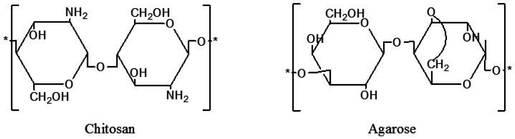
:1. Introduction
2. Results and Discussion
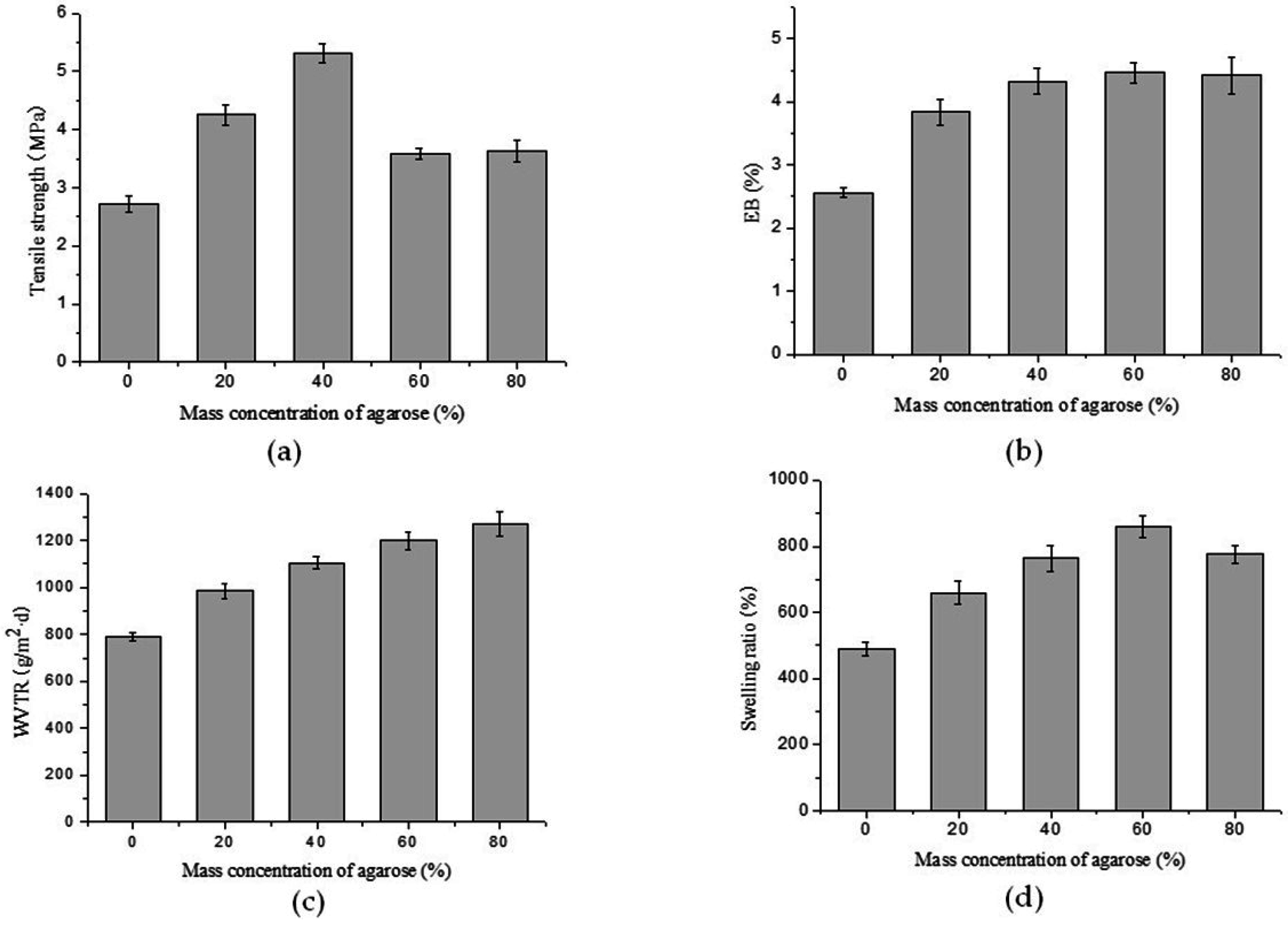
2.1. Mechanical Properties
2.2. Water Vapor Transmission Rate (WVTR)
2.3. Swelling Test
2.4. Antibacterial Properties
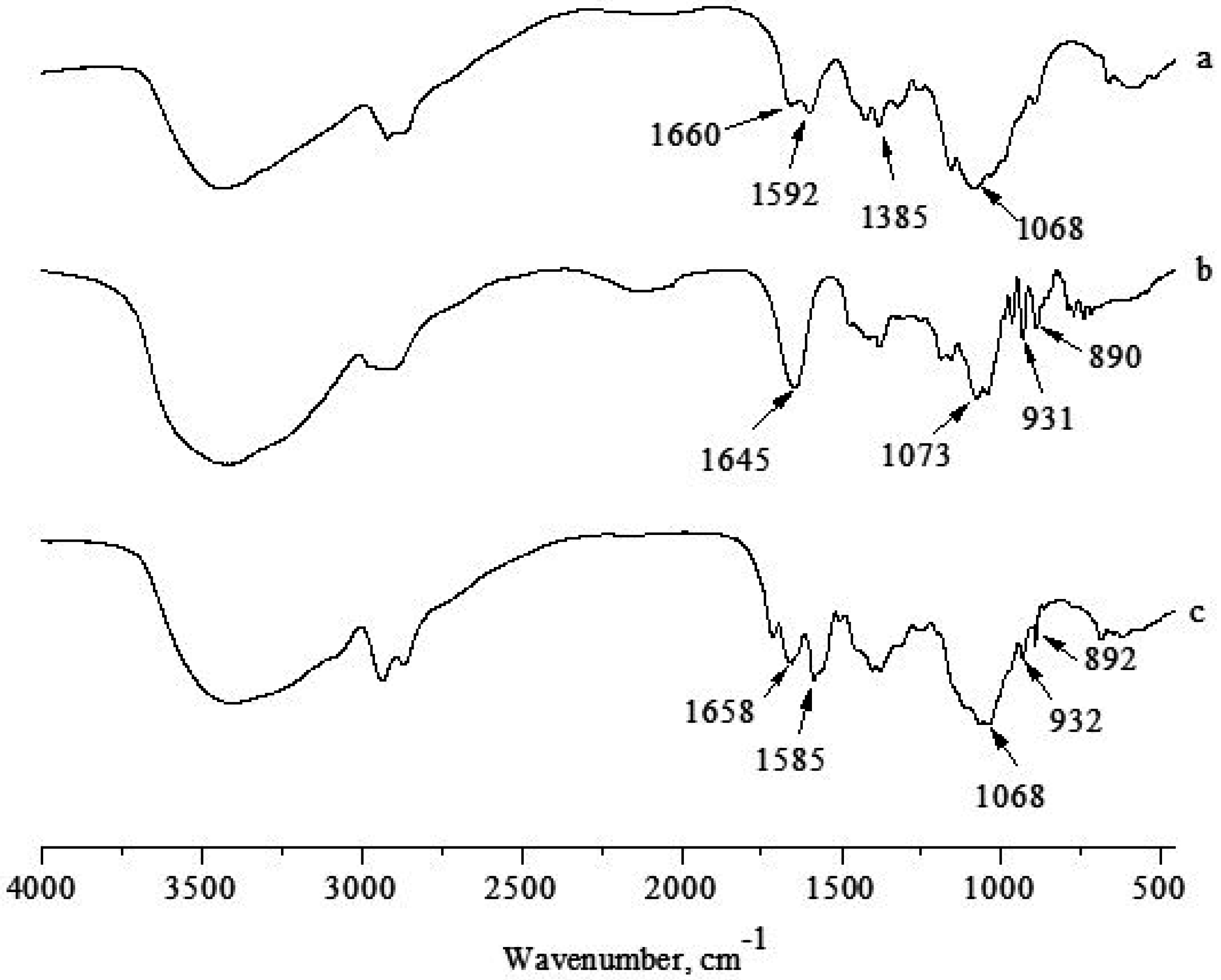
2.5. FTIR-ATR Spectroscopy
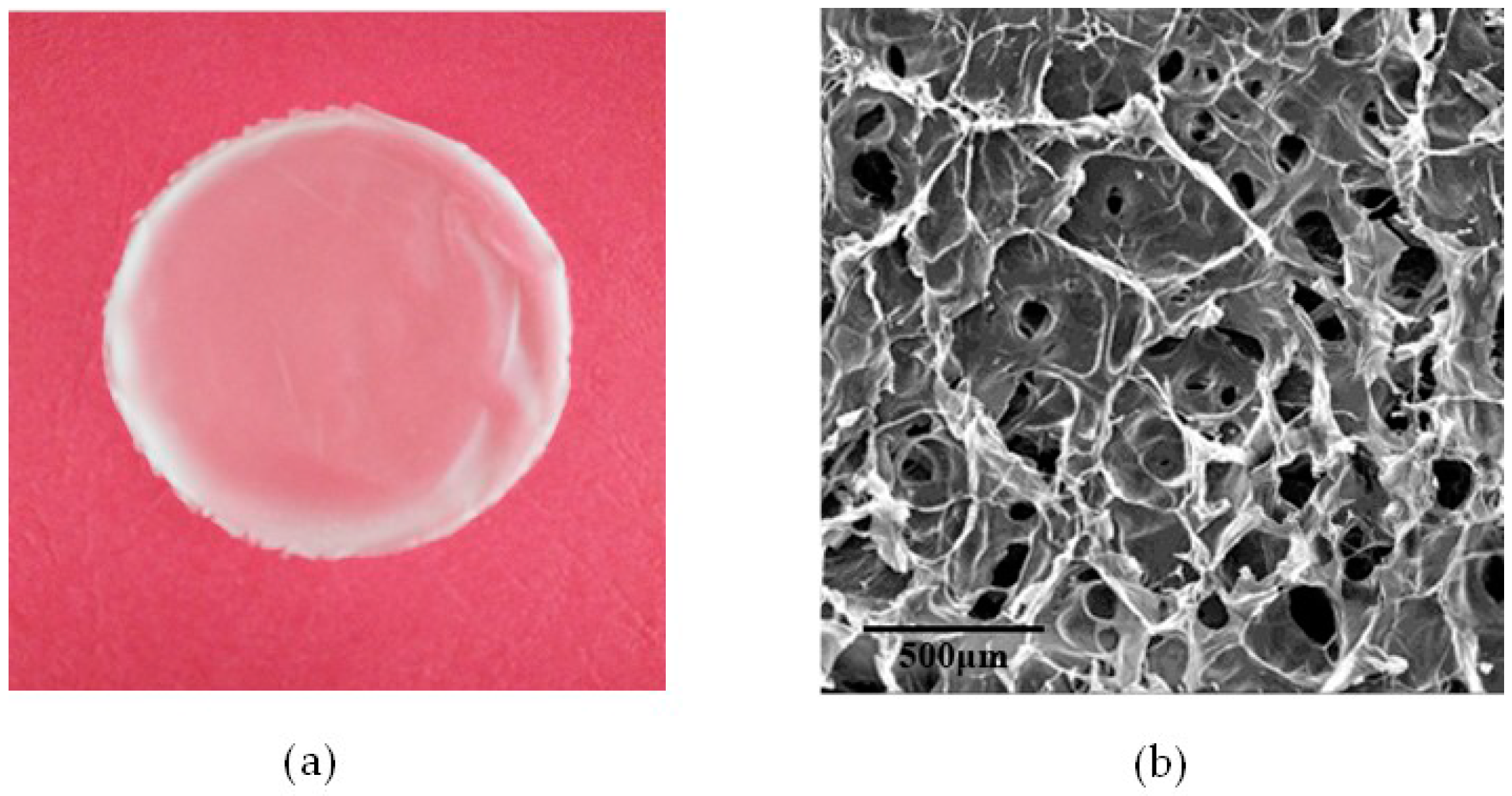
2.6. Morphology Studies
3. Materials and Methods
3.1. Materials
3.2. Film Preparation
3.3. Mechanical Properties
3.4. Water Vapor Transmission Rate (WVTR)
3.5. Swelling Test
3.6. Antibacterial Test
3.7. FTIR Analysis
3.8. Morphology Studies
4. Conclusions
Acknowledgments
Author Contributions
Conflicts of Interest
References
- Dutta, P.K.; Tripathi, S.; Mehrotra, G.K.; Dutta, J. Perspectives for chitosan based antimicrobial films in food applications. Food Chem. 2009, 114, 1173–1182. [Google Scholar] [CrossRef]
- Leceta, I.; Guerrero, P.; Caba, K.D.L. Functional properties of chitosan-based films. Carbohydr. Polym. 2013, 93, 339–346. [Google Scholar] [CrossRef] [PubMed]
- Murray, C.A.; Dutcher, J.R. Effect of changes in relative humidity and temperature on ultrathin chitosan films. Biomacromolecules 2006, 7, 3460–3465. [Google Scholar] [CrossRef] [PubMed]
- Ramya, R.; Venkatesan, J.; Kim, S.K.; Sudha, P.N. Biomedical applications of chitosan: An overview. J. Biomater. Tissue Eng. 2012, 2, 100–111. [Google Scholar] [CrossRef]
- Ravi Kumar, M.N.V.; Muzzarelli, R.A.A.; Muzzarelli, C.; Sashiwa, H.; Domb, A.J. Chitosan chemistry and pharmaceutical perspectives. Chem. Rev. 2004, 104, 6017–6084. [Google Scholar] [CrossRef] [PubMed]
- Agulló, E.; Rodríguez, M.S.; Ramos, V.; Albertengo, L. Present and future role of chitin and chitosan in food. Macromol. Biosci. 2003, 3, 521–530. [Google Scholar] [CrossRef]
- Chien, P.J.; Sheu, F.; Yang, F.H. Effects of edible chitosan coating on quality and shelf life of sliced mango fruit. J. Food Eng. 2007, 78, 225–229. [Google Scholar] [CrossRef]
- Jeon, Y.J.; Park, P.J.; Kim, S.K. Antimicrobial effect of chitooligosaccharides produced by bioreactor. Carbohydr. Polym. 2001, 44, 71–76. [Google Scholar] [CrossRef]
- No, H.K.; Park, N.Y.; Lee, S.H.; Meyers, S.P. Antibacterial activity of chitosans and chitosan oligomers with different molecular weights. Int. J. Food Microbiol. 2002, 74, 65–72. [Google Scholar] [CrossRef]
- Kong, M.; Chen, X.G.; Xing, K.; Park, H.J. Antimicrobial properties of chitosan and mode of action: A state of the art review. Int. J. Food Microbiol. 2010, 144, 51–63. [Google Scholar] [CrossRef] [PubMed]
- Deszczynski, M.; Kasapis, S.; Mitchelle, J.R. Rheological investigation of the structural properties and aging effects in the agarose/co-solute mixture. Carbohydr. Polym. 2003, 53, 85–93. [Google Scholar] [CrossRef]
- Lahooti, S.; Sefton, M.V. Effect of an immobilization matrix and capsule membrane permeability on the viability of encapsulated HEK cells. Biomaterials 2000, 21, 987–995. [Google Scholar] [CrossRef]
- Watase, M.; Nishinari, K.; Clark, A.H.; Ross-Murphy, S.B. Differential scanning calorimetry, rheology, X-ray, and NMR of very concentrated agarose gels. Macromolecules 1989, 22, 1196–1201. [Google Scholar] [CrossRef]
- Shih, C.M.; Shieh, Y.T.; Twu, Y.K. Preparation and characterization of cellulose/chitosan blend films. Carbohydr. Polym. 2009, 78, 169–174. [Google Scholar] [CrossRef]
- Ke, G.; Xu, W.; Yu, W. Preparation and properties of drug-loaded chitosan–sodium alginate complex membrane. Int. J. Polym. Mater. 2010, 59, 184–191. [Google Scholar] [CrossRef]
- Liu, F.; Qin, B.; He, L.; Song, R. Novel starch/chitosan blending membrane: Antibacterial, permeable and mechanical properties. Carbohydr. Polym. 2009, 78, 146–150. [Google Scholar] [CrossRef]
- Da Silva, M.A.; Krause, A.C.; Kieckbush, T.G. Alginate and pectin composite films crosslinked with Ca2+ ions: Effect of the plasticizer concentration. Carbohydr. Polym. 2009, 77, 736–742. [Google Scholar] [CrossRef]
- Hu, Z.; Li, S.; Yang, L. Preparation of berbamine loaded chitosan-agarose microspheres and in vitro release study. Polímeros 2012, 22, 422–426. [Google Scholar] [CrossRef]
- Rao, M.S.; Kanatt, S.R.; Chawla, S.P.; Sharma, A. Chitosan and guar gum composite films: Preparation, physical, mechanical and antimicrobial properties. Carbohydr. Polym. 2010, 82, 1243–1247. [Google Scholar] [CrossRef]
- Martínez-Camacho, A.P.; Cortez-Rocha, M.O.; Ezquerra-Brauer, J.M.; Graciano-Verdugo, A.Z.; Rodriguez-Félix, F.; Castillo-Ortega, M.M.; Yépiz-Gómez, M.S.; Plascencia-Jatomea, M. Chitosan composite films: Thermal, structural, mechanical and antifungal properties. Carbohydr. Polym. 2010, 82, 305–315. [Google Scholar] [CrossRef]
- Bhat, S.; Tripathi, A.; Kumar, A. Supermacroprous chitosan–agarose–gelatin cryogels: In vitro characterization and in vivo assessment for cartilage tissue engineering. J. R. Soc. Interface 2011, 8, 540–554. [Google Scholar] [CrossRef] [PubMed]
- Elsabee, M.Z.; Abdou, E.S. Chitosan based edible films and coatings: A review. Mater. Sci. Eng. C 2013, 33, 1819–1841. [Google Scholar] [CrossRef] [PubMed]
- Ziani, K.; Oses, J.; Coma, V.; Maté, J.I. Effect of the presence of glycerol and Tween 20 on the chemical and physical properties of films based on chitosan with different degree of deacetylation. LWT-Food Sci. Technol. 2008, 41, 2159–2165. [Google Scholar] [CrossRef]
- Hu, Z.; Hong, P.; Li, S.; Yang, L.; Xie, J. Study on the preparation of quaternized chitosan/agarose microspheres for berbamine delivery. J. Mater. Sci. 2012, 29, 459–464. [Google Scholar]
- Arzate-Vázquez, I.; Chanona-Pérez, J.J.; Calderón-Domínguez, G.; Terres-Rojas, E.; Garibay-Febles, V.; Martínez-Rivas, A.; Gutiérrez-López, G.F. Microstructural characterization of chitosan and alginate films by microscopy techniques and texture image analysis. Carbohydr. Polym. 2012, 87, 289–299. [Google Scholar] [CrossRef]
- Bueno, C.Z.; Dias, A.M.A.; Cipriano de Sousa, H.J.; Braga, E.M.; Moraes, Â.M. Control of the properties of porous chitosan–alginate membranes through the addition of different proportions of Pluronic F68. Mater. Sci. Eng. C 2014, 44, 117–125. [Google Scholar] [CrossRef] [PubMed]
- Zhang, L.; Guo, J.; Peng, X.; Jin, Y. Preparation and release behavior of carboxymethylated chitosan/alginate microspheres encapsulating bovine serum albumin. J. Appl. Polym. Sci. 2004, 92, 878–882. [Google Scholar] [CrossRef]


| Films with Agarose Mass Concentrations (%) | Areas of the Antibacterial Zone (A, mm2) | |
|---|---|---|
| S. aureus | E. coli | |
| 0 | 89.29 ± 1.23 | 83.83 ± 0.95 |
| 20 | 78.89 ± 0.89 | 51.52 ± 2.11 |
| 40 | 71.64 ± 2.06 | 37.38 ± 1.58 |
| 60 | 70.56 ± 1.55 | 30.43 ± 0.73 |
| 80 | 22.09 ± 1.17 | 13.91 ± 1.35 |
© 2016 by the authors; licensee MDPI, Basel, Switzerland. This article is an open access article distributed under the terms and conditions of the Creative Commons Attribution (CC-BY) license (http://creativecommons.org/licenses/by/4.0/).
Share and Cite
Hu, Z.; Hong, P.; Liao, M.; Kong, S.; Huang, N.; Ou, C.; Li, S. Preparation and Characterization of Chitosan—Agarose Composite Films. Materials 2016, 9, 816. https://doi.org/10.3390/ma9100816
Hu Z, Hong P, Liao M, Kong S, Huang N, Ou C, Li S. Preparation and Characterization of Chitosan—Agarose Composite Films. Materials. 2016; 9(10):816. https://doi.org/10.3390/ma9100816
Chicago/Turabian StyleHu, Zhang, Pengzhi Hong, Mingneng Liao, Songzhi Kong, Na Huang, Chunyan Ou, and Sidong Li. 2016. "Preparation and Characterization of Chitosan—Agarose Composite Films" Materials 9, no. 10: 816. https://doi.org/10.3390/ma9100816