Article

The π-Electron Delocalization in 2-Oxazolines Revisited: Quantification and Comparison with Its Analogue in Esters †

1
Polymer Competence Center Leoben, Roseggerstrasse 12, 8700 Leoben, Austria
2
Institute for Chemistry and Technology of Materials, Graz University of Technology, NAWI Graz, Stremayrgasse 9, 8010 Graz, Austria
3
Institute of Inorganic Chemistry, Graz University of Technology, NAWI Graz, Stremayrgasse 9, 8010 Graz, Austria
4
MTA-BME Lendület Quantum Chemistry Research Group, Department of Physical Chemistry and Materials Science, Budapest University of Technology and Economics, P.O. Box 91, 1521 Budapest, Hungary
*
Author to whom correspondence should be addressed.
This publication is dedicated to Professor Hubert Schmidbaur on the occasion of his 80th birthday.
These authors contributed equally to this work.
Materials 2015, 8(8), 5385-5397; https://doi.org/10.3390/ma8085249
Submission received: 24 June 2015 / Accepted: 13 August 2015 / Published: 21 August 2015

Abstract

:
The single crystal X-ray analysis of the ester-functionalized 2-oxazoline, methyl 3-(4,5-dihydrooxazol-2-yl)propanoate, revealed π-electron delocalization along the N–C–O segment in the 2-oxazoline pentacycle to significant extent, which is comparable to its counterpart along the O–C–O segment in the ester. Quantum chemical calculations based on the experimental X-ray geometry of the molecule supported the conjecture that the N–C–O segment has a delocalized electronic structure similar to an ester group. The calculated bond orders were 1.97 and 1.10 for the N=C and C–O bonds, and the computed partial charges for the nitrogen and oxygen atoms of −0.43 and −0.44 were almost identical. In the ester group, the bond orders were 1.94 and 1.18 for the C–O bonds, while the partial charges of the oxygen atom are −0.49 and −0.41, which demonstrates the similar electronic structure of the N–C–O and O–C–O segments. In 2-oxazolines, despite the higher electronegativity of the oxygen atom (compared to the nitrogen atom), the charges of the hetero atoms oxygen and nitrogen are equalized due to the delocalization, and it also means that a cationic attack on the nitrogen is possible, enabling regioselectivity during the initiation of the cationic ring-opening polymerization of 2-oxazoline monomers, which is a prerequisite for the synthesis of materials with well-defined structures.

Graphical Abstract

1. Introduction

Since the introduction of microwave reactors dedicatedly designed for usage in laboratories, considering the chemists’ and material scientists’ requirements [1,2,3,4], the class of poly(2-oxazoline)s has been reawakened from its hibernation (Scheme 1): discovered in 1966/67 by four research groups almost simultaneously [5,6,7,8], the versatility of this class of polymers, and its derived materials, has been under constant investigation, while the commonly-low polymerization rates have been the bottleneck for a long time in terms of applicability of these materials. With the exclusion of acceleration-limiting factors such as temperatures (boiling points), microwave reactors have helped to successfully accelerate the polymerizations of 2-oxazolines, and, currently, they are under thorough investigation, in particular, for usage in medical and sanitary applications [9,10,11,12,13,14,15] and as cross-linkable materials [16,17,18,19,20,21,22].
In particular for medical and medicinal applications, a precise knowledge of the materials’ structures is of key importance, which makes living or at least pseudo-living polymerizations and their inherent access to polymers with narrow molecular weight distributions favorite synthetic strategies. For the polymerization of 2-oxazolines, it has been shown that the highly reactive methyl tosylate is one of the initiators that can start pseudo-living polymerizations [23,24,25]. Due to its high reactivity, it has been argued whether the initiation by methyl cations occurs regioselectively at the nitrogen atom (Scheme 1) when the polymerization times experience accelerations by a factor of up to 400 [26].
Scheme 1. Methyl tosylate-initiated polymerization of 2-oxazolines.
Materials 08 05249 g005
In a precedent study [27], we could show that, due to π-electron delocalization, the partial negative charge at the oxygen atom of the 2-oxazoline ring is lessened (Scheme 2). The negative charge of the nitrogen atom, on the other hand, is enhanced and, hence, the nitrogen atom is an ideal reaction partner for the methyl tosylate. In order to expand the understanding of this π-electron delocalization in 2-oxazolines, we aimed for a correlation/comparison with its counterparts in esters, where the C–O “single” bond as well has been reported to show an intermediate value between that of a C–O double and single bond. In this study, we therefore present the single crystal X-ray analysis of an ester-functionalized 2-oxazoline and the corresponding ring-opened ester-functionalized amino acid.
Scheme 2. Delocalization of π-electrons in 2-oxazolines (left) and esters (right).
Materials 08 05249 g006

2. Experimental Section

2.1. Materials

All chemicals were used as received. Chloroethylamine hydrochloride, hydrochloric acid solution (0.1 mol/L), methyl chlorobutyrate, triethylamin, and calcium carbonate were purchased from Sigma Aldrich (Vienna, Austria), while ethanol, methanol, dichloromethane, and sodium hydroxide were bought from Carl Roth (Karlsruhe, Germany).

2.2. Instrumentation

IR spectra were recorded with 48 scans per sample on a Bruker Alpha FT-IR spectrometer (Bruker Optics Inc., Billerica, MA, USA) equipped with the ALPHA’s Platinum attenuated total reflection (ATR) single reflection diamond ATR module. The spectral range was set from 500 to 4000 cm−1. 1H NMR spectra were measured in deuterated chloroform or deuterium dioxide on a Bruker 300 MHz spectrometer (Bruker BioSpin Corporation, Billerica, MA, USA) with 32 scans and relaxation delays of 5 s. The solvent residual peaks were used for referencing the spectra to 7.26 ppm and 4.80 ppm, respectively.

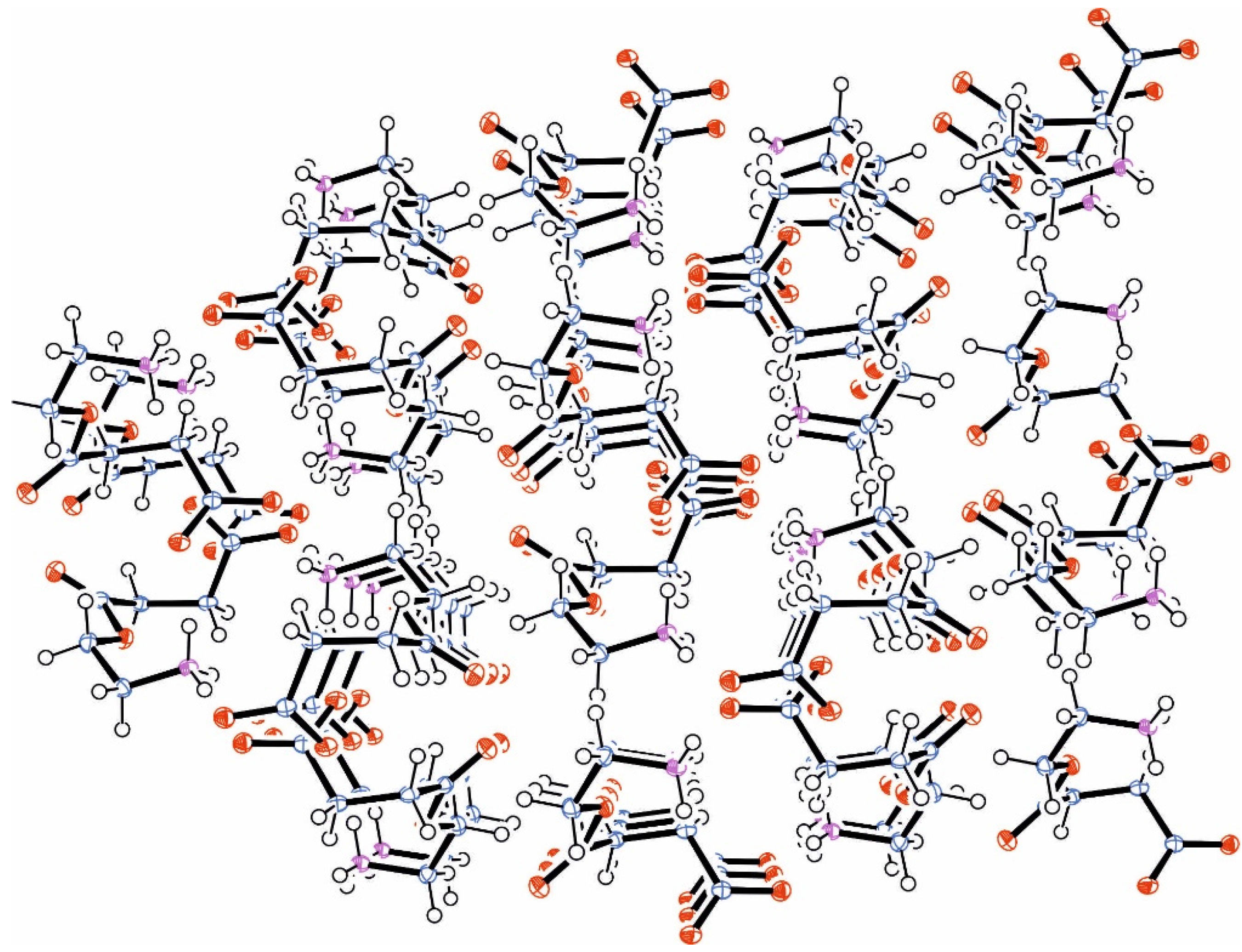
2.3. Single Crystal X-ray Diffraction Analyses

The crystalline samples were placed in inert oil, mounted on a glass pin and transferred to the cold gas stream of the diffractometer. Crystal data were collected and integrated with a Bruker APEX-II CCD system (Bruker AXS GmbH, Karlsruhe, Germany) with monochromated Mo-Kα (λ = 0.71073 Å) radiation at 100(2) K. The structures were solved by direct methods using SHELXS-97 [28] and refined by full matrix least squares calculations on F2 with SHELXL-97 [29]. The space group assignments and structural solutions were evaluated using PLATON [30]. Non-H-atoms were refined with anisotropic thermal parameters. All protons located on carbon atoms were calculated and allowed to ride on their parent atoms with fixed isotropic contributions; protons on nitrogen atoms were located and refined with isotropic contributions. Extinction corrections were applied for all compounds using SADABS [31]. A summary of the crystal data, experimental details and refinement results is listed in Table 1. Important interatomic distances and angles are given in the figure captions. Thermal parameters and complete tables of interatomic distances and angles have been deposited with the Cambridge Crystallographic Data Centre, 12 Union Road, Cambridge CB2 1 EZ, UK. The data are available on request on quoting CCDS-1418758/1418759 and in the Supporting Information.
Table 1. Crystal data, data collection, absorption and structure refinement of methyl 3-(4,5-dihydrooxazol-2-yl)propanoate EstOx (ester-functionalized 2-oxazoline) and 4-(2-aminoethoxy)-4-oxobutanoic acid EstAA (ester-functionalized amino acid).
Crystal Structure AnalysisMethyl 3-(4,5-dihydrooxazol-2-yl) Propanoate, EstOx4-(2-Aminoethoxy)-4-oxo-butanoic Acid, EstAA
Crystal data
CCDC No.14187581418759
Crystal formblockblock
Crystal colourcolorlesscolorless
Empirical formulaC7H11NO3C6H11NO4
Formula weight157.17161.16
Crystal systemmonoclinicmonoclinic
Space groupP21P21/c
a (Å)5.547 (2)10.3132 (8)
b (Å)6.765 (3)9.0304 (7)
c (Å)9.993 (4)8.0012 (7)
α (°)9090
β (°)91.583 (13)96.688 (5)
γ (°)9090
V3)374.9 (3)740.10 (10)
ρcalc (g·cm−3)1.2681.446
Z24
F(0 0 0)152344
µ (Mo-Kα) (cm−1)0.1020.122
Data Collection
Measured reflections45591786
Unique reflections12781786
Rint0.06590.000
Absorption
Tmin/Tmax0.9690/0.98080.9574/0.9855
Refinement Results
Refined parameters101101
R1 a,b, wR2 a;c0.0530; 0.13520.0630; 0.1432
a, b0.0887; 0.04570,000; 2.2816
ρ (e·Å−3)0.306; −0.2190.362; −0.325
a I > 2σ(I); b R1 = Σ(||Fo| − |Fc||/Σ|Fo|; c wR2 = {[Σw(Fo2Fc2)2]/Σ[w(Fo2)2]}0.5; w = 1/[σ2(Fo2) + (ap)2 + bp]; p = (Fo2 + 2Fc2)/3; a and b: free variables.

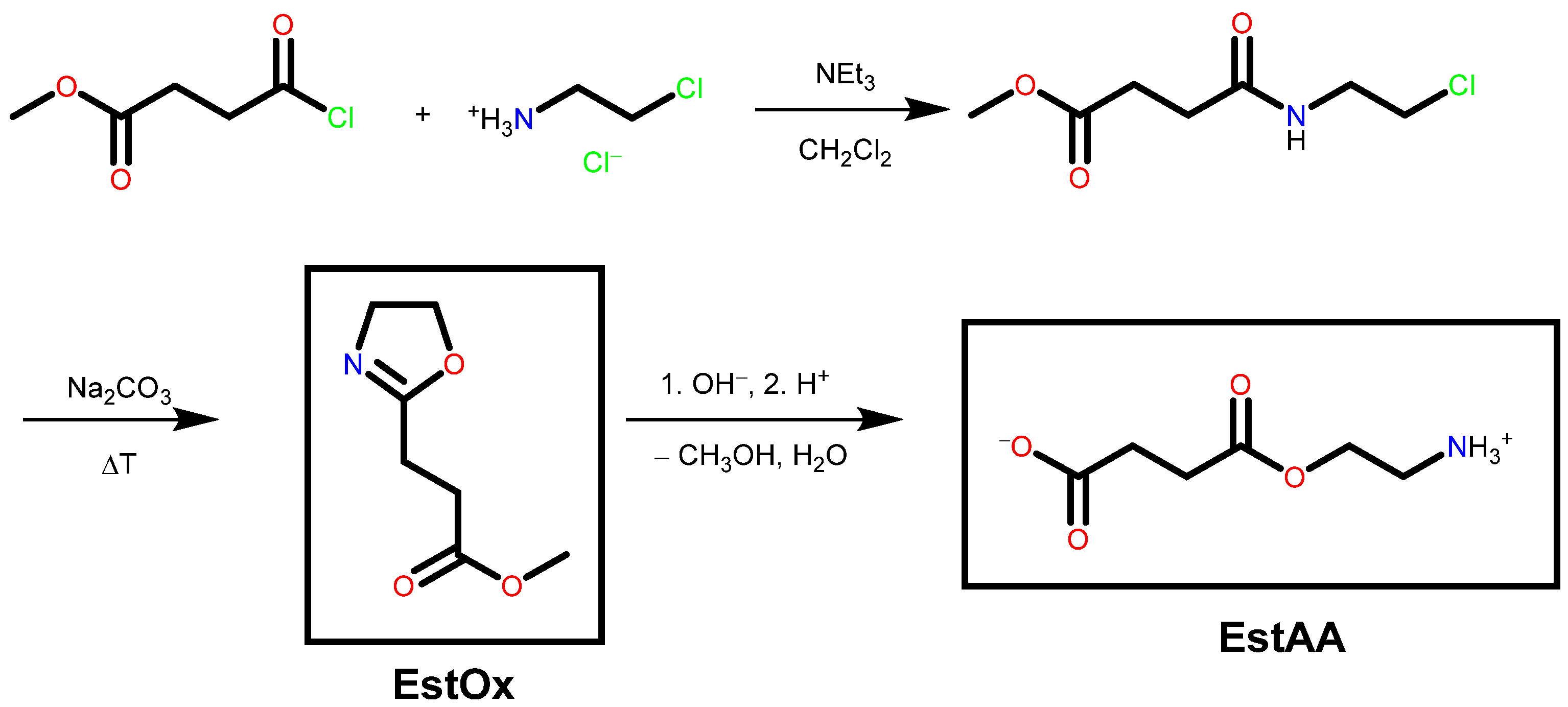
2.4. Preparation of Methyl 3-(4,5-Dihydrooxazol-2-yl)propanoate EstOx

Ten millilitres (0.13 mol) of methyl 4-chloro-4-oxobutanoate and 15.41 g (0.13 mol) of chloroethylamine hydrochloride were dissolved in 130 mL of dichloromethane under inert conditions and cooled to 0 °C. Forty-one millilitres of triethylamine, dissolved in 20 mL of dichloromethane, were added dropwise within 1 h, and the reaction mixture was stirred overnight. The organic phase was extracted twice with deionized water and once with brine, prior to drying with sodium sulphate. Subsequently, the solvent was removed under reduced pressure. 17.40 g of the dry intermediate product were recovered (0.09 mol, 69% yield). 9.537 g (1 equiv.) of sodium carbonate were added and the mixture was stirred overnight under reduced pressure. The mixture was subsequently filtrated. 7.11 g (0.063 mol, 48% yield) of the final product were recovered as colorless liquid by distillation under reduced pressure. The product crystallized at 6 °C.
NMR (300 MHz, CDCl3): δ (ppm) = 2.55 (2 H, t, 3JH–H = 6.6 Hz, H4a and H4b), 2.64 (2 H, t, 3JH–H = 6.6 Hz, H5a and H5b), 3.67 (3 H, s, H7a, H7b and H7c), 3.78 (2 H, t, 3JH–H = 9.3 Hz, H3a and H3b), 4.21 (2 H, t, 3JH–H = 9.3 Hz, H2a and H2b) (for atomic labelling, see Figure 1).
IR (ATR, cm−1): ν = 2985 m, 2948 m, 2906 m, 2884 m, νstr (CH); 1734 s, νstr (C=O); 1669 s, νstr (N=C); 1438 m, νstr (CO); 1364 m, 1350 m, νdef (CH3); 1204 m, 1161 s, νasym str (COCH3); 657 w, νbend (COCH3); 583 w, νdef (CH2COCH3).

2.5. Preparation of 4-(2-Aminoethoxy)-4-oxobutanoic Acid EstAA

A solution of 0.5 g (0.0032 mol, 1 equiv.) of EstOx in 50 mL of methanol and 32 mL of an aqueous solution of sodium hydroxide (0.1 mol/L) were mixed and stirred for 1 h. The solvents were subsequently removed under reduced pressure. The crude product was dissolved in methanol, and 0.0032 mol of hydrochloric acid (aqueous 0.1 M solution) were added. After 10 min of stirring under reflux conditions, the mixture was stored overnight at 6 °C. The solvents were removed under reduced pressure, and 0.467 g (0.0029 mol, 91% yield) of the final product were recovered by recrystallization from ethanol.
1H NMR (20 °C, CDCl3, 300 MHz): δ (ppm) = 2.50 (2 H, t, 3JH–H = 6.5 Hz, H3a and H3b), 2.66 (2 H, t, 3JH–H = 6.5 Hz, H2a and H2b), 3.34 (2 H, t, 3JH–H = 5.1 Hz, H6a and H6b), 4.38 (2 H, t, 3JH–H = 5.1 Hz, H5a and H5b) (for atomic labelling, see Figure 3).
IR (ATR, cm−1): ν = 3438 m, νstr (NH2); 2986 w, 2961 w, 2925 w, 2848 w, νstr (CH); 2524 m νstr (OH); 1729 s, νstr (C=O); 1611 s, 1571 s, νdef (NH2); 1312 s, νdef (OH); 1248 s, 1155 s, νasym str (COC); 1013 s, νstr(NH2); 957 m, ν(COOH).

3. Results and Discussion

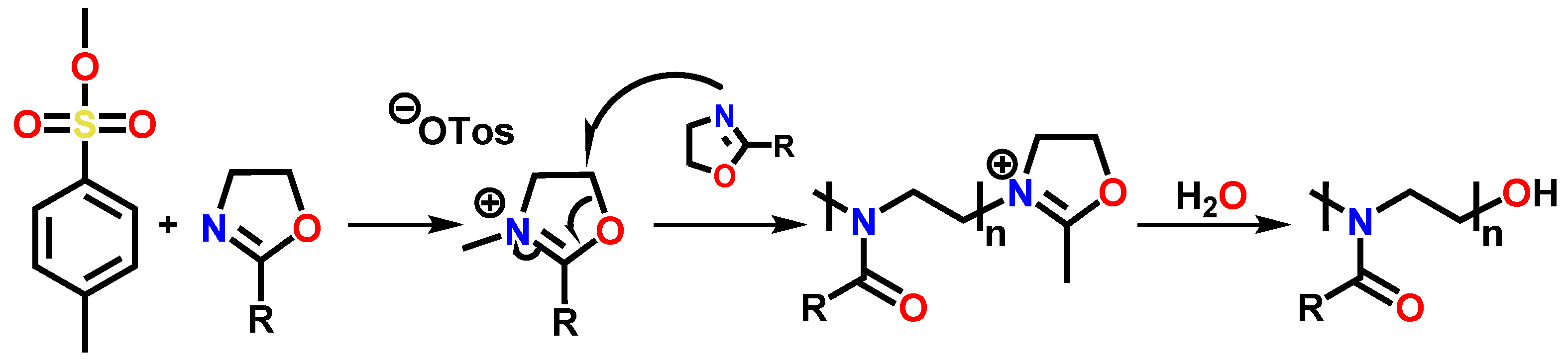
3.1. Synthesis of the Compounds EstOx and EstAA

Methyl 3-(4,5-dihydrooxazol-2-yl)propanoate EstOx can be prepared from the reaction of methyl 4-chloro-4-oxobutanoate and chloroethylamine hydrochloride and subsequent ring-closure under alkaline conditions (Scheme 3, top), following a literature protocol [32]. Hydrolyses of the ester bond and the 2-oxazoline ring yield the ester-functionalized amino acid 4-(2-aminoethoxy)-4-oxobutanoic acid EstAA (Scheme 3, bottom). Single crystals from both compounds were grown at 6 °C.
Scheme 3. Synthesis of EstOx from methyl 4-chloro-4-oxobutanoate and chloroethylamine hydrochloride (top) as well as hydrolyses of the 2-oxazoline under alkaline and acidic conditions yielding EstAA (bottom).
Materials 08 05249 g007

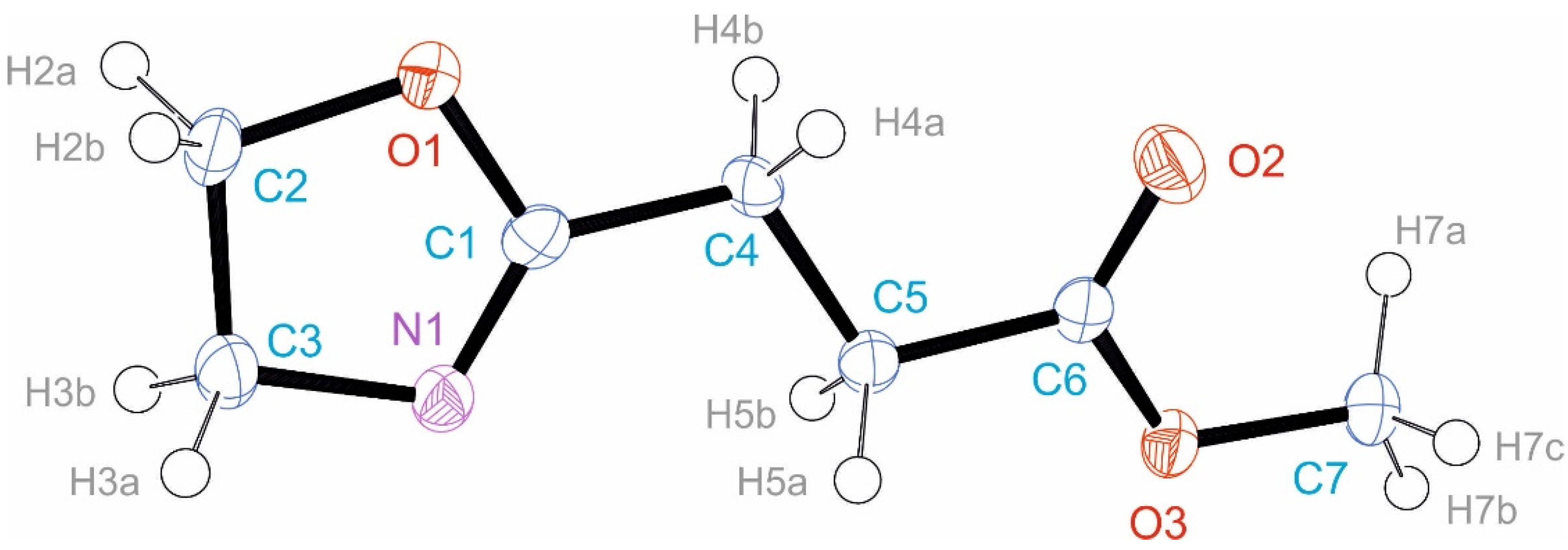
3.2. Crystal Structure of EstOx

EstOx crystallizes in the monoclinic space group P21 with Z = 2 formula units in the unit cell. The asymmetric unit contains 1 formula unit (Figure 1). A detailed analysis of the dihedral angles reveals that the 2-oxazoline C3N1O1-pentacycle is almost planar [O1–C2–C3–N1: −4.2(3)°, N1–C1–O1–C2: −1.8(4)°, C3–C2–O1–C1: 3.6(3)°, O1–C1–N1–C3: −1.1(4)°, C2–C3–N1–C1: 3.3(3)°], and the carbon atoms of the side-chain are in trans alignment [C1–C4–C5–C6: −174.0(2)°]. Minor deviations of the overall trans alignment can only be observed around the ester group [C4–C5–C6–O2: 12.7(4)°, C4–C5–C6–O3: −168.4(2)°, C7–O3–C6–C5: −178.4(2)°].
Figure 1. Asymmetric unit in the crystalline structure of EstOx (ORTEP drawing [33] with 50% probability ellipsoids). Selected bond lengths (Å): C1–N1: 1.263(4), C1–O1: 1.376(3), C2–O1: 1.458(3) C3–N1: 1.483(4), C2–C3: 1.529(4), C1–C4: 1.497(3), C4–C5: 1.515(4), C5–C6: 1.507(3), C6–O2: 1.211(3), C6–O3: 1.338(3), C7–O3: 1.448(3). Selected bond angles (°): N1–C1–O1: 118.6(2), N1–C1–C4: 128.9(2), O1–C1–C4: 112.5(2), O1–C2–C3: 104.0(2), N1–C3–C2: 105.1(2).
Materials 08 05249 g001
Notably, like in the crystalline structures of 2-phenyl-2-oxazoline, 2-nnonyl-2-oxazoline and 2,2'-tetramethylenebis(2-oxazoline) [27], the two C–O bonds in the 2-oxazoline pentacycle differ significantly: While the C2–O1 bond with a length of 1.458(3) Å has the expected length of a C–O single bond [34,35], the C1–O1 bond with a length of 1.376(3) Å is significantly shorter and exhibits a value intermediate between the expected bond lengths of a C–O single and double bond. This phenomenon can be explained by the delocalization the π-electrons along the N–C–O segment of the C3N1O1-pentacycle. Along the O–C–O segment of the ester group of the side-chain, comparable delocalization can be observed: while the O2–C6 bond with a length of 1.211(3) exhibits a bond length typical for a C=O double bond, the O3–C6 bond with a length of 1.338(3) Å shows a value intermediate between the expected bond lengths of a C–O single and double bond.
Hence, the lengths of the C1–O1 bond and the O3–C6 bond are of very comparable value. While the potential (hetero) keto-enol tautomerism of the ester bond cannot be elucidated from the C–C bond lengths [C4–C5: 1.515(4) Å, C5–C6: 1.507(3) Å], it can be stated that the extent of π-electron delocalization along the N–C–O segment in 2-oxazolines is very comparable to that along the O–C–O segment in esters. The π-electron delocalization in esters is less pronounced than in amides (Scheme 2), but nonetheless significant: In 2-oxazolines, it renders the partial charge of the oxygen atom less negative, and the partial negative charge of the nitrogen atom more negative.
Packing of the EstOx molecules in the crystalline phase seems to be controlled by steric factors only (Figure 2): The EstOx molecules are aligned in parallel fashion, with molecule-to-molecule distances of 5.547 Å; for comparison: Distances of adjacent molecules of 2,2'-tetramethylenebis(2-oxazoline) in the crystalline phase (that showed a packaging very similar to that of EstOx) exhibited a value of 5.084 Å [27].
Figure 2. Arrangement of EstOx molecules in the crystalline phase.
Materials 08 05249 g002

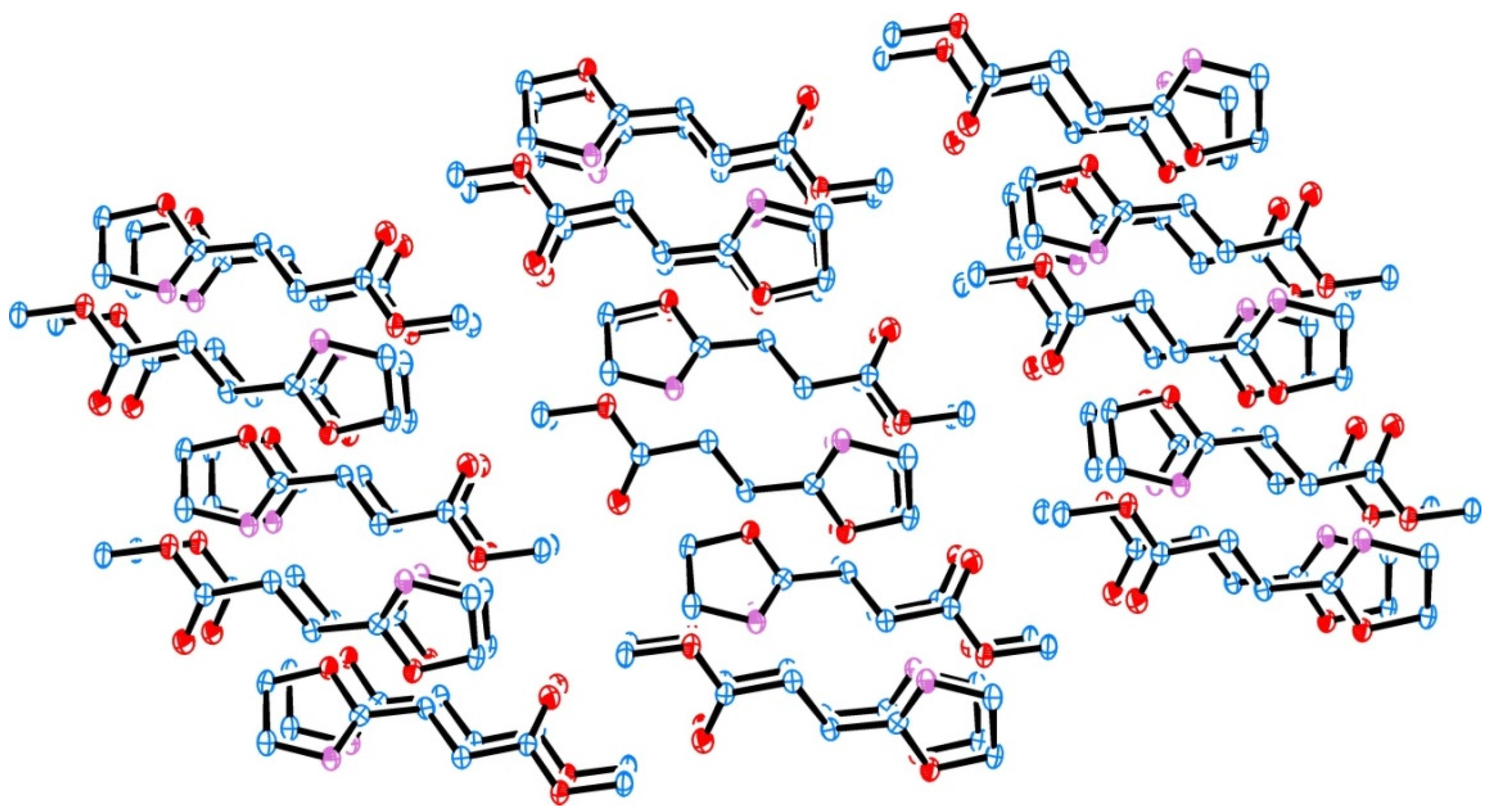
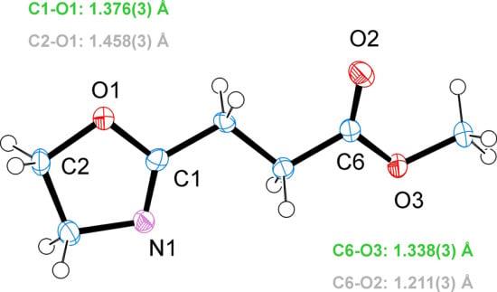
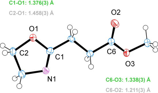
3.3. Crystal Structure of EstAA
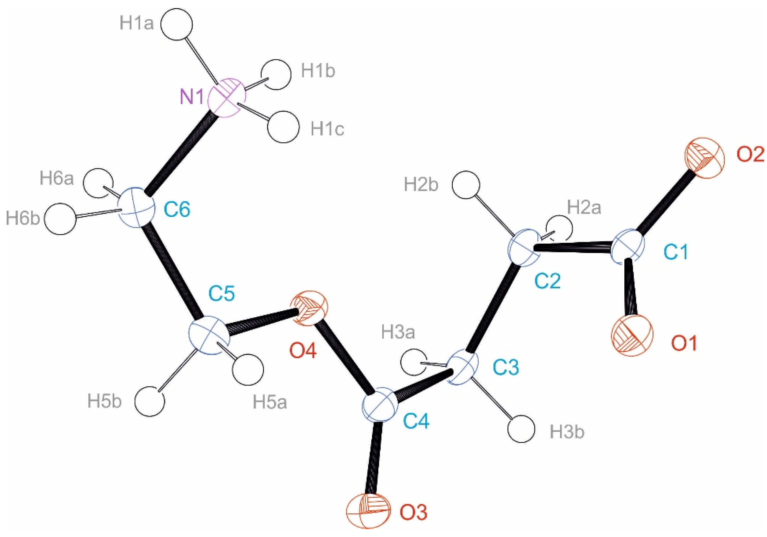
EstAA crystallizes in its zwitterionic form in the monoclinic space group P21/c with Z = 4 formula units in the unit cell. The asymmetric unit contains 1 formula unit (Figure 3). A detailed analysis of the dihedral angles shows that the C6–C5–O4–C4(O3)–C3 segment of the formula unit is in trans alignment [C4–O4–C5–C6: 179.6(2)°, C5–O4–C4–C3: 176.2(2)°, C5–O4–C4–O3: 0.2(4)°, while the ammonium group and the C2–C1(O1O2) segment deviate from that alignment [C1–C2–C3–C4: 68.7(3)°, O4–C5–C6–N1: −57.6(3)°]. In addition to its ester group with C–O bond lengths of 1.205(4) and 1.354(3) Å (which are almost identical to the C–O ester bond lengths of 1.211(3) and 1.338(3) Å in EstOx), EstAA also contains a carboxylate group that exhibits C–O bond lengths of 1.251(3) and 1.268(3) Å, which correspond in close proximity to the bond lengths of C–O double bonds [34,35]. The very minor difference among the two C–O bond lengths in the carboxylate group is assumed to originate from a different involvement of the oxygen atoms O1 and O2 atoms in the formation of hydrogen bonds (Table 2).
Figure 3. Asymmetric unit in the crystalline structure of EstAA (ORTEP drawing [33] with 50% probability ellipsoids). Selected bond lengths (Å): C1–O1: 1.251(3), C1–O2: 1.268(3), C4–O3: 1.205(4), C4–O4:1.354(3), C5–O4: 1.447(3), C6–N1: 1.489(3). Selected bond angles (°): O1–C1–C2 118.1(2), O3–C4–O4: 122.8(3), C1–C2–C3: 113.2(2).
Materials 08 05249 g003
Table 2. Hydrogen bonds in the crystalline phase of EstAA.
Atom NamesBond Length N–HDistance H···ODistance N···OAngle NHO
N1–H1A···O2 A0.91 Å1.84 Å2.739(3) Å171.1°
N1–H1B··O2 B0.91 Å1.86 Å2.741(3) Å162.7°
N1–H1C··O1 C0.91 Å1.84 Å2.747(3) Å172.2°
A,B,C: Symmetry operations used to generate equivalent atoms; A: −x + 2, y − 0.5, −z + 1.5; B: x, −y + 1.5, z − 0.5; C: −x + 2, −y + 1, −z + 2.
All acidic protons, namely the protons of the ammonium group, are involved in hydrogen bonds jointly with the oxygen atoms of the carboxylate group. The hydrogen bonds are likely to cause the deviation of the ammonium group and the carboxylate group from the overall trans alignment of the carbon chain of EstAA. The oxygen atoms of the ester group do not participate in the formation of hydrogen bonds. Correspondingly, packing of the EstAA molecules in the crystalline phase (Figure 4) is controlled by the formation of hydrogen bonds and large molecule-to-molecule distances of 8.001 Å.
Figure 4. Arrangement of EstAA molecules in the crystalline phase. The orientation of the ammonium groups towards the carboxylate groups is indicative of hydrogen bond formation.
Materials 08 05249 g004

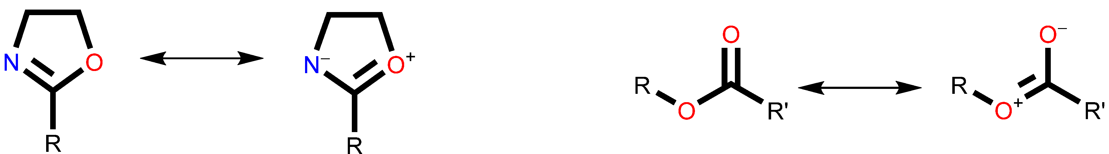
3.4. Quantum Chemical Calculations of EstOx

In order to interpret the experimental findings, quantum chemical calculations were performed for the EstOx model system using the MRCC program [36,37]. Mulliken atomic charges [38] and Mayer bond orders [39] were computed with the density-fitting Hartree-Fock method using the correlation-consistent valence quadruple-zeta (cc-pVQZ) basis set [40] and the corresponding auxiliary basis sets [41]. The calculations were carried out at the experimental X-ray geometry of the molecule.
The theoretical results support the conjecture that the N–C–O segment has a delocalized electronic structure similar to an ester group. The calculated bond orders are 1.97 and 1.10, respectively, for the N=C and C–O bonds indicating that the former bond order is lower than a double bond, while the latter bond order is higher than a typical single bond. The computed partial charges for the nitrogen and oxygen atoms, namely −0.43 and −0.44, respectively, are comparable: despite the higher electronegativity of the oxygen atom (compared to the nitrogen atom), the charges of the hetero atoms are equalized due to the delocalization, and it also means that a cationic attack on the nitrogen atom is possible. It is interesting to compare the above numbers with the corresponding results for the ester group. The bond orders are 1.94 and 1.18 for the O=C and C–O bonds, while the partial charges of the carbonyl and other oxygen atom are −0.49 and −0.41, respectively, which demonstrate the similar electronic structure of the N–C–O and O–C–O segments.

4. Conclusions

The single crystal x-ray analysis of EstOx reveals that the π-electron delocalization along the N–C–O segment in 2-oxazolines occurs at a content comparable to the π-electron delocalization in esters along the O–C–O segment: while the observed C=N bond length in 2-oxazolines and the measured C=O bond length in the ester group are in good agreement with the literature values for the corresponding C=X double bonds [33,34], the C–O “single” bonds are similar to each other and exhibit a value intermediate between a single and double C–O bond. Quantum chemical calculations revealed calculated bond orders of 1.97 and 1.10 for the N=C and C–O bonds of the 2-oxazoline ring, and almost identical partial charges for the nitrogen and oxygen atoms of −0.43 and −0.44. As the charges of the hetero atoms are equalized due to the delocalization, the cationic attack on the nitrogen atom during the initiation of the cationic ring-opening polymerization is possible. The ester group exhibits bond orders of 1.94 and 1.18 for the O=C and C–O bonds, which demonstrates the similar electronic structure of the N–C–O and O–C–O segments.

Supplementary Materials

Supplementary materials can be accessed at: https://www.mdpi.com/1996-1944/8/8/5385/s1.

Acknowledgments

This research has been performed in the Austrian-French project “MimiFlow”, jointly supported by the Austrian Science Fund FWF and the French National Research Agency ANR. Financial support within the project I1123-N19 (FWF) is gratefully acknowledged. Stéphanie Reynaud (Université de Pau et des Pays de l’Adour) is gratefully acknowledged for numerous and scientifically stimulating discussions. The research work was performed at the Polymer Competence Center Leoben GmbH (PCCL, Austria) within the framework of the COMET-program of the Federal Ministry for Transport, Innovation and Technology and Federal Ministry for Economy, Family and Youth with contributions by the Graz University of Technology and NAWI Graz. The PCCL is funded by the Austrian Government and the State Governments of Styria, Lower Austria and Upper Austria.

Author Contributions

Martin Fimberger and Klaus P. Luef performed the syntheses and analyses of the compounds; Claudia Payerl performed the initial syntheses of the compounds; Roland C. Fischer performed the X-ray analyses of the single crystals. Franz Stelzer was actively involved in numerous and scientifically stimulating discussions. Mihály Kállay and Frank Wiesbrock discussed the results and prepared the manuscript.

Conflicts of Interest

The authors declare no conflict of interest.

References

  1. Kappe, C.O. Controlled microwave heating in modern organic synthesis. Angew. Chem. Int. Ed. 2004, 43, 6250–6284. [Google Scholar] [CrossRef] [PubMed]
  2. Ebner, C.; Bodner, T.; Stelzer, F.; Wiesbrock, F. One decade of microwave-assisted polymerizations: Quo vadis? Macromol. Rapid Commun. 2011, 32, 254–288. [Google Scholar] [CrossRef] [PubMed]
  3. Rossegger, E.; Schenk, V.; Wiesbrock, F. Design strategies for functionalized poly(2-oxazoline)s and derived materials. Polymers 2013, 5, 956–1011. [Google Scholar] [CrossRef]
  4. Kempe, K.; Becer, C.R.; Schubert, U.S. Microwave-assisted polymerizations: Recent status and future perspectives. Macromolecules 2011, 44, 5825–5842. [Google Scholar] [CrossRef]
  5. Tomalia, D.A.; Sheetz, D.P. Homopolymerization of 2-alkyl- and 2-aryl-2-oxazolines. J. Polym. Sci. A Polym. Chem. 1966, 4, 2253–2265. [Google Scholar] [CrossRef]
  6. Seeliger, W.; Aufderhaar, E.; Diepers, W.; Feinauer, R.; Nehring, R.; Thier, W.; Hellmann, H. Recent syntheses and reactions of cyclic imidic esters. Angew. Chem. Int. Ed. 1966, 5, 875–888. [Google Scholar] [CrossRef] [PubMed]
  7. Kagiya, T.; Narisawa, S.; Maeda, T.; Fukui, K. Ring-opening polymerisation of 2-substituted 2-oxazolines. J. Polym. Sci. B Polym. Lett. 1966, 4, 441–445. [Google Scholar] [CrossRef]
  8. Bassiri, T.G.; Levy, A.; Litt, M. Polymerization of cyclic imino ethers. I. Oxazolines. J. Polym. Sci. B Polym. Lett. 1967, 5, 871–879. [Google Scholar] [CrossRef]
  9. Hoogenboom, R. Poly(2-oxazoline)s: A polymer class with numerous potential applications. Angew. Chem. Int. Ed. 2009, 48, 7978–7994. [Google Scholar] [CrossRef] [PubMed]
  10. Hoogenboom, R.; Schlaad, H. Bioinspired poly(2-oxazoline)s. Polymers 2011, 3, 467–488. [Google Scholar] [CrossRef]
  11. Schlaad, H.; Diehl, C.; Gress, A.; Meyer, M.; Demirel, A.L.; Nur, Y.; Bertin, A. Poly(2-oxazoline)s as smart bioinspired polymers. Macromol. Rapid Commun. 2010, 31, 511–525. [Google Scholar] [CrossRef] [PubMed]
  12. Gaertner, F.C.; Luxenhofer, R.; Blechert, B.; Jordan, R.; Essler, M. Synthesis, biodistribution and excretion of radiolabeled poly(2-alkyl-2-oxazoline)s. J. Control. Release 2007, 119, 291–300. [Google Scholar] [CrossRef] [PubMed]
  13. Waschinski, C.J.; Tiller, J.C. Poly(oxazoline)s with telechelic antimicrobial functions. Biomacromolecules 2005, 6, 235–243. [Google Scholar] [CrossRef] [PubMed]
  14. Waschinski, C.J.; Barnert, S.; Theobald, A.; Schubert, R.; Kleinschmidt, F.; Hoffmann, A.; Saalwächter, K.; Tiller, J.C. Insights in the antibacterial action of poly(methyloxazoline)s with a biocidal end group and varying satellite groups. Biomacromolecules 2008, 9, 1764–1771. [Google Scholar] [CrossRef] [PubMed]
  15. Kelly, A.; Kaltenhauser, V.; Mühlbacher, I.; Rametsteiner, K.; Kren, H.; Slugovc, C.; Stelzer, F.; Wiesbrock, F. Poly(2-oxazoline)-derived contact biocides: Contributions to the understanding of antimicrobial activity. Macromol. Biosci. 2013, 13, 116–125. [Google Scholar] [CrossRef] [PubMed]
  16. Kelly, A.M.; Wiesbrock, F. Strategies for the synthesis of poly(2-oxazoline)-based hydrogels. Macromol. Rapid Commun. 2012, 33, 1632–1647. [Google Scholar] [CrossRef] [PubMed]
  17. Schenk, V.; Ellmaier, L.; Rossegger, E.; Edler, M.; Griesser, T.; Weidinger, G.; Wiesbrock, F. Water-developable poly(2-oxazoline)-based negative photoresists. Macromol. Rapid Commun. 2012, 33, 396–400. [Google Scholar] [CrossRef] [PubMed]
  18. Fijten, M.W.M.; Haensch, C.; van Lankvelt, B.M.; Hoogenboom, R.; Schubert, U.S. Clickable poly(2-oxazoline)s as versatile building blocks. Macromol. Chem. Phys. 2008, 209, 1887–1895. [Google Scholar] [CrossRef]
  19. Zschoche, S.; Rueda, J.; Boyko, V.; Krahl, F.; Arndt, K.-F.; Voit, B. Thermo-responsive nanogels based on poly[NIPAAm-graft-(2-alkyl-2-oxazoline)]s crosslinked in the micellar state. Macromol. Chem. Phys. 2011, 211, 1035–1042. [Google Scholar] [CrossRef]
  20. Kempe, K.; Hoogenboom, R.; Jaeger, M.; Schubert, U.S. Three-fold metal-free efficient “click” reactions onto a multifunctional poly(2-oxazoline) designer scaffold. Macromolecules 2011, 44, 6424–6432. [Google Scholar] [CrossRef]
  21. Kelly, A.M.; Hecke, A.; Wirnsberger, B.; Wiesbrock, F. Synthesis of poly(2-oxazoline)-based hydrogels with tailor-made swelling degrees capable of stimuli-triggered compound release. Macromol. Rapid Commun. 2011, 32, 1815–1819. [Google Scholar] [CrossRef] [PubMed]
  22. Ten Brummelhuis, N.; Schlaad, H. Stimuli-responsive star polymers through thiol-yne core functionalization/crosslinking of block copolymer micelles. Polym. Chem. 2011, 2, 1180–1184. [Google Scholar] [CrossRef]
  23. Wiesbrock, F.; Hoogenboom, R.; Leenen, M.A.M.; Meier, M.A.R.; Schubert, U.S. Investigation of the living cationic ring-opening polymerization of 2-methyl-, 2-ethyl-, 2-nonyl-, and 2-phenyl-2-oxazoline in a single-mode microwave reactor. Macromolecules 2005, 38, 5025–5034. [Google Scholar] [CrossRef]
  24. Hoogenboom, R.; Fijten, M.W.M.; Kickelbick, G.; Schubert, U.S. Synthesis and crystal structures of multifunctional tosylates as basis for star-shaped poly(2-ethyl-2-oxazoline)s. Beilstein J. Org. Chem. 2010, 6, 773–783. [Google Scholar] [CrossRef] [PubMed]
  25. Luxenhofer, R.; Bezen, M.; Jordan, R. Kinetic investigations on the polymerization of 2-oxazolines using pluritriflate initiators. Macromol. Rapid Commun. 2008, 29, 1509–1513. [Google Scholar] [CrossRef]
  26. Wiesbrock, F.; Hoogenboom, R.; Abeln, C.H.; Schubert, U.S. Single-mode microwave ovens as new reaction devices: Accelerating the living polymerization of 2-ethyl-2-oxazoline. Macromol. Rapid Commun. 2004, 25, 1895–1899. [Google Scholar] [CrossRef]
  27. Bodner, T.; Ellmaier, L.; Schenk, V.; Albering, J.; Wiesbrock, F. Delocalized π-electrons in 2-oxazoline rings resulting in negatively charged nitrogen atoms: Revealing the selectivity during the initiation of cationic ring-opening polymerizations. Polym. Int. 2011, 60, 1173–1179. [Google Scholar] [CrossRef]
  28. SHELXS-97, Program for structure solution; University of Göttingen: Göttingen, Germany, 1997.
  29. SHELXL-97, Program for crystal structure analysis; University of Göttingen: Göttingen, Germany, 1997.
  30. Spek, A.L. Single-crystal structure validation with the program PLATON. J. Appl. Crystallogr. 2003, 36, 7–13. [Google Scholar] [CrossRef]
  31. Area-Detector Absorption Correction; Siemens Industrial Automation, Inc.: Madison, WI, USA, 1996.
  32. Zarka, M.T.; Nuyken, O.; Weberskirch, R. Amphiphilic polymer supports for the asymmetric hydrogenation of amino acid precursors in water. Chem. Eur. J. 2003, 9, 3228–3234. [Google Scholar] [CrossRef] [PubMed]
  33. Johnson, C.K. ORTEP; Report ORNL-5138; Oak Ridge National Laboratory: Oak Ridge, TN, USA, 1976. [Google Scholar]
  34. Cottrell, T.L. The Strengths of Chemical Bonds, 2nd ed.; Butterworths: London, UK, 1958. [Google Scholar]
  35. Benson, S.W. III - Bond energies. J. Chem. Educ. 1965, 42, 502–518. [Google Scholar] [CrossRef]
  36. Kállay, M.; Rolik, Z.; Csontos, J.; Ladjánszki, I.; Szegedy, L.; Ladóczki, B.; Samu, G.; MRCC. A Quantum Chemical Program Suite. Budapest University of Technology and Economics: Budapest, Hungary. Available online: http://www.mrcc.hu (accessed on 28 May 2015).
  37. Rolik, Z.; Szegedy, L.; Ladjánszki, I.; Ladóczki, B.; Kállay, M. An efficient linear-scaling CCSD(T) method based on local natural orbitals. J. Chem. Phys. 2013, 139, 094105. [Google Scholar] [CrossRef] [PubMed]
  38. Mulliken, R.S. Electronic population analysis on LCAO-MO molecular wave functions. J. Chem. Phys. 1955, 23, 1833–1840. [Google Scholar] [CrossRef]
  39. Mayer, I. Charge, bond order and valence in the ab initio SCF theory. Chem. Phys. Lett. 1983, 97, 270–274. [Google Scholar] [CrossRef]
  40. Dunning, T.H. Gaussian basis sets for use in correlated molecular calculations. I. The atoms boron through neon and hydrogen. J. Chem. Phys. 1989, 90, 1007–1023. [Google Scholar] [CrossRef]
  41. Weigend, F. Hartree-Fock exchange fitting basis sets for H to Rn. J. Comp. Chem. 2008, 29, 167–175. [Google Scholar] [CrossRef] [PubMed]

