
Cholesterol-Enhanced Polylactide-Based Stereocomplex Micelle for Effective Delivery of Doxorubicin

 ,
,
Abstract
:
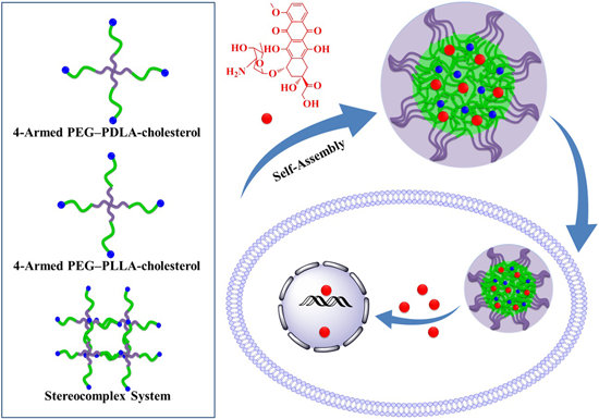
1. Introduction

2. Results and Discussion
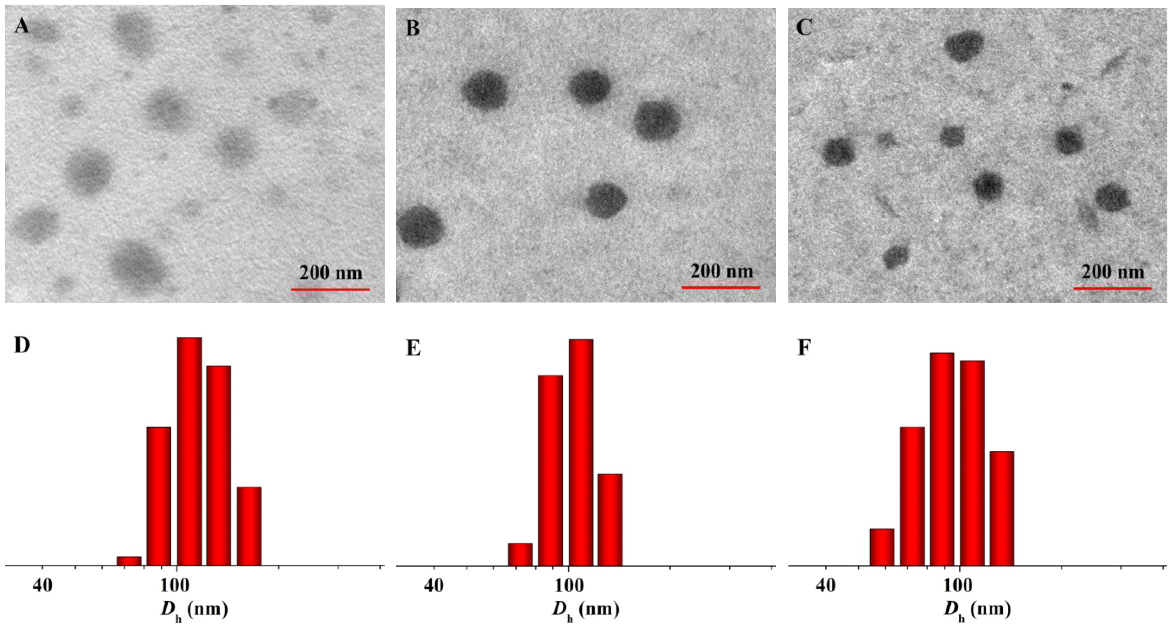
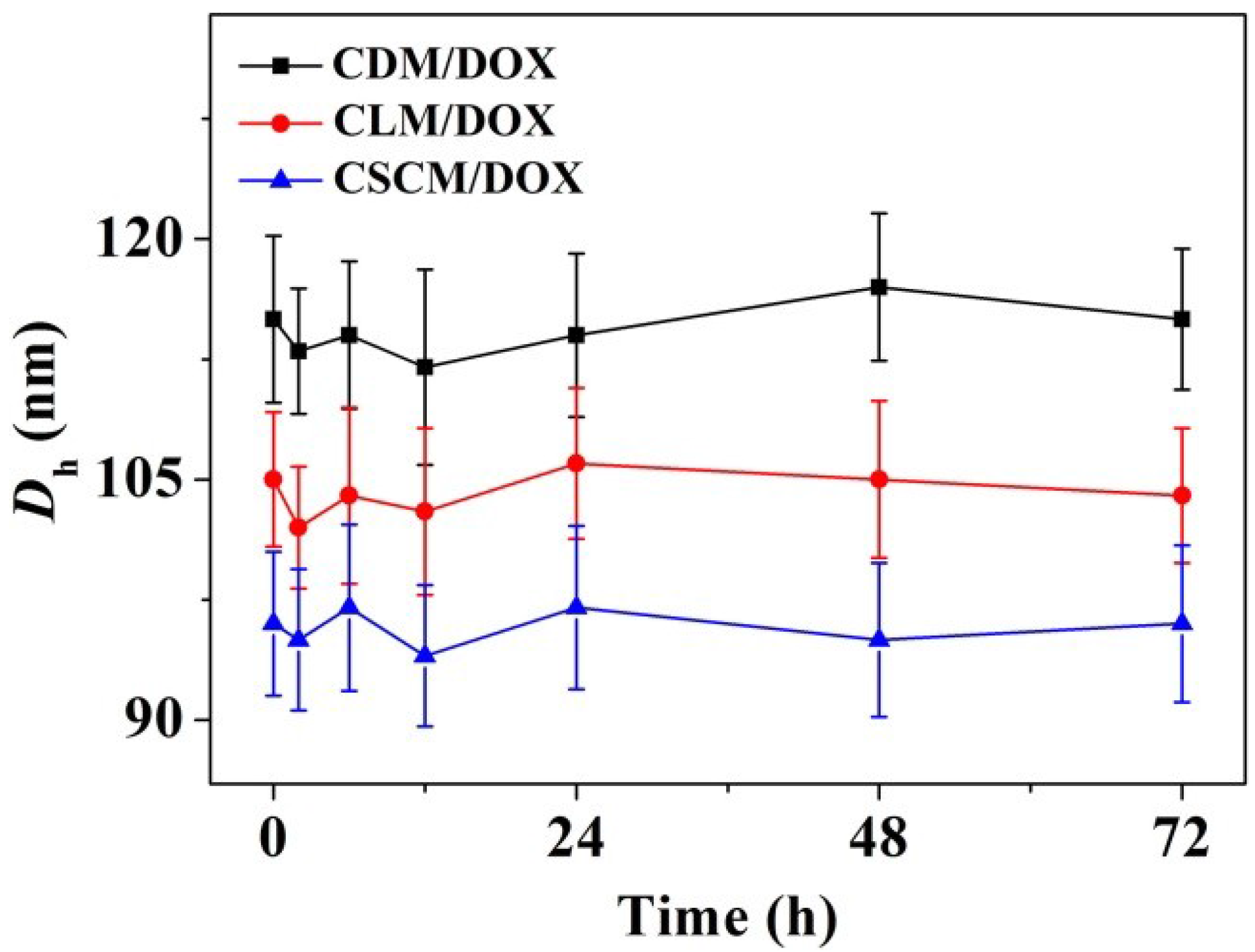
2.1. Fabrication and Characterization of DOX-Loaded Micelles
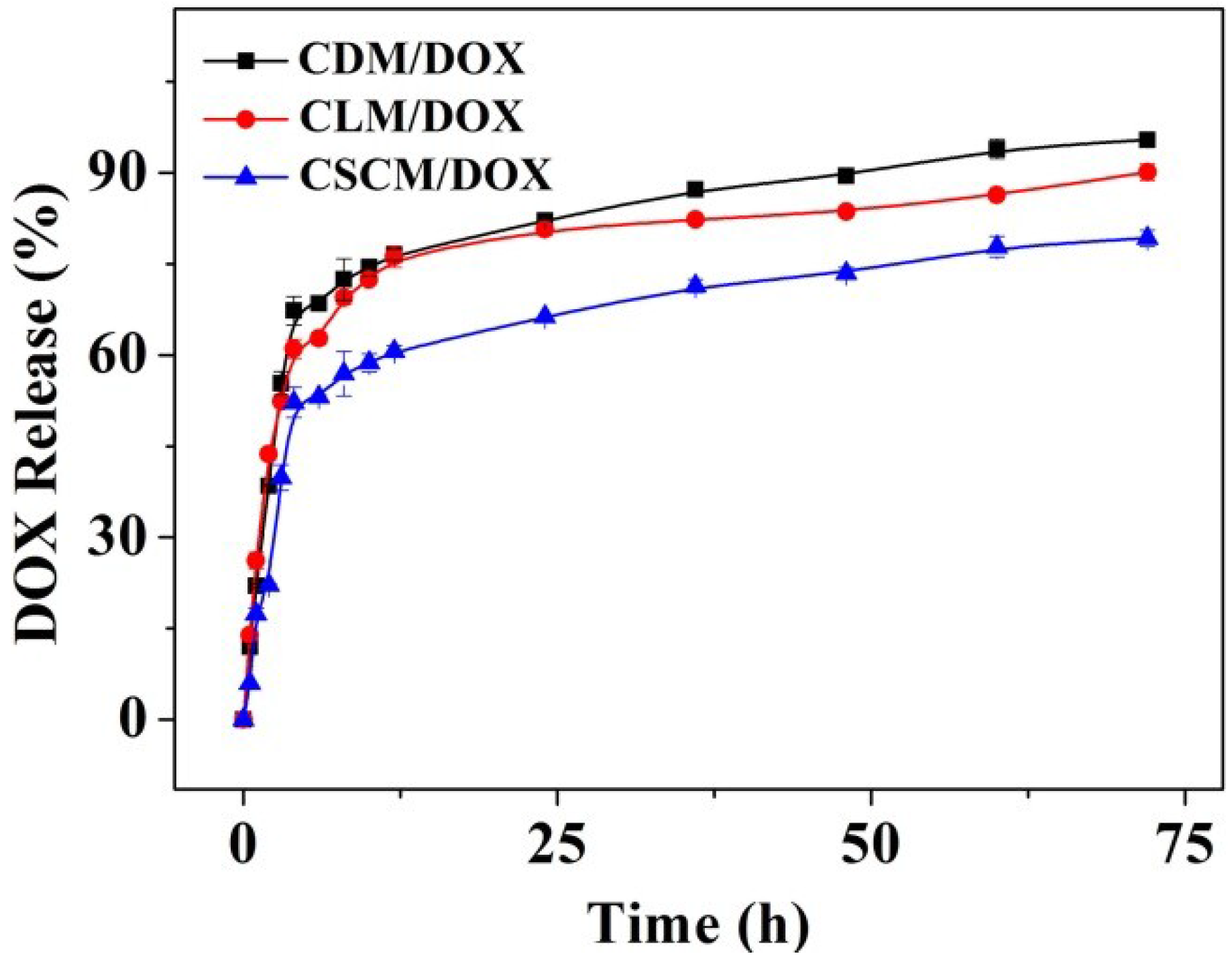
2.2. DOX Release from Various Formulations
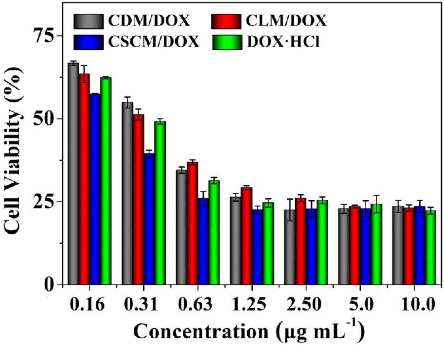
2.3. In Vitro Assessment of Cell Viability
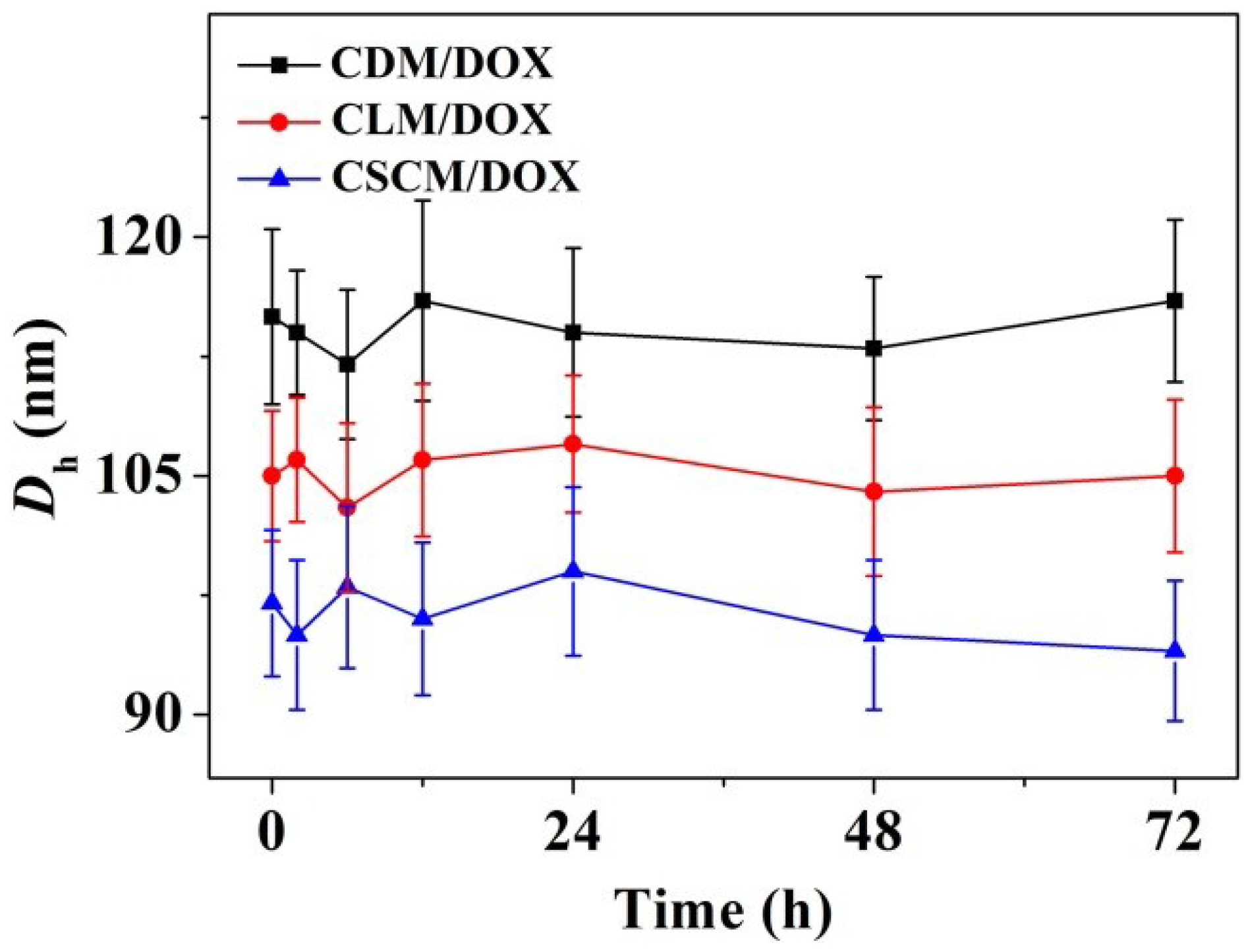
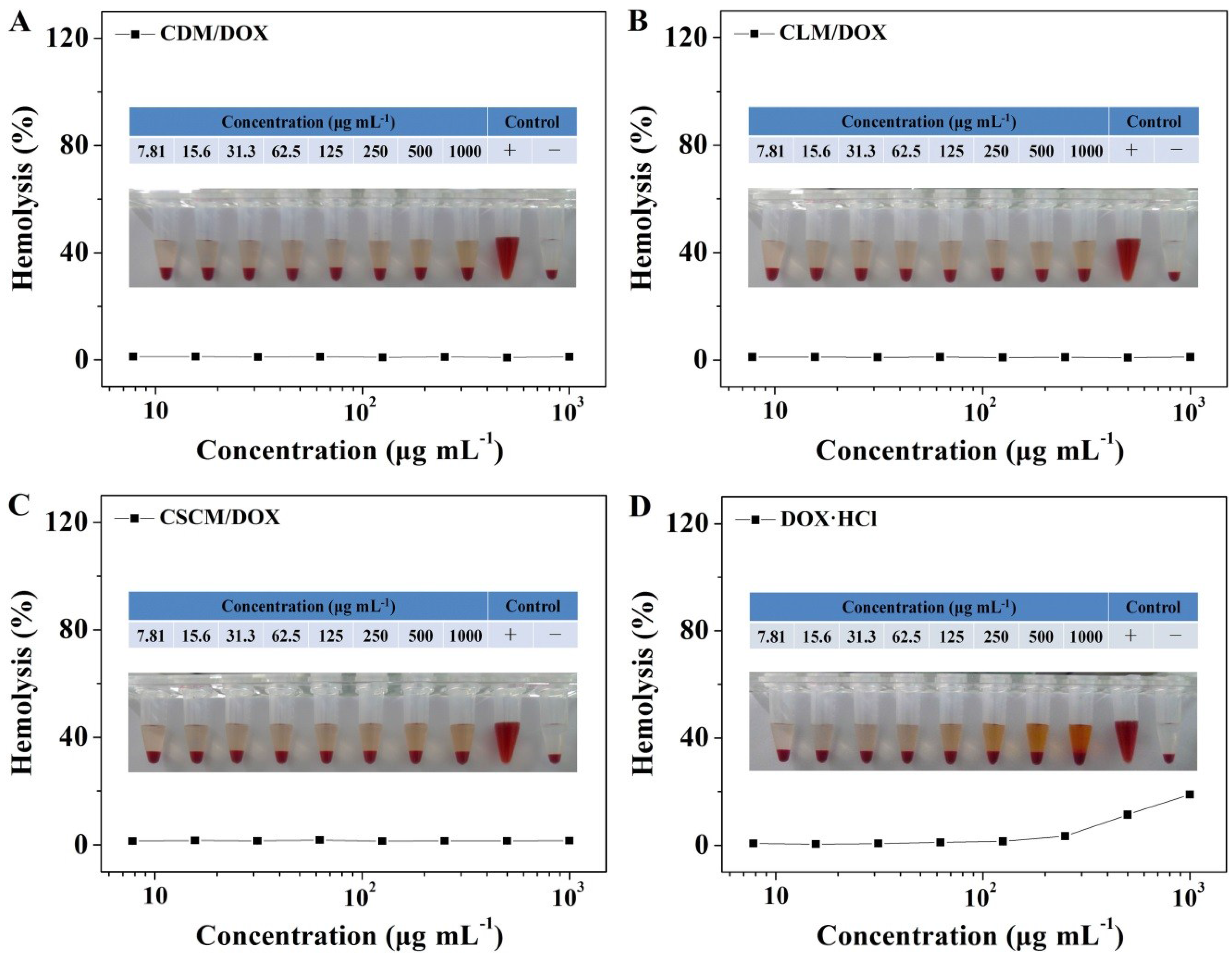
2.4. Evaluations of Serum Albumin-Tolerance Stability and Hemocompatibility
3. Experimental Section
3.1. Materials
3.2. DOX Encapsulation
3.3. Measurements
3.4. In Vitro DOX Release
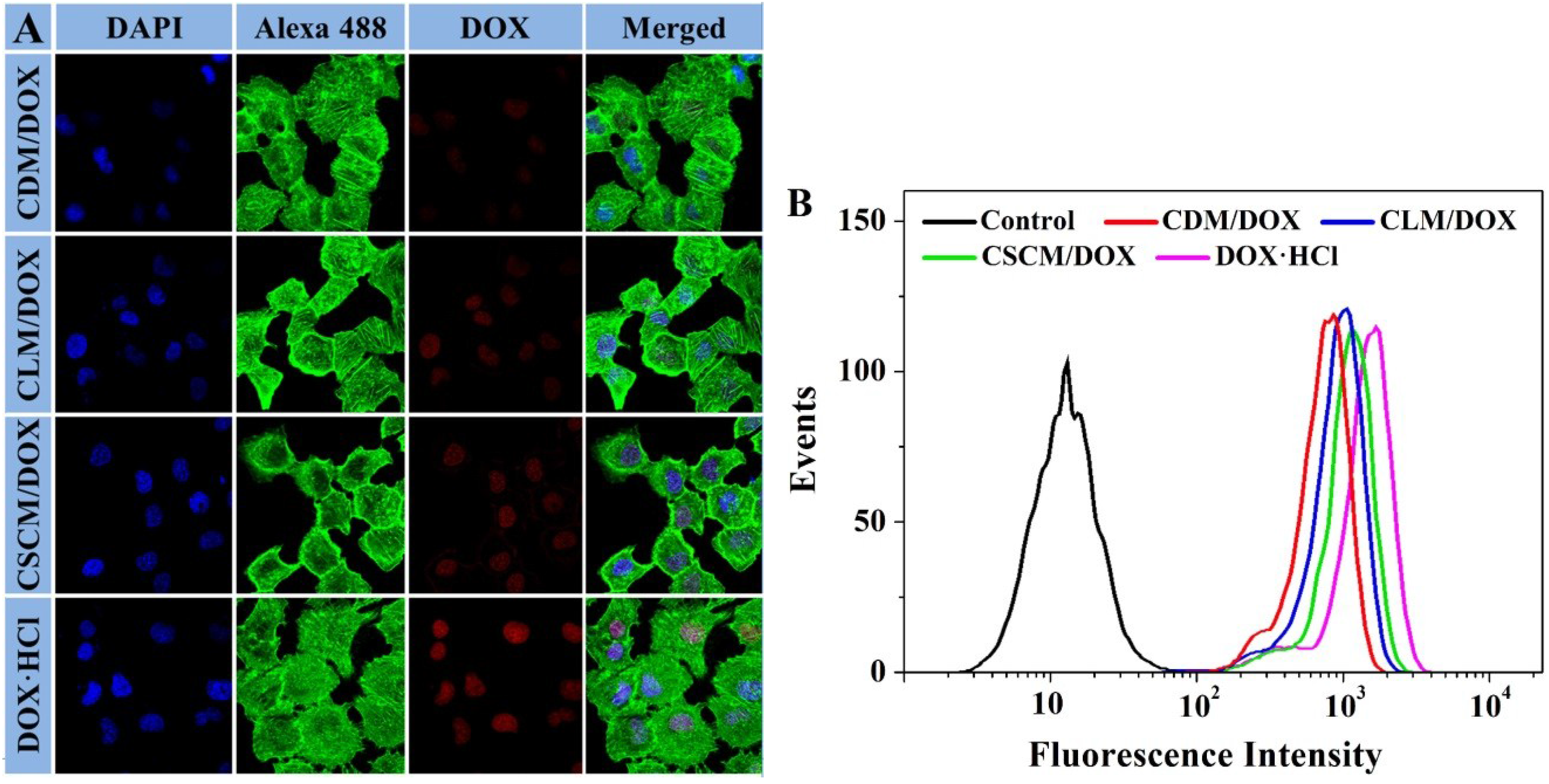
3.5. Intracellular DOX Release Analyses
3.5.1. CLSM
3.5.2. FCM
3.6. Cytotoxicity Assays
3.7. Serum Albumin-Tolerance Stability Assays
3.8. Hemolysis Activity Tests
4. Conclusions
Acknowledgments
Author Contributions
Conflicts of Interest
References
- Shi, F.H.; Ding, J.X.; Xiao, C.S.; Zhuang, X.L.; He, C.L.; Chen, L.; Chen, X.S. Intracellular microenvironment responsive PEGylated polypeptide nanogels with ionizable cores for efficient doxorubicin loading and triggered release. J. Mater. Chem. 2012, 22, 14168–14179. [Google Scholar] [CrossRef]
- Hao, Y.B.; Yi, S.Y.; Ruan, J.; Zhao, L.; Nan, K.J. New insights into metronomic chemotherapy-induced immunoregulation. Cancer Lett. 2014, 354, 220–226. [Google Scholar] [CrossRef] [PubMed]
- Malam, Y.; Loizidou, M.; Seifalian, A.M. Liposomes and nanoparticles: Nanosized vehicles for drug delivery in cancer. Trends Pharmacol. Sci. 2009, 30, 592–599. [Google Scholar] [CrossRef] [PubMed]
- Koren, E.; Apte, A.; Jani, A.; Torchilin, V.P. Multifunctional PEGylated 2C5-immunoliposomes containing pH-sensitive bonds and TAT peptide for enhanced tumor cell internalization and cytotoxicity. J. Control. Release 2012, 160, 264–273. [Google Scholar] [CrossRef] [PubMed]
- Ninomiya, K.; Kawabata, S.; Tashita, H.; Shimizu, N. Ultrasound-mediated drug delivery using liposomes modified with a thermosensitive polymer. Ultrason. Sonochem. 2014, 21, 310–316. [Google Scholar] [CrossRef] [PubMed]
- Ding, J.X.; Chen, J.J.; Li, D.; Xiao, C.S.; Zhang, J.C.; He, C.L.; Zhuang, X.L.; Chen, X.S. Biocompatible reduction-responsive polypeptide micelles as nanocarriers for enhanced chemotherapy efficacy in vitro. J. Mater. Chem. B 2013, 1, 69–81. [Google Scholar] [CrossRef]
- Liang, J.; Wu, W.L.; Xu, X.D.; Zhuo, R.X.; Zhang, X.Z. pH responsive micelle self-assembled from a new amphiphilic peptide as anti-tumor drug carrier. Colloid. Surf. B 2014, 114, 398–403. [Google Scholar] [CrossRef]
- Ding, J.X.; Shi, F.H.; Li, D.; Chen, L.; Zhuang, X.L.; Chen, X.S. Enhanced endocytosis of acid-sensitive doxorubicin derivatives with intelligent nanogel for improved security and efficacy. Biomater. Sci. 2013, 1, 633–646. [Google Scholar] [CrossRef]
- Nukolova, N.V.; Oberoi, H.S.; Cohen, S.M.; Kabanov, A.V.; Bronich, T.K. Folate-decorated nanogels for targeted therapy of ovarian cancer. Biomaterials 2011, 32, 5417–5426. [Google Scholar] [CrossRef] [PubMed]
- Kobayashi, H.; Watanabe, R.; Choyke, P.L. Improving conventional enhanced permeability and retention (EPR) effects; What is the appropriate target? Theranostics 2014, 4, 81–89. [Google Scholar] [CrossRef]
- Gou, P.F.; Liu, W.W.; Mao, W.W.; Tang, J.B.; Shen, Y.Q.; Sui, M.H. Self-assembling doxorubicin prodrug forming nanoparticles for cancer chemotherapy: Synthesis and anticancer study in vitro and in vivo. J. Mater. Chem. B 2013, 1, 284–292. [Google Scholar] [CrossRef]
- Kamimura, M.; Furukawa, T.; Akiyama, S.-i.; Nagasaki, Y. Enhanced intracellular drug delivery of pH-sensitive doxorubicin/poly(ethylene glycol)-block-poly(4-vinylbenzylphosphonate) nanoparticles in multi-drug resistant human epidermoid KB carcinoma cells. Biomater. Sci. UK 2013, 1, 361–367. [Google Scholar] [CrossRef]
- Tang, Z.H.; Chen, X.S.; Yang, Y.K.; Pang, X.; Sun, J.R.; Zhang, X.F.; Jing, X.B. Stereoselective polymerization of rac-lactide with a bulky aluminum/Schiff base complex. J. Polym. Sci. Pol. Chem. 2004, 42, 5974–5982. [Google Scholar] [CrossRef]
- Zhao, Z.W.; Yao, X.M.; Zhang, Z.; Chen, L.; He, C.L.; Chen, X.S. Boronic acid shell-crosslinked dextran-b-PLA micelles for acid-responsive drug delivery. Macromol. Biosci. 2014, 14, 1609–1618. [Google Scholar] [CrossRef] [PubMed]
- Li, Y.; Lin, J.Y.; Wu, H.J.; Jia, M.M.; Yuan, C.H.; Chang, Y.; Hou, Z.Q.; Dai, L.Z. Novel methotrexate prodrug-targeted drug delivery system based on PEG-lipid-PLA hybrid nanoparticles for enhanced anticancer efficacy and reduced toxicity of mitomycin C. J. Mater. Chem. B 2014, 2, 6534–6548. [Google Scholar] [CrossRef]
- Serra, T.; Planell, J.A.; Navarro, M. High-resolution PLA-based composite scaffolds via 3-D printing technology. Acta. Biomater. 2013, 9, 5521–5530. [Google Scholar] [CrossRef] [PubMed]
- Lu, Y.M.; Cheng, L.M.; Pei, G.X.; Cai, Z.; Pan, L.; Su, J.; Zhang, K.H.; Guo, L.L.; Yu, Q.S.; Guo, Y.R. Experimental study of repairing femoral bone defects with nHA/RHLC/PLA scaffold composite with endothelial cells and osteoblasts in canines. Chin. Med. J. 2013, 93, 1335–1340. [Google Scholar]
- Kulkarni, R.K.; Pani, K.C.; Neuman, C.; Leonard, F. Polylactic acid for surgical implants. Arch. Surg. 1966, 93, 839–843. [Google Scholar] [CrossRef] [PubMed]
- Bradley, A.J.; Murad, K.L.; Regan, K.L.; Scott, M.D. Biophysical consequences of linker chemistry and polymer size on stealth erythrocytes: Size does matter. BBA Biomembr. 2002, 1561, 147–158. [Google Scholar] [CrossRef]
- Kim, K.; Yu, M.; Zong, X.H.; Chiu, J.; Fang, D.F.; Seo, Y.S.; Hsiao, B.S.; Chu, B.; Hadjiargyrou, M. Control of degradation rate and hydrophilicity in electrospun non-woven poly(D,L-lactide) nanofiber scaffolds for biomedical applications. Biomaterials 2003, 24, 4977–4985. [Google Scholar] [CrossRef] [PubMed]
- Gref, R.; Luck, M.; Quellec, P.; Marchand, M.; Dellacherie, E.; Harnisch, S.; Blunk, T.; Muller, R.H. ‘Stealth’ corona-core nanoparticles surface modified by polyethylene glycol (PEG): Influences of the corona (PEG chain length and surface density) and of the core composition on phagocytic uptake and plasma protein adsorption. Colloid. Surface B 2000, 18, 301–313. [Google Scholar] [CrossRef]
- Klibanov, A.L.; Maruyama, K.; Torchilin, V.P.; Huang, L. Amphipathic polyethyleneglycols effectively prolong the circulation time of liposomes. FEBS Lett. 1990, 268, 235–237. [Google Scholar] [CrossRef] [PubMed]
- Riley, T.; Stolnik, S.; Heald, C.R.; Xiong, C.D.; Garnett, M.C.; Illum, L.; Davis, S.S.; Purkiss, S.C.; Barlow, R.J.; Gellert, P.R. Physicochemical evaluation of nanoparticles assembled from poly(lactic acid)-poly(ethylene glycol) (PLA-PEG) block copolymers as drug delivery vehicles. Langmuir 2001, 17, 3168–3174. [Google Scholar] [CrossRef]
- Heald, C.R.; Stolnik, S.; De Matteis, C.; Garnett, M.C.; Illum, L.; Davis, S.S.; Leermakers, F.A.M. Characterisation of poly(lactic acid): Poly(ethyleneoxide) (PLA:PEG) nanoparticles using the self-consistent theory modelling approach. Colloid. Surf. A 2003, 212, 57–64. [Google Scholar] [CrossRef]
- Fu, C.H.; Sun, X.L.; Liu, D.H.; Chen, Z.J.; Lu, Z.J.; Zhang, N. Biodegradable tri-block copolymer poly(lactic acid)-poly(ethylene glycol)-poly(L-lysine)(PLA-PEG-PLL) as a non-viral vector to enhance gene transfection. Int. J. Mol. Sci. 2011, 12, 1371–1388. [Google Scholar] [CrossRef] [PubMed]
- Ocal, H.; Arica-Yegin, B.; Vural, I.; Goracinova, K.; Calis, S. 5-fluorouracil-loaded PLA/PLGA PEG-PPG-PEG polymeric nanoparticles: Formulation, in vitro characterization and cell culture studies. Drug Dev. Ind. Pharm. 2014, 40, 560–567. [Google Scholar] [CrossRef] [PubMed]
- Mukose, T.; Fujiwara, T.; Nakano, J.; Taniguchi, I.; Miyamoto, M.; Kimura, Y.; Teraoka, I.; Lee, C.W. Hydrogel formation between enantiomeric B-A-B-type block copolymers of polylactides (PLLA or PDLA: A) and polyoxyethylene (PEG: B); PEG-PLLA-PEG and PEG-PDLA-PEG. Macromol. Biosci. 2004, 4, 361–367. [Google Scholar] [CrossRef] [PubMed]
- Hiemstra, C.; Zhong, Z.; Li, L.; Dijkstra, P.J.; Feijen, J. In-situ formation of biodegradable hydrogels by stereocomplexation of PEG-(PLLA) 8 and PEG-(PDLA) 8 star block copolymers. Biomacromolecules 2006, 7, 2790–2795. [Google Scholar] [CrossRef] [PubMed]
- Fujiwara, T.; Mukose, T.; Yamaoka, T.; Yamane, H.; Sakurai, S.; Kimura, Y. Novel thermo-responsive formation of a hydrogel by stereo-complexation between PLLA-PEG-PLLA and PDLA-PEG-PDLA block copolymers. Macromol. Biosci. 2001, 1, 204–208. [Google Scholar] [CrossRef]
- Ouahab, A.; Cheraga, N.; Onoja, V.; Shen, Y.; Tu, J.S. Novel pH-sensitive charge-reversal cell penetrating peptide conjugated PEG-PLA micelles for docetaxel delivery: In vitro study. Int. J. Pharm. 2014, 466, 233–245. [Google Scholar] [CrossRef] [PubMed]
- Chen, L.; Xie, Z.G.; Hu, J.L.; Chen, X.S.; Jing, X.B. Enantiomeric PLA–PEG block copolymers and their stereocomplex micelles used as rifampin delivery. J. Nanopart. Res. 2007, 9, 777–785. [Google Scholar] [CrossRef]
- Liu, D.H.; Ding, J.X.; Xu, W.G.; Song, X.F.; Zhuang, X.L.; Chen, X.S. Stereocomplex micelles based on 4-armed poly(ethylene glycol )-polylactide enantiomeric copolymers for drug delivery. Acta Polym. Sin. 2014, 1265–1273. [Google Scholar]
- Lee, J.L.; Ahn, J.H.; Park, S.H.; Lim, H.Y.; Kwon, J.H.; Ahn, S.; Song, C.; Hong, J.H.; Kim, C.S.; Ahn, H. Phase II study of a cremophor-free, polymeric micelle formulation of paclitaxel for patients with advanced urothelial cancer previously treated with gemcitabine and platinum. Invest. New Drug. 2012, 30, 1984–1990. [Google Scholar] [CrossRef]
- Ahn, H.K.; Jung, M.; Sym, S.J.; Shin, D.B.; Kang, S.M.; Kyung, S.Y.; Park, J.W.; Jeong, S.H.; Cho, E.K. A phase II trial of cremorphor EL-free paclitaxel (Genexol-PM) and gemcitabine in patients with advanced non-small cell lung cancer. Cancer Chemoth. Pharm. 2014, 74, 277–282. [Google Scholar] [CrossRef]
- Simons, K.; Ikonen, E. Cell biology—How cells handle cholesterol. Science 2000, 290, 1721–1726. [Google Scholar] [CrossRef] [PubMed]
- Lee, A.L.Z.; Venkataraman, S.; Sirat, S.B.M.; Gao, S.J.; Hedrick, J.L.; Yang, Y.Y. The use of cholesterol-containing biodegradable block copolymers to exploit hydrophobic interactions for the delivery of anticancer drugs. Biomaterials 2012, 33, 1921–1928. [Google Scholar] [CrossRef] [PubMed]
- Pucadyil, T.J.; Chattopadhyay, A. Role of cholesterol in the function and organization of G-protein coupled receptors. Prog. Lipid Res. 2006, 45, 295–333. [Google Scholar] [CrossRef] [PubMed]
- Laskar, P.; Samanta, S.; Ghosh, S.K.; Dey, J. In vitro evaluation of pH-sensitive cholesterol-containing stable polymeric micelles for delivery of camptothecin. J. Colloid Interface Sci. 2014, 430, 305–314. [Google Scholar] [CrossRef] [PubMed]
- Yang, B.; Lv, Y.; Zhu, J.Y.; Han, Y.T.; Jia, H.Z.; Chen, W.H.; Feng, J.; Zhang, X.Z.; Zhuo, R.X. A pH-responsive drug nanovehicle constructed by reversible attachment of cholesterol to PEGylated poly(L-lysine) via catechol-boronic acid ester formation. Acta Biomater. 2014, 10, 3686–3695. [Google Scholar] [CrossRef] [PubMed]
- Cameron, D.J.A.; Shaver, M.P. Aliphatic polyester polymer stars: Synthesis, properties and applications in biomedicine and nanotechnology. Chem. Soc. Rev. 2011, 40, 1761–1776. [Google Scholar] [CrossRef] [PubMed]
- Ding, J.X.; Xu, W.G.; Zhang, Y.; Sun, D.K.; Xiao, C.S.; Liu, D.H.; Zhu, X.J.; Chen, X.S. Self-reinforced endocytoses of smart polypeptide nanogels for “on-demand” drug delivery. J. Control. Release 2013, 172, 444–455. [Google Scholar] [CrossRef] [PubMed]
- Liu, L.; Li, C.X.; Li, X.C.; Yuan, Z.; An, Y.L.; He, B.L. Biodegradable polylactide/poly(ethylene glycol)/polylactide triblock copolymer micelles as anticancer drug carriers. J. Appl. Polym. Sci. 2001, 80, 1976–1982. [Google Scholar] [CrossRef]
- Kang, N.; Perron, M.E.; Prud’homme, R.E.; Zhang, Y.B.; Gaucher, G.; Leroux, J.C. Stereocomplex block copolymer micelles: Core-shell nanostructures with enhanced stability. Nano Lett. 2005, 5, 315–319. [Google Scholar] [CrossRef] [PubMed]
- Li, M.Q.; Tang, Z.H.; Lv, S.X.; Song, W.T.; Hong, H.; Jing, X.B.; Zhang, Y.Y.; Chen, X.S. Cisplatin crosslinked pH-sensitive nanoparticles for efficient delivery of doxorubicin. Biomaterials 2014, 35, 3851–3864. [Google Scholar] [CrossRef] [PubMed]
- Lee, M.; Rentz, J.; Han, S.O.; Bull, D.A.; Kim, S.W. Water-soluble lipopolymer as an efficient carrier for gene delivery to myocardium. Gene Ther. 2003, 10, 585–593. [Google Scholar] [CrossRef] [PubMed]
- Maxfield, F.R.; Meer, G.V. Cholesterol, the central lipid of mammalian cells. Curr. Opin. Cell Biol. 2010, 22, 422–429. [Google Scholar] [CrossRef] [PubMed]
- Dai, J.; Lin, S.D.; Cheng, D.; Zou, S.Y.; Shuai, X.T. Interlayer-crosslinked micelle with partially hydrated core showing reduction and pH dual sensitivity for pinpointed intracellular drug release. Angew. Chem. Int. Edit. 2011, 50, 9404–9408. [Google Scholar] [CrossRef]
- Sun, D.K.; Ding, J.X.; Xiao, C.S.; Chen, J.J.; Zhuang, X.L.; Chen, X.S. Preclinical evaluation of antitumor activity of acid-sensitive PEGylated doxorubicin. ACS Appl. Mater. Inter. 2014, 23, 21202–21214. [Google Scholar] [CrossRef]
- Ding, J.X.; Zhao, L.; Li, D.; Xiao, C.S.; Zhuang, X.L.; Chen, X.S. Thermo-responsive “hairy-rod” polypeptides for smart antitumor drug delivery. Polym. Chem. 2013, 4, 3345–3356. [Google Scholar] [CrossRef]
- Wang, Y.; Wang, H.B.; Liu, G.Y.; Liu, X.S.; Jin, Q.; Ji, J. Self-assembly of near-monodisperse redox-sensitive micelles from cholesterol-conjugated biomimetic copolymers. Macromol. Biosci. 2013, 13, 1084–1091. [Google Scholar] [CrossRef] [PubMed]
- Benival, D.M.; Devarajan, P.V. Lipomer of doxorubicin hydrochloride for enhanced oral bioavailability. Int. J. Pharm. 2012, 423, 554–561. [Google Scholar] [CrossRef] [PubMed]
© 2015 by the authors; licensee MDPI, Basel, Switzerland. This article is an open access article distributed under the terms and conditions of the Creative Commons Attribution license (http://creativecommons.org/licenses/by/4.0/).
Share and Cite
Wang, J.; Xu, W.; Ding, J.; Lu, S.; Wang, X.; Wang, C.; Chen, X. Cholesterol-Enhanced Polylactide-Based Stereocomplex Micelle for Effective Delivery of Doxorubicin. Materials 2015, 8, 216-230. https://doi.org/10.3390/ma8010216
Wang J, Xu W, Ding J, Lu S, Wang X, Wang C, Chen X. Cholesterol-Enhanced Polylactide-Based Stereocomplex Micelle for Effective Delivery of Doxorubicin. Materials. 2015; 8(1):216-230. https://doi.org/10.3390/ma8010216
Chicago/Turabian StyleWang, Jixue, Weiguo Xu, Jianxun Ding, Shengfan Lu, Xiaoqing Wang, Chunxi Wang, and Xuesi Chen. 2015. "Cholesterol-Enhanced Polylactide-Based Stereocomplex Micelle for Effective Delivery of Doxorubicin" Materials 8, no. 1: 216-230. https://doi.org/10.3390/ma8010216