Article

Flux-Aided Synthesis of Lu2O3 and Lu2O3:Eu—Single Crystal Structure, Morphology Control and Radioluminescence Efficiency

by
Justyna Zeler
1,
Lucjan B. Jerzykiewicz
1,† and
Eugeniusz Zych
1,2,*,†
1
Faculty of Chemistry, University of Wroclaw, 14. F. Joliot-Curie Street, 50-383 Wroclaw, Poland
2
Wroclaw Research Centre EIT+, 147 Stablowicka Street, 54-066 Wroclaw, Poland
*
Author to whom correspondence should be addressed.
These authors contributed equally to this work.
Materials 2014, 7(10), 7059-7072; https://doi.org/10.3390/ma7107059
Submission received: 21 July 2014 / Revised: 11 September 2014 / Accepted: 22 September 2014 / Published: 20 October 2014

Abstract

:
Li2SO4 or (Li2SO4 + SiO2)-mixture fluxes were used to prepare a Lu2O3:Eu powder phosphor as well as an undoped Lu2O3 utilizing commercial lutetia and europia as starting reagents. SEM images showed that the fabricated powders were non-agglomerated and the particles sizes varied from single microns to tens of micrometers depending largely on the flux composition rather than the oxide(s)-to-flux ratio. In the presence of SiO2 in the flux, certain grains grew up to 300–400 μm. The lack of agglomeration and the large sizes of crystallites allowed making single crystal structural measurements and analysis on an undoped Lu2O3 obtained by means of the flux technique. The cubic structure with a = 10.393(2) Å, and Ia3 space group at 298 K was determined. The most efficient radioluminescence of Lu2O3:Eu powders reached 95%–105% of the commercial Gd2O2S:Eu.

Graphical Abstract

1. Introduction

At the end of 20th century, the Lu2O3:Eu X-ray phosphor became a subject of thorough research [1,2,3,4,5,6,7,8,9,10,11,12,13,14,15,16,17,18,19]. Also, other activators were used, especially for lasing in Lu2O3 sintered transparent bodies [20,21]. With its impressive density of 9.84 g/cm3, high effective Z-number (63.7) and light yield similar to Gd2O2S-based phosphors [3,22,23,24] both lutetia powders and sintered ceramics were considered attractive for X-ray scintillator detectors [3,22,25]. Furthermore, with an energy band gap of about 5.85 eV [25], Lu2O3:Eu was recognized to have potential to produce as much as about 75,000 ph/MeV upon ionizing radiation excitation. This is well beyond the performance reported in literature [25], which gives hope that there still is room for significant improvement. The extraordinary absorption coefficient of X-rays by photoelectric effect rather than Compton scattering [26,27] allows using thinner Lu2O3:Eu phosphor layers, which translates into reduced light scattering, i.e., image smudging [27]. Clearly, Lu2O3:Eu may offer important advantages as X-ray phosphor if only its light yield could be improved and morphology well controlled.
High quality screens, with uniformly and densely packed grains, require spherical, rather uniform in size particles. It was shown that the spherical size of particles allows reducing the screen thickness by 1/3 [17,28]. Also, effective sintering of powders into translucent or transparent bodies, especially using pressureless techniques, is very much dependent on the starting powder morphology and its thermal history [17,29,30,31,32]. Yet, literature data on controlling the Lu2O3:Eu powders morphology are only scant [33,34,35,36] and those that are available were never confronted with the phosphors performance under X-rays. With this paper we try to partially fill this gap. We thus shall report on the evolution of morphology of Lu2O3:Eu powders produced using Li2SO4/(Li2SO4 + SiO2)-flux-aided preparation technique. Since this fabrication procedure gave occasionally crystallites with sizes sufficient for single crystal structure determination, we took advantage of that and preformed such analysis. Up to now, the only direct determination of Lu2O3 structure was performed on single crystals obtained by a micro-pulling down (μ-PD) technique as well as a laser heated pedestal growth (LHPG) [37].

2. Results and Discussion

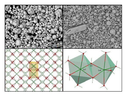
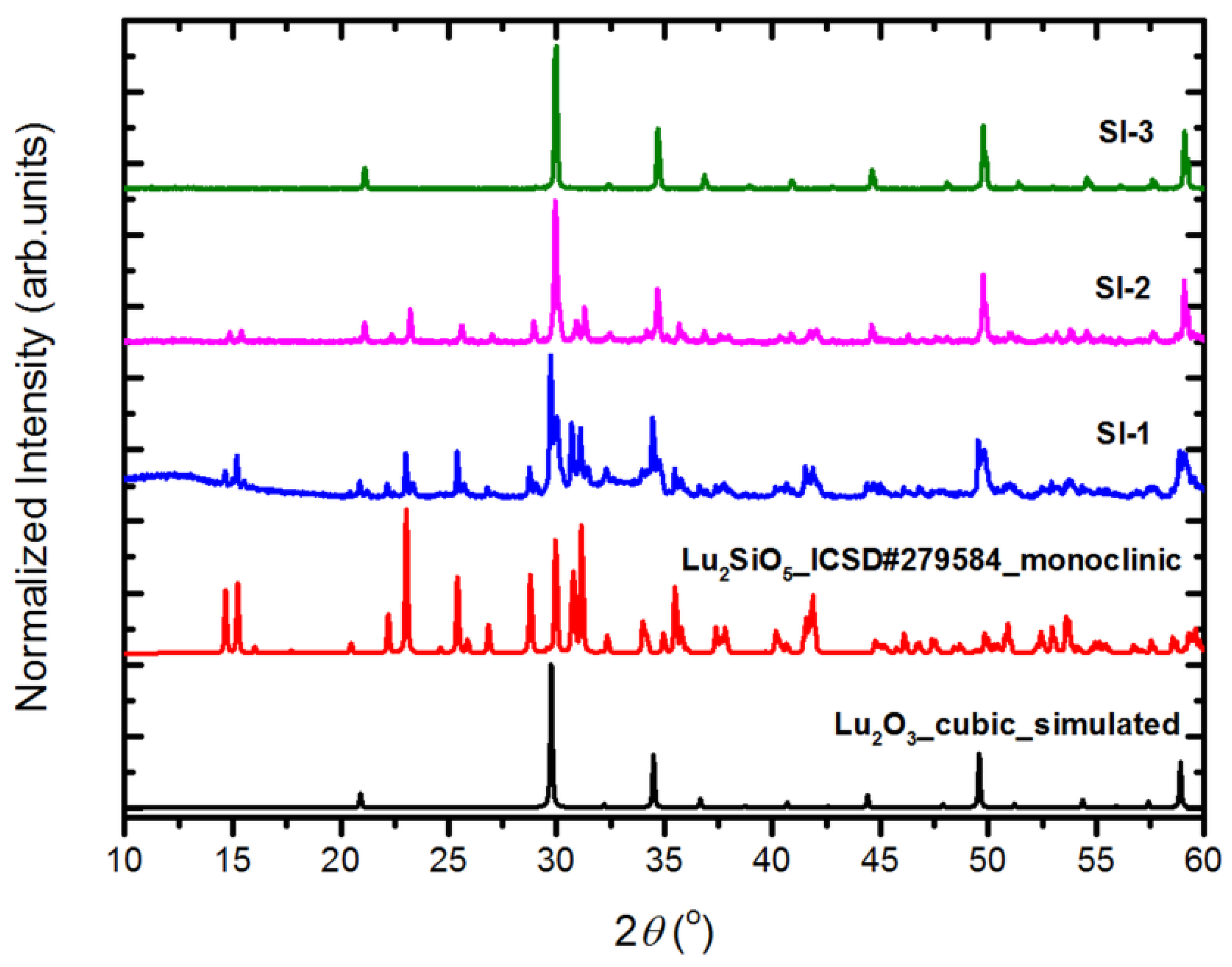
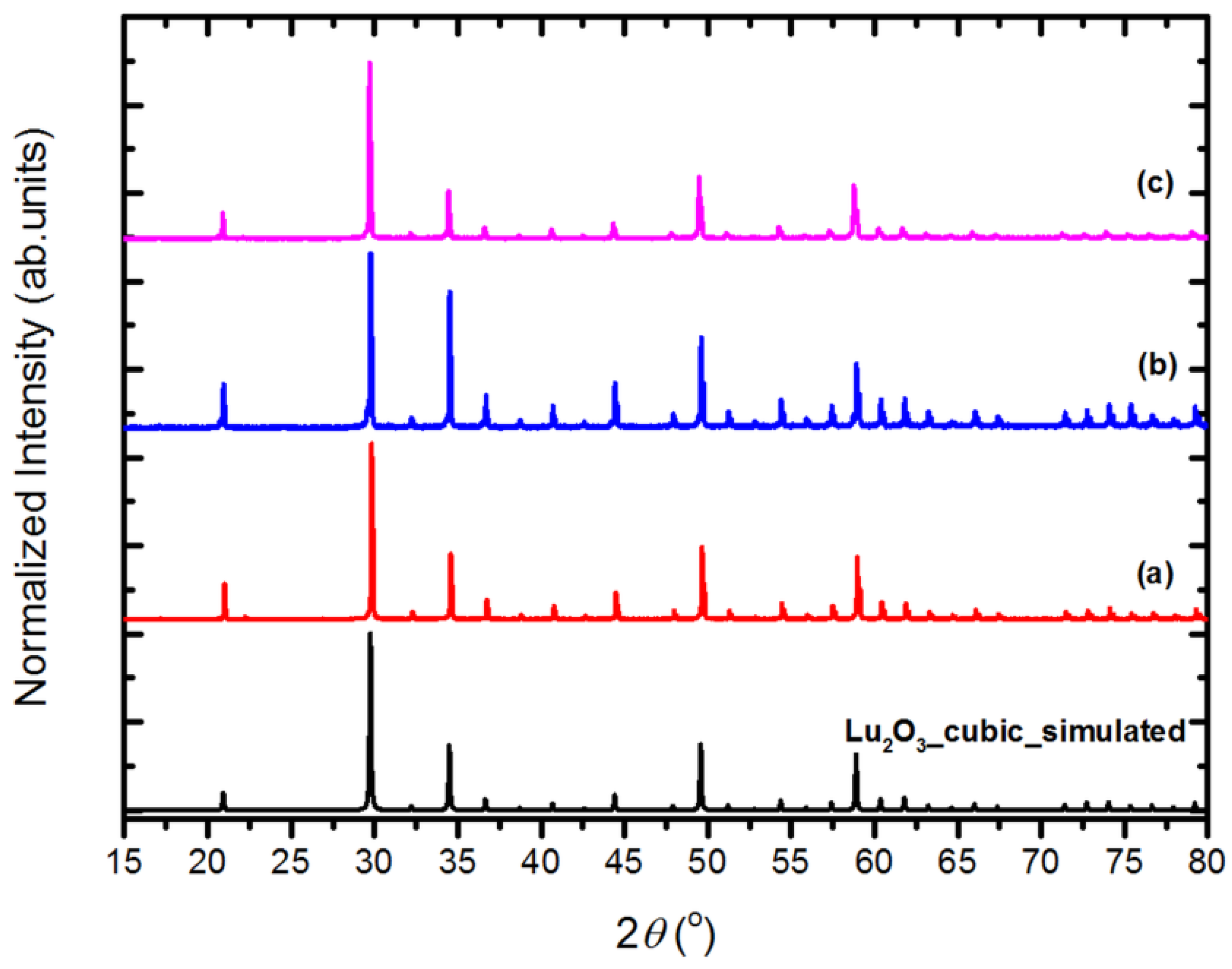
2.1. Structural Analysis

Let us start by presenting the X-ray diffraction (XRD) patterns of powders of Series I (Figure 1), which supposedly should/might provide Lu2SiO5 (LSO) powders, as the lutetia and silica were mixed in 1:1 molar ratio. Indeed, when the synthesis was only 1-h long (sample SI-1) a main fraction of the product was LSO, with Lu2O3 present as an impurity, readily detected but not a massive fraction. In the case of the sample heated for 2 h (SI-2), the powder was already largely composed of Lu2O3, while LSO existed only as an impurity phase. A 20-h prolonged heating at 1300 °C (SI-3) ended up with a single phase cubic Lu2O3 powder without any traces of LSO, as seen by the XRD technique. Hence, the XRD data of powders of Series I proves, that in the presence of significant amount of Li2SO4-flux, silica is effectively eliminated from the batch upon prolonged heating at elevated temperatures. Additional experiments, results of which we do not present here, proved that the process of removal of SiO2 from the reacting mixture of Lu2O3 and SiO2 was faster with increasing Li2SO4 flux content and with increasing temperature—at 900 °C it was much slower, while at 1400 °C it was even faster than at 1300 °C. This we took into account by proceeding with synthesis of samples of Series II–IV. The structural measurements of all powders of Series II–IV prove that each of the products is cubic Lu2O3. Yet, we noted that prolonged washing with hot water to remove the Li2SO4 flux may end up with slight contamination by LuO(OH). This should be taken into account when recovering the lutetia powder from the flux. Heat treatment at 900 °C for 2 h effectively eliminated the LuO(OH), and only Lu2O3 was seen in XRD patterns.
Figure 1. The measured XRD patterns of the powders of Series I: SI-1, SI-2, SI-3, together with simulated pattern of a cubic Lu2O3 (data from this work) and the pattern of a monoclinic Lu2SiO5.
Materials 07 07059 g001
In Figure 2, we present selected XRDs of samples of Series II–IV. They are representative of the other powders of the three series. Also, the powders of Series III, in which the most important change compared to Series II was addition of some SiO2 to the flux, contain only cubic Lu2O3, and no silica or LSO was detected. Addition of 5 mol% of Eu2O3 to the reacting mixture (Series IV) did not affect the crystallization process and the product was a solid solution of Lu2O3:Eu. In this case, the diffraction lines were slightly shifted towards smaller angles due to the larger size of Eu3+ ion (0.947 Å) compared to Lu3+ (0.861 Å). Hence, already the structural measurements indicated that Eu2O3 dissolved in the Lu2O3 host during the synthesis process giving activated Lu2O3:Eu phosphor.
Figure 2. Representative XRD patterns of Lu2O3 powders of Series II, III and IV. (a) SII-1; (b) SIII-3; (c) SIV-2, together with simulated pattern of a cubic Lu2O3 (data from this work).
Materials 07 07059 g002

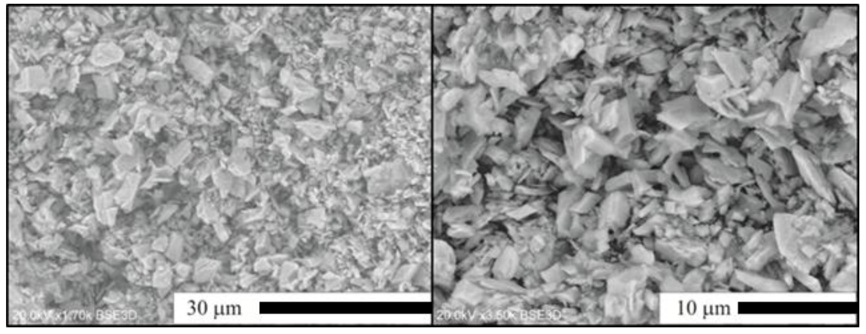
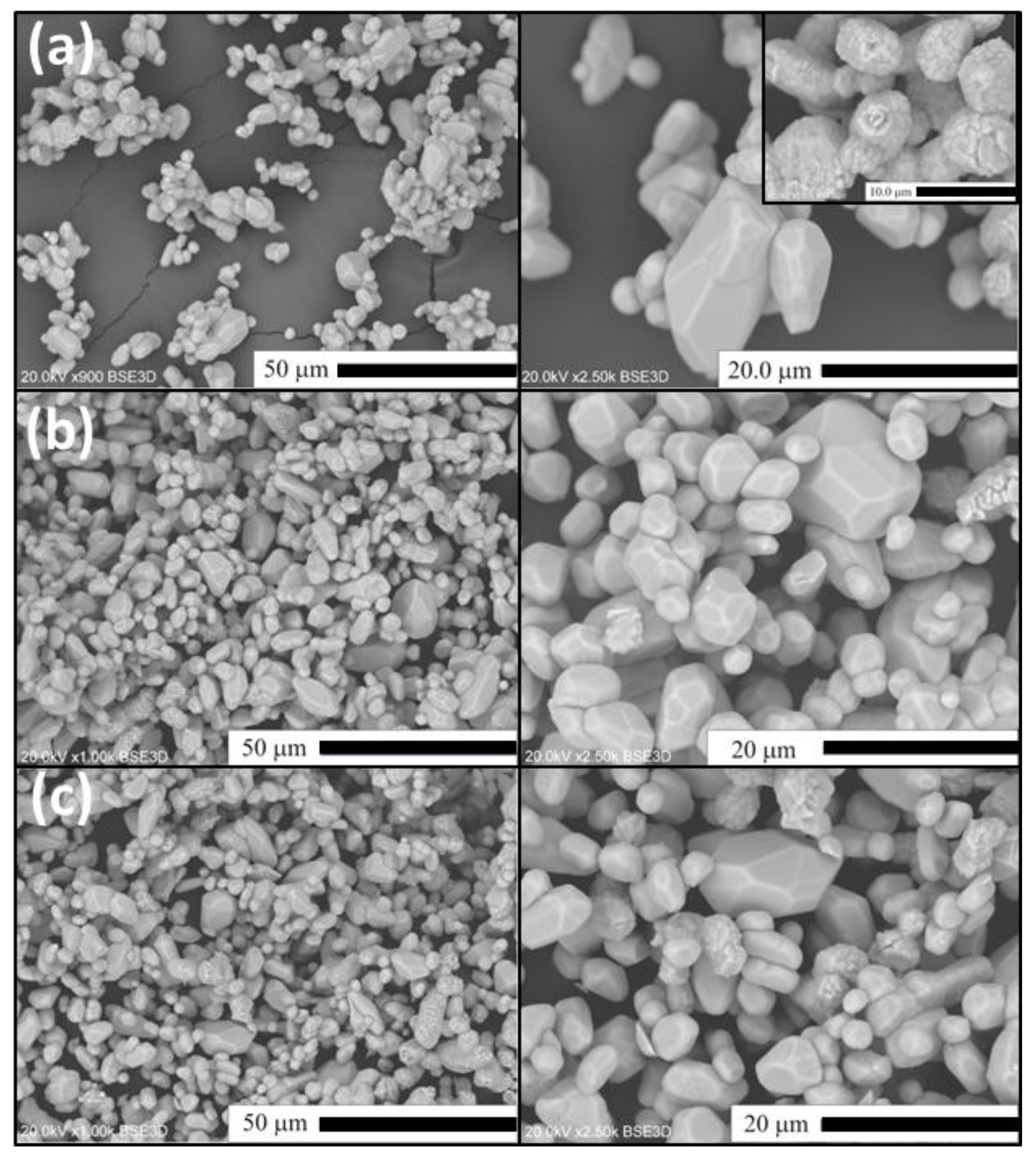
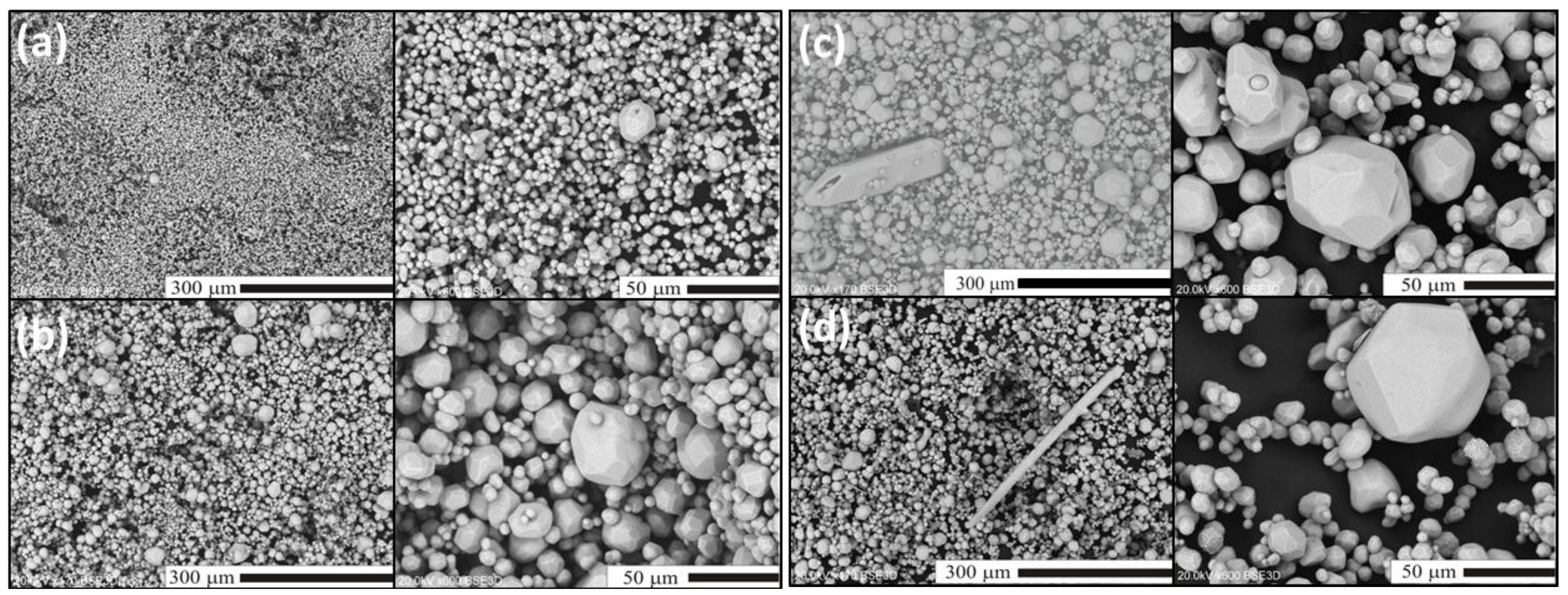
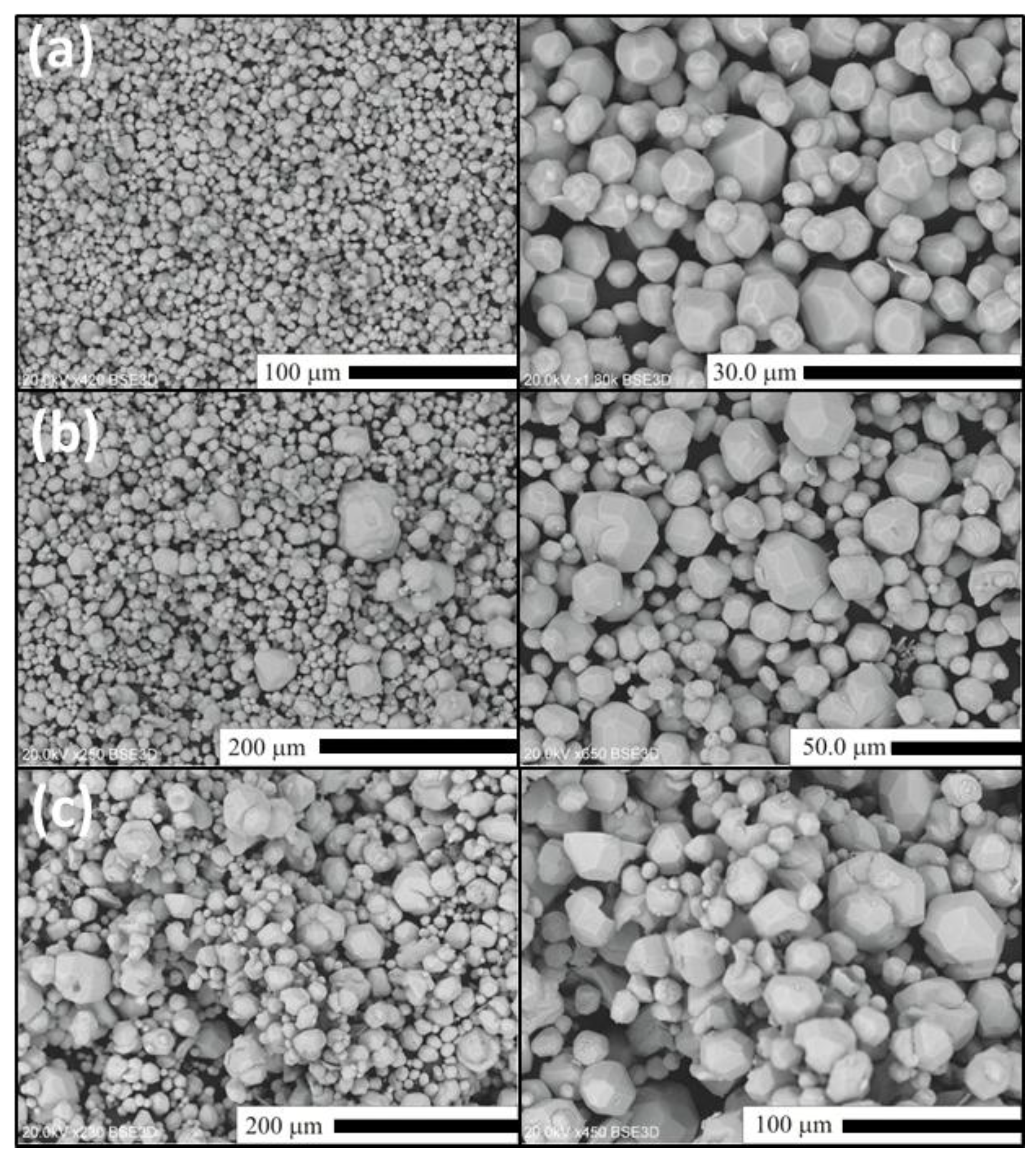
2.2. Flux-Stimulated Evolution of the Powders Morphology

In this section, we shall present and discuss differences in the morphology of Lu2O3 powders of the Series II–IV as well as evolution of the morphology within each series. For comparison, in Figure 3 we show scanning electron microscope (SEM) images of Lu2O3 starting material used in our synthesis processes. As we shall see, the morphology of the raw lutetia is much different from all morphology of the powders we synthesized using the flux. Figure 4 presents SEM images of Lu2O3 powders of Series II in which the flux consisted of Li2SO4 exclusively, and was used in different proportions to the Lu2O3. Clearly, alteration of the ratio of the flux against Lu2O3 did not affect the morphology to any significant degree. Basically, all powders of Series II consist of grains of similar sizes of 3–4 μm, though the largest of them are of about 20 μm in diameter. The grains are not very uniform when size is taken into account. The grains are mostly monocrystalline and form polyhedra of regular and similar shapes. Some granules consist of aggregated smaller crystallites (see the upper top image). These, though not very numerous, were seen in each specimen of Series II.
Figure 3. SEM images of the Lu2O3 starting materials (Stanford Materials Corporation).
Materials 07 07059 g003
Figure 4. SEM images of Lu2O3 were prepared using Series II synthesis for (a) SII-1; (b) SII-2; (c) SII-3.
Materials 07 07059 g004
In Series III (Figure 5), when synthesized using a mixture of Li2SO4 and SiO2 as the flux, the grains become more uniform in size, compared to Series II. For the SIII-1 powder the sizes are typically 3–6 μm and only occasionally 20 μm grains are observed (Figure 5a). In the case of sample SIII-2 (Figure 5b), when the content of SiO2 in the flux (compared to SIII-1) was roughly tripled, the grains become clearly larger. A six-fold increase of the SiO2 in the flux (compared to SIII-1) further increases, though not significantly, the fraction of the largest grains (Figure 5c), among which we can easily select 70–100 μm monocrystals. Rarely, needle-shaped grains are observed as presented in Figure 5c. The needles are as long as about 200–300 μm. Their fraction gets larger with an increasing amount of the flux compared to Lu2O3 (compare Figure 5c,d). Since for cubic structures the needle-like shape of monocrystalline grains is unexpected, we decided to perform a full structural analysis of such a crystal to unambiguously resolve if Lu2O3 can crystallize in a different structure in the conditions we applied for the preparations. The results will be presented in the next section.
We already mentioned when discussing XRD patterns that by using a mixture of Lu2O3 and Eu2O3 it was possible to produce a Lu2O3:Eu phosphor by means of the Li2SO4-SiO2 flux technique. The morphology of the material (see Figure 6) was not much different from its undoped counterparts presented above. Interestingly, it was routinely indicated by inductively coupled plasma (ICP) analysis that only about 60%–65% of Eu indeed entered the lutetia host during the flux-aided synthesis.
Figure 5. SEM images of Lu2O3 of samples of Series III. (a) SIII-1; (b) SIII-2; (c) SIII-3; (d) SIII-4. Note that some grans in SIII-3 and SIII-4 powders have the unexpected shape of needles.
Materials 07 07059 g005
Figure 6. SEM images of Lu2O3:Eu of samples of Series IV. (a) SIV-1; (b) SIV-2; (c) SIV-3.
Materials 07 07059 g006

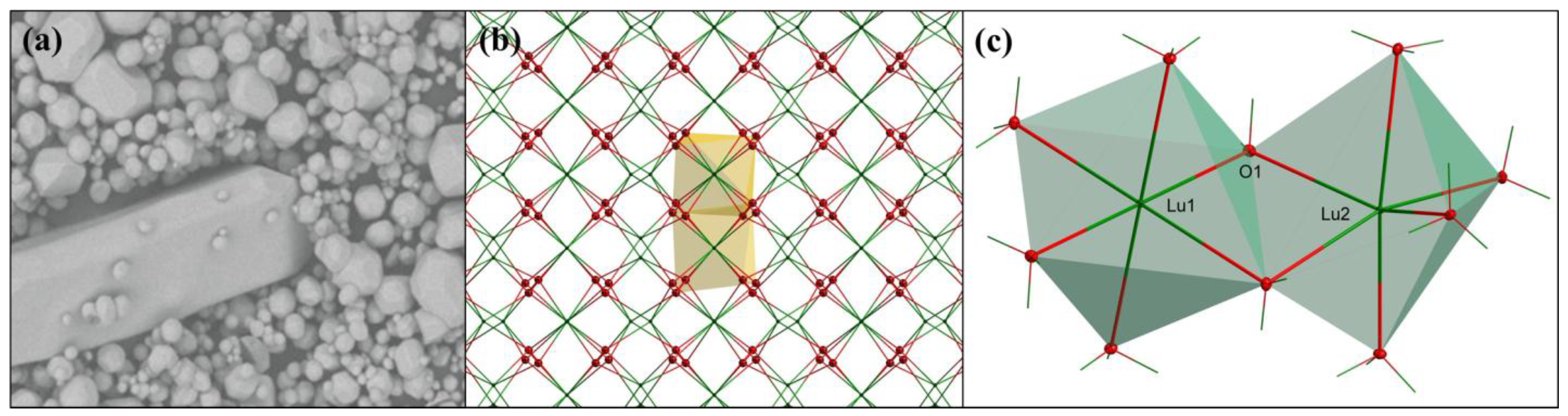
2.3. Single Crystal Structural Analysis

As we already saw, see Figure 5c,d, in some cases needle-shaped crystals could be selected within a batch. This observation encouraged us to verify the crystal structure of both the needles and the more regular, polyhedral particles. Both types of crystals gave practically the same results and here we shall present those for the needle-shaped crystal selected from the powder SIII-3 (see Figure 5c and Figure 7a). Below a detailed description of the results is given. It was unambiguously confirmed that this Lu2O3 crystal, despite its specific shape, crystallized in a cubic system, the space group is Ia 3 with a = 10.393(2) Å, V = 1122.6(6) Å3, Z = 16, T = 298 K. Other crystal data are listed in Table 1, together with refinement details. Complete crystallographic data for the structural analysis have been deposited with the Fachinformationszentium Karlsruhe (FIZ), CSD No. 428012. Further details of the crystal structure investigations may be obtained from Fachinformationszentrum Karlsruhe, Eggenstein-Leopoldshafen, Germany [38] on quoting the appropriate CSD number. The cubic cell parameter calculated using powder XRD measured for sample SIII-3 gave = 10.4216(9) Å. This is in good agreement with the value obtained for single crystal measurements.
Table 1. Crystal data and structure refinement for Lu2O3.
Chemical FormulaLu2O3
Formula Mass397.94
Crystal systemregular
a10.393(2)
Unit cell volume/Å31122.6(6)
Temperature/K298(2)
Space groupIa3
No. of formula units per unit cell, Z16
No. of reflections measured8753
No. of independent reflections1789
Rint0.0425
Final R1 values (I > 2σ(I))0.0287
Final wR(F2) values (I > 2σ(I))0.0555
Final R1 values (all data)0.0307
Final wR(F2) values (all data)0.0560
The Lu2O3 represents the rare-earth sequioxide C-type of structure, isostructural to Y2O3. Figure 7c shows that the crystal structure of Lu2O3 offers two different positions for the metal ion (Lu1 and Lu2). Lutetium atoms are connected by bridging O1 and O1i oxygen atoms. Coordination numbers (CN) of both Lu1 and Lu2 are CN = 6. Lu1 occupies a perfectly centrosymmetric position (C3i) in the lattice with all Lu1-O1with symmetry codes: (i) −y + 1/2, −z + 1/2, −x + 1/2; (ii) y, z, x; (iii) −x + 1/2, −y + 1/2, −z + 1/2; (iv) −z + 1/2, −x + 1/2, −y + 1/2; (v) z, x, y; distances equal 2.2392(18) Å. Lu2 atom possesses non-centrosymmetric C2 local site symmetry in the cubic structure of Lu2O3. The population of the Lu1 is 25% and the fraction of Lu2 is 75% of all metal sites. Figure 7b shows the packing of Lu2O3. Two types of layers of Lu sites are present. One of them contains only Lu2 atoms showing the non-centrosymmetric geometry (C2), while the other consists of the same number of Lu1 (C3i) and Lu2 (C2) ions. The crystal structure of Lu2O3 is stabilized by bridging oxygen atoms between lutetium atoms. Table 2 presents all geometric parameters of the structure.
Figure 7. (a) The grain of the powder SIII-3 selected for the monocrystalline structural measurements; (b) a packing diagram of Lu2O3. For clarity, only two polyhedra were shown; (c) coordination environment of Lu1 and Lu2 in Lu2O3.
Materials 07 07059 g007
Table 2. Geometric parameters (Å, °).
Atoms involved Distance (Å), Angle (°)
Lu1—O12.2392(18)
Lu1—Lu23.4395(7)
Lu2—O12.2277(19)
Lu2—O1i2.2945(19)
Lu2—O1x2.1954(18)
O1—Lu1—O1i80.10(6)
O1—Lu1—O1ii99.90(6)
O1—Lu1—O1iii180.0
O1x—Lu2—O1xi87.14(10)
O1x—Lu2—O199.66(6)
O1xi—Lu2—O1109.94(8)
O1—Lu2—O1xii138.89(9)
O1x—Lu2—O1i79.57(7)
O1xi—Lu2—O1i165.12(5)
O1—Lu2—O1i79.15(9)
O1x—Lu2—O1xiii165.12(5)
O1—Lu2—O1xiii78.91(6)
O1i—Lu2—O1xiii114.36(9)
Symmetry codes: (i) −y + 1/2, −z + 1/2, −x + 1/2; (ii) y, z, x; (iii) −x + 1/2, −y + 1/2, −z + 1/2; (x) −z + 1, −x + 1/2, y; (xi) −z + 1, x−1/2, −y + 1/2; (xii) x, −y, −z + 1/2; (xiii) −y + 1/2, z − 1/2, x.
The results presented here are generally very similar to the recently published data on the single crystal structure of Lu2O3 [37]. It may be surprising that for our crystal made with the Li2SO4 flux the final R1 (I > 2σ(I)) value is even smaller than for the two crystals reported in [37], which were made by a μ-PD and LHPG techniques. The unit cell of the one obtained by the μ-PD showed a slightly smaller size of a = 10.364(2) Å compared to that fabricated by LHPG, which was a = 10.403(2) Å. The difference was suggested to result from O-vacancies supposedly present in the slightly yellow μ-PD crystal formed in a reducing atmosphere. The size of the unit cell obtained for our crystal made by the flux technique is much closer (10.393(2) Å) to the size of the LHPG crystal, and is almost identical with the size of the unit cell obtained for sintered ceramics (10.39120(6) Å) [37].

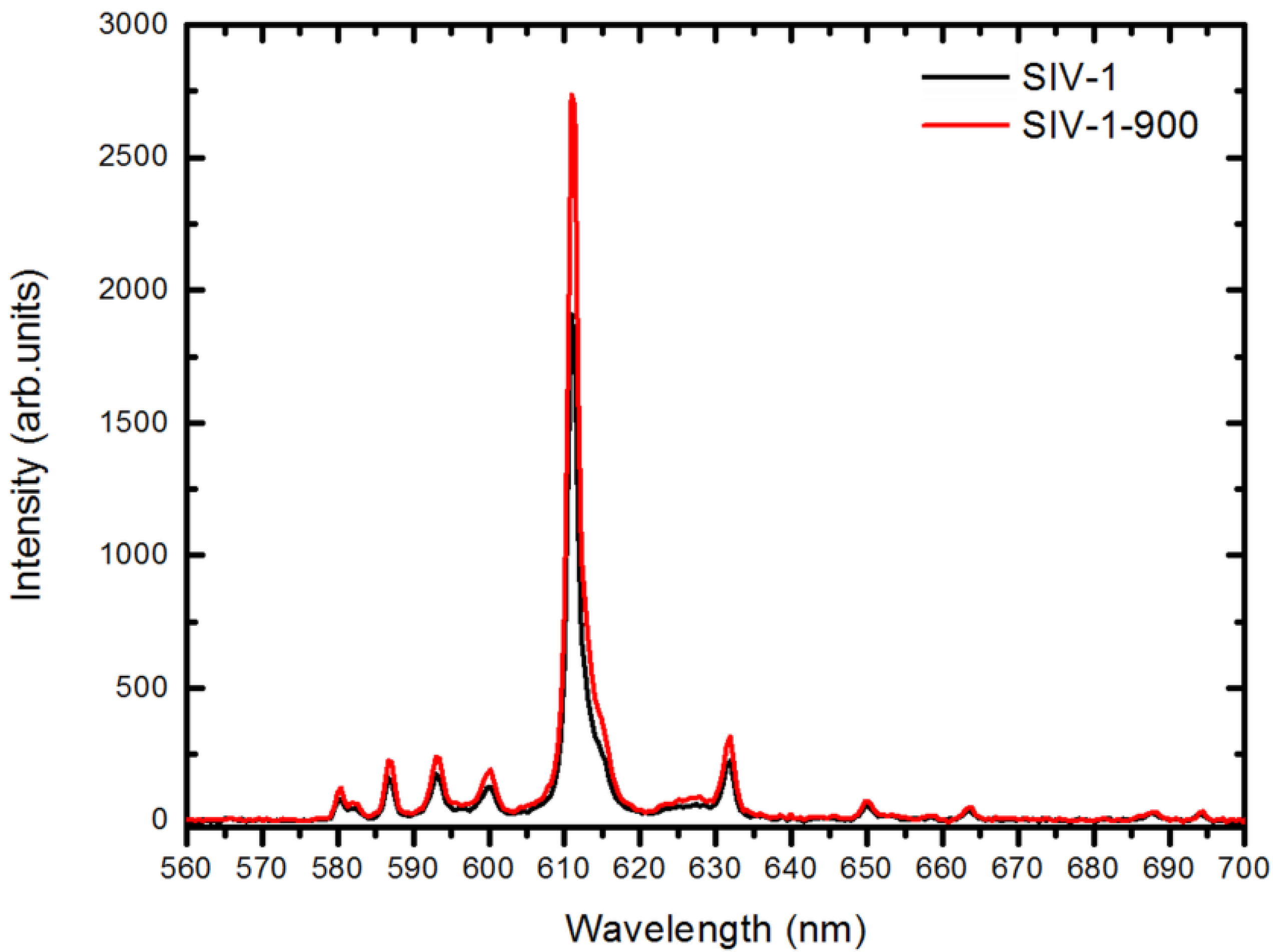
2.4. Radioluminescent Properties

As we mentioned in the Introduction, Lu2O3:Eu arouses interest mostly for its radioluminescent properties. Thus, having the possibility to control the morphology of its powders, we also were interested in testing their performance under excitation with X-rays. Figure 8 shows the X-ray excited luminescence (XEL) spectra of Lu2O3:Eu powders of Series IV. The spectral distribution of the emitted photons is typical for Lu2O3:Eu, with the main luminescence located at 610 nm and resulting from the 5D07F2 transition. The overall XEL efficiencies of Lu2O3:Eu were related to the performance of a commercial Gd2O2S:Eu (GOS:Eu) measured at the same conditions. All as-made samples performed similarly and on average their light yield reached 70%–80% of the GOS:Eu efficiency. Thus, the phosphor performance was not affected by the particle sizes to any significant degree. When the raw powders were additionally heated at 900 °C (SIV-1-900 and SIV-2-900) and 1200 °C (SIV-1-1200), the XEL efficiencies increased to 95%–105% of the commercial phosphor (see Figure 8 and Table 3). An increase in the formal concentration of Eu from 5% to 8% (samples SIV-3 versus SIV-4) in raw Lu2O3:Eu did not further affect the phosphor performance. Since Lu2O3:Eu can be used in layers when at least 1.5-fold thinner—due to its higher absorption of X-rays than Gd2O2S:Eu—it provides some advantage over GOS. On the other hand, its significantly higher price reduces its competitiveness. Hence, only a further enhancement of the luminescence efficiency under X-rays might make Lu2O3:Eu a real competitor for GOS:Eu. While using the (Li2SO4-SiO2) flux allows improving the phosphor morphology, the XEL efficiency, though high, is still too low to beat up the effective and reasonably priced GOS.
Figure 8. XEL emission spectra of the sample SIV-1 (black line) and SIV-1-900 (red line). See also Table 3.
Materials 07 07059 g008
Table 3. XEL efficiency of Lu2O3:Eu vs. commercially offered Gd2O2S:Eu.
Lu2O3:Eu sample #XEL Efficiency (%)
SIV-172
SIV-1-900105
SIV-1-120097
SIV-266
SIV-2-90095
SIV-380
SIV-475

3. Experimental Section

Lu2O3 and Eu-doped Lu2O3 powders were prepared by means of a Li2SO4 or (Li2SO4 + SiO2) flux (Li2SO4, Alfa Aesar, 99.7%, Karlsruhe, Germany; SiO2, Sigma Aldrich, ~99%, St. Louis, MO, USA) method using a mixture of commercially offered Lu2O3 (Stanford Materials Corporation, 99.995%, Aliso Viejo, CA, USA) and Eu2O3 (Stanford Materials Corporation, 99.999%). Four series of compounds were prepared and details are given in Table 4. Powders of series I–III contain no activator. Taking advantage of the knowledge on the results of Series I–III, fourth Eu-activated powders, Lu2O3:Eu (5 mol%), were prepared using mixtures of Li2SO4 and SiO2 as the flux (see Table 4 for details). These products gave Series IV of powders. To document the influence of Li2SO4 flux on the continuous removing of SiO2 from the reacting mixture, three samples (Series I) were prepared using the molar ratio of Lu2O3:SiO2 = 1:1, as in the Lu2SiO5, see Table 4 for other details. In typical synthesis, the mixture of the reagents and the flux was transferred to a Pt-Ir (90%–10%) crucible which was next heated up at 1400 °C for 50 h in air. In the case of Series I, the temperature was lowered to 1300 °C and the time varied from 1 up to 20 h. After heating, the mixture was cooled down to room temperature (RT). To recover the product, the Li2SO4 flux was washed out with hot water and finally the powder was dried at ~80 °C in air for a few hours. For radioluminescent measurements, fractions of samples SIV-1 and SIV-2 were additionally heated at 900 °C and 1200 °C to see how this treatment affects their performance. These samples are named SIV-1-900 and SIV-2-900.
The powder X-ray diffraction patterns were measured using a D8 Advance Diffractometer from Bruker (Billerica, MA, USA) with Ni-filtered CuKα radiation (λ = 1.540596 Å) in the range of 2θ = 10°–100°, and with the step of 2θ = 0.008°. Single crystal X-ray diffraction data for undoped Lu2O3 were collected at room temperature using the ω-scan technique on Xcalibur diffractometer (Agilent Technologies, Santa Clara, CA, USA) equipped with Ruby CCD-detector using graphite-monochromatized MoKα radiation (λ = 0.71073 Å) [39]. The data were corrected for Lorentz-polarization effects and for absorption. The structure was refined with the full-matrix least-squares procedure on F2 by SHELXL [40] on coordinates of atoms were taken from a previously reported isomorphous crystal of Y2O3. All atoms were refined anisotropically. The products’ morphology was tested by means of SEM imaging with Hitachi S-3400N scanning electron microscope (Hitachi High-Technologies, Tokyo, Japan) equipped with an energy dispersive X-ray spectroscopy (EDX) EDAX analyzer. The room temperature X-ray excited luminescence—measurements of the Eu-activated powders of Series IV were performed using white radiation from a Cu X-ray tube working under the voltage of 40 kV and a current of 10 mA. The generated emission photons were collected with a 74-UV lens connected to a QP600-2-SR-BX waveguide which transferred the luminescent light to an Ocean Optics HR2000CG-UV-NIR Spectrometer (Ocean Optics, Dunedin, FL, USA). Efficiency of the XEL was estimated using a commercially offered powder of Gd2O2S:Eu as the benchmark kindly supplied by Phosphor Technology. The real content of Eu in Lu2O3:Eu powders was determined by means of Inductively Coupled Plasma (ICP) technique using an ARL 3410 ICP Spectrometer (Fisons Instruments, Ecublens, Switzerland).
Table 4. Exemplary amounts of starting materials for the four series of synthesized powders.
SeriesSample #Batch Composition (g)Process ParametersProduct Composition
Lu2O3Eu2O3Li2SO4SiO2Temperature (°C)/Time (h)
ISI-11.758513.970.26611300/1Lu2SiO5, Lu2O3
SI-21.758513.970.26611300/2Lu2SiO5, Lu2O3
SI-31.758513.970.26611300/20Lu2O3
IISII-1221400/50Lu2O3
SII-224.77Lu2O3
SII-329.54Lu2O3
IIISIII-124.770.045Lu2O3
SIII-224.770.15Lu2O3
SIII-324.770.3Lu2O3
SIII-429.540.3Lu2O3
IVSIV-11.90.08844.770.045Lu2O3
SIV-21.90.08849.540.3Lu2O3
SIV-31.90.08844.770.3Lu2O3
SIV-41.840.14154.770.3Lu2O3

4. Conclusions

In this paper, we showed that Li2SO4-flux-aided synthesis of Lu2O3 and Lu2O3:Eu gives non-agglomerated powders with particles whose sizes may be controlled to some extent by addition of some SiO2 to the flux. No traces of silica were detected in the final product. Large enough single crystals could be selected to perform structural analysis. This confirmed that lutetia is isostructural with cubic yttria and a = 10.393(2) Å. This is in agreement with very recently published data on Lu2O3 structure determined on single crystals made by μ-PD and LHPG techniques. X-ray excited luminescence spectra were typical for Lu2O3:Eu and the light yield reached 95%–105% of the yield of commercial GOS:Eu, which is not enough to compete effectively with this phosphor in practical applications.

Acknowledgments

The work was supported by Wroclaw Research Centre EIT+ within the project “The Application of Nanotechnology in Advanced Materials”—NanoMat (POIG.01.01.02-02-002/08) co-financed by the European Regional Development Fund (Innovative Economy Operational Program 1.1.2).

Author Contributions

Justyna Zeler fabricated the powders, recorded the SEM images and measured the RL spectra, Lucjan B. Jerzykiewicz preformed the structural measurements and related calculations, Eugeniusz Zych directed the project. All authors analyzed the results and contributed to writing.

Conflicts of Interest

The authors declare no conflict of interest.

References

  1. Miller, S.R.; Nagarkar, V.V.; Tipnis, S.V.; Shestakova, I.; Brecher, C.; Lempicki, A.; Lingertat, H. Lu2O3:Eu scintillator screen for X-ray imaging. Proc. SPIE 2004, 5199, 167–172. [Google Scholar]
  2. Nagarkar, V.V.; Tipnis, S.V.; Miller, S.R.; Lempicki, A.; Brecher, C.; Szczupryczynski, P.; Lingertat, H. A new X-ray scintillator for digital radiography. IEEE Trans. Nucl. Sci. 2003, 50, 297–300. [Google Scholar] [CrossRef]
  3. Nagarkar, V.V.; Miller, S.R.; Tipnis, S.V.; Lempicki, A.; Brecher, C.; Lingertat, H. A new large area scintillator screen for X-ray imaging. Nucl. Instrum. Methods Phys. Res. Sect. B Beam Interact. Mater. Atoms 2004, 213, 250–254. [Google Scholar] [CrossRef]
  4. Marton, Z.; Bhandari, H.; Brecher, C.; Miller, S.R.; Singh, B.; Nagarkar, V.V. High performance microstructured Lu2O3:Eu thin film scintillator for X-ray computed tomography. Proc. SPIE 2013, 8668. [Google Scholar] [CrossRef]
  5. Garcia, A.; Le Luyer, C.; Pedrini, C.; Mugnier, J. Synthesis and properties of Lu2O3 sol-gel films. J. Alloy. Compd. 2001, 323, 4–77. [Google Scholar]
  6. Garcia, A.; Le Luyer, C.; Dujardin, C.; Martin, T.; Garapon, C.; Pédrini, C.; Mugnier, J. Elaboration and scintillation properties of Eu3+-doped Gd2O3 and Lu2O3 sol-gel films. Nucl. Instrum. Methods Phys. Res. Sect. A Accel. Spectrom. Detect. 2002, 486, 181–185. [Google Scholar] [CrossRef]
  7. Daldosso, M.; Sokolnicki, J.; Kepinski, L.; Legendziewicz, J.; Speghini, A.; Bettinelli, M. Preparation and optical properties of nanocrystalline Lu2O3:Eu3+ phosphors. J. Lumin. 2007, 122–123, 858–861. [Google Scholar]
  8. Sokolnicki, J. Photoluminescence and structural characteristics of Lu2O3:Eu3+ nanocrystallites in silica matrix. J. Solid State Chem. 2007, 180, 2400–2408. [Google Scholar] [CrossRef]
  9. Yermolayeva, Y.V.; Tolmachev, A.V.; Dobrotvorskaya, M.V.; Vovk, O.M. Preparation and structural properties of Lu2O3:Eu3+ submicrometer spherical phosphors. J. Alloy. Compd. 2011, 509, 5320–5325. [Google Scholar] [CrossRef]
  10. Dulina, N.A.; Baumer, V.N.; Danylenko, M.I.; Mateychenko, P.V.; Tolmachev, A.V.; Vovk, O.M.; Yavetskiy, R.P. Effects of phase and chemical composition of precursor on structural and morphological properties of (Lu0.95Eu0.05)2O3 nanopowders. Ceram. Int. 2013, 39, 2397–2404. [Google Scholar] [CrossRef]
  11. Antic, Z.; Krsmanovic, R.; Wojtowicz, M.; Zych, E.; Bartova, B.; Dramicanin, M.D. Preparation, structural and spectroscopic studies of (YxLu1−x)2O3:Eu3+ nanopowders. Opt. Mater. 2010, 32, 1612–1617. [Google Scholar] [CrossRef]
  12. Trojan-Piegza, J.; Zych, E. Preparation of nanocrystalline Lu2O3:Eu phosphor via a molten salts route. J. Alloy. Compd. 2004, 380, 118–122. [Google Scholar] [CrossRef]
  13. Trojan-Piegza, J.; Zych, E.; Hreniak, D.; Strek, W. Comparison of spectroscopic properties of nanoparticulate Lu2O3:Eu synthesized using different techniques. J. Alloy. Compd. 2004, 380, 123–129. [Google Scholar] [CrossRef]
  14. Li, R.; Gai, S.; Wang, L.; Wang, J.; Yang, P. Facile synthesis and multicolor luminescent properties of uniform Lu2O3:Ln (Ln = Eu3+, Tb3+, Yb3+/Er3+, Yb3+/Tm3+, and Yb3+/Ho3+) nanospheres. J. Colloid Interface Sci. 2012, 368, 165–171. [Google Scholar] [CrossRef]
  15. Xu, M.; Zhang, W.; Dong, N.; Jiang, Y.; Tao, Y.; Yin, M. Preparation and characterization of optical spectroscopy of Lu2O3:Eu nanocrystals. J. Solid State Chem. 2005, 178, 477–482. [Google Scholar] [CrossRef]
  16. Guo, H.; Yin, M.; Dong, N.; Xu, M.; Lou, L.; Zhang, W. Effect of heat-treatment temperature on the luminescent properties of Lu2O3:Eu film prepared by Pechini sol–gel method. Appl. Surf. Sci. 2005, 243, 245–250. [Google Scholar] [CrossRef]
  17. Wang, Z.; Zhang, W.; Lin, L.; You, B.; Fu, Y.; Yin, M. Preparation and spectroscopic characterization of Lu2O3:Eu3+ nanopowders and ceramics. Opt. Mater. 2008, 30, 1484–1488. [Google Scholar] [CrossRef]
  18. Chen, Q.; Shi, Y.; An, L.; Wang, S.; Chen, J.; Shi, J. A novel co-precipitation synthesis of a new phosphor Lu2O3:Eu3+. J. Eur. Ceram. Soc. 2007, 27, 191–197. [Google Scholar] [CrossRef]
  19. Liu, X.-J.; Li, H.-L.; Xie, R.-J.; Hirosaki, N.; Xu, X.; Huang, L.-P. Synthesis, characterization, and luminescent properties of Lu2O3:Eu phosphors. J. Lumin. 2007, 127, 469–473. [Google Scholar] [CrossRef]
  20. Sanghera, J.; Kim, W.; Villalobos, G.; Shaw, B.; Baker, C.; Frantz, J.; Sadowski, B.; Aggarwal, I. Ceramic laser materials. Materials 2012, 5, 258–277. [Google Scholar] [CrossRef]
  21. Kim, W.; Baker, C.; Bowman, S.; Florea, C.; Villalobos, G.; Shaw, B.; Sadowski, B.; Hunt, M.; Aggarwal, I.; Sanghera, J. Laser oscillation from Ho3+ doped Lu2O3 ceramics. Opt. Mater. Express 2013, 3. [Google Scholar] [CrossRef]
  22. Lempicki, A.; Brecher, C.; Szczupryczynski, P.; Lingertat, H.; Nagarkar, V.V.; Tipnis, S.V.; Miller, S.R. A new lutetia-based ceramic scintillator for X-ray imaging. Nucl. Instrum. Methods Phys. Res. Sect. A Accel. Spectrom. Detect. Assoc. Equip. 2003, 488, 579–570. [Google Scholar] [CrossRef]
  23. Zych, E.; Trojan-Piegza, J.; Kepinski, L. Homogeneously precipitated Lu2O3:Eu nanocrystalline phosphor for X-ray detection. Sens. Actuators B Chem. 2005, 109, 112–118. [Google Scholar] [CrossRef]
  24. Farman, T.T.; Vandre, R.H.; Pajak, J.C.; Miller, S.R.; Lempicki, A.; Farman, A.G. Effects of scintillator on the detective quantum efficiency (DQE) of a digital imaging system. Oral Surg. Oral Med. Oral Pathol. Oral Radiol. Endod. 2006, 101, 219–223. [Google Scholar] [CrossRef]
  25. Zych, E. Spectroscopy of Eu-activated Lu2O3 X-ray phosphors. In Frontal Semiconductor Research; Nova Science Publishers: Hauppauge, NY, USA, 2006; pp. 1–24. [Google Scholar]
  26. Lempicki, A.; Wojtowicz, A.J.; Brecher, C. Wide-Gap Luminescent Materials: Theory and Applilcations; Kluwer AcademicPublishers: Dordrecht, The Netherlands, 1966; pp. 235–301. [Google Scholar]
  27. Zych, E. Luminescence and Scintillation of Inorganic Phosphor Materials. In Handbook of Luminescence, Display Materials, and Devices. Inorganic Display Materials; Vollume 2, Academic Scientific: San Francisco, CA, USA, 2003; pp. 251–300. [Google Scholar]
  28. Seeley, Z.; Cherepy, N.; Payne, S. Two-step sintering of Gd0.3Lu1.6Eu0.1O3 transparent ceramic scintillator. Opt. Mater. Express 2013, 3. [Google Scholar] [CrossRef]
  29. Seeley, Z.M.; Dai, Z.R.; Kuntz, J.D.; Cherepy, N.J.; Payne, S.A. Phase stabilization in transparent Lu2O3:Eu ceramics by lattice expansion. Opt. Mater. 2012, 35, 74–78. [Google Scholar] [CrossRef]
  30. Seeley, Z.M.; Kuntz, J.D.; Cherepy, N.J.; Payne, S.A. Transparent Lu2O3:Eu ceramics by sinter and HIP optimization. Opt. Mater. 2011, 33, 1721–1726. [Google Scholar] [CrossRef]
  31. Kopylov, Y.L.; Kravchenko, V.B.; Dulina, N.A.; Lopin, A.V.; Parkhomenko, S.V.; Tolmachev, A.V.; Yavetskiy, R.P.; Zelenskaya, O.V. Fabrication and characterization of Eu3+-doped Lu2O3 scintillation ceramics. Opt. Mater. 2013, 35, 812–816. [Google Scholar] [CrossRef]
  32. Zych, E.; Hreniak, D.; Strek, W.; Kepinski, L.; Domagala, K. Sintering properties of urea-derived Lu2O3-based phosphors. J. Alloy. Compd. 2002, 341, 391–394. [Google Scholar] [CrossRef]
  33. Wang, J.; Liu, Q.; Liu, Q. Controlled synthesis of europium-doped lutetium compounds: Nanoflakes, nanoquadrels, and nanorods. J. Mater. Chem. 2005, 15, 4141–4146. [Google Scholar] [CrossRef]
  34. Zhao, Q.; Guo, N.; Jia, Y.; Lv, W.; Shao, B.; Jiao, M.; You, H. Facile surfactant-free synthesis and luminescent properties of hierarchical europium-doped lutetium oxide phosphors. J. Colloid Interface Sci. 2013, 394, 216–222. [Google Scholar] [CrossRef]
  35. Cascales, C.; Zaldo, C.; Esteban-Betegon, F.; Calderon-Villajos, R. From porous to dense Tm3+-Lu2O3 micro-and nanosized crystalline morphologies designed through hydrothermal precursors: Assessment on infrared emission properties. CrystEngComm 2012, 14, 3577–3585. [Google Scholar] [CrossRef]
  36. Dulina, N.A.; Yermolayeva, Y.V.; Tolmachev, A.V.; Sergienko, Z.P.; Vovk, O.M.; Vovk, E.A.; Matveevskaya, N.A.; Mateychenko, P.V. Synthesis and characterization of the crystalline powders on the basis of Lu2O3:Eu3+spherical submicron-sized particles. J. Eur. Ceram. Soc. 2010, 30, 1717–1724. [Google Scholar] [CrossRef]
  37. Guzik, M.; Pejchal, J.; Yoshikawa, A.; Ito, A.; Goto, T.; Siczek, M.; Lis, T.; Boulon, G. Structural investigations of Lu2O3 as single crystal and polycrystalline transparent ceramic. Cryst. Growth Des. 2014, 14, 3327–3334. [Google Scholar] [CrossRef]
  38. FIZ Karlsruhe. Available online: http://www.fiz-karlsruhe.de/request_for_deposited_data.html (accessed on 26 September 2014).
  39. Agilent, CrysAlis PRO; Agilent Technologies: Santa Clara, CA, USA, 2011.
  40. Sheldrick, G.M. A Short History of SHELX. Acta Crystallogr 2008, A64, 112–122. [Google Scholar] [CrossRef]

