Review

Effect of Lignocellulose Related Compounds on Microalgae Growth and Product Biosynthesis: A Review

1
AgricultureIsLife Platform, University of Liege-Gembloux Agro-Bio Tech, Passage des Déportés 2, B-5030 Gembloux, Belgium
2
Genetics of Microorganisms, Institute of Botany, University of Liege, B22, 27, Bld du Rectorat, B-4000 Liège, Belgium
3
Unit of Biological and Industrial Chemistry, University of Liege-Gembloux Agro-Bio Tech, Passage des Déportés 2, B-5030 Gembloux, Belgium
4
Cellule Innovation et Créativité, University of Liege-Gembloux Agro-Bio Tech, Passage des Déportés, 2, B-5030 Gembloux, Belgium
*
Author to whom correspondence should be addressed.
Energies 2014, 7(7), 4446-4481; https://doi.org/10.3390/en7074446
Submission received: 4 May 2014 / Revised: 30 June 2014 / Accepted: 1 July 2014 / Published: 11 July 2014
(This article belongs to the Special Issue Algae Based Technologies)

Abstract

:
Microalgae contain valuable compounds that can be harnessed for industrial applications. Lignocellulose biomass is a plant material containing in abundance organic substances such as carbohydrates, phenolics, organic acids and other secondary compounds. As growth of microalgae on organic substances was confirmed during heterotrophic and mixotrophic cultivation, lignocellulose derived compounds can become a feedstock to cultivate microalgae and produce target compounds. In this review, different treatment methods to hydrolyse lignocellulose into organic substrates are presented first. Secondly, the effect of lignocellulosic hydrolysates, organic substances typically present in lignocellulosic hydrolysates, as well as minor co-products, on growth and accumulation of target compounds in microalgae cultures is described. Finally, the possibilities of using lignocellulose hydrolysates as a common feedstock for microalgae cultures are evaluated.

1. Microalgae: A Source of Valuable Compounds

Microalgae include several groups of microorganisms that belong to the Prokaryota or Eukaryota, typically found in fresh water or marine systems in single cell forms or in groups. They are capable of performing photosynthesis, producing approximately half of the atmospheric oxygen while using the greenhouse gas carbon dioxide to grow photoautotrophically [1]. Microalgae contain valuable compounds such as lipids, proteins and pigments (Table 1) which have substantial potential for commercial applications. Microalgae cells accumulate lipids which include triacylglycerides (TAGs), polyunsaturated fatty acids (PUFAs) and sterols [2]. These lipids constitute storage materials or membrane structural components in microalgae and can be used for biofuel, food supplement and pharmaceutical production. Indeed, fossil fuels are nowadays still the main source of carbon based fuels, the exploitation of which causes emission of greenhouse CO2. Biodiesel production from oil crops is seen as currently the best alternative, but still presents the main drawback of competing with food production for arable land. Therefore, production of biodiesel from TAGs present in microalgae can become an environmentally friendly alternative as microalgae produce oil and fix CO2 from atmosphere without the necessity of implementing vast arable areas for cultivation [3]. On the other hand, consumption of PUFAs in the human diet can help prevent the development of cardiovascular and mental diseases [4]. Fish are a rich source of PUFAs, but uncontrolled fishing has led to a substantial decrease in the worldwide fish population [5]. Production of PUFAs from microalgae may overcome this problem. On the other hand, sterols from microalgae are important part of the diet for juvenile scallops or prawns in aquaculture hatcheries [6]. Moreover, the high protein content in microalgae makes them a possible fodder for agricultural livestock [7]. In addition, microalgae cells also possess pigments such as chlorophylls and carotenoids. Chlorophylls harvest solar light in the process of photosynthesis while carotenoids are accessory pigments that increase the range of sun light used for photosynthesis (β-carotene) or protect the photosynthetic mechanism against photodamage induced due to environmental stress conditions (astaxanthin, lutein) [8]. Those pigments can be used as food colorants and cosmetic additives against UV light or as pharmacological agents because of their wound healing and anticancer properties [9,10].
In order to make production of value-added compounds from microalgae an economically feasible process, it is necessary to produce high amount of microalgae biomass. Microalgae are cultivated in open ponds or different types of photobioreactors (see below, Section 5).
However, high culture densities are not achievable in scaled-up systems due to light limitations [11]. Many strains of microalgae are able to consume sugars, alcohols and organic acids as a source of carbon during heterotrophic or mixotrophic cultivation [12,13,14]. During heterotrophic growth, microalgae are cultivated in the dark, assimilate organic substances from the medium to cover energy requirements and release carbon dioxide. During mixotrophic cultivation, microalgae consume CO2 using light energy as well as external organic compounds from environment. High microalgae biomass concentrations can be achieved during cultivation in large-volume bioreactors when organic substances are applied [15]. The ability of microalgae to grow on organic substrates raises the possibility of cultivating microalgae on lignocellulose feedstock and thus reduce cultivation costs and increase productivity. Lignocellulose is the world’s most abundantly available raw plant material that can become a promising feedstock for microorganisms such as bacteria, yeasts and fungi to produce high value added products and biofuels [16,17,18].
Table 1. Taxonomy classification of microalgae with cellular component content *.
TaxonomyMicroalgae
DomainEucaryota
DivisionChlorophyta
ClassChlorophyceaeTrebouxiophyceae
OrderSphaeroplealesVolvocaes 1ChlamydomonadalesChlorellales
FamilyScenedesmaceaeHaematococcaceaeDunaliellaceaeChlamydomonadaceaeChlorellaceae
GenusAcutodesmusHaematococcusDunaliellaChlamydomonasChlorella
SpeciesScenedesmus obliquusHaematococcus pluvialisDunaliella salinaChlamydomonas reinhardtiiChlorella prothotecoidesChlorella zoofingiensisChlorella vulgaris
ContentProteins 51%Pigments 1.5%Pigments 11%Lipids 65%Lipids 62%Lipids 54%Proteins 46%
Reference[19][20][21][22][23][24][25]
DomainEucaryota
DivisionHeterokontophytaDinophyta 2EuglenophytaHeterokontophytaChlorophyta
ClassBacillarophyceaeDinophyceaeEuglenophyceaeEustigmatophyceaeChlorophyceaeChlorophyceae 3
OrderNaviculalesDinotrichales 2EuglenalesEustigmatalesSphaeroplealesChlorococcalesChlorococcales 3
FamilyPhaeodactylaceaeCrypthecodiniaceaeEuglenaceaeMonodopsidaceaeSelenastraceaeChlorococcaceaeDictyosphaeriaceae 3
GenusPhaeodactylumCrypthecodiniumEuglenaNannochloropsisMonoraphidiumNeochlorisBotryococcus
SpeciesPhaeodyctylum tricornutumCrypthecodinium cohniiEuglena gracilisNannochloropsis oculataMonoraphidium contortumNeochloris oleoabundansBotryococcus braunii
ContentLipids 20%Lipids 20%Lipids 29%Lipids 32%Lipids 30%Lipids 52%Lipids 65%
Reference[26][27][28][29][30][31][32]
DomainProcaryota
DivisionCyanobacteria
ClassCyanophyceae
OrderNostocales 3Chroococcales 4ChroococcalesChroococcales 5Oscillatoriales 6
FamilyNostocaceae 3Spirulinaceae 4MicrocystaceaeOscillatoriaceae
GenusAnabaenaSpirulinaMicrocystisThermosynechococcusOscillatoria
SpeciesAnabaena azollaeSpirulina platensisMicrocystis aeruginosaThermosynechococcus elongatesOscillatoria acuminata
ContentProteins 40%Proteins 67%Lipids 28%Lipids 20%Lipids 25%
Reference[33][34][35][36][35]
* Cellular content values are expressed as % of dry weight and may vary markedly depending on growth conditions such as light intensity, CO2 concentration, N deprivation, temperature or the presence of organic substrates; Source: AlgaeBase, Integrated Taxonomic Information System (ITIS) Report, PATRIC (Pathosystems Resource Integration Center). 1 According to ITIS Report Order: Volvocales, according to AlgaeBase Order: Chlamydomonadales; 2 According to ITIS Report Division: Pyrrophycophyta, Order: Gonyaulacales, according to AlgaeBase: Division: Dinophyta, Order: Dinotrichales; 3 According to ITIS Report; 4 According to AlgaeBase; 5 According to PATRIC; 6 According to Algae Base Order: Oscillatoriales, according to ITIS Report Order: Nostocales.
In this publication, the effect of lignocellulose feedstocks on microalgae growth and production of target compounds from microalgae culture is evaluated.

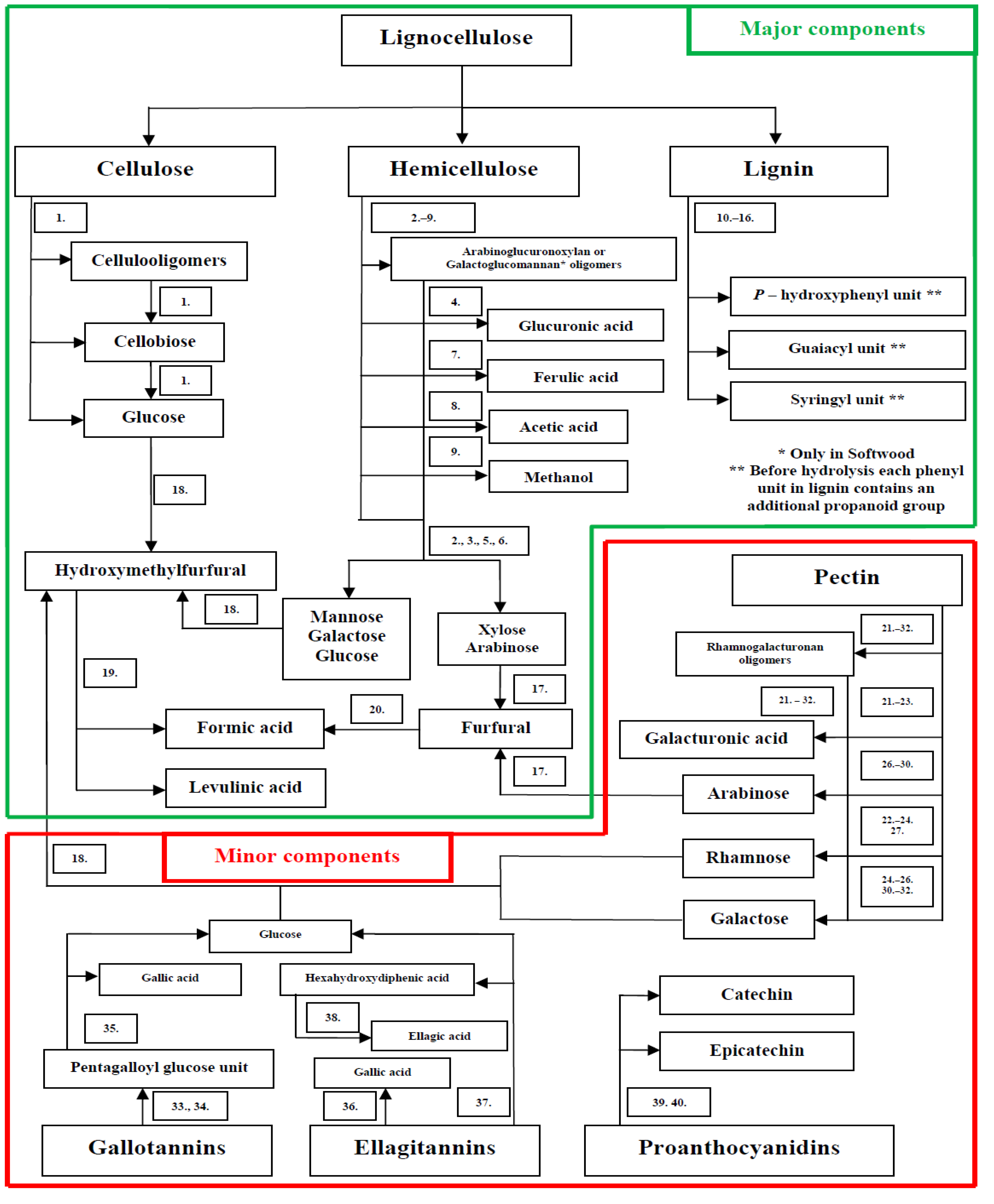
2. Composition and Treatment of Lignocellulose Materials

Lignocellulosic materials can be found in a large variety of plants such as coniferous trees (softwood), broad leave trees (hardwood), grasses and agricultural residues (Table 2). Lignocellulose is composed of three main biopolymers: namely cellulose, hemicelluloses and lignin. Cellulose, is a non-branched polymer consisting of d–glucopyranose units (hexoses) connected via β-(1,4)–glycosidic linkages. Hemicellulose is a complex carbohydrate polymer containing pentoses (mainly xyloses in the case of xylan—the main constituent of hardwood, grasses and agricultural wastes) and hexoses (typically mannoses in the case of mannan found principally in softwood), as the main sugars, bonded with β-(1,4)–glycosidic linkages. Unlike cellulose, many compounds such as saccharide residues and organic acids (glucuronic acid GluA), organic acid groups (acetyl) or lignin components are attached to the main sugar chain giving hemicellulose a branched structure. Finally, lignin is a complex biopolymer that consists of phenylpropanoid units such as hydroxyphenyl, guaiacyl and syringyl, which are connected to each other via various ether and carbon—carbon bonds. Cellulose chains are arranged in bundles and interlinked with hemicellulose. Lignin is cross-linked with hemicellulose and occupies space between cellulose bundles [37,38]. Plant materials also contain starch and small amounts of pectins, proteins, minerals, lipids, terpenoids, polyphenols and alkaloids. Starch is composed of amylose and amylopectin and serves as a storage material. When photosynthesis can not take place, glucose from starch provides energy that is used by plants to perform survival functions [39]. Starch constitutes 0.4% of straw [40], but its content in wheat bran can be 34% [41]. Pectin is a polysaccharide possessing in structure with d–galacturonic acid (GalA) as main units that are connected via α-(1,4)–glycosidic linkages. Additionally, regions composed of galacturonic acids are connected together via rhamnose (Rha) to which galactose (Gal) and arabinose (Ara) chains are also attached [42]. In plant material, mineral elements: Ca, K, Mg, Na, P, Fe, Mn and Si, are combined with organic molecules or are present in a form of inorganic salts [43,44]. Lipids in lignocellulose comprise a wide range of different compounds including fatty acids, glycerides, sterols and waxes [45]. Terpenoids such as monoterpenes and diterpenes can be found in softwood residues [46,47] and triterpenes are constituents of hardwood bark [48]. Polyphenols are found in softwood and hardwood and are represented by a wide range of compounds including gallotannins and ellagitannins, proanthocyanidins, flavonoids, lignans and stilbenes [49,50,51]. A variety of alkaloids can also be present in grasses [52] or the wood of tropical plants [53]. Lignocellulose sugars constitute a feedstock for bacteria, yeast and fungi to produce a variety of target compounds. Xylitol, a sweetener, was produced from xylose by Candida guilliermondii [17], while Saccharomyces cerevisae [54] and Clostridium beijerinckii [55] convert sugars to ethanol and butanol, respectively—second generation biofuels. Itaconic acid, a building block for fibers and rubbers, was produced by Ustilago maydis from xylose or glucose [18] and hydrogen, a third generation biofuel, is obtained from sugars with the use of Caldicellulosiruptor saccharolyticus [16]. However, the presence of three major polymers and other minor substances in lignocellulose, their rigidity and strong structure make the access to valuable carbohydrates very complicated. A number of methods have been developed to successfully breakdown this recalcitrant polymer structure and efficiently hydrolyse lignocellulosic materials (Figure 1).
Table 2. Exemplary composition of different lignocellulosic materials.
Component *Triticum Aestivum BranCorn StoverCynodon Dactylon GrassHordeum Vulgare Brewer’s Spent GrainOryza Sativa StrawPicea Abies SoftwoodSaccharum Officinarum BagasseSalix Hardwood
Glucan 110.536.130.416.735.940.935.843.0
Xylan18.321.422.619.919.05.121.214.9
Mannan1.80.010.10.793.2
Galactan1.12.51.81.90.742.0
Arabinan10.13.54.98.43.11.01.941.2
Klason lignin5.017.2 318.822.913.627.7 316.624.2
AS lignin 24.44.83.31.62.4
Reference[41][56][57][58][59][60][61][62]
* Values are expressed as % of dry material; 1 From cellulose; 2 AS—Acid Soluble; 3 A sum of Acid Soluble and Klason lignin.
Figure 1. Lignocellulose structure breakdown: hydrolysis of specific bonds and possible pathways for release of lignocellulose derived compounds. 1. Hydrolysis of O–glycosidic bond β (C1→C4) between Carbon 1 of Glu and Carbon 4 of another Glu. 2. Hydrolysis of O–glycosidic bond β (C1→C4) between Carbon 1 of one Xyl and Carbon 4 of another Xyl. 3. Hydrolysis of O–glycosidic bond β (C1→C4) between Carbon 1 of Man or Glu and Carbon 4 of Man or Glu. 4. Hydrolysis of O–glycosidic bond (C1→C2) between Carbon 1 of GluA and Carbon 2 of Xyl. 5. Hydrolysis of O–glycosidic bond α (C1→C2) or α (C1→C3) between Carbon 1 of Ara and Carbon 2 or 3 of Xyl. 6. Hydrolysis of O–glycosidic bond α (C1→C6) between Carbon 1 of Gal and Carbon 6 of Man. 7. Hydrolysis of feruloyl group Fr–O–C on Carbon 5 of Ara. 8. Hydrolysis of acetyl group Ac–O–C on Carbon 2 or 3 of Xyl. 9. Hydrolysis of methyl group Met–O–C on Carbon 4 of GluA. 10. Hydrolysis of ether bond β–O–C4 between Carbon β in propanoid group of one unit and Carbon 4 in phenyl structure of the second unit. 11. Hydrolysis of ether bond α–O–C4 between Carbon α in propanoid group of one unit and Carbon 4 in phenyl structure of the second unit. 12. Hydrolysis of ether bond C4–O–C5 between Carbon 4 in phenyl structure of one unit and Carbon 5 in phenyl structure of the second unit. 13. Hydrolysis of carbon bond β–C5 between Carbon β in propanoid group of one unit and Carbon 5 in phenyl structure of the second unit. 14. Hydrolysis of carbon bond β–C1 between Carbon β in propanoid group of one unit and Carbon 1 in phenyl structure of the second unit. 15. Hydrolysis of carbon bond C5–C5 between Carbon 5 in phenyl structure of one unit and Carbon 5 in phenyl structure of the second unit. 16. Hydrolysis of carbon bond β–β between Carbon β in propanoid group of one unit and Carbon β in propanoid group of the second unit. 17. Dehydration of pentose structure with the loss of 3 molecules of water. 18. Dehydration of hexose structure with the loss of 3 molecules of water. 19. Hydrolysis of ring in hydroxymethylfurfural structure and its conversion to levulinic acid and formic acid. 20. Hydrolysis of formyl group and its oxidation to formic acid. 21. Hydrolysis of α O–glycosidic bond (C1→C4) between Carbon 1 of GalA and Carbon 4 of another GalA. 22. Hydrolysis of O–glycosidic bond (C1→C2) between Carbon 1 of GalA and Carbon 2 of Rha. 23. Hydrolysis of O–glycosidic bond (C1→C4) between Carbon 1 of Rha and Carbon 4 of GalA. 24. Hydrolysis of O–glycosidic bond (C1→C4) between Carbon 1 of Gal and Carbon 4 of Rha. 25. Hydrolysis of O–glycosidic bond (C1→C4) between Carbon 1 of Gal and Carbon 4 of Gal. 26. Hydrolysis of O–glycosidic bond (C1→C3) between Carbon 1 of Ara and Carbon 3 of Gal. 27. Hydrolysis of O–glycosidic bond (C1→C4) between Carbon 1 of Ara and Carbon 4 of Rha. 28. Hydrolysis of O–glycosidic bond (C1→C5) between Carbon 1 of Ara and Carbon 5 of Ara. 29. Hydrolysis of O–glycosidic bond (C1→C3) between Carbon 1 of Ara and Carbon 3 of Ara. 30. Hydrolysis of O–glycosidic bond (C1→C6) between Carbon 1 of Ara and Carbon 6 of Gal. 31. Hydrolysis of O–glycosidic bond (C1→C3) between Carbon 1 of Gal and Carbon 3 of Gal. 32. Hydrolysis of O–glycosidic bond (C1→C6) between Carbon 1 of Gal and Carbon 6 of Gal. 33. Hydrolysis of meta ester bond between hydroxyl group and carboxyl group of different gallic acids. 34. Hydrolysis of para ester bond between hydroxyl group and carboxyl group of different gallic acids. 35. Hydrolysis of ester bond between carboxyl group of gallic acid and Carbon 1,2,3,4 or 6 of glucose in pentagalloyl glucose unit. 36. Hydrolysis of ester bond between carboxyl group of gallic acid and Carbon 1 of glucose. 37. Hydrolysis of two ester bonds between two carboxyl groups of hexahydroxydiphenic acid and C2 and C3 or C4 and C6 of glucose. 38. Lactonization of hexahydroxydiphenic acid to ellagic acid. 39. Hydrolysis of carbon bond C4–(C8 or C6) between Carbon 4 of one flavanol unit and Carbon 8 (or 6) of the second unit. 40. Hydrolysis of ether bond C2–O–C7 between Carbon 2 of one flavanol unit and Carbon 7 of the second unit.
Energies 07 04446 g001
Lignocellulose biomass at first is pretreated with mechanical (mill, screw press) [63] or physical (steam explosion, etc.) [56] methods to “open up” the lignocellulose structure and make the material more accessible for further chemical hydrolysis. Hot water extraction [57] and dilute acid treatment [58,64,65] are implemented to release sugars and organic acids localized in the hemicellulose structure. Xylans in hemicellulose can also be selectively extracted by organic solvents [18] such as oxalic acid combined with methyltetrahydrofuran (2-MTHF) or hydrolyzed enzymatically by xylanases into oligomers and simple sugars [66]. Alkaline treatment with NaOH or Ca(OH)2 [16] and Na2CO3 [59] breaks linkages in the lignin structure and removes phenolic compounds. Biological treatment of lignin with fungi was also reported as these microorganisms are able to decompose lignin via enzymatic action of ligninases [67]. Lignin can be also “extracted” by dilute acid [68], oxidation/alkaline [60], ethanolysis/alkaline [61], acetic/formic acid [69] and ionic liquid [70] treatment. Pretreatment methods increase the accessibility of cellulose towards cellulosic enzymes that subsequently hydrolyze cellulose into simple sugars as final products [16,57,70,71]. However, during chemical treatment of lignocellulose, sugars can undergo degradation to furans: 2-furfural (2-F) and 5-hydroxymethylfurfural (5-HMF) [72]. Further decomposition of these furans leads to the formation of formic acid. Additionally, 5-hydroxymethylfurfural can be cleaved to levulinic acid [72,73]. Final selection of hydrolysis methods depends on the material type, expected degree of hydrolysis, targeted products, environmental and economic considerations.

3. Effect of Lignocellulose Components on Microalgae Cultures

Compounds available in lignocellulosic hydrolysates can be an attractive feedstock for microalgal cultivation as positive effects of sugars, acetates and phenolics on microalgae growth have been reported. Also the influence of numerous minor co-products such as sugar acids, alcohols, furans and their degradation products, fatty acids, terpenoids, polyphenols, alkaloids as well as impurities on microalgae cultures has been mentioned in many publications (Summary in Section 4).

3.1. Sugars

Growth of Chlorella zofingiensis and astaxanthin synthesis were confirmed during heterotrophic cultivation with 50 g/L of glucose, mannose or galactose. However, Chlorella cultivation with 50 g/L of galactose gave four times less biomass density and 27% less astaxanthin content when compared to growth with glucose or mannose [74]. In another study, heterotrophic growth of Chlorella strains was improved on 10 g/L glucose with a growth rate increase of 40%–85% compared to autotrophic cultures. However, the lipid content in Chlorella cells cultivated in the dark was decreased by 14%–39% compared to photoautotrophic cultivation [75]. Heterotrophic cultivation of Neochloris oleoabundans also showed the ability of this strain to grow on 10 g/L glucose or 10 g/L cellobiose with a biomass productivity that was 32% higher for glucose if compared to cellobiose. However, no Neochloris growth was observed when using xylose or arabinose as carbon sources [31]. When Chlorella sorokiniana was cultivated, addition of 8 g/L glucose resulted in a 3-fold and 5-fold increase in growth rate and almost 2-fold and 4-fold increase in total fatty acid content for Chlorella culture grown on heterotrophic and mixotrophic mode, respectively [76]. Also mixotrophic cultivation of Chlorella sorokiniana with 18 g/L glucose gave a 60% higher biomass density, but the lutein content in Chlorella cells was 30% smaller when compared to autotrophic cultivation [77]. Cultivation of Phaeodactylum tricornutum in mixotrophic mode and using 0.5–1 g/L glucose increased the growth rate by 38% and cell lipid content by 144%–161% in comparison to autotrophic control [78]. Chlorella strains upon xylose enhancement were able to grow on pentose sugars, but only in the presence of light, with Chlorella showing improved growth on xylose and no growth changes on arabinose. What is more, addition of glucose improved xylose utilization in Chlorella culture [79]. Rhamnose was reported to support Chlorella vulgaris growth at 1.64 g/L, with Chlorella culture density at the end of cultivation reaching the same level as in case of using 1.5 g/L xylose and being 20% smaller when compared to 1.8 g/L glucose [80].

3.2. Acetates

Acetates in lignocellulosic hydrolysates come from acetyl groups which are localized on the main hemicellulose chains. Acetates can constitute 2.9%–4.2% [62], [69] of lignocellulosic materials and are easily released together with hemicellulose sugars during hot water or dilute acid treatment [64]. Some strains of microalgae are able to use acetate as an organic carbon source. A proper combination of light intensity and acetate concentration (2.46 g/L) resulted in enhancement of Haematococcus pluvialis growth in mixotrophic cultures. However, overdoses of acetate (above 4.1 g/L) caused cell bleaching and had a lethal effect on Haematococcus pluvialis cells [81]. Comparably, in another study acetate (2.5 g/L) increased growth of mixotrophically cultivated Haematococcus pluvialis by 24% and cell carotenoid content by 80%. Increases in acetate concentration up to 10–20 g/L caused growth inhibition, but carotenoid content in Haematococcus cells increased three times when compared to control [82]. Addition of acetate (up to 3.28 g/L) for mixotrophic cultivation of Chlorella sorokiniana gave 20% more biomass and cell lutein content increased. An increase in acetate concentration (4.1–4.9 g/L) caused that biomass density remained at the same level as in case of 3.28 g/L acetate, but cell lutein content decreased, when compared to control or acetate concentrations up to 3.28 g/L [77]. Chlamydomonas reinhardtii can use acetate in the dark as the only carbon and energy source, thereby leading to high microalgae densities [83]. Chlamydomonas reinhardtii can also grow in the presence of acetate under mixotrophic conditions. Chlamydomonas cultivation in 1 g/L acetate (TAP) medium gave almost 2-fold increase in growth rate, when compared to photoautotrophic cultivation without acetate. Additionally, acetate cultivation resulted in larger cell size as well as higher chlorophyll cell content and oxygen production (by 31% and 52%, respectively). On the other hand, although Chlamydomonas cultivation on acetate showed a 34% higher growth rate, chlorophyll cell content was smaller by 24% and oxygen production was 2.2-fold smaller when compared to photoautotrophic cultivation, with an additional 5% CO2 supply [84].

3.3. Methanol

Methanol is generated in lignocellulose hydrolysates as a result of proton attack on methyl groups attached to glucuronic acid in hemicellulosic structures [64]. Methanol at a concentration of 7.9 g/L increased Chlorella biomass culture by 90% and lipid accumulation by 40%, under mixotrophic cultivation with additional 5% CO2 supply. Mixotrophic Chlorella cultivation with 7.9 g/L methanol, but without 5% CO2 decreased biomass culture by 65% and lipids accumulation by 61%, in comparison to photoautotrophic cultivation with 5% CO2 [85]. In another study, mixotrophic cultivation of Scenedesmus obliquus with 3.9 g/L methanol resulted in an increase in biomass by 340%, when compared to photoautotrophic control. However no Scenedesmus growth enhancement, with respect to control, was detected during heterotrophic cultivation on methanol [86]. Methanol concentrations such as those mentioned above cannot be obtained directly from lignocellulose, but external addition of methanol could greatly improve mixotrophic growth of microalgae on lignocellulosic hydrolysates.

3.4. Sugar Acids

Glucuronic acid—a constituent of hemicellulose—and galacturonic acid—a constituent of pectin—can also be released from plant materials due to chemical treatment. Acid treatment of Eucalyptus wood gave a hydrolysate containing 1.5 g/L of glucuronic acid and 1 g/L of galacturonic acid [87]. In animals, glucuronic acid is an intermediate in the l-ascorbic acid biosynthesis pathway. In plants, galacturonic instead of glucuronic acid participates in the biosynthesis of l-ascorbic acid [88]. Addition of 2.5 g/L galacturonic acid to the microalga Ochromonas danica cultivated on 1 g/L glucose in the presence of light resulted in 3.3 fold increase in ascorbic acid production when compared to experiments, where Ochromonas was cultivated mixotrophically on glucose. Addition of 2.5 g/L glucuronic acid failed to enhance ascorbic acid synthesis [89]. However, mixotrophic cultivation of microalga Euglena gracilis caused 2-fold and 4-fold increase in ascorbic acid production with 2.5 g/L glucuronic and galacturonic acid, respectively, as a comparison to the control grown in the presence of light, but without any added sugars or sugar acids. Euglena was thus shown to possess both “animal–like” and “plant–like” pathways for ascorbic acid synthesis [90].

3.5. Phenolics

Phenolic compounds are known to exert inhibitory activity against microorganisms. Growth and ethanol production were inhibited in Candida shehatae by 33% and 53% and in Pichia stipitis by 88% and 91%, respectively, when 0.5 g/L vanillin was used [91]. Microalgae show various growth responses when exposed to phenolic compounds. Strains of microalgae such as Chlorella saccharophila and Scenedesmus quadricauda showed full resistance against catechol but Chlorella zofingiensis, Coelastrum microporum and Mesotaenium caldarorium were completely inhibited in the presence of catechol. Additionally, the green microalga Scenedesmus quadricauda was able to metabolize 0.4 g/L phenolic compounds of different structure with 95% removal of catechol, p-hydroxybenzoic acid, p-coumaric acid and caffeic acid and 85% removal of ferulic acid [92]. In another study, the effect of three isomers of hydroxybenzoic acid (13.8 mg/L) on the growth of Chlorella vulgaris was tested. o-Hydroxybenzoic acid was shown to possess stimulatory effect on Chlorella growth together with an increased amount of protein, sugar, pigment and nucleic acid content in microalgae cells. p-Hydroxybenzoic acid also showed a growth-enhancing effect but to a smaller extent than o-hydroxybenzoic acid. m-Hydroxybenzoic acid exerted an inhibitory effect on growth of Chlorella vulgaris [93]. The stimulating activity of p-hydroxybenzoic acid was mentioned in a few reports. p-Hydroxybenzoic as well as vanillic acid and syringic acid had stimulating effect towards Chlorella pyrenoidosa growth. Particularly, there was a shift from inhibition to stimulation for p-hydroxybenzoic acid at 41–55 mg/L and vanillic acid at 50–67 mg/L [94]. p–Hydroxybenzoic acid had stimulatory effects at lower concentration (up to 138 mg/L) and inhibitory effects at high concentration (1.36 g/L) on the growth of Pseudokirchneriella subcapitata. o–Hydroxybenzoic acid was toxic (13.8–138 mg/L) to Pseudokirchneriella, but the presence of p-hydroxybenzoic acid decreased the negative effect of o–hydroxybenzoic acid [95].
A few mechanisms can be proposed to explain the effect of phenolic compounds on microalgae growth. Phenolic compounds are known to have regulatory effects on enzyme activity, structure of cellular membranes and synthesis of macromolecules [93,94]. On the other hand, phenolic compounds under aerobic conditions are biodegraded to basic organic molecules and inorganic carbon dioxide which can be consumed by algae [96]. Metabolism of phenols was also reported for Coniochaeta ligniaria, a fungus strain which was able to purify dilute acid hydrolysate of cornstover from phenolic compounds [97]. Such phenolic compounds were found in lignocellulose hydrolysates obtained after spruce, willow or brewer´s spent grain treatment [68,98,99] (Table 3).
Table 3. Phenolic compounds in hydrolysates from lignocellulosic materials.
Phenolic CompoundConcentration in Hydrolysate (mg/L)Treatment MethodMaterialReferences
Vanillin36, 430Dilute acid treatment Spruce[68,98]
Steam explotion + SO2 impregnationWillow
Vanilic acid3, 33Alkaline hydrolysis Dilute acid treatmentBrewer’s spent grain Spruce[68,99]
Catechol440Steam explotion + SO2 impregnationWillow[98]
Ferulic acid145Alkaline hydrolysisBrewer’s spent grain[99]
p-Hydroxybenzoic acid27, 81Alkaline hydrolysis Dilute acid treatmentBrewer’s spent grain Spruce[68,99]
p-Coumaric acid139Alkaline hydrolysisBrewer’s spent grain[99]
Syringic acid8Alkaline hydrolysisBrewer’s spent grain[99]

3.6. Furans

Furfural and hydroxymethylfurfural (5-HMF) are furans which are formed from sugars during dilute acid treatment of lignocellulose [100]. The concentration of furans in lignocellulosic hydrolysates was reported to range between 0.26 and 5.7 g/L for furfural and from 0.49 to 7.3 g/L for HMF [68,101]. So far, information about the effect of furans on microalgae growth has been rather scarce. Furfural up to 2 g/L and HMF up to 5 g/L were reported to cause strong inhibition of growth and ethanol production in Saccharomyces cerevisiae, Zymomonas mobilis, Pichia stipites and Candida shehatae cultures [91]. Furfural and hydroxymethylfurfural were examined for their effect on Spirulina maxima growth. Both types of furans exerted an inhibitory effect on Spirulina growth with full inhibition for furfural at 0.67 g/L and for HMF at 1.13 g/L. Inhibition of photosynthesis was detected and shown by the decrease in oxygen production. Additionally, it was concluded that furans could interfere in metabolic processes and cause lysis of Spirulina cells [102]. Recently, it has been reported that furfural up to 0.6 g/L can cause 30% biomass reduction during mixotrophic acetate-based cultivation of Chlamydomonas reinhardtii [103].

3.7. Levulinic Acid

Levulinic acid is generated upon cleavage of HMF [73] and its effect on microalgae was presented in a few reports. It was stated that levulinic acid at concentrations above 1.16 g/L inhibited growth and chlorophyll synthesis in photoautotrophically cultivated Sceletonema costatum, Chlorella vulgaris and Agmenellum quadruplicatum cells [104,105,106]. Inhibition of growth and chlorophyll synthesis was accompanied by accumulation of aminolevulinic acid—an intermediate for chlorophyll synthesis, as levulinic acid inhibits enzymatic conversion of 5-aminolevulinic acid in chlorophyll synthesis pathway.

3.8. Fatty Acids

Lipids constitute the extractable fraction of lignocellulose and their overall content in wood of oleaginous trees such as Eucalyptus is up to 0.2% [45,107]. Fatty acids content in Eucalyptus wood is about around 0.03%–0.04%, with palmitic acid (C16:0), oleic acid (C18:1) and linoleic acid (C18:2) as the most common representatives [107]. These fatty acids can be found in effluents released during pulping and bleaching treatment of wood and were tested in terms of their effect on Selenastrum capricornutum growth [108]. Oleic acid (C18:1) was the strongest inhibitor, as this fatty acid decreased Selenastrum growth by 50% at a concentration of 0.47 mg/L. Palmitic acid (C16:0) and linoleic acid (C18:2) also caused 50% inhibition, but at higher concentrations, 3.87 mg/L and 1.55 mg/L, respectively. On the other hand, a triglyceride of oleic acid had almost no inhibitory effect on Selenastrum growth, even at a concentration of 5 mg/L. In another study [109], growth of Monoraphidium contortum and Chlorella vulgaris was also inhibited by 50% in the presence of the fatty acids mentioned above, but inhibitory concentrations differed considerably. For Monoraphidium growth, linoleic acid (8 mg/L) was a stronger inhibitor than palmitic acid (9.2 mg/L) or oleic acid (12.1 mg/L). For Chlorella growth, linoleic acid (9.4 mg/L) also exerted stronger inhibitory effects than oleic acid (12.4 mg/L), but palmitic acid (59.1 mg/L) was shown to be a very poor inhibitor. Additionally, a leakage of K+ ions from Monoraphidium and Chlorella cells was detected upon exposure to the tested fatty acids and it was suggested that fatty acids caused damages to the membranes of microalgae cells.

3.9. Terpenoids

Terpenoids are a large class of hydrocarbons based on the isoprene structure and include monoterpenoids, diterpenoids and triterpenoids (Table 4).
Monoterpenoids such as α–pinene, β–pinene and limonene can be obtained from fir (Abies) residues by steam distillation [46]. Diterpenoids such as abietic acid or palustric acid were extracted from Scots pine (Pinus sylvestris) or Norway spruce (Picea abies) residues with the use of acetone [47]. Triterpenoids such as betulin can be found in an extract from White Birch (Betula papyrifera) upon mixed organic solvent—water extraction [48]. (+)-Limonene, (−)-α–pinene and (−)-β–pinene were tested in terms of their effect on Chlorella pyrenoidosa growth, but no inhibition was observed [110]. However, α–pinene was reported to be an efficient bio-solvent used, instead of n-hexane, for lipid extraction from Chlorella vulgaris [111].
Table 4. Extraction of terpenoids, polyphenols and alkaloids from natural sources.
NameGroupSourceExtraction SolventContentRef.
α–Pinene (1)MonoterpenoidsAbies alba woodWater0.2% A[46]
Abies alba knots26.4% A
β-PineneMonoterpenoidsAbies balsamea knots Water0.4% A [46]
Abies alba knots2.3% A
LimoneneMonoterpenoidsAbies alba knotsWater2.1% A[46]
Abietic acidDiterpenoidsPinus sylvestris wood Acetone0.65%–1.43% A [47]
Pinus sylvestris knots 2.1%–3.9% A
Picea abies wood0.017% A
Picea abies knots0% A
Palustric acidDiterpenoidsPinus sylvestris wood Acetone0.25%–0.67% A [47]
Pinus sylvestris knots 0.43%–1.7% A
Picea abies wood0.045% A
Picea abies knots0.014% A
BetulinTriterpenoidsBetula papyrifera barkEtOAC— Ethanol-Water15.4% A[48]
Gallic acid (2)GallotanninsTerminalia paniculata barkWater-Chloroform0.068% B[112]
Ellagic acid (3)EllagitanninsTerminalia paniculata barkWater-Chloroform0.061% B[112]
Catechin (4)ProanthocyanidinsAcacia catechu woodWater4.5% A[113]
Quercetin (5)FlavonoidsTerminalia paniculata barkWater-Chloroform0.019% B[112]
Rutin (6)FlavonoidsTerminalia paniculata barkWater-Chloroform0.049% B[112]
PinosylvinStilbenesPinus sylvestris wood Acetone0.12%–0.98% A [47]
Pinus sylvestris knots0.91%–3.5% A
ResveratrolStilbenesPicea mariana barkWater0.01% A[114]
Pterostilbene (7)StilbenesPterocarpus marsupium woodEtOAcNo data[115]
SecoisolariciresinolLignansAraucaria araucana woodMethanol32.99% C[116]
LariciresinolLignansAraucaria araucana woodMethanol10.09% C[116]
PinoresinolLignansAraucaria araucana woodMethanol7.32% C[116]
Eudesmin (8)LignansAraucaria araucana woodMethanol18.24% C[116]
Gramine (9)AlkaloidsHordeum vulgare shoots
Phalaris arundinacea samples
No data Chloroform0.7% A
0.011% A
[117]
[52]
Berberine (10)AlkaloidsPhellodendron barkWater or MethanolNo data[118]
Flindersine (11)AlkaloidsFlindersia australis wood
Hortia colombiana wood
No data EthanolNo data
0.009% A
[119]
[120]
A expressed as % of dried tested material; B expressed as % of water bark extract; C expressed as % of aqueous MeOH extract. Energies 07 04446 i001

3.10. Polyphenols

Polyphenols available mainly in wood and bark can be divided into hydrolysable tannins, condensed tannins or smaller flavonoids, stilbenes and lignans (Table 4). Hydrolysable tannins contain esters of gallic acids with glucose (gallotannin) or esters of hexahydroxydiphenic acids and gallic acids with glucose (ellagitannins) as basic units [121]. Condensed tannins are proanthocyanidins [122] composed of flavanol units (catechin or epicatechin). Flavonoids found in bark are quercetin (flavonol) or rutin (quercetin glycoside) [112]. Stilbenes are diphenylethylene substances [123] such as pinosylvin present in wood or knots [47], resveratrol present in bark [114] or pterostilbene present in wood [115]. Lignans available in wood are derivatives of phenylpropanoid dimers and include secoisolariciresinol, lariciresinol, pinoresinol and eudesmin [116].
Release of tannins can be achieved by means of water extraction [113,124], also with addition of inorganic salt (Na2CO3, NaHSO3) [125], as well as hexane, ethyl acetate, ethanol [126], methanol [127] or water‒ethanol [124] and water‒chloroform mixtures [112]. The extract prepared from Terminalia paniculata bark after water‒chloroform extraction treatment contained gallic acid, ellagic acid, quercetin and rutin [112]. The effect of gallic acid, a simple phenol molecule that forms ester bonds with glucose in the gallotannin structure, was tested on the cyanobacterium Nostoc sp. [128]. Gallic acid at 10 mg/L caused 40% growth inhibition, 84% protein content decrease and 98% chlorophyll content reduction. Additionally, the enzymatic activity of glutamine synthetase and nitrate reductase in Nostoc cells was inhibited by 30% and 68%. In cyanobacteria, nitrate (NO3) is reduced by nitrate reductase and nitrite reductase into ammonium, which is combined with 2-oxoglutarate via glutamine synthetase/glutamate synthase action to form glutamic acid [129]. This amino acid possesses a primary role in the synthesis of other amino acids—building blocks for peptides and proteins, and is also converted to 5-aminolevulinic acid—a precursor of chlorophyll synthesis [130]. Inhibition of enzymes involved in glutamic acid synthesis pathway caused inhibition of chlorophyll and pigment production. Ellagic acid, a lactonized product of hexahydroxydiphenic acid present in ellagitannin, was tested in terms of its effect on the cyanobacterium Microcystis aeruginosa [131]. Growth of Microcystis was inhibited by 50% in the presence of 5 mg/L ellagic acid. As a comparison, gallic acid caused 50% growth inhibition of Microcystis at a concentration of 1 mg/L. Quercetin was reported to inhibit the photosynthetic mechanism in the diatoms Thalassiosira pseudonana, Phaeodactylum tricornutum and Thalassiosira weissflogii, 67%, 62% and 55%, respectively, at 6 mg/L. In contrast, the green microalgae Chlamydomonas sp. and Dunaliella tetriolecta were not inhibited even in the presence of 12 mg/L quercetin [132]. Rutin, a glycoside composed of quercetin and rutinose, showed to exert 50% inhibition of Sceletonema costatum growth at 0.4 mg/L [133]. Catechin, a flavanol present in condensed tannins, can be produced from Acacia catechu wood upon water extraction [113]. An exposure of Microcystis aeruginosa and Pseudokirchneriella subcapitata to catechin (25–100 mg/L) caused an increased formation of reactive oxygen species (ROS) in cells of both tested strains cultivated in dark or light conditions. Catechin, upon cell uptake, was suggested to be converted to quinone with generation of ROS, which can damage structural cell components. ROS formation in cells exposed to catechin was higher in light than in dark conditions. Presumably, oxygen (O2) and reducing power (NAD(P)H) generated during photosynthesis can enhance catechin to quinone conversion and ROS formation [134]. Pinosylvin, a dihydroxyl derivative of stilbene, was found in acetone extracts from Scots pine (Pinus sylvestris) wood and knots [47]. Resveratrol, a trihydroxyl derivative of stilbene, can be released from Black Spruce (Picea mariana) bark upon hot water extraction [114]. Pterostilbene, a dimethylated derivative of resveratrol, can be extracted from Pterocarpus marsupium wood with the use of ethyl acetate [115]. Inhibitory effect of pinosylvin, resveratrol and pterostilbene was tested on Selenastrum capricornutum and cyanobacterium Oscillatoria perornata growth [135]. Pterostilbene negatively affected Selenastrum and Oscillatoria growth at concentrations of 2.5 mg/L and 25.6 mg/L, respectively, thereby showing higher Selenastrum susceptibility. On the other hand, pinosylvin (21.2 mg/L) or resveratrol (22.8 mg/L) did not affect growth of tested strains. Eudesmin, a tetramethylated furofuran derivative of phenylpropanoid dimers, was present in aqueous methanol extract from Araucaria araucana wood [116]. Eudesmin negatively affected the growth of Oscillatoria perornata at 3.86 mg/L and Selenastrum capricornutum at 38.6 mg/L, thereby showing the higher susceptibility of O. perornata. As a contrast, eudesmin exerted no effect on Oscillatoria agardhii even at 38.6 mg/L [136].

3.11. Alkaloids

Gramine, an indole alkaloid (Table 4) present in young barley shoots (up to 0.7%) [117] or reed canary grass (up to 0.01%) [52], is synthetized in a self-defense mechanism against animal grazers [137]. Microcystis aeruginosa cultivated in the presence of various gramine concentrations and exposure times showed structure breakage (2 mg/L, 24–60 h) and DNA fragmentation (1 mg/L, 5 days; 8 mg/L, 1 or 5 days) [138]. Growth of Chlorella vulgaris was also inhibited by 50% when 65 mg/L gramine was added [139]. Berberine, an isoquinoline alkaloid extracted from Phellodendron bark [118], inhibited growth of Scenedesmus quadriqauda, Microcystis aeruginosa, Synechococcus nidulans and Aphanothece clathrata by 50% at 0.75 mg/L, 0.27 mg/L, 0.57 mg/L and 0.64 mg/L, respectively. On the other hand, 50% inhibition of Pseudokirchneriella subcapitata and Chlorella vulgaris growth could be achieved only if berberine concentrations higher than 1 mg/L, were applied [140]. Flindersine, a pyranoquinoline alkaloid isolated from wood of Flindersia australis [119] and Hortia colombiana [120], caused 50% inhibition of Oscillatoria perornata and Selenastrum capricornutum growth at a dosage of 3.6 and 4 mg/L, respectively. No effect on Oscillatoria agardhii growth was detected, even with 22.7 mg/L [136]. The mode of inhibitory action of alkaloids against microalgae may be attributed to their ability to cause oxidative damage in cells. Berberine, in an experiment on Microcystis aeruginosa growth, was reported to inhibit activity of superoxide dismutase (SOD), the enzyme responsible for converting O2 into H2O2 and O2. O2 is a ROS causing damage to macromolecules in cellular structures. Because berberine inhibited by 43% SOD activity, cell O2 content was elevated up to 7 times [141]. However, concentration of berberine used in that study (0.2 g/L) was much higher than in another growth inhibitory report [140].

3.12. Impurities

Some compounds can be present in lignocellulosic hydrolysates as contaminants from environment (heavy metals) or as remnants after chemical pretreatment (ionic liquids). Their possible effect on microalgae should be also taken into consideration.

3.12.1. Heavy Metal Ions

Heavy metal ions can be released into lignocellulose hydrolysate from corroded equipment used for hydrolysis [142] or from hydrolyzed lignocellulose materials, as plants accumulate heavy metals [143] during cultivation on polluted areas (post-industrial terrains, roadsides etc.). Metal ions such as lead (Pb), chromium (Cr), cadmium (Cd) and nickel (Ni) have an influence on microalgae growth. Pb at a concentration of 0.5 mg/L caused 50% inhibition of Selenastrum capricornutum, Chlorella pyrenoidosa, Chlorella ellipsoidea and Chlorella vulgaris cultivated in phosphate limited medium [144]. The presence of 10 mg/L Ni reduced growth of the cyanobacterium Synechococcus sp. and was accompanied by cell morphological changes and elevated Ni content [145]. Cd at a concentration of 17 mg/L inhibited the growth by 51% and photosynthetic oxygen evolution by 30% in Scenedesmus armatus culture cultivated in the presence of 0.1% CO2, but the increase in CO2 to 2% improved protection of Scenedesmus cells against cadmium, as only 8% inhibition of oxygen evolution and 27% growth inhibition was observed [146]. Cr at 0.97 mg/L was also shown to strongly suppress growth and the photosynthetic mechanism in Chlorella vulgaris cells [147].

3.12.2. Ionic Liquids

Ionic liquids are a class of new organic solvents used for hydrolysis of polymers present in lignocellulose. Treatment of legume straw with 1-butyl-3-methylimidazolium chloride (BMIM Cl) gave two solid fractions, a residue fraction with decreased by 31% lignin content and a lignin-cellulose flocculated fraction containing no hemicellulose [148]. The use of 1-ethyl-3-methylimidazolium chloride (EMIM Cl) with H2SO4 resulted in 73% and 77% conversion of cellulose and hemicellulose from hydrolyzed Miscanthus grasses [149]. If solid fractions containing ionic liquid remnants [150] are further hydrolyzed with enzymes, ionic liquid molecules can be released into hydrolysates. The presence of ionic liquids in lignocellulose hydrolysate should be avoided, as inhibitory effect of EMIM Cl and BMIM Cl was also reported for microalgae [151]. Growth of Chlorella vulgaris was inhibited by 56% with 1.46 g/L EMIM Cl and by 66% with 0.17 g/L BMIM Cl. Oocystis submarina growth was suppressed by 70% with 1.83 g/L EMIM Cl and by 67% with 0.26 g/L BMIM Cl. Growth of diatom Cyclotella meneghiniana was reduced by 66% with 14.6 mg/L EMIM Cl and by 68% with 1.74 mg/L BMIM Cl. 1-Butyl-3-methylimidazolium chloride proved to be a stronger inhibitor of microalgae than 1-ethyl-3-methylimidazolium chloride and the diatom Cyclotella was shown to be more sensitive to ionic liquids than the green microalgae Chlorella or Oocystis. Additionally, a decrease in growth inhibition to less than 10% with the increase in salinity of the growth medium used for Chlorella, Oocystis and Cyclotella cultivation was observed. It was suggested that salts created ion pairing with methylimidazolium cation molecules, thereby diminishing their interaction with negatively charged components of cell wall structure and alleviating growth inhibitory effects.

4. Effect of Lignocellulose Hydrolysates on Microalgae

Many lignocellulose related compounds have been tested separately in terms of their effect (Table 5) on microalgae cultures, but so far only few works have been reported about the direct effect of lignocellulosic hydrolysates on microalgae. In one study [152], rice straw, upon organosolvent treatment and a further hydrolysis with cellulosic enzymes to produce glucose from cellulose, was used as a feedstock for mixotrophic cultivation of Chlorella pyrenoidosa. Interestingly, the biomass productivity of Chlorella growing on rice straw hydrolysate medium containing 11 g/L sugars was three times higher than for Chlorella cultivated on synthetic medium containing 11 g/L glucose. It was suggested that non-sugar substances present in the rice straw hydrolysate could be responsible for acceleration of Chlorella pyrenoidosa growth. However, lipid content in Chlorella cells was only slightly higher (56.3%) for the growth on rice straw hydrolysate when compared to lipid content in Chlorella (50.3%) grown on glucose enriched medium. In another report [19], wheat bran material was biologically treated with fungal strains to produce reducing sugars which were used to enhance the growth of Chlorella vulgaris and Scenedesmus obliquus. Experiments showed that 0.25%–1.5% of wheat bran hydrolysate in the medium improved the growth of microalgae strains cultivated under mixotrophic or heterotrophic mode. Additionally, the presence of wheat bran hydrolysate increased carbohydrate and protein content in Chlorella and Scenedesmus cells when mixotrophic or heterotrophic cultivation was applied. Lipid content in Chlorella and Scenedesmus cells growing on wheat bran hydrolysate under mixotrophic conditions were higher than under heterotrophic mode. Chlorophyll content in microalgae cultures growing mixotrophically was up to 10 times higher than in the same cultures growing heterotrophically. However, during mixotrophic cultivation on wheat bran hydrolysate, chlorophyll content in Chlorella culture was higher in comparison to the control but chlorophyll content in Scenedesmus culture was smaller than in the control and decreased with the increase of wheat bran hydrolysate content. Recently, it has been mentioned that Chlamydomonas reinhardtii is able to excrete cellulosic enzymes which hydrolyze exogenous cellulose into cellobiose [153]. Consequently, cellobiose was consumed by Chlamydomonas under mixotrophic CO2 limiting conditions, but the effect of cellobiose was better seen under heterotrophic conditions.
Table 5. Effect of lignocellulosic hydrolysate related compounds on microalgae under various cultivation conditions: a summary.
CompoundConcentrationMicroalgaeLightCultivation TimeEffect on MicroalgaeRef.
Glucose
Mannose
Galactose
50 g/L aChlorella zofingiensisNoNot mentionedGrowth confirmed
Astaxanthin synthesis confirmed
[74]
Glucose10 g/LChlorella vulgarisNo6 daysIncreased growth 1
Decreased lipid content 1
[75]
Glucose
Cellobiose
10 g/L aNeochloris oleoabundansNo5 daysGrowth confirmed[31]
Xylose
Arabinose
10 g/L aNeochloris oleoabundansNo5 daysNo effect on growth[31]
Glucose8 g/L 8 g/LChlorella sorokinianaYes
No
6 days
6 days
Growth acceleration 1
Increased total fatty acid content 1
Growth acceleration 1
Increased total fatty acid content 1
[76]
Glucose18 g/LChlorella sorokinianaYes10 daysIncreased biomass density 1
Decreased lutein content 1
[77]
Glucose0.5–1 g/LPhaeodactylum tricornutumYes10 daysIncreased growth 1 Increased lipid content 1[78]
Xylose0.15 g/LChlorellaYes2 weeksIncreased growth 2[79]
Glucose
Rhamnose
Xylose
1.8 g/L a
1.64 g/L a
1.5 g/L a
Chlorella vulgarisNo15 daysGrowth confirmed[80]
Acetate2.46 g/L
over 4.1 g/L
Haematococcus pluvialisYes8 daysGrowth confirmed
Decreased growth 3
[81]
Acetate2.5 g/L
10–20 g/L
Haematococcus pluvialisYes10 daysIncreased growth 1
Increased carotenoid content 1
Decreased growth 1
Increased carotenoid content 1
[82]
Acetateup to 3.28 g/L
4.1–4.9 g/L
Chlorella sorokinianaYes10 daysIncreased biomass concentration 1
Increased lutein content 1
Increased biomass concentration 1
Decreased lutein content 1
[77]
Acetate1 g/LChlamydomonas reinhardtiiYes2 daysIncreased growth 1
Chlorophyll content increased 1
Cell size increased 1
Oxygen production increased 1
Increased growth 4
Chlorophyll content decreased 4
Cell size unchanged 4
Oxygen production decreased 4
[84]
Methanol7.9 g/L + 5% CO2
7.9 g/L without 5% CO2
Chlorella sp.Yes
Yes
45 days
45 days
Increased biomass growth 4
Increased lipid content 4
Decreased biomass growth 4
Decreased lipid content 4
[85]
Methanol3.9 g/LScenedesmus obliquusYes
No
40 h
24 h
Biomass growth enhancement 1
No growth enhancement 1
[86]
Glucuronic acid2.5 g/L bOchromonas danicaYes6 hNo increase in ascorbic acid synthesis 5[89]
Glucuronic acid2.5 g/LEuglena gracilisYes4 hEnhanced ascorbic acid synthesis 6[90]
Galacturonic acid2.5 g/L bOchromonas danicaYes6 hEnhanced ascorbic acid synthesis 5[89]
Galacturonic acid2.5 g/LEuglena gracilisYes4 hEnhanced ascorbic acid synthesis 6[90]
Catechol0.05 μg cChlorella zofingiensis
Coelastrum microporum
Mesotaenium caldarorium
YesNot mentionedGrowth inhibition 7a[92]
Catechol0.05 μg cChlorella saccharophila
Scenedesmus quadricauda
YesNot mentionedNo effect on growth 7b[92]
Catechol
P-hydroxybenzoic acid
P-coumaric acid
Caffeic acid
Ferulic acid
0.4 g/L aScenedesmus quadricaudaYes5 or 10 daysRemoval of compounds from growth medium[92]
O-hydroxybenzoic acid
P-hydroxybenzoic acid
13.8 mg/L aChlorella vulgarisYes6–9 daysGrowth stimulation 8
Increased pigment content 8
Increased protein content 8
Increased RNA and DNA content 8
[93]
M-hydroxybenzoic acid13.8 mg/LChlorella vulgarisYes6–9 daysGrowth inhibition 8[93]
P-hydroxybenzoic acid13.8–55 mg/LChlorella pyrenoidosaYes16 daysGrowth stimulation 8[94]
Vanillic acid16.8–67 mg/LChlorella pyrenoidosaYes16 daysGrowth stimulation 8[94]
Syringic acid19.8–79 mg/L
99 mg/L
Chlorella pyrenoidosaYes16 daysGrowth stimulation 8
Culture death
[94]
P-hydroxybenzoic acid13.8–138 mg/L
1.36 g/L
Pseudokirchneriella subcapitata+Yes72 hGrowth stimulation 8
Growth inhibition 8
[95]
O-hydroxybenzoic acid13.8–138 mg/LPseudokirchneriella subcapitata+Yes72 hGrowth inhibition 8[95]
2-Furfural0.67 g/LSpirulina maximaYes144 hGrowth inhibition 8
Photosynthesis inhibition 8
[102]
2-Furfural0.6 g/L + acetateChlamydomonas reinhardtiiYesNot mentionedGrowth inhibition 9[103]
5-HMF1.13 g/LSpirulina maximaYes144 hGrowth inhibition 8
Photosynthesis inhibition 8
[102]
Levulinic acid1.16–11.6 g/LSceletonema costatumYes96 hGrowth inhibition 8
Aminolevulinic acid accumulation 8
Chlorophyll synthesis inhibited 8
[105]
Levulinic acid1.16–5.8 g/LChlorella vulgarisYes24 hGrowth inhibition 8
Aminolevulinic acid accumulation8
Chlorophyll synthesis inhibited 8
[104]
Levulinic acid6.96 g/LAgmenellum quadruplicatumYes14 hGrowth inhibition 8
Aminolevulinic acid accumulation 8
Chlorophyll synthesis inhibited 8
[106]
Palmitic acid C16:03.87 mg/LSelenastrum capricornutumYes72 hGrowth inhibition 8[108]
Palmitic acid C16:059.1 mg/LChlorella vulgarisYes24 hGrowth inhibition 8
K+ leakage from cells
[109]
Palmitic acid C16:09.2 mg/LMonoraphidium contortumYes24 hGrowth inhibition 8
K+ leakage from cells
[109]
Oleic acid C18:10.47 mg/LSelenastrum capricornutumYes72 hGrowth inhibition 8[108]
Oleic acid C18:112.4 mg/LChlorella vulgarisYes24 hGrowth inhibition 8
K+ leakage from cells
[109]
Oleic acid C18:112.1 mg/LMonoraphidium contortumYes24 hGrowth inhibition 8
K+ leakage from cells
[109]
Linoleic acid C18:21.55 mg/LSelenastrum capricornutumYes72 hGrowth inhibition 8[108]
Linoleic acid C18:29.4 mg/LChlorella vulgarisYes24 hGrowth inhibition 8
K+ leakage from cells
[109]
Linoleic acid C18:28.0 mg/LMonoraphidium contortumYes24 hGrowth inhibition 8
K+ leakage from cells
[109]
α–Pinene
β–Pinene
Limonene
10 g/L dChlorella pyrenoidosaYes2 daysNo effect on growth 7b[110]
α–PineneAnalytical gradeChlorella vulgaris stored as dried paste7–8 h of extractionExtraction of lipids from Chlorella[111]
Gallic acid10 mg/LNostoc sp.Yes5 daysGrowth inhibition 8
Protein content reduction 8
Chlorophyll content reduction 8
Inhibition of glutamine synthetase activity 8
Inhibition of nitrate reductase activity 8
[128]
Gallic acid1 mg/LMicrocystis aeruginosaYes15 daysGrowth inhibition 8[131]
Ellagic acid5 mg/LMicrocystis aeruginosaYes15 daysGrowth inhibition 8[131]
Quercetin6 mg/LThalassiosira pseudonana
Phaeodactylum tricornutum
Thalassiosira weissflogii
YesNot mentionedPhotosynthetic mechanism inhibited 8[132]
Quercetin12 mg/LChlamydomonas sp.
Dunaliella tetriolecta
YesNot mentionedNo inhibition of photosynthetic mechanism 8[132]
Rutin0.4 mg/LSceletonema costatumYes3 daysGrowth inhibition 8[133]
Catechin25–100 mg/LMicrocystis aeruginosa
Pseudokirchneriella subcapitata
Yes
No
Yes
No
2 h
2 h
2 h
2 h
Formation of ROS~ in cells
[134]
Pinosylvin21.2 mg/L
21.2 mg/L
Selenastrum capricornutum
Oscillatoria perornata
Yes
Yes
4 days
4 days
No effect on growth 8
[135]
Resveratrol22.8 mg/L
22.8 mg/L
Selenastrum capricornutum
Oscillatoria perornata
Yes
Yes
4 days
4 days
No effect on growth 8
[135]
Pterostilbene2.5 mg/L
25.6 mg/L
Selenastrum capricornutum
Oscillatoria perornata
Yes
Yes
4 days
4 days
Growth inhibition 8
Growth inhibition 8
[135]
Eudesmin3.8 mg/L
38.6 mg/L
38.6 mg/L
Oscillatoria perornata
Oscillatoria agardhii
Selenastrum capricornutum
Yes
Yes
Yes
4 days
4 days
4 days
Growth inhibition 8
No effect on growth 8
Growth inhibition 8
[136]
Gramine2 mg/L
1 mg/L
8 mg/L
Microcystis aeruginosaYes24–60 h
5 days
1 day or 5 days
Breakage of cell wall structure 8
DNA fragmentation 8
DNA fragmentation 8
[138]
Gramine65 mg/LChlorella vulgarisYes10 daysGrowth inhibition 8[139]
Berberine1 mg/L
1 mg/L
0.75 mg/L
0.27 mg/L
0.57 mg/L
0.64 mg/L
Pseudokirchneriella subcapitata+
Chlorella vulgaris
Scenedesmus quadricauda
Microcystis aeruginosa
Synechococcus nidulans
Aphanothece clathrata
Yes
Yes
Yes
Yes
Yes
Yes
4 days
4 days
4 days
4 days
4 days
4 days
Not stated
Not stated
Growth inhibition 8
Growth inhibition 8
Growth inhibition 8
Growth inhibition 8
[140]
Berberine0.2 g/LMicrocystis aeruginosaYes3 daysInhibition of SOD activity 8
Increased O2 content in cells 8
[141]
Flindersine3.6 mg/L
22.7 mg/L
4 mg/L
Oscillatoria perornata
Oscillatoria agardhii
Selenastrum capricornutum
Yes
Yes
Yes
4 days
4 days
4 days
Growth inhibition 8
No effect on growth 8
Growth inhibition 8
[136]
Lead Pb (added as PbCl2)0.5 mg/LSelenastrum capricornutum
Chlorella pyrenoidosa
Chlorella ellipsoidea
Chlorella vulgaris
Yes7 daysGrowth inhibition 8[144]
Cadmium Cd (added as CdCl2)17 mg/LScenedesmus armatusYes24 hGrowth inhibition 10a
Inhibition of photosynthetic mechanism 10a
Growth inhibition 10b
Inhibition of photosynthetic mechanism 10b
[146]
Nickel Ni (added as NiCl2)10 mg/LSynechococcus sp.Yes10 daysGrowth inhibition 8[145]
Chromium Cr (added as K2CrO4)0.97 mg/LChlorella vulgarisYes96 hGrowth inhibition 8
Photosynthetic mechanism inhibited 8
[147]
EMIM Cl1.46 g/LChlorella vulgarisYes72 hGrowth inhibition 8[151]
EMIM Cl1.83 g/LOocystis submarinaYes72 hGrowth inhibition 8[151]
EMIM Cl14.6 mg/LCyclotella meneghinianaYes72 hGrowth inhibition 8[151]
BMIM Cl0.17 g/LChlorella vulgarisYes72 hGrowth inhibition 8[151]
BMIM Cl0.26 g/LOocystis submarinaYes72 hGrowth inhibition 8[151]
BMIM Cl1.74 mg/LCyclotella meneghinianaYes72 hGrowth inhibition 8[151]
Rice straw hydrolysate11 g/L sugars eChlorella pyrenoidosaYes60 hIncreased growth 11
Increased lipid content 11
[152]
Wheat bran hydrolysate0.25%–1.5% fChlorella vulgarisYes6 daysIncreased biomass growth 12
Increased protein content 12
Increased pigment content 12
[19]
Wheat bran hydrolysate0.25%–1.5% fChlorella vulgarisNo6 daysIncreased biomass growth 13
Increased protein content 13
Increased pigment content 13
[19]
Wheat bran hydrolysate0.25%–1.5% fScenedesmus obliquusYes8 daysIncreased biomass growth 12
Increased protein content 12
Decreased pigment content 12
[19]
Wheat bran hydrolysate0.25%–1.5% fScenedesmus obliquusNo8 daysIncreased biomass growth 13
Increased protein content 13
Decreased pigment content 13
[19]
1 when compared to photoautotrophic cultivation; 2 when compared to “non xylose enhanced” strains; 3 when compared to experiments with lower acetate concentrations; 4 when compared to photoautotrophic cultivation with 5% CO2 supplied; 5 when compared to mixotrophic cultivation with 1% glucose; 6 when compared to experiments without any sugars or sugar acids added; 7a diameters of inhibition zone observed on filter paper disk; 7b no inhibition observed on filter paper disk; 8 when compared to experiments without tested compound added; 9 when compared to mixotrophic acetate-based cultivation; 10a when compared to control during cultivation with 0.1% CO2; 10b when compared to control during cultivation with 2% CO2; 11 when compared to mixotrophic conditions with synthetic medium containing glucose; 12 when compared to photoautotrophic cultivation without wheat bran hydrolysate in growth medium; 13 when compared to cultivation in dark without wheat bran hydrolysate in growth medium; a compounds tested separately; b as an addition to 1 g/L of glucose; c expressed as weight on filter paper disk; d concentration in ethanol used to saturate paper disks on agar plates; e sugars from rice straw hydrolysate in growth medium; f % of wheat bran hydrolysate in growth medium; + formerly known as Selenastrum capricornutum; ~ Reactive Oxygen Species.

5. Strategies for Implementing Lignocellulose Extracts into Microalgae Cultivation Systems

In this review, a new approach to use lignocellulose hydrolysates as a feedstock for microalgae culture is presented. Such an approach requires many processing steps, including lignocellulose hydrolysis, detoxification of lignocellulosic hydrolysates with their implementation in microalgae cultivation systems and downstream processing of microalgae cultures. Many systems such as open ponds, or closed-up photobioreactors or bioreactors have been developed to cultivate microalgae [11]. Open ponds are the simplest cultivation systems where algae growing in ponds covering wide areas, are exposed to sun irradiation and convert light into biomass. Closed-up photobioreactors are systems where parameters such as pH, temperature, O2 tension, concentration of CO2 added or nutrient availability can be strictly controlled during cultivation. Such photobioreactors can be situated outdoors with light energy supplied from sun or can be placed indoors where light energy is provided by artificial lamps. In bioreactors, similar to photobioreactors, all cultivation parameters are controlled but a lack of light source makes this system only suitable for cultivation of microalgae that can use organic carbon sources, instead of light. Lignocellulosic hydrolysates contain organic carbon, in the form of sugars and acetate, which can be added to the bioreactor to support heterotrophically cultivated microalgae. Hydrolysates can be also implemented in photobioreactor cultivation, as addition of organic carbon in mixotrophic cultures was proved to increase biomass growth, if compared to photoautotrophic cultures. Whether production of compounds from microalgae cultures enriched with lignocellulosic hydrolysates can be incorporated on a commercial scale, depends strictly on economic factor. A long chain of processing steps generates costs due to energy input for material transport, hydrolysis, detoxification and cultivation system operations. Also the cost of chemical usage and labor have to be taken into consideration. All costs contribute to the final price of a desirable product and if this price is too high, the product cannot appear on the market. Therefore, efforts should be made to design systems that can provide efficient production process along with reduced energy input and maintenance costs. Microalgae are commercially cultivated in open ponds because of their economic feasibility and simplicity of maintenance. However in open systems, the light provided to microalgae cells is not sufficient and as a result, biomass density is not higher than 0.5 g/L [11]. Nowadays microalgae research is focused on closed-up photobioreactors, where better light utilization by microalgae cells results in achievable biomass densities between 5 and 25 g/L, however due to light limitation, this is not possible in scale-up systems [154]. Heterotrophic cultivation of microalgae in light independent bioreactors can also give biomass densities as high as 100 g/L in scale-up systems, when organic carbon source is supplied [15]. Implementation of lignocellulosic hydrolysates into open pond systems is rather doubtful, as bacteria can contaminate systems, consume supplied organic carbon, grow and overcome the microalgae cultures. This major problem could be solved by addition of organic solvents such as methanol or ethanol, together with lignocellulosic hydrolysates, into open systems. Methanol, which enhanced Chlorella [85] and Scenedesmus [86] growth and ethanol, which improved growth of Euglena gracilis [155], would also provide a sterility factor to prevent contamination by wild strains. However, not all microalgae strains are capable of utilizing methanol or ethanol and at higher concentration these solvents could also become inhibitory for microalgae [156,157]. Combination of some of processing steps into one process could be an interesting approach. Hydrolysates from lignocellulose hydrolysis can contain inhibitory substances such as furans, levulinic acid, fatty acids, etc. Hydrolysate sugars can support microalgae growth, but inhibitors present in the same hydrolysate can cause negative effects, making implementation of lignocellulosic hydrolysate a useless process. In order to overcome this barrier, detoxification methods such as evaporation, precipitation, active charcoal or ion exchange resin treatment are used [158]. A promising method could be adaptation of microalgae to inhibitors as in case of a Chlamydomonas reinhardtii strain, which was cultivated for 170 days in the presence of gradually increasing concentrations of a bio-oil fraction that contained furfural and phenolics. As a result, an increased tolerance of Chlamydomonas towards toxic substances was achieved [103]. Some microalgae are also capable of removing phenolics from the growth medium [92]. Therefore, the possibility of using microalgae cultures as a detoxification treatment for lignocellulosic hydrolysates could be considered. Such hydrolysates containing phenolic compounds could be implemented into growth media during cultivation of microalgae in open ponds or enclosed photobioreactors. However, microalgae cultivated in open ponds are at risk of contamination from wild strains. Hence closed-up photobioreactors, where culture sterility can be maintained with less difficulty, seem to be more suitable for detoxification process with the use of microalgae. On the other hand, the presence of wild strains can be beneficial for detoxification process, as microalgae in consortium with bacteria can also degrade phenols [159,160]. The concentration of the added phenolic fraction should be also taken into consideration, in order to not cause lethal effects on the microalgae. Phenolics could be also implemented at lower concentration to increase biomass production, as these compounds were mentioned to stimulate growth of microalgae [93]. Again closed up systems are more suitable for this purpose, as the amount of the implemented phenolic fraction can be optimized in response to detoxification rate and culture growth rate, during cultivation on batch or fed‒batch mode [161]. Heterotrophic cultivation in bioreactors can also be harnessed for detoxification, as the microalga Ochromonas danica was able to grow in a medium containing phenol or phenol with sugars, in the dark, with complete phenol removal from the medium [162]. Finally, the capability of producing cellulosic enzymes by Chlamydomonas reinhardtii [153] allows consideration of new possibilities of cultivating microalgae on lignocellulose without the need of using lignocellulose pretreatment methods or specific commercial hydrolysis enzymes.

6. Conclusions

Lignocellulose extracts have potential to enhance the growth of microalgae and stimulate accumulation of specific products in mixotrophic or heterotrophic cultures, but the selection of suitable strains and adjustment of cultivation conditions should be properly combined. Sugars and acetates present in lignocellulosic hydrolysates can be used by microalgae cells as carbon sources. Improved growth of some strains and production of target compounds in the presence of sugars or acetates can be expected in some strains [76,78,82], but in other strains increased growth can only be achieved at the expense of decreased content of target compounds in microalgae cells [75,77]. The concentration of organic substances should be also taken into consideration as acetates at higher concentrations can inhibit growth or product synthesis [77,81,82]. Moreover, cultivation conditions such as availability of light strongly influences production of specific substances in microalgae cultures [19]. In addition to sugar and acetates, other substances such as phenolics and furans are constituents of lignocellulose hydrolysates [98,99,100,101]. Phenolic compounds can be stimulatory or inhibitory for microalgae, but the final effect depends strictly on phenolic structure and concentration, as well as on microalgae strains used [93,94,95]. Furans show inhibitory activity toward microalgae [102,103], but this effect has been scarcely investigated. Furans originate from sugars during lignocellulose treatment and it seems necessary to either maintain furan concentration below the inhibitory threshold or apply a pre-adaptation step to increase the resistance of microalgae strains to furans in the cultivation medium enriched with lignocellulose derived compounds [103]. Finally, the composition of lignocellulosic hydrolysates is dependent on the lignocellulose treatment method implemented. Methods and process hydrolysis conditions should be selected in such a way to achieve optimal feedstock substrates for microalgae cultivation, without generation of growth inhibitors.

Acknowledgments

This work was financed by AgricultureIsLife Platform at University of Liege-Gembloux Agro-Bio Tech.

Conflict of Interests

The authors declare no conflict of interest.

References

  1. Hallmann, A. Algal transgenics and biotechnology. Trans. Plant. J. 2007, 1, 81–98. [Google Scholar]
  2. Cardozo, K.H.M.; Guaratini, T.; Barros, M.P.; Falcão, V.R.; Tonon, A.P.; Lopes, N.P.; Campos, S.; Torres, M.A.; Souza, A.O.; Colepicolo, P.; et al. Metabolites from algae with economical impact. Comp. Biochem. Physiol. C 2007, 146, 60–78. [Google Scholar] [CrossRef]
  3. Sakthivel, R.; Elumalai, S.; Mohommadarif, M. Microalgae lipid research, past, present: A critical review for biodiesel production, in the future. J. Exp. Sci. 2011, 2, 29–49. [Google Scholar]
  4. Khozin-Goldberg, I.; Iskandarov, U.; Cohen, Z. LC-PUFA from photosynthetic microalgae: Occurrence, biosynthesis, and prospects in biotechnology. Appl. Microbiol. Biotechnol. 2011, 91, 905–915. [Google Scholar] [CrossRef]
  5. Myers, R.A.; Worm, B. Rapid worldwide depletion of predatory fish communities. Nature 2003, 423, 280–283. [Google Scholar] [CrossRef]
  6. Reitan, K.I. Digestion of lipids and carbohydrates from microalgae (Chaetoceros muelleri Lemmermann and Isochrysis aff. galbana clone T-ISO) in juvenile scallops (Pecten maximus L.). Aquac. Res. 2011, 42, 1530–1538. [Google Scholar] [CrossRef]
  7. Becker, E.W. Micro-algae as a source of protein. Biotechnol. Adv. 2007, 25, 207–210. [Google Scholar] [CrossRef]
  8. Jin, E.; Polle, J.E.W.; Lee, H.K.; Hyun, S.M.; Chang, M. Xanthophylls in microalgae: From biosynthesis to biotechnological mass production and application. J. Microbiol. Biotechnol. 2003, 13, 165–174. [Google Scholar]
  9. Mortensen, A. Carotenoids and other pigments as natural colorants. Pure Appl. Chem. 2006, 8, 1477–1491. [Google Scholar]
  10. Stahl, W.; Sies, H. Bioactivity and protective effects of natural carotenoids. Biochim. Biophys. Acta 2005, 1740, 101–107. [Google Scholar] [CrossRef]
  11. Borowitzka, M.A. Commercial production of microalgae: Ponds, tanks, tubes and fermenters. J. Biotechnol. 1999, 70, 313–321. [Google Scholar] [CrossRef]
  12. Chojnacka, K.; Noworyta, A. Evaluation of Spirulina sp. growth in photoautotrophic, heterotrophic and mixotrophic cultures. Enzyme Microb. Technol. 2003, 34, 461–465. [Google Scholar] [CrossRef]
  13. Perez-Garcia, O.; Escalante, F.M.E.; de-Bashan, L.E.; Bashan, Y. Heterotrophic cultures of microalgae: Metabolism and potential products. Water Res. 2011, 45, 11–36. [Google Scholar] [CrossRef]
  14. Park, K.C.; Whitney, C.; McNichol, J.C.; Dickinson, K.E.; MacQuarrie, S.; Skrupski, B.P.; Zou, J.; Wilson, K.E.; O’Leary, S.J.B.; McGinn, P.J. Mixotrophic and photoautotrophic cultivation of 14 microalgae isolates from Saskatchewan, Canada: Potential applications for wastewater remediation for biofuel production. J. Appl. Phycol. 2012, 24, 339–348. [Google Scholar] [CrossRef]
  15. Running, J.A.; Huss, R.J.; Olson, P.T. Heterotrophic production of ascorbic acid by microalgae. J. Appl. Phycol. 1994, 6, 99–104. [Google Scholar] [CrossRef]
  16. De Vrije, T.; Bakker, R.R.; Budde, M.A.W.; Lai, M.H.; Mars, A.E.; Claassen, P.A.M. Efficient hydrogen production from the lignocellulosic energy crop Miscanthus by the extreme thermophilic bacteria Caldicellulosiruptor saccharolyticus and Thermotoga neapolitana. Biotechnol. Biofuels 2009, 2. [Google Scholar] [CrossRef]
  17. Villarreal, M.L.M.; Prata, A.M.R.; Felipe, M.G.A.; Almeida, E.; Silva, J.B. Detoxification procedures of eucalyptus hemicellulose hydrolysate for xylitol production by Candida guilliermondii. Enzyme Microb. Technol. 2006, 40, 17–24. [Google Scholar] [CrossRef]
  18. Klement, T.; Milker, S.; Jäger, G.; Grande, P.M.; de María, P.D.; Büchs, J. Biomass pretreatment affects Ustilago maydis in producing itaconic acid. Microb. Cell. Fact. 2012, 11. [Google Scholar] [CrossRef] [Green Version]
  19. EL-Sheekh, M.M.; Bedaiwy, M.Y.; Osman, M.E.; Ismail, M.M. Mixotrophic and heterotrophic growth of some microalgae using extract of fungal-treated wheat bran. Int. J. Rec. Org. Waste Agric. 2012, 1, 121–129. [Google Scholar]
  20. García-Malea, M.C.; Brindley, C.; del Río, E.; Acien, F.G.; Fernandez, J.M.; Molina, E. Modelling of growth and accumulation of carotenoids in Haematococcus pluvialis as a function of irradiance and nutrients supply. Biochem. Eng. J. 2005, 26, 107–114. [Google Scholar] [CrossRef]
  21. El-Baky, A.; El Baz, F.K.; El-Baroty, G.S. Production of antioxidant by the green alga Dunaliella salina. Int. J. Agric. Biol. 2004, 6, 49–57. [Google Scholar]
  22. James, G.O.; Hocart, C.H.; Hillier, W.; Chen, H.; Kordbacheh, F.; Price, G.D.; Djordjevic, M.A. Fatty acid profiling of chlamydomonas reinhardtii under nitrogen deprivation. Bioresour. Technol. 2011, 102, 3343–3351. [Google Scholar] [CrossRef]
  23. Chen, Y.H.; Walker, T.H. Biomass and lipid production of heterotrophic microalgae Chlorella protothecoides by using biodiesel-derived crude glycerol. Biotechnol. Lett. 2011, 33, 1973–1983. [Google Scholar] [CrossRef]
  24. Feng, P.; Deng, Z.; Hu, Z.; Fan, L. Lipid accumulation and growth of Chlorella zofingiensis in flat plate photobioreactors outdoors. Bioresour. Technol. 2011, 102, 10577–10584. [Google Scholar] [CrossRef]
  25. Seyfabadi, J.; Ramezanpour, Z.; Khoeyi, Z.A. Protein, fatty acid, and pigment content of Chlorella vulgaris under different light regimes. J. Appl. Phycol. 2011, 23, 721–726. [Google Scholar] [CrossRef]
  26. Gatenby, C.M.; Orcutt, D.M.; Kreeger, D.A.; Parker, B.C. Biochemical composition of three algal species proposed as food for captive freshwater mussels. J. Appl. Phycol. 2003, 15, 1–11. [Google Scholar] [CrossRef]
  27. Couto, R.M.; Simoes, P.C.; Reis, A.; da Silva, T.L.; Martins, V.H.; Sanchez-Vicente, Y. Supercritical fluid extraction of lipids from the heterotrophic microalga Crypthecodinium cohnii. Eng. Life Sci. 2010, 10, 158–164. [Google Scholar]
  28. Rezić, T.; Filipović, J.; Šantek, B. Photo-mixotrophic cultivation of algae Euglena gracilis for lipid production. Agric. Conspec. Sci. 2013, 78, 65–69. [Google Scholar]
  29. Gu, N.; Lin, Q.; Li, G.; Tan, Y.; Huang, L.; Lin, J. Effect of salinity on growth, biochemical composition, and lipid productivity of Nannochloropsis oculata CS 179. Eng. Life Sci. 2012, 12, 631–637. [Google Scholar] [CrossRef]
  30. Sathya, S.; Srisudha, S. Isolation and identification of freshwater microalgal strains—Potential for biofuel production. Int. J. Rec. Sci. Res. 2013, 4, 1432–1437. [Google Scholar]
  31. Morales-Sánchez, D.; Tinoco-Valencia, R.; Kyndt, J.; Martinez, A. Heterotrophic growth of Neochloris oleoabundans using glucose as a carbon source. Biotechnol. Biofuels 2013, 6. [Google Scholar] [CrossRef]
  32. Tran, H.L.; Kwon, J.S.; Kim, Z.H.; Oh, Y.; Lee, C.G. Statistical optimization of culture media for growth and lipid production of Botryococcus braunii LB572. Biotechnol. Bioprocess. Eng. 2010, 15, 277–284. [Google Scholar] [CrossRef]
  33. Venugopal, V.; Prasanna, R.; Sood, A.; Jaiswal, P.; Kaushik, B.D. Stimulation of pigment accumulation in Anabaena azollae strains: Effect of light intensity and sugars. Folia Microbiol. 2006, 1, 50–56. [Google Scholar]
  34. Chauhan, U.K.; Pathak, N. Effect of different conditions on the production of chlorophyll by Spirulina platensis. J. Algal Biomass Utln. 2010, 1, 89–99. [Google Scholar]
  35. Sharathchandra, K.; Rajashekhar, M. Total lipid and fatty acid composition in some freshwater cyanobacteria. J. Algal Biomass Utln. 2011, 2, 83–97. [Google Scholar]
  36. Eberly, J.O.; Ely, R.L. Photosynthetic accumulation of carbon storage compounds under CO2 enrichment by the thermophilic cyanobacterium thermosynechococcus elongates. J. Ind. Microbiol. Biotechnol. 2012, 39, 843–850. [Google Scholar] [CrossRef] [Green Version]
  37. Hamelinck, C.N.; van Hooijdonk, G.; Faaij, A.P.C. Ethanol from lignocellulosic biomass: Techno-economic performance in short-, middle- and long-term. Biomass Bioenergy 2005, 28, 384–410. [Google Scholar] [CrossRef]
  38. Lee, J. Biological conversion of lignocellulosic biomass to ethanol. J. Biotechnol. 1997, 56, 1–24. [Google Scholar] [CrossRef]
  39. Morales, J.B.; Jiménez, L.A.P.; Chiang, F. Seasonal fluctuations of starch in wood and bark of trees from a tropical deciduous forest in Mexico. Anales. Inst. Biol. Univ. Nac. Auton. Mexico Ser. Bot. 1997, 68, 7–19. [Google Scholar]
  40. Linde, M.; Galbe, M.; Zacchi, G. Simultaneous saccharification and fermentation of steam-pretreated barley straw at low enzyme loadings and low yeast concentration. Enzyme Microb. Technol. 2007, 40, 1100–1107. [Google Scholar] [CrossRef]
  41. Palmarola-Adrados, B.; Choteborska, P.; Galbe, M.; Zacchi, G. Ethanol production from non-starch carbohydrates of wheat bran. Bioresour. Technol. 2005, 96, 843–850. [Google Scholar] [CrossRef]
  42. Mohnen, D. Pectin structure and biosynthesis. Curr. Opin. Plant. Biol. 2008, 11, 266–277. [Google Scholar] [CrossRef]
  43. Ragland, K.W.; Aerts, D.J.; Baker, A.J. Properties of wood for combustion analysis. Bioresour. Technol. 1991, 37, 161–168. [Google Scholar] [CrossRef]
  44. Sarnklong, C.; Cone, J.W.; Pellikaan, W.; Hendriks, W.H. Utilization of rice straw and different treatments to improve its feed value for ruminants: A review. Asian Aust. J. Anim. Sci. 2010, 23, 680–692. [Google Scholar] [CrossRef]
  45. Gutiérrez, A.; del Río, J.C.; González-Vila, F.J.; Martín, F. Chemical composition of lipophilic extractives from eucalyptus globulus labill wood. Holzforschung 1999, 53, 481–486. [Google Scholar]
  46. Sekine, N.; Shibutani, S.; Yatagai, M. Chemical composition of the terpenoids in wood and knots of Abies species. Eur. J. Wood. Prod. 2013, 71, 679–682. [Google Scholar] [CrossRef]
  47. Hovelstad, H.; Leirset, I.; Oyaas, K.; Fiksdahl, A. Screening analyses of pinosylvin stilbenes, resin acids and lignans in norwegian conifers. Molecules 2006, 11, 103–114. [Google Scholar]
  48. Lugemwa, F.N. Extraction of betulin, trimyristin, eugenol and carnosic acid using water-organic solvent mixtures. Molecules 2012, 17, 9274–9282. [Google Scholar] [CrossRef]
  49. Kraus, T.E.C.; Dahlgren, R.A.; Zasoski, R.J. Tannins in nutrient dynamics of forest ecosystems—A review. Plant Soil 2003, 256, 41–66. [Google Scholar] [CrossRef]
  50. Luostarinen, K.; Mottonen, V. Effects of log storage and drying on birch (Betula pendula) wood proanthocyanidin concentration and discoloration. J. Wood Sci. 2004, 50, 151–156. [Google Scholar]
  51. Conde, E.; Fang, W.; Hemming, J.; Willfor, S.; Moure, A.; Dominguez, H.; Parajo, J.C. Water-soluble components of Pinus pinaster wood. Bioresources 2013, 8, 2047–2063. [Google Scholar]
  52. Majak, W.; McDiarmid, R.E.; Ryswyk, A.L.; Broersma, K.; Bonin, S.G. Alkaloid levels in reed canarygrass grown on wet meadows in British Columbia. J. Range Manag. 1979, 32, 322–326. [Google Scholar] [CrossRef]
  53. Chaturvedula, V.S.P.; Schilling, J.K.; Miller, J.S.; Andriantsiferana, R.; Rasamison, V.E.; Kingston, D.G.I. New cytotoxic alkaloids from the wood of Vepris punctata from the madagascar rainforest. J. Nat. Prod. 2003, 66, 532–534. [Google Scholar] [CrossRef]
  54. Shafiei, M.; Zilouei, H.; Zamani, A.; Taherzadeh, M.J.; Karimi, K. Enhancement of ethanol production from spruce wood chips by ionic liquid pretreatment. Appl. Energy 2013, 102, 163–169. [Google Scholar] [CrossRef]
  55. Qureshi, N.; Saha, B.C.; Cotta, M.A. Butanol production from wheat straw hydrolysate using Clostridium beijerinckii. Bioprocess. Biosyst. Eng. 2007, 30, 419–427. [Google Scholar] [CrossRef]
  56. Ohgren, K.; Bura, R.; Lesnicki, G.; Saddler, J.; Zacchi, G. A comparison between simultaneous saccharification and fermentation and separate hydrolysis and fermentation using steam-pretreated corn stover. Process. Biochem. 2007, 42, 834–839. [Google Scholar] [CrossRef]
  57. Lee, J.M.; Shi, J.; Venditti, R.A.; Jameel, H. Autohydrolysis pretreatment of Coastal Bermuda grass for increased enzyme hydrolysis. Bioresour. Technol. 2009, 100, 6434–6441. [Google Scholar] [CrossRef]
  58. Mussatto, S.I.; Roberto, I.C. Chemical characterization and liberation of pentose sugars from brewer’s spent grain. J. Chem. Technol. Biotechnol. 2006, 81, 268–274. [Google Scholar] [CrossRef]
  59. Yang, L.; Cao, J.; Jin, Y.; Chang, H.; Jameel, H.; Phillips, R.; Li, Z. Effects of sodium carbonate pretreatment on the chemical compositions and enzymatic saccharification of rice straw. Bioresour. Technol. 2012, 124, 283–291. [Google Scholar] [CrossRef]
  60. Kallioinen, A.; Hakola, M.; Riekkola, T.; Repo, T.; Leskelä, M.; von Weymarn, N.; Siika-aho, M. A novel alkaline oxidation pretreatment for spruce, birch and sugar cane bagasse. Bioresour. Technol. 2013, 140, 414–420. [Google Scholar] [CrossRef]
  61. De Carvalho, D.M.; Perez, A.; Garcia, J.C.; Colodette, J.L.; Lopez, F.; Diaz, M.J. Ethanol-soda pulping of sugarcane bagasse and straw. Cellul. Chem. Technol. 2014, 48, 355–364. [Google Scholar]
  62. Sassner, P.; Galbe, M.; Zacchi, G. Bioethanol production based on simultaneous saccharification and fermentation of steam-pretreated Salix at high dry-matter content. Enzyme Microb. Technol. 2006, 39, 756–762. [Google Scholar] [CrossRef]
  63. Yan, Q.; Modigell, M. Mechanical pretreatment of lignocellulosic biomass using a screw press as an essential step in the biofuel production. Chem. Eng. Trans. 2012, 29, 601–606. [Google Scholar]
  64. Hu, R.; Lin, L.; Liu, T.; Liu, S. Dilute sulfuric acid hydrolysis of sugar maple wood extract at atmospheric pressure. Bioresour. Technol. 2010, 101, 3586–3594. [Google Scholar]
  65. Sun, Y.; Cheng, J.J. Dilute acid pretreatment of rye straw and bermudagrass for ethanol production. Bioresour. Technol. 2005, 96, 1599–1606. [Google Scholar] [CrossRef]
  66. Silveira, F.Q.P.; Ximenes, F.A.; Cacais, A.O.G.; Milagres, A.M.F.; Medeiros, C.L.; Puls, J.; Filho, E.X.F. Hydrolysis of xylans by enzyme systems from solid cultures of Trichoderma harzianum strains. Braz. J. Med. Biol. Res. 1999, 32, 947–952. [Google Scholar]
  67. Lee, J.W.; Gwak, K.S.; Park, J.Y.; Park, M.J.; Choi, D.H.; Kwon, M.; Choi, I.-G. Biological pretreatment of softwood Pinus densiflora by three white rot fungi. J. Microbiol. 2007, 45, 485–491. [Google Scholar]
  68. Miyafuji, H.; Danner, H.; Neureiter, M.; Thomasser, C.; Bvochora, J.; Szolar, O.; Braun, R. Detoxification of wood hydrolysates with wood charcoal for increasing the fermentability of hydrolysates. Enzyme Microb. Technol. 2003, 32, 396–400. [Google Scholar] [CrossRef]
  69. Simon, M.; Brostaux, Y.; Vanderghem, C.; Jourez, B.; Paquot, M.; Richel, A. Optimization of a formic/acetic acid delignification treatment on beech wood and its influence on the structural characteristics of the extracted lignins. Chem. Technol. Biotechnol. 2013, 89. [Google Scholar] [CrossRef]
  70. Lee, S.H.; Doherty, T.V.; Linhardt, R.J.; Dordick, J.S. Ionic liquid-mediated selective extraction of lignin from wood leading to enhanced enzymatic cellulose hydrolysis. Biotechnol. Bioeng. 2009, 102, 1368–1376. [Google Scholar] [CrossRef]
  71. Van Dyk, J.S.; Pletschke, B.I. A review of lignocellulose bioconversion using enzymatic hydrolysis and synergistic cooperation between enzymes—Factors affecting enzymes, conversion and synergy. Biotechnol. Adv. 2012, 30, 1458–1480. [Google Scholar] [CrossRef]
  72. Jönsson, L.J.; Alriksson, B.; Nilvebrant, N.O. Bioconversion of lignocellulose: Inhibitors and detoxification. Biotechnol. Biofuels 2013, 6. [Google Scholar] [CrossRef]
  73. Galletti, A.M.R.; Antonetti, C.; de Luise, V.; Licursi, D.; di Nasso, N.N. Levulinic acid production from waste biomass. BioResources 2012, 7, 1824–1834. [Google Scholar]
  74. Sun, N.; Wang, Y.; Li, Y.-T.; Huang, J.-C.; Chen, F. Sugar-based growth, astaxanthin accumulation and carotenogenic transcription of heterotrophic Chlorella zofingiensis (Chlorophyta). Process. Biochem. 2008, 43, 1288–1292. [Google Scholar] [CrossRef]
  75. Kim, D.G.; Hur, S.B. Growth and fatty acid composition of three heterotrophic Chlorella species. Algae 2013, 28, 101–109. [Google Scholar] [CrossRef]
  76. Li, T.; Zheng, Y.; Yu, L.; Chen, S. Mixotrophic cultivation of a Chlorella sorokiniana strain for enhanced biomass and lipid production. Biomass Bioenergy 2014, 66, 204–213. [Google Scholar] [CrossRef]
  77. Cordero, B.F.; Obraztsova, I.; Couso, I.; Leon, R.; Vargas, M.A.; Rodriguez, H. Enhancement of lutein production in Chlorella sorokiniana (Chorophyta) by improvement of culture conditions and random mutagenesis. Mar. Drugs 2011, 9, 1607–1624. [Google Scholar] [CrossRef] [Green Version]
  78. Wang, H.; Fu, R.; Pei, G. A study on lipid production of the mixotrophic microalgae Phaeodactylum tricornutum on various carbon sources. Afr. J. Microbiol. Res. 2012, 6, 1041–1047. [Google Scholar]
  79. Hawkins, R.L. Utilization of xylose for growth by the eukaryotic alga, Chlorella. Curr. Microbiol. 1999, 38, 360–363. [Google Scholar] [CrossRef]
  80. Gim, G.H.; Kim, J.K.; Kim, H.S.; Kathiravan, M.N.; Yang, H.; Jeong, S.H.; Kim, S.W. Comparison of biomass production and total lipid content of freshwater green microalgae cultivated under various culture conditions. Bioprocess. Biosyst. Eng. 2013. [Google Scholar] [CrossRef]
  81. Jeon, Y.-C.; Cho, C.-W.; Yun, Y.-S. Combined effects of light intensity and acetate concentration on the growth of unicellular microalga Haematococcus pluvialis. Enzyme Microb. Technol. 2006, 39, 490–495. [Google Scholar] [CrossRef]
  82. Orosa, M.; Franqueira, D.; Cid, A.; Abalde, J. Carotenoid accumulation in Haematococcus pluvialis in mixotrophic growth. Biotechnol. Lett. 2001, 23, 373–378. [Google Scholar] [CrossRef]
  83. Zhang, X.W.; Chen, F.; Johns, M.R. Kinetic models for heterotrophic growth of Chlamydomonas reinhardtii in batch and fed-batch cultures. Proc. Biochem. 1999, 35, 385–389. [Google Scholar] [CrossRef]
  84. Fischer, B.B.; Wiesendanger, M.; Eggen, R.I.L. Growth condition-dependent sensitivity, photodamage and stress response of Chlamydomonas reinhardtii exposed to high light conditions. Plant. Cell. Physiol. 2006, 47, 1135–1145. [Google Scholar] [CrossRef]
  85. Choi, W.Y.; Oh, S.H.; Seo, Y.C.; Kim, G.B.; Kang, D.H.; Lee, S.Y.; Jung, K.H.; Cho, J.S.; Ahn, J.H.; Choi, G.P.; et al. Effects of methanol on cell growth and lipid production from mixotrophic cultivation of Chlorella sp. Biotechnol. Bioprocess. Eng. 2011, 16, 946–955. [Google Scholar] [CrossRef]
  86. Theodoridou, A.; Dornemann, D.; Kotzabasis, K. Light-dependent induction of strongly increased microalgal growth by methanol. Biochim. Biophys. Acta 2002, 1573, 189–198. [Google Scholar]
  87. Ferrari, M.D.; Neirotti, E.; Albornoz, C.; Saucedo, E. Ethanol production from eucalyptus wood hemicellulose hydrolysate by Pichia stipitis. Biotechnol. Bioeng. 1992, 40, 753–759. [Google Scholar]
  88. Cruz-Rus, E.; Amaya, I.; Sanchez-Sevilla, J.F.; Botella, M.A.; Valpuesta, V. Regulation of l-ascorbic acid content in strawberry fruits. J. Exp. Bot. 2011, 1–11. [Google Scholar] [CrossRef]
  89. Helsper, J.P.; Kagan, L.; Hilby, C.L.; Maynard, T.M.; Loewus, F.A. l-Ascorbic acid biosynthesis in Ochromonas Danica. Plant. Physiol. 1982, 69, 465–468. [Google Scholar] [CrossRef]
  90. Shigeoka, S.; Nakano, Y.; Kitaoka, S. The biosynthetic pathway of l-ascorbic acid in Euglena gracilis Z. J. Nutr. Sci. Vitaminol. (Tokyo) 1979, 25, 299–307. [Google Scholar] [CrossRef]
  91. Delgenes, J.P.; Moletta, R.; Navarro, J.M. Effects of lignocellulose degradation products on ethanol fermentations of glucose and xylose by Saccharomyces cerevisiae, Zymomonas mobilis, Pichia stipitis, and Candida shehatae. Enzyme Microb. Technol. 1996, 19, 220–225. [Google Scholar] [CrossRef]
  92. Pinto, G.; Pollio, A.; Previtera, L.; Temussi, F. Biodegradation of phenols by microalgae. Biotechnol. Lett. 2002, 24, 2047–2051. [Google Scholar] [CrossRef]
  93. Bajguz, A.; Czerpak, R.; Piotrowska, A.; Polecka, M. Effect of isomers of hydroxybenzoic acid on the growth and metabolism of Chlorella vulgaris Beijerinck (Chlorophyceae). Acta Soc. Bot. Pol. 2001, 70, 253–259. [Google Scholar]
  94. Larson, L.J. Effect of phenolic acids on growth of Chlorella pyrenoidosa. Hydrobiologia 1989, 183, 217–222. [Google Scholar] [CrossRef]
  95. Kamaya, Y.; Tsuboi, S.; Takada, T.; Suzuki, K. Growth stimulation and inhibition effects of 4-hydroxybenzoic acid and some related compounds on the freshwater green alga Pseudokirchneriella subcapitata. Arch. Environ. Contam. Toxicol. 2006, 51, 537–541. [Google Scholar] [CrossRef]
  96. Lika, K.; Papadakis, I.A. Modeling the biodegradation of phenolic compounds by microalgae. J. Sea Res. 2009, 62, 135–146. [Google Scholar]
  97. Nichols, N.N.; Sharma, L.N.; Mowery, R.A.; Chambliss, C.K.; van Walsum, G.P.; Dien, B.S.; Iten, L.B. Fungal metabolism of fermentation inhibitors present in corn stover dilute acid hydrolysate. Enzyme Microb. Technol. 2008, 42, 624–630. [Google Scholar] [CrossRef]
  98. Jonsson, L.J.; Palmqvist, E.; Nilvebrant, N.O.; Hahn-Hagerdal, B. Detoxification of wood hydrolysates with laccase and peroxidase from the white-rot fungus Trametes versicolor. Appl. Microbiol. Biotechnol. 1998, 49, 691–697. [Google Scholar] [CrossRef]
  99. Mussatto, S.I.; Dragone, G.; Roberto, I.C. Ferulic and p-coumaric acids extraction by alkaline hydrolysis of brewer’s spent grain. Ind. Crops Prod. 2007, 25, 231–237. [Google Scholar] [CrossRef]
  100. Taherzadeh, M.J.; Karimi, K. Acid-based hydrolysis processes for ethanol from lignocellulosic materials: A review. Bioresources 2007, 3, 472–499. [Google Scholar]
  101. Taherzadeh, M.J.; Niklasson, C.; Liden, G. Conversion of dilute-acid hydrolyzates of spruce and birch to ethanol by fed-batch fermentation. Bioresour. Technol. 1999, 69, 59–66. [Google Scholar] [CrossRef]
  102. Yu, S.; Forsberg, A.; Kral, K.; Pedersen, M. Furfural and hydroxymethylfurfural inhibition of growth and photosynthesis in Spirulina. Brit. Phys. J. 1990, 25, 141–148. [Google Scholar]
  103. Liang, Y.; Zhao, X.; Chi, Z.; Rover, M.; Johnston, P.; Brown, R.; Jarboe, L.; Wen, Z. Utilization of acetic acid-rich pyrolytic bio-oil by microalga Chlamydomonas reinhardtii: Reducing bio-oil toxicity and enhancing algal toxicity tolerance. Bioresour. Technol. 2013, 133, 500–506. [Google Scholar] [CrossRef]
  104. Beale, S.I. Studies on the biosynthesis and metabolism of δ-aminolevulinic acid in Chlorella. Plant Physiol. 1971, 48, 316–319. [Google Scholar] [CrossRef]
  105. Owens, T.G.; Riper, D.M.; Falkowski, P.G. Studies of delta-aminolevulinic acid dehydrase from Skeletonema costatum, a marine plankton diatom. Plant Physiol. 1978, 62, 516–521. [Google Scholar]
  106. Kipe-Nolt, J.A.; Stevens, S.E., Jr. Effect of levulinic acid on pigment biosynthesis in Agmenellum quadruplicatum. J. Bacteriol. 1979, 137, 146–152. [Google Scholar]
  107. Rencoret, J.; Gutierrez, A.; del Rio, J.C. Lipid and lignin composition of woods from different eucalypt species. Holzforschung 2007, 61, 165–174. [Google Scholar]
  108. Kamaya, Y.; Kurogi, Y.; Suzuki, K. Acute toxicity of fatty acids to the freshwater green alga Selenastrum capricornutum. Inc. Environ. Toxicol. 2003, 18, 289–294. [Google Scholar] [CrossRef]
  109. Wu, J.T.; Chiang, Y.R.; Huang, W.Y.; Jane, W.N. Cytotoxic effects of free fatty acids on phytoplankton algae and cyanobacteria. Aquat. Toxicol. 2006, 80, 338–345. [Google Scholar] [CrossRef]
  110. Ikawa, M.; Mosley, S.P.; Barbero, L.J. Inhibitory effects of terpene alcohols and aldehydes on growth of green alga Chlorella pyrenoidosa. J. Chem. Ecol. 1992, 18, 1755–1760. [Google Scholar] [CrossRef]
  111. Tanzi, C.D.; Vian, M.A.; Ginies, C.; Elmaataoui, M.; Chemat, F. Terpenes as green solvents for extraction of oil from microalgae. Molecules 2012, 17, 8196–8205. [Google Scholar] [CrossRef]
  112. Talwar, S.; Jagani, H.V.; Nayak, P.G.; Kumar, N.; Kishore, A.; Bansal, P.; Shenoy, R.R.; Nandakumar, K. Toxicological evaluation of Terminalia paniculata bark extract and its protective effect against CCl4-induced liver injury in rodents. BMC Complement. Altern. Med. 2013, 13. [Google Scholar] [CrossRef]
  113. Hye, M.A.; Taher, M.A.; Ali, M.Y.; Ali, M.U.; Zaman, S. Isolation of (+)-catechin from Acacia Catechu (Cutch Tree) by a convenient method. J. Sci. Res. 2009, 1, 300–305. [Google Scholar]
  114. García-Pérez, M.E.; Royer, M.; Herbette, G.; Desjardins, Y.; Pouliot, R.; Stevanovic, T. Picea mariana bark: A new source of trans-resveratrol and other bioactive polyphenols. Food Chem. 2012, 135, 1173–1182. [Google Scholar] [CrossRef]
  115. Maurya, R.; Ray, A.B.; Duah, F.K.; Slatkin, D.J.; Schiff, P.L., Jr. Constituents of Pterocarpus Marsupium. J. Nat. Prod. 1984, 47, 179–181. [Google Scholar] [CrossRef]
  116. Cespedes, C.L.; Avila, J.G.; Garcıa, A.M.; Becerra, J.; Flores, C. Antifungal and antibacterial activities of Araucaria araucana (Mol.) K. Koch heartwood lignans. Z. Naturforsch. 2006, 61, 35–43. [Google Scholar]
  117. Hanson, A.D.; Ditz, K.M.; Singletary, G.W.; Leland, T.J. Gramine accumulation in leaves of barley grown under high-temperature stress. Plant. Physiol. 1983, 71, 896–904. [Google Scholar] [CrossRef]
  118. Ahn, C.; Zeng, X.; Obendorf, S.K. Analysis of dye extracted from Phellodendron bark and its identification in archaeological textiles. Text. Res. J. 2012, 82, 1645–1658. [Google Scholar] [CrossRef]
  119. Mathes, H.; Schreiber, E. Ber dert pharm ges. 1914, 24, 385.
  120. Cuca, S.L.E.; Martínez, V.J.C.; Monache, F.D. Alcaloides presentes en Hortia colombiana. Rev. Colomb. Quím. 1998, 27, 23–29. (In Spanish) [Google Scholar]
  121. Khanbabaee, K.; van Ree, T. Tannins: Classification and definition. Nat. Prod. Rep. 2001, 18, 641–649. [Google Scholar] [CrossRef]
  122. He, F.; Pan, Q.H.; Shi, Y.; Duan, C.Q. Biosynthesis and genetic regulation of proanthocyanidins in plants. Molecules 2008, 13, 2674–2703. [Google Scholar] [CrossRef]
  123. Okuda, T.; Ito, H. Tannins of constant structure in medicinal and food plants—Hydrolyzable tannins and polyphenols related to tannins. Molecules 2011, 16, 2191–2217. [Google Scholar] [CrossRef]
  124. Gironi, F.; Piemonte, V. Temperature and solvent effects on polyphenol extraction process from chestnut tree wood. Chem. Eng. Res. Des. 2011, 89, 857–862. [Google Scholar] [CrossRef]
  125. Vieira, M.C.; Lelis, R.C.C.; da Silva, B.C.; Oliveira, G.L. Tannin extraction from the bark of Pinus oocarpa var. oocarpa with sodium carbonate and sodium bisulfite. Florest. Ambient. 2011, 18, 1–8. [Google Scholar] [CrossRef]
  126. Janceva, S.; Dizhbite, T.; Telisheva, G.; Spulle, U.; Klavinsh, L.; Dzenis, M. Tannins of deciduous trees bark as a potential source for obtaining ecologically safe wood adhesives. Environ. Technol. Res. Proceedings of the 7th International Scientific and Practical Conference 2011, 1, 265–270. [Google Scholar]
  127. Chiarini, A.; Micucci, M.; Malaguti, M.; Budriesi, R.; Ioan, P.; Lenzi, M.; Fimognari, C.; Toschi, T.G.; Comandini, P.; Hrelia, S. Sweet Chestnut (Castanea sativa Mill.) bark extract: Cardiovascular activity and myocyte protection against oxidative damage. Oxid. Med. Cell. Longev. 2013, 2013. [Google Scholar] [CrossRef]
  128. Zhao, G.; Watson, J.; Crowder, C.; Stevens, S.E., Jr. Changes in biological production of the cyanobacterium, Nostoc sp. strain MAC, under subinhibitory concentrations of tannic acid and related compounds. J. Appl. Phycol. 1998, 10, 1–7. [Google Scholar] [CrossRef]
  129. Flores, E.; Frıas, J.E.; Rubio, L.R.; Herrero, A. Photosynthetic nitrate assimilation in cyanobacteria. Photosynt. Res. 2005, 83, 117–133. [Google Scholar] [CrossRef]
  130. Moat, A.G.; Foster, J.W.; Spector, M.P. Biosynthesis and metabolism of amino acids. Microb. Physiol. 2002, 15, 503–544. [Google Scholar]
  131. Nakai, S.; Inoue, Y.; Hosomi, M.; Murakami, A. Myriophyllum Spicatum-released allelopathic polyphenols inhibiting growth of blue-green algae Microcystis Aeruginosa. Water Res. 2000, 34, 3026–3032. [Google Scholar] [CrossRef]
  132. McGinn, P.J.; Morel, F.M.M. Expression and inhibition of the carboxylating and decarboxylating enzymes in the photosynthetic C4 pathway of marine diatoms. Plant Physiol. 2008, 146, 300–309. [Google Scholar] [CrossRef]
  133. Jiang, D.; Huang, L.F.; Lin, Y.Q.; Nie, L.L.; Lv, S.L.; Kuang, T.Y.; Li, Y.X. Inhibitory effect of Salicornia europaea on the marine alga Skeletonema costatum. Sci. China Life Sci. 2012, 55, 551–558. [Google Scholar]
  134. Wang, J.; Zhu, J.; Liu, S.; Liu, B.; Gao, Y.; Wu, Z. Generation of reactive oxygen species in cyanobacteria and green algae induced by allelochemicals of submerged macrophytes. Chemosphere 2011, 85, 977–982. [Google Scholar] [CrossRef]
  135. Mizuno, C.S.; Schrader, K.K.; Rimando, A.M. Algicidal activity of stilbene analogues. J. Agric. Food Chem. 2008, 56, 9140–9145. [Google Scholar] [CrossRef]
  136. Cantrell, C.L.; Schrader, K.K.; Mamonov, L.K.; Sitpaeva, G.T.; Kustova, T.S.; Dunbar, C.; Wedge, D.E. Isolation and identification of antifungal and antialgal alkaloids from Haplophyllum sieversii. J. Agric. Food Chem. 2005, 53, 7741–7748. [Google Scholar] [CrossRef]
  137. Cheeke, P.R. Endogenous toxins and mycotoxins in forage grasses and their effects on livestock. J. Anim. Sci. 1995, 73, 909–918. [Google Scholar]
  138. Hong, Y.; Hu, H.Y.; Sakoda, A.; Sagehashi, M. Effects of allelochemical gramine on metabolic activity and ultrastructure of cyanobacterium Microcystis aeruginosa. World Acad. Sci. Eng. Technol. 2010, 47, 826–830. [Google Scholar]
  139. Bravo, H.R.; Iglesias, M.J.; Copaja, S.V.; Argandoña, V.H. Phytotoxicity of indole alkaloids from cereals. Rev. Latinoam. Quím. 2010, 38, 123–129. [Google Scholar]
  140. Jancula, D.; Gregorova, J.; Marsalek, B. Algicidal and cyanocidal effects of selected isoquinoline alkaloids. Aquac. Res. 2010, 41, 598–601. [Google Scholar] [CrossRef]
  141. Zhang, S.; Zhang, B.; Dai, W.; Zhang, X. Oxidative damage and antioxidant responses in Microcystis aeruginosa exposed to the allelochemical berberine isolated from golden thread. J. Plant. Physiol. 2011, 168, 639–643. [Google Scholar] [CrossRef]
  142. Berggren, D.; Bertling, S.; Heijerick, D.; Herting, G.; Koundakjian, P.; Leygraf, C.; Wallinder, I.O. Release of Chromium, Nickel and Iron from Stainless Steel Exposed under Atmospheric Conditions and the Environmental Interaction of these Metals; European Confederation of Iron and Steel Industries: Brussel, Belgium, 2004. [Google Scholar]
  143. Malá, J.; Cvrčková, H.; Máchová, P.; Dostál, J.; Šíma, P. Heavy metal accumulation by willow clones in short-time hydroponics. J. For. Sci. 2010, 56, 28–34. [Google Scholar]
  144. Monahan, T.J. Lead inhibition of chlorophycean microalgae. J. Phycol. 1976, 12, 358–362. [Google Scholar]
  145. Nohomovich, B.; Nguyen, B.T.; Quintanilla, M.; Lee, L.H.; Murray, S.R.; Chu, T.C. Physiological effects of nickel chloride on the freshwater cyanobacterium Synechococcus sp. IU 625. Adv. Biosci. Biotechnol. 2013, 4, 10–14. [Google Scholar] [CrossRef]
  146. Tukaj, Z.; Bascik-Remisiewicz, A.; Skowronski, T.; Tukaj, C. Cadmium effect on the growth, photosynthesis, ultrastructure and phytochelatin content of green microalga Scenedesmus armatus: A study at low and elevated CO2 concentration. Environ. Exp. Botany 2007, 60, 291–299. [Google Scholar] [CrossRef]
  147. Ouyang, H.L.; Kong, X.Z.; He, W.; Qin, N.; He, Q.S.; Wang, Y.; Wang, R.; Xu, F.L. Effects of five heavy metals at sub-lethal concentrations on the growth and photosynthesis of Chlorella vulgaris. Chin. Sci. Bull. 2012, 57, 3363–3370. [Google Scholar] [CrossRef]
  148. Wei, L.; Li, K.; Ma, Y.; Hou, X. Dissolving lignocellulosic biomass in a 1-butyl-3-methylimidazolium chloride—Water mixture. Ind. Crops Prod. 2012, 37, 227–234. [Google Scholar] [CrossRef]
  149. Dee, S.; Bell, A.T. Effects of reaction conditions on the acid-catalyzed hydrolysis of miscanthus dissolved in an ionic liquid. Green Chem. 2011, 13, 1467–1475. [Google Scholar] [CrossRef]
  150. Engel, P.; Krull, S.; Seiferheld, B.; Spiess, C.A. Rational approach to optimize cellulase mixtures for hydrolysis of regenerated cellulose containing residual ionic liquid. Bioresour. Technol. 2012, 115, 27–34. [Google Scholar] [CrossRef]
  151. Latała, A.; Nedzi, M.; Stepnowski, P. Toxicity of imidazolium ionic liquids towards algae. Influence of salinity variations. Green. Chem. 2010, 12, 60–64. [Google Scholar] [CrossRef]
  152. Li, P.; Miao, X.; Li, R.; Zhong, J. In situ biodiesel production from fast-growing and high oil content Chlorella pyrenoidosa in rice straw hydrolysate. J. Biomed. Biotechnol. 2011, 2011. [Google Scholar] [CrossRef]
  153. Blifernez-Klassen, O.; Klassen, V.; Doebbe, A.; Kersting, K.; Grimm, P.; Wobbe, L.; Kruse, O. Cellulose degradation and assimilation by the unicellular phototrophic eukaryote Chlamydomonas reinhardtii. Nat. Commun. 2012, 3. [Google Scholar] [CrossRef]
  154. Suh, I.S.; Lee, S.B. A light distribution model for an internally radiating photobioreactor. Biotechnol. Bioeng. 2003, 82, 180–189. [Google Scholar] [CrossRef]
  155. Afiukwa, C.A.; Ogbonna, J.C. Effects of mixed substrates on growth and vitamin production by Euglena gracilis. Afr. J. Biotech. 2007, 6, 2612–2615. [Google Scholar]
  156. Stratton, G.W.; Smith, T.M. Interaction of organic solvents with the green alga Chlorella pyrenoidosa. Bull. Environ. Contam. Toxicol. 1988, 40, 736–742. [Google Scholar] [CrossRef]
  157. El Jay, A. Effects of organic solvents and solvent-atrazine interactions on two algae, Chlorella vulgaris and Selenastrum capricornutum. Arch. Environ. Contam. Toxicol. 1996, 31, 84–90. [Google Scholar] [CrossRef]
  158. Mussatto, S.I.; Roberto, I.C. Alternatives for detoxification of diluted-acid lignocellulosic hydrolyzates for use in fermentative processes: A review. Bioresour. Technol. 2004, 93, 1–10. [Google Scholar] [CrossRef]
  159. Borde, X.; Guieysse, B.; Delgado, O.; Munoz, R.; Hatti-Kaul, R.; Nugier-Chauvin, C.; Patin, H.; Mattiasson, B. Synergistic relationships in algal–bacterial microcosms for the treatment of aromatic pollutants. Bioresour. Technol. 2003, 86, 293–300. [Google Scholar] [CrossRef]
  160. Munoz, R.; Guieysse, B. Algal–bacterial processes for the treatment of hazardous contaminants: A review. Water Res. 2006, 40, 2799–2815. [Google Scholar]
  161. Collins, L.D.; Daugulis, A.J. Biodegradation of phenol at high initial concentrations in two-phase partitioning batch and fed-batch bioreactors. Biotechnol. Bioeng. 1997, 55, 155–162. [Google Scholar] [CrossRef]
  162. Semple, K.T.; Cain, R.B. Biodegradation of phenols by the alga Ochromonas Danica. Appl. Environ. Microbiol. 1996, 62, 1265–1273. [Google Scholar]

