Article

Acclimation to Low Level Exposure of Copper in Bufo arenarum Embryos: Linkage of Effects to Tissue Residues

by
Jorge Herkovits
1,* and
Cristina Silvia Pérez-Coll
1,2
1
Members of the Consejo Nacional de Investigaciones Científicas y Técnicas (CONICET), Argentina
2
Escuela de Ciencia y Tecnología, Universidad Nacional de San Martín, Provincia de Buenos Aires, Argentina
*
Author to whom correspondence should be addressed.
Int. J. Environ. Res. Public Health 2007, 4(2), 166-172; https://doi.org/10.3390/ijerph2007040012
Submission received: 4 December 2006 / Accepted: 30 April 2007 / Published: 30 June 2007

Abstract

:
The acclimation possibilities to copper in Bufo arenarum embryos was evaluated by means of three different low level copper exposure conditions during 14 days. By the end of the acclimation period the copper content in control embryos was 1.04 ± 0.09 μg.g−1 (wet weight) while in all the acclimated embryos a reduction of about 25% of copper was found. Thus copper content could be considered as a biomarker of low level exposure conditions. Batches of 10 embryos (by triplicate) from each acclimation condition were challenged with three different toxic concentrations of copper. As a general pattern, the acclimation protocol to copper exerted a transient beneficial effect on the survival of the Bufo arenarum embryos. The acclimation phenomenon could be related to the selection of pollution tolerant organisms within an adaptive process and therefore the persistence of information within an ecological system following a toxicological stressor.

Introduction

Although most toxicity studies focus on lethal or sublethal effects, from an environmental point of view, low level concentrations are probably the most frequent background exposure scenarios. It is well known that some low level exposure to specific harmful physical or chemical agents may exert a beneficial effect on certain parameters such as longevity, cell division rate, regeneration processes [1] and an enhanced resistance to subsequent challenge to the same environmental agent at toxic doses/concentrations [2, 3] In the case of low level exposure, within two orders of magnitude below the no-observable-effect-level (NOEL) values, the Arndt-Schulz law predicts the obtaining of a dose-response β curve with a low dose stimulation-high dose inhibition effect referred sometimes as hormesis [4]. Although a large number of experimental and epidemiological evidence supports an adaptive response to low level exposure conditions, consensus on low level exposure effects have been hampered by the limited possibilities either to conduct statistically significant experiments or to collect sufficient epidemiological data [5].
Environmental contamination with copper comes from its extensive use in commercial and industrial products, i.e., agricultural use in fertilizers and different biocides, animal feed additives and growth promoters, electroplating, textile products, petroleum refining, manufacture of copper compounds, etc. [6]. The homeostasis of copper involves both the essentiality and toxicity of the element. Its essentiality arises from its specific incorporation into a large number of proteins for catalytic and structural purposes. At least 12 major proteins require copper as an integral part of their structure [7]. In spite of this, copper can be very toxic producing a wide diversity of adverse effects on biochemical mechanisms and physiology affecting reproduction, behavior, bone, skin, etc. Although in unpolluted freshwater bodies, the copper contents are in the range of 1–20μg.L−1[8] it was reported that copper concentrations as low as 1–2 μg /L could exert adverse effects in aquatic biota. As the degree of copper toxicity is highly variable, it seems that bioavailability of copper dictates its toxicity to a large extent [6]. The adverse effects of copper at the biochemical level occur on the structure and function of biomolecules such as DNA, membranes and proteins directly or through oxygen-radical mechanisms [9].
Aquatic organisms tend to accumulate metals and the uptake rate as well as the tissue residue was associated to their adverse effects in a wide range of organisms in both environmental scenarios [1014] and laboratory conditions [15, 16]. A reduction in metal-uptake as a physiological mechanism for metal-resistance has been reported for a wide diversity of organisms from bacteria to mammalian cells [14, 1721]. In the case of essential metals, it was reported for Zn that the tissue residue in liver and ovary of low level treated amphibian females’ with this metal resulted in an increase or reduction of Zn respectively [22] while for Cu a reduced accumulation has been reported in a natural population of black-banded rainbow fish Melanotaemia nigrans in Australia, which has been exposed to elevated Cu concentrations for more than 40 years than in fish from a reference site [14].
Copper and zinc as essential trace elements have similar internal concentrations (+/−10mg/kg Zn2+ and 1 mg/kg Cu2+) in all living systems [23]. However, it has been reported that both copper and zinc exhibit changes related to type of tissue, sex, and age [24,25]. A large number of interactions among copper and zinc have been established including the possibility to antagonize copper toxicity by administrating zinc previously, simultaneously or even after the exposure of Bufo arenarum embryos to copper [26,27]. This result was related to the fact that copper transport could be inhibited by the presence of zinc as both cations compete for common carriers [28], a physiological feature that was used to reduce the copper burden in Wilson’s disease [6, 29]. Conversely, copper supplementation may interfere with zinc absorption [30]. However, as in other studies conducted with different zinc/copper ratios, no significant effects on the absorption of these essential metals was reported [31] the mechanism involved in the competition between copper and zinc remains controversial. Therefore, the zinc contents in amphibian embryos exposed to low level concentrations of copper could provide additional information on the interactions among these essential trace elements. Copper and zinc ions are very efficient inducers of metallothioneins (MTs) also in amphibians [32, 33]. In the cellular milieu, both metals bind to MTs which become saturated before the occurrence of any toxic effects [6]. On the other hand, MTs, also have been suggested to act as intracellular antioxidants, thereby protecting cells by the direct scavenging of reactive oxygen species as one of the mechanism involved in copper and zinc toxicity [34].
There is a significant current interest to quantitatively associate toxic effects to residue concentrations as a powerful approach that integrates exposure and kinetics and directly address a variety of uncertainties in terms of contaminant bioavailability. In fact, accurate prediction of contaminant bioavailability in aquatic organisms can be greatly influenced by a large number of physicochemical factors that tend to be fairly specific to a given chemical or environmental condition. Moreover, stage-dependent uptake features could be also related to toxic effects as was reported for cadmium during developmental stages of amphibian embryos [16, 35]. Thus, the tissue-residue-based approach could provide a more accurate prediction of dose and, hence, effects of contaminants on aquatic organisms [3639]. In a database on tissue residue of aquatic organisms exposed to chemical substances including copper, no data on low level exposure conditions for this metal on amphibians was registered [15]. It is noteworthy that although copper is an essential element, normal development can be achieved in spite of no copper supply in the maintaining media [40]. The case of low level exposure to copper to early life stages of amphibian embryos could be of particular interest because could reveal a regulatory mechanism in the copper content in amphibian embryos modulated by Cu itself and could help to understand the linkage of effects to tissue residue in low level exposure scenarios. Moreover, it was reported that pre-exposure to metals may substantially modify the kinetics of metal uptake [41].
There is an increasing concern due to the decline of amphibian populations and the large number of malformations found in many geographic regions [42, 43]. This fact could be related to water quality. For instance, by means of early life stage toxicity test of copper to endangered and surrogate species, a safety factor of 0.5 was recommended to apply to the current chronic water quality criterion (WQC) values in order to protect the most sensitive fishes [44]. Amphibian embryos can be more susceptible to chemical stress than fishes [45] and moreover some studies point out that even water quality features in unpolluted places could be also related to adverse effects on amphibian embryos [46, 47].
The main aim of this study was to evaluate acclimation possibilities to copper in Bufo arenarum embryos exposed to different low level concentrations of this metal and the copper and zinc contents (as tissue residue) in acclimated embryos for their potential value as biomarkers of low level copper exposure in the amphibian embryos. The eventual role of the acclimation phenomenon toward an enhanced resistance to chemical stress during the evolutionary process is discussed.

Material and Methods

Bufo arenarum adult females weighing 200 – 250 g were obtained in Moreno (Buenos Aires province). Ovulation was induced by means of i.p injection of homologous hypophysis extract. After in vitro fertilization, embryos were maintained in AMPHITOX solution, AS, [48] until the complete operculum stage (stage 25) that is the end of embryonic development according to Del Conte and Sirlin [49].
In order to set up the acclimation protocol, the Cu 24-h LC90, LC50 and LC10 were previously obtained by means of PROBITS applied to results provided by the acute AMPHITOX toxicity test. For acclimation, three groups each containing 300 Bufo arenarum embryos at stage 25 were maintained in 3 L of AS with three different low level copper exposure protocols during 14 days as follows: the treatments started with 40 (A); 115 (B) and 190 (C) ng Cu2+.L−1 respectively for each experimental group and concentrations were gradually increased up to final concentrations of 270 (A); 350 (B); and 420 (C) ng Cu2+.L−1 respectively. This last Cu concentration was 180 times lower than the 24-h LC100 of copper for these embryos (0.075 mg Cu2+.L−1) and 70 times lower than the NOEC value (0.03 mg Cu2+.L−1). A fourth batch with 300 embryos was simultaneously maintained in AS without additions. The maintaining media were changed every other day coincident with the increase of the copper concentration in the solution. Experiments were carried out at 20 +/−1°C. At day 15, batches of the acclimated embryos exposed to the different low level copper concentrations were selected to evaluate metal contents and to conduct challenge experiments. To quantify Cu and Zn contents, 50 embryos (by triplicate) from each acclimation protocol and control (AS) were processed as follows: the organisms were digested with 3 ml of sulfonitric acid 1:1 (v/v) until complete mineralization. Digested samples were diluted to 5 ml with bidistilled water, and metals were quantified with a Perkin-Elmer atomic absorption spectrophotometer with flame (Zn) and graphite furnace (Cu). For challenge experiments, batches of 10 embryos (by triplicate) from each acclimation condition were challenged with the following LCs of copper: 0.075 (A1, A2 and A3), 0.115 (B1, B2 and B3) and 0.155 mg Cu2+.L−1 (C1, C2 and C3) in Petri dishes with 40 mL of solution at 20+/−1°C. Controls were embryos maintained in AS and exposed to the different LCs of Cu2+ employed: 0.075 (E); 0.115 (F) and 0.155 mg Cu2+.L−1 (G) and absolute control (no copper exposure). The survival of the embryos was evaluated each hour up to 8 hours and then at 24 hours of exposure.

Statistical Analysis

Results of copper and zinc contents and the surviving embryos after challenge were expressed as mean ± SE. The standard one-way analysis of variance (ANOVA) was followed by LSD or Tukey’s test for multiple comparisons. For all statistical tests, the fiducial limit of 0.05 (two-tailed) was used as the minimum criterion for significance.

Solutions and Reagents
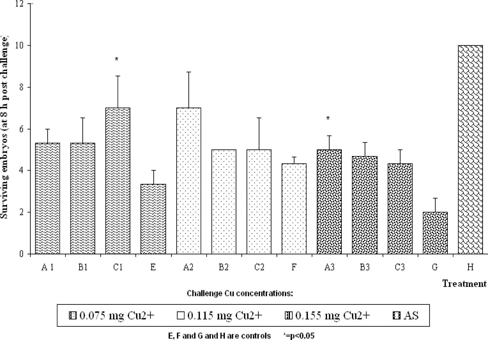
CuCl2 solutions were prepared by diluting a standard solution for atomic absorption spectrophotometry (Riedel-de Haën) in AS. All other chemicals used were of analytical purity. The copper concentration of solutions employed for acclimation and challenge were measured with a Perkin-Elmer atomic absorption spectrophotometer.

Results

The PROBIT values corresponding to Cu2+ toxicity on Bufo arenarum embryos at stage 25 for 24-h LC10, LC50 and LC90 were 0.035, 0.050 and 0.075 mg Cu2+.L−1 respectively while 0.03 mg.L−1 did not exert lethal effect. As a general pattern, the acclimation protocol to copper exerted a transient beneficial effect on the survival of the Bufo arenarum embryos exposed to different high copper concentrations. For certain cases a significant (p<0.05) protective effect in the embryo survival up to 8 hours of exposure was registered (Figure 1). However, the results at 24 hours of exposure point out no differences in survival among the acclimated and control embryos. Table 1 shows the copper and zinc contents in acclimated and control Bufo arenarum embryos. A reduction of approximately 25% of copper was found in the embryos treated with low copper concentrations respect to the copper content in control embryos (1.04 ± 0.09 μg Cu.g−1 wet weigh). In all cases, the differences between control and all the acclimation conditions were significant (p<0.05). In contrast, no significant differences in the embryonic zinc contents, among control and the acclimation conditions were found (control: 17.92 ± 0.42 μg Zn.g−1 wet weight).

Discussion

The effect of low level exposure to environmental pollutants is a matter of particular importance for environmental and human health protection purposes. Extrapolation from experimental high-dose effects to often much lower environmental relevant concentrations, or the assumption that effects at high doses clearly do not occur in the same manner as at low doses introduces uncertainties in the risk assessment process. Moreover the range of concentrations and the criteria to define the concept of low level exposure is not well established [24, 50]. For this study, low level exposure to copper was selected in relation to pristine fresh water concentrations and the NOEC / 24-h LC100 values of this metal for Bufo arenarum embryos being 30 and 75 μg.L−1 Cu2+ respectively. These results were similar to those previously reported and it is noteworthy that they almost did not change in spite of extending the exposure to 168 h [26]. The threshold concentration is 1.5 times higher than the maximal value found in unpolluted aquatic ecosystems. Although in unpolluted freshwater bodies the copper contents are in the range of 1–20μg.L−1[8], it is noteworthy that within these concentrations significant toxic effects were reported in diatoms and some invertebrates, notably cladocerans. Moreover, low pH and hardness conditions enhance copper toxicity to a large number of organisms including fishes [6]. Thus, it could be meaningful to explore the possibility to evaluate the potential of copper concentrations lower than those found in pristine conditions for acclimation purposes. The range of final copper concentration used during the acclimation protocols in this study was between 0.27 and 0.42 μg Cu2+.L−1, that is at least 2 times lower than the minimal concentration found in pristine freshwater. This analysis could provide a contribution to define more accurately the concept of “low level exposure” for both experimental and environmental scenarios. In the case of this acclimation study, the copper concentration was gradually increased in order to enhance the response of amphibian embryos to copper as it was demonstrated in the case of cadmium [51].
The acclimated embryos exhibit only a transient but in some cases statistically significant beneficial effect (p<0.05) to copper concentrations exerting 100% of lethality within 24 hours of exposure. This result contrasts with the higher increase in the resistance achieved by means of a similar acclimation protocol employing cadmium [3]. As a general pattern, the acclimation to copper achieved by means of exposures to low level concentrations of this metal could result in an enhanced resistance in case of challenges to very high copper concentrations (e.g. two times over the 24-h LC100) evaluated in this study. The transient protection registered could be related to a delay to reach internal Cu concentrations which result in lethality within 24 hours of exposure. In fact, a decreased uptake of another heavy metal, cadmium, due to changes in the membrane permeability was associated to acclimation phenomena both in “in vitro” and “in vivo” experiments in kidney cells [52]. On the other hand, the acclimation processes seem to include the increased synthesis and accumulation of “stress proteins” [53] like heat shock proteins [54] and metallothioneins (MTs), the last ones usually associated with heavy metal exposure [51, 55]. In the case of Bufo arenarum embryos exposed to low level Cu concentration, an increase in MTs was also reported [33].
It is noteworthy that Bufo arenarum embryos were able to react to the very low concentrations of copper employed in this acclimation experiment by significantly reducing the content of this metal. In fact, a reduction of about 25% of copper was found in the embryos treated with low copper concentrations in respect to the copper content in control embryos (1.04 ± 0.09 μg Cu.g−1 wet weigh, Table 1).
This reduction indicates that in cases of low level exposure to copper, the amphibian embryo can react; enhancing mechanisms of excretion of this heavy metal which is in line with the reduction in metal-uptake as a physiological mechanism for metal-resistance reported for a wide diversity of organisms from bacteria to mammalian cells [14, 1721]. The tissue residue results point out that Bufo arenarum embryo can achieve normal development in spite of a reduction of about 25% on its copper content and this reduction could be considered as a biomarker for low level exposure to copper, at least for early life stages of amphibians. Therefore it seems that also in amphibian embryos the copper transport can be modulated by the metal itself [6]. Bufo arenarum embryos treated with copper in toxic concentrations increase their copper contents (unpublished data from our laboratory) as was reported for a large number of other species [15]. Thus, the amphibian embryo could be a good model to study the feedback mechanism for copper transport at different concentrations. The fact that a significant decrease in the copper residue of the acclimated embryos was found without changes in the zinc content seems to confirm a homeostatic mechanism for the copper itself [6].
The capability of adapting to environmental stress is an inherent property of life and could be viewed as the persistence of information within an ecological system following a toxicological stressor [56]. Adaptation to metal concentrations in field populations was interpreted as the dynamic interaction between the selective pressure of elevated pollutants and gene flow [5760]. Moreover, living organisms at ontogenetic stages could be considered as biomarkers of environmental signatures of the evolutionary process [61]. In this context, the reduction in copper content even in embryos exposed to very low level concentrations of copper could be interpreted as an archaic mechanism of defense against a very toxic metal. Thus, it can be suggested that during the evolutionary process the rise of free copper in the environment occurred from a very low level compared to actual concentrations in pristine environments and living forms still conserve the capability to react to those exposure conditions.
Figure 1:. Survival of Bufo arenarum embryos treated with low copper concentrations and challenged to different lethal concentrations of this metal.
Ijerph 04 00166f1
Table 1:. Copper and zinc contents in Bufo arenarum embryos acclimated to Cu.
Ranges of Cu concentration during the acclimation protocols (ng.L1)μg Cu/g wet weight (a)μg Zn/g wet weight (a)
Control1.035 (0.09)17.92 (0.42)
40 – 2700.785 (0.02)*17.42 (0.84)
115 – 3500.800 (0,03)*17.93 (0.92)
190 – 4200.805 (0.04)*17.44 (0.71)
(a)Values are means from three different samples of mineralized embryos.
*p<0.05

Acknowledgments

This project was support by grants from the Agencia Nacional de Promoción Científica y Tecnológica (PICT 14375), Consejo Nacional de Investigaciones Científicas y Tecnológicas (CONICET, PIP 2316, 2317). We wish to thank Olga Domínguez for skilful technical assistance.

References

  1. Weis, P; Weis, JS. Cadmium acclimation and hormesis in Fundulus heteroclitus during fin regeneration. Environ. Res. 1986, 39(2), 356–363. [Google Scholar]
  2. van Straalen, NM; Vaal, MA. Physiological mechanism underlying adaptation to environmental stress. Sci. Total Environ. (Supp.) 1993, 1783–1787. [Google Scholar]
  3. Herkovits, J; Pérez-Coll, CS. Increased resistance against cadmium toxicity by means of pretreatment with low cadmium/zinc concentrations in Bufo arenarum embryos. Biol. Trace Elem. Res. 1995, 49, 171–175. [Google Scholar]
  4. Calabrese, EJ; Baldwin, LA. Hormesis as a biological hypothesis. Environ. Health Perspect. 1998, 106 Suppl. 1, 357–362. [Google Scholar]
  5. Davis, JM; Farland, WH. Biological effects of low level exposures: A perspective from U.S. EPA Scientist. Environ. Health Perspect. 1998, 106 Suppl. 1, 379–381. [Google Scholar]
  6. IPCS Environmental Health Criteria 200. Copper; Geneva; WHO, International Programme on Chemical Safety, 1998. [Google Scholar]
  7. Linder, MC; Hazegh-Azam, M. Copper biochemistry and molecular biology. Am. J. Clin. Nutr. 1996, 63, 797–811. [Google Scholar]
  8. Nriagu, JO. Global inventory of natural and anthropogenic emissions of trace metals to the atmosphere. Nature (Lond) 1979, 279, 409–411. [Google Scholar]
  9. Alt, ER; Sternlieb, I; Goldfisher, S. The cytopathology of metal overload. Int. Rev. Exp. Pathol. 1990, 31, 165–188. [Google Scholar]
  10. Noel-Lambot, F; Bouquegneau, JM; Frankenne, F; Dieteche, A. Cadmium, zinc and copper accumulation in limpets (Patella vulgata) from the Bristol Channel with special reference to metallotioneins. Mar. Ecol. Pro. Ser. 1980, 2, 81–89. [Google Scholar]
  11. Coimbra, J; Carraça, S. Accumulation of Fe, Zn, Cu and Cd during the different stages of the reproductive cycle in Mytilus edulis. Comp. Biochem. Physiol. 1990, 95C, 265–270. [Google Scholar]
  12. Kannan, K; Yasunaga, Y; Iwata, H; Ichihashi, H; Tanabe, S; Tatsukawa, R. Concentrations of heavy metals, organochlorines and organotins in horseshoe crab Tachypleus tridentatus, from Japanese coastal waters. Arch. Environ. Contam. Toxicol. 1995, 28, 40–47. [Google Scholar]
  13. Favero, N; Cattalini, F; Bertaggia, D; Albergoni, V. Metal accumulation in a biological indicator (Ulva rigida) from the lagoon of Venice (Italy). Arch. Environ. Contam. Toxicol. 1996, 31, 9–18. [Google Scholar]
  14. Gale, SA; Smith, SV; Lim, RP; Jeffree, RA; Petocz, P. Insights into the mechanisms of copper tolerance of a population of black-banded rainbow fish (Melanotaenia nigrans) (Richardson) exposed to mine leachate, using64/67Cu. Aquat. Toxicol. 2003, 62, 135–153. [Google Scholar]
  15. Jarvinen, AW; Ankley, GT. Linkage of effects to tissue residues: Development of a comprehensive database for aquatic organisms exposed to inorganic and organic chemicals. In SETAC Technical Publications Series; SETAC Press, 1999. [Google Scholar]
  16. Herkovits, J; Cardellini, P; Pavanati, C; Pérez-Coll, CS. Cadmium uptake and bioaccumulation in Xenopus laevis embryos at different developmental stages. Ecotoxicol. Environ. Saf. 1998, 39, 21–26. [Google Scholar]
  17. Ashida, J. Adaptation of fungi to metal toxicants. Annu. Rev. Phytopatol. 1965, 3, 153–174. [Google Scholar]
  18. Chopra, I. Decreased uptake of cadmium by a resistant strain of Staphylococcus aureus. J. Gen. Microbiol. 1971, 63, 265–267. [Google Scholar]
  19. Baker, AJM. Metal Tolerance. N. Phytol 1987, 106, S93–S111. [Google Scholar]
  20. Tsuchiya, N; Ochi, T. Mechanisms for the decrease in the accumulation of cadmium (Cd) in Cd-resistant Chinese hamster V79 cells. Arch. Toxicol. 1994, 68, 325–331. [Google Scholar]
  21. Xie, L; Klerks, PL. Changes in cadmium accumulation as a mechanism for cadmium resistance in the least killifish Heterandria formosa. Aquatic. Toxicol. 2004, 66, 73–81. [Google Scholar]
  22. Naab, F; Volcominsky, M; Burlón, A; Caraballo, ME; Debray, M; Kesque, JM; Kreiner, AJ; Ozafrán, MJ; Schuff, JA; Stoliar, P; Vázquez, ME; Davidson, J; Davidson, M; Fonovich de Schroeder, TM. Metabolic alterations without metal accumulation in the ovary of adult Bufo arenarum females, observed after long-term exposure to Zn2+, followed by toxicity to embryos. Arch. Environ. Contam. Toxicol. 2001, 41, 201–207. [Google Scholar]
  23. Van Tilborg, WJM; Van Assche, F. Risk assessment of essential elements: proposal for a fundamentally new approach. SETAC NEWS 1996, 16, 28–29. [Google Scholar]
  24. Cousins, RJ; Hempe, JM. Brown, ML, Ed.; Washington, DC, 1990; pp. 251–260.
  25. Bertazzo, A; Costa, C; Biasiolo, M; Allegri, G; Cirrincione, G; Presti, G. Determination of copper and zinc levels in human hair. Biol. Trace Elem. Res. 1996, 52, 37–53. [Google Scholar]
  26. Herkovits, J; Helguero, LA. Copper toxicity and copper-zinc interactions in amphibian embryos. Sci. Total Environ 1998, 221, 1–10. [Google Scholar]
  27. Herkovits, J; Pérez-Coll, CS; Dominguez, O. Cu-Zn antagonism in Bufo arenarum embryos: the time thresholds for Zn to protect against lethality exerted by copper. SETAC 21st Annual Meeting, Abstract Book 2000, 273. [Google Scholar]
  28. Gachot, B; Poujeol, P. Effects of cadmium and copper on Zn transport kinetics by isolated renal proximal cells. Biol. Trace Elem. Res. 1992, 35, 93–103. [Google Scholar]
  29. WHO. Copper. In Trace elements in human nutrition and health; Geneva; World Health Organization, 1996; chapter 7; pp. 123–143. [Google Scholar]
  30. Salim, S; Farquharson, J; Arneil, GC; Cockburn, F; Forbes, GI; Logan, RW; Sherloc, JC; Wilson, TS. Dietary copper intake in artificially fed infants. Arch. Dis. Chil. 1986, 61(11), 1068–1075. [Google Scholar]
  31. August, D; Janghorbani, M; Young, VR. Determination of zinc and copper absorption at three dietary Zn-Cu ratios by using stable isotope methods in young adult and elderly subjects. Am. J. Clin. Nutr. 1989, 50, 1457–1463. [Google Scholar]
  32. Muller, JP; Wouters-Tyrou, D; Erraiss, N; Vedel, M; Touzet, N; Mesnard, J; Sautiere, P; Wegnez, M. Molecular cloning and expression of a metallothionein mRNA inXenopus laevis. DNA and Cell. Biol. 1993, 12(4), 341–349. [Google Scholar]
  33. Pérez-Coll, CS; Herkovits, J; Fridman, O; D′Eramo, JL; Corró, L. Acclimation of Bufo arenarum embryos to copper: effects on survival and the induction of metallothioneins. 21st SETAC Annual Meeting; Abstract Book. 2000. PWP140. p. 274. [Google Scholar]
  34. Liu, Y; Liu, J; Iszard, MB; Andrews, GK; Palmiteer, RD; Klaassen, CD. Transgenic mice that over-express metallothionein-I are protected from cadmium lethality and toxicity. Toxicol. Appl. Pharmacol. 1995, 13, 222–228. [Google Scholar]
  35. Pérez-Coll, CS; Herkovits, J. Stage dependent uptake of cadmium in Bufo arenarum embryos. Bull. Environ. Contam. Toxicol. 1996, 56(4), 663–669. [Google Scholar]
  36. McCarty, LS. The relationship between aquatic toxicity QSARs and bioconcentration for some organic chemicals. Environ. Toxicol. Chem., 1986, 5, 1071–1080. [Google Scholar]
  37. McCarty, LS. Toxicant body residues: Implications for aquatic bioassays with some organic chemicals. Aquat. Toxicol. 1991, 14, 183–192. [Google Scholar]
  38. Cook, PM; Erickson, RJ; Spehar, L; Bradbury, SP; Ankley, GT. Interim report on data and methods for assessment of 2,3,7,8-tetrachlorodibenzo-p-dioxin risk to aquatic life and associated wildlife; Duluth MN; U.S. Environmental Protection Agency; EPA-600/R-93/055; 1993. [Google Scholar]
  39. McCarty, LS; Mackay, D. Enhancing ecotoxicological modeling and assessment: body residues and modes of toxic action. Environ. Sci. Technol. 1993, 27, 1719–1728. [Google Scholar]
  40. Herkovits, J; Pérez-Coll, CS; Herkovits, FD. Ecotoxicological studies of environmental samples from Buenos Aires area using standardized amphibian embryo toxicity test (AMPHITOX). Environ. Poll. 2002, 116, 177–183. [Google Scholar]
  41. Zang, L; Wang, W. Alteration of dissolved cadmium and zinc uptake kinetics by metal pre- exposure in the Black Sea bream (Acanthopagrus schlegeli). Environm. Toxicol. Chem. 2006, 25, 1312–1321. [Google Scholar]
  42. Blaustein, AR; Wake, DB; Sousa, WP. Amphibian declines: judging stability, persistence and susceptibility of populations to local and global extinctions. Conserv. Biol. 1994, 8, 60–71. [Google Scholar]
  43. Boyer, R; Grue, CE. The need for water quality criteria for frogs. Environ. Health Perspect. 1995, 103, 352–355. [Google Scholar]
  44. Besser, JM; Dwyer, FJ; Ingersoll, CG; Wang, N. Early Life-stage toxicity of copper to endangered and surrogate fish species; Washington, DC; U.S. Environmental Protection Agency; EPA/600/R-01/051; 2001. [Google Scholar]
  45. Herkovits, J; Pérez-Coll, CS; Herkovits, FD. Ecotoxicity in Reconquista River (Province of Buenos Aires, Argentina): A preliminary study. Environ. Health Perspect. 1996, 104(2), 186–189. [Google Scholar]
  46. Burkhart, JG; Ankley, G; Bell, H; Carpenter, H; Fort, D; Gardner, D; Gardner, H; Hale, R; Helgen, JC; Jepson, P; Johnson, D; Lannoo, M; Lee, D; Lary, J; Levey, R; Magner, J; Meteyer, C; Shelby, MD; Lucier, G. Strategies for assessing the implications of malformed frogs for environmental health. Environ. Health Perspect. 2000, 108(1), 83–90. [Google Scholar]
  47. Tietge, JE; Ankley, GT; DeFoe, DL; Holcombe, GW; Jensen, KM. Effects of water quality on development of Xenopus laevis: a frog embryo teratogenesis assay-Xenopus assessment of surface water associated with malformations in native anurans. Environ. Toxicol. Chem. 2000, 19, 2114–2121. [Google Scholar]
  48. Herkovits, J; Pérez-Coll, CS. AMPHITOX: A customized set of toxicity tests employing amphibian embryos. “Symposium on multiple stressor effects in relation to declining amphibian populations”. In “Multiple Stressor Effects in Relation to Declining Amphibian Populations” ASTM International STP 1443; Linder, GL, Krest, S, Sparling, D, Little, EE, Eds.; USA, 2003; pp. 46–60. [Google Scholar]
  49. Del Conte, E; Sirlin, L. The first stages of Bufo arenarum development. Acta Zool. Lilloana 1951, 12, 495–499. [Google Scholar]
  50. Herkovits, J; Pérez-Coll, CS. Dose-response relationship within the potentized microdoses phenomenon. Complementary Therapies in Medicine 1993, 1, 7–9. [Google Scholar]
  51. Pérez-Coll, CS; Herkovits, J; Fridman, O; Daniel, P; D′Eramo, JL. Metallothionein induction and cadmium uptake in Bufo arenarum embryos following an acclimation protocol. Environ. Poll 1999, 106, 443–448. [Google Scholar]
  52. Jin, T; Nordberg, GF. Cadmium toxicity in kidney cells. Resistance induced by short-term pretreatment in vitro and in vivo. Acta Pharmacol. Toxicol. 1986, 58, 137–143. [Google Scholar]
  53. Sanders, BM. Stress proteins, potential as multitiered biomarkers. In Biomarkers of Environmental Contamination; Lewis: Chelsea, MI, USA, 1990. [Google Scholar]
  54. Schelesinger, MJ. Heat shock proteins. J. Biol. Chem. 1986, 265, 12111–12114. [Google Scholar]
  55. Kägi, JHR. Evolution, structure and chemical activity of class I metallothioneins: An overview. In Metallothionein III: Biological Roles and Medical Implications; Suzuki, KT, Imura, N, Kimura, M, Eds.; Berlin; Birkhauser Verlag, 1993; pp. 29–56. [Google Scholar]
  56. Clements, FE. Plant succession: An analysis of the development of vegetation; Publication 42; Carnegie Institute: Washington DC, USA, 1916. [Google Scholar]
  57. Brandon, RN. Adaptation and environment; Princeton University Press: Princeton, New Jersey, USA, 1990. [Google Scholar]
  58. Postma, JF; Groenendijk, D. Adaptation to metals in the midge Chironomus riparius: a case study in the river Dommel. In Genetics and Ecotoxicology; Forbes, VE, Ed.; Taylor & Francis: London, UK, 1999; pp. 79–101. [Google Scholar]
  59. Herkovits, J. Paleoecotoxicology: The impact of chemical and physical stress in the evolutionary process. Environ. Health Perspect. 2001, 109(12), 564–566. [Google Scholar]
  60. Groenendijk, D; Lucker, SMG; Plans, M; Kraak, MHS; Admiraal, W. Dynamics of metal adaptation in riverine chironomids. Environ. Poll 2002, 117, 101–109. [Google Scholar]
  61. Herkovits, J. Evoecotoxicology: environmental changes and life features development during the evolutionary process – the record of the past at developmental stages of living organisms. Environ. Health Perspect. 2006, 114, 1139–1142. [Google Scholar]

