Article

Ultraviolet Radiation Increases the Toxicity of Pyrene, 1-Aminopyrene and 1-Hydroxypyrene to Human Keratinocytes

by
Stephen I. N. Ekunwe
1,2,3,*,
Rochelle D. Hunter
1,3,4 and
Huey-Min Hwang
2,3
1
Microbial Molecular Genetics, Cancer and Phytonutriceuticals Research Laboratory
2
NIH–Center for Environmental Health, College of Science, Engineering and Technology, Jackson, Mississippi, USA
3
Department of Biology, Jackson State University, 1400 J. R. Lynch Street, P.O. Box 18540, Jackson, MS 39217, USA
4
Pharmaceutical Products Development, Inc. (PPD), 980 Harvest Dr., Suite 130, Blue Bell, PA 19422, USA
*
Author to whom correspondence should be addressed.
Int. J. Environ. Res. Public Health 2005, 2(1), 58-62; https://doi.org/10.3390/ijerph2005010058
Submission received: 15 November 2004 / Accepted: 6 February 2005 / Published: 30 April 2005

Abstract

:
Over the past several years, a great deal of interest has been focused on the harmful effects of ultraviolet (UV) radiation to human skin. UV light has been implicated in aging, sunburn and skin cancer. Few studies, however, have been done to determine the effects that UV light, in conjunction with other environmental contaminants, may have on human skin. Polycyclic Aromatic Hydrocarbons (PAHs) are a class of compounds that have been reported to be toxic, mutagenic and carcinogenic to many eukaryotic organisms. UV light is also known to increase the toxicity of PAHs through photo-activation and photo-modification. The purpose of this study was to assess the effects of UV-A irradiated pyrene (Pyr), 1-aminopyrene (1-AP) and 1-hydroxypyrene (1-HP) on human keratinocytes, the skin primary site of UV irradiated PAH exposure. Our findings indicate that simultaneous treatment of human keratinocyte cell line, HaCaT, with 1.0μg/ml pyrene, 1-AP or 1-HP and 3.9 J/cm2/min UV-A light resulted in significant inhibition of cell proliferation. Approximately 100% of the cells died in the case of UV-A irradiated 1-AP and 1-HP. In the case of UV-A irradiated pyrene, more than 70% of the cells died, indicating that UV-A is able to transform these PAHs into more harmful intermediates.

Introduction

Over the past several years, a great deal of interest has been focused on the harmful effects of ultraviolet (UV) radiation to human skin. UV light has been implicated in aging, sunburn and skin cancer. Few studies, however, have been done to determine the effects that UV light, in conjunction with other environmental contaminants, may have on human skin. The ubiquitous environmental contaminants known as Polycyclic Aromatic Hydrocarbons (PAHs) are a class of compounds that have been reported to be toxic, mutagenic and carcinogenic to many eukaryotic organisms [13]. UV light is also known to increase the toxicity of PAHs through photo-activation and photo-modification [4, 5]. The purpose of this study was to assess the effects of UV-A irradiated pyrene (Pyr), 1-aminopyrene (1-AP) and 1-hydroxypyrene (1-HP) on human keratinocytes, which are the primary site of UV-A irradiated PAH exposure to humans.

Materials and Methods

Chemicals and Reagents

Dulbecco’s Modified Eagle Medium (DMEM) 1X with high glucose, L- glutamine, sodium pyruvate, pyridoxine hydrochloride, Fetal Bovine Serum (FBS) and 0.25% Trypsin were purchased from Gibco Invitrogen, Corporation, USA while Penicillin/Streptomycin (P/S) antibiotic 10,000 I.U./mL was purchased from Mediatech, Inc. USA. Pyrene, 1-aminopyrene, 1-hydroxyprene and benzo[a]pyrene, HPLC grade N, and N-Dimethylformamide (DMF) were purchased from Sigma-Aldrich Chemical Co., (St. Louis, Missouri).

Preparation of Growth Media

Dulbecco’s Modified Eagle Medium (DMEM) supplemented with 10% Fetal Bovine Serum (FBS) and 1% Penicillin/Streptomycin (P/S) antibiotic served as the complete growth medium (CGM). DMEM plus 1% P/S is the serum free medium. Stock solutions (1.0 mg/mL) of pyrene, 1-aminopyrene, 1-hydroxyprene and benzo[a]pyrene were prepared in HPLC grade N, N-Dimethylformamide (DMF).

Human Keratinocyte Cell Line, HaCaT

Spontaneously immortalized human keratinocyte cell line, HaCaT, was the cell model used in the cytotoxicity studies. They were a generous gift from Dr. Norbert Fusenig of the Division of Differentiation & Carcinogenesis of the German Cancer Research Center (DKFZ). HaCaT cells were prepared from the surgical excision of full thickness adult human skin from the distant periphery of a melanoma [6].

Culturing HaCaT Cells

Initially, a vial of frozen HaCaT cells containing 106cells/mL was rapidly thawed in room temperature water. The entire 1.0mL cell suspension was plated in 4.0 mL of complete growth medium in a T-25 canted-neck culture flask. The cell culture was allowed to grow at 37°C in a humidified (5% CO2, 95% air) incubator until cells were completely confluent, usually 7–9 days, with change of growth medium every 48 hours. Cells were trypsinized, counted using a hemocytometer, and passaged at 1:10 split ratio.

DNA Synthesis – [3H]Thymidine Incorporation Assay

DNA synthesis is used as an indirect measure of cell proliferation. DNA synthesis by HaCaT cells is assessed with [3H]thymidine incorporation assay. Briefly the assay involves growing cells to sub-confluence (≈60%), synchronizing cells by overnight serum starvation, followed by labeling with [3H]thymidine at 1μCi/mL for 4–6 hours. Cells are then fixed in 10% trichloroacetic acid (TCA) and solubilized with 2.0mL/well of 0.5M NaOH solution. One milliliter of the solubilized cells in 5.0mL scintillation cocktail is counted in Packard Tri-Carb TR 2700 Liquid Scintillation Analyzer.

HaCaT Exposure to Ultraviolet Radiation

HaCaT cells were plated in Primaria™ 6-well plates at 105 cells/mL in CGM and allowed to grow at 37°C in a humidified (5% CO2, 95% air) incubator until sub-confluent (≈60% confluent). HaCaT cells were serum starved for 24 hours and then exposed to only UV-A light (3.9 J/cm2/min) for 20, 60, 80, 100, 120, 140, 160, 180 and 200 minutes at room temperature. Except for a thin coating of medium to prevent cells from drying up, all medium was withdrawn from the cells during UV irradiation. Fresh medium was added after UV-irradiation and cells were incubated for 18hours before being labeled with 3H[thymidine] at 1μCi/mL for 4–6 hours. One milliliter of the solubilized cells in 5.0 mL scintillation cocktail is counted in Hewlett Packard Tri-Carb 2700 TR Liquid Scintillation Analyzer. Cells treated with CGM and serum free DMEM served as the positive and negative controls, respectively. Each treatment was done in triplicates.

Treatment of HaCaT Cells with PAH

HaCaT cells were assessed for survival after exposure to various concentrations of pyrene, 1-aminopyrene and 1-hydroxypyrene. Cells were plated in Primaria™ 6-well plates at 105cells/2ml in CGM and allowed to grow until sub-confluent. Cells were serum starved for 24 hours and then treated with pyrene, 1-AP or 1-HP at 0.01, 0.1, 1.0 and 10.0μg/ml for 18 hours before being labeled with 3H[thymidine] at 1μCi/mL for 4–6 hours. Cells treated with CGM and serum free DMEM served as the positive and negative controls, respectively. Each treatment was done in triplicates.

Effect of UV-A irradiated PAH on HaCaT Cells

HaCaT cells were assessed for survival after exposure to UV-A irradiated pyrene, 1-HP and 1-AP. Cells were plated in Primaria™ 6-well plates at 105 cells/2ml in CGM and allowed to grow until sub-confluent. Cells were serum starved for 24 hours and then simultaneously exposed to pyrene, 1-AP or 1-HP (1.0μg/ml) and UV-A light (3.9J/cm2/min) for 60 minutes and then incubated at 37°C for 18 hours prior to treatment with 3H[thymidine] at 1μCi/mL for 4–6 hours. Cells treated with CGM and serum free DMEM served as the positive and negative controls, respectively. Each treatment was done in triplicates.

Results

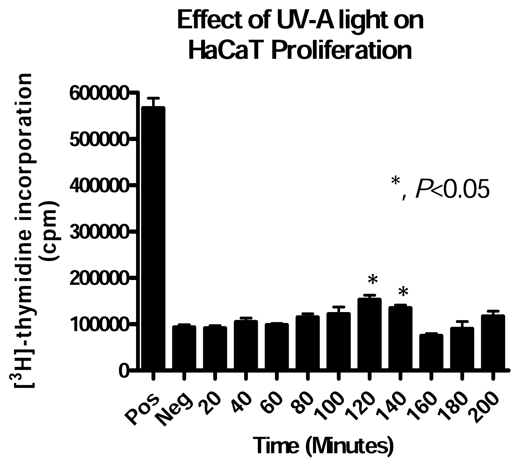
Effect of UV-A Alone Exposure on HaCaT Cells

The results show that HaCaT cells responded to UV-A light in a biphasic manner (Figure 1). The growth of HaCaT cells exposed for 120 and 140 minutes was significantly different from the unexposed control. It has been established that UV-A light causes damage to cellular DNA, resulting in cell death [78]. The present study, however, did not show significant cell death after UV-A exposure. It is reasonable to say, therefore, that the UV-A exposure periods at an energy of 3.9 J/cm2/min were not adequate enough to stop DNA synthesis. This could be because the DNA was not damaged in a way to cause breaks in the DNA, although significant mutations could have occurred.

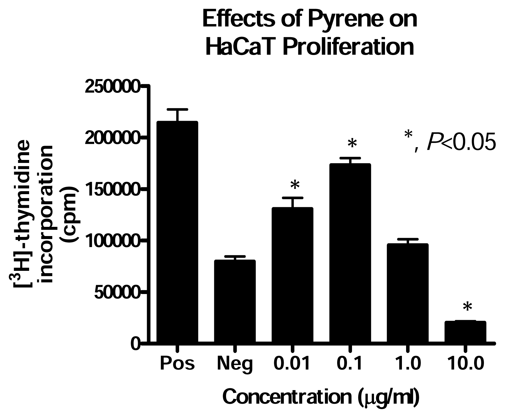
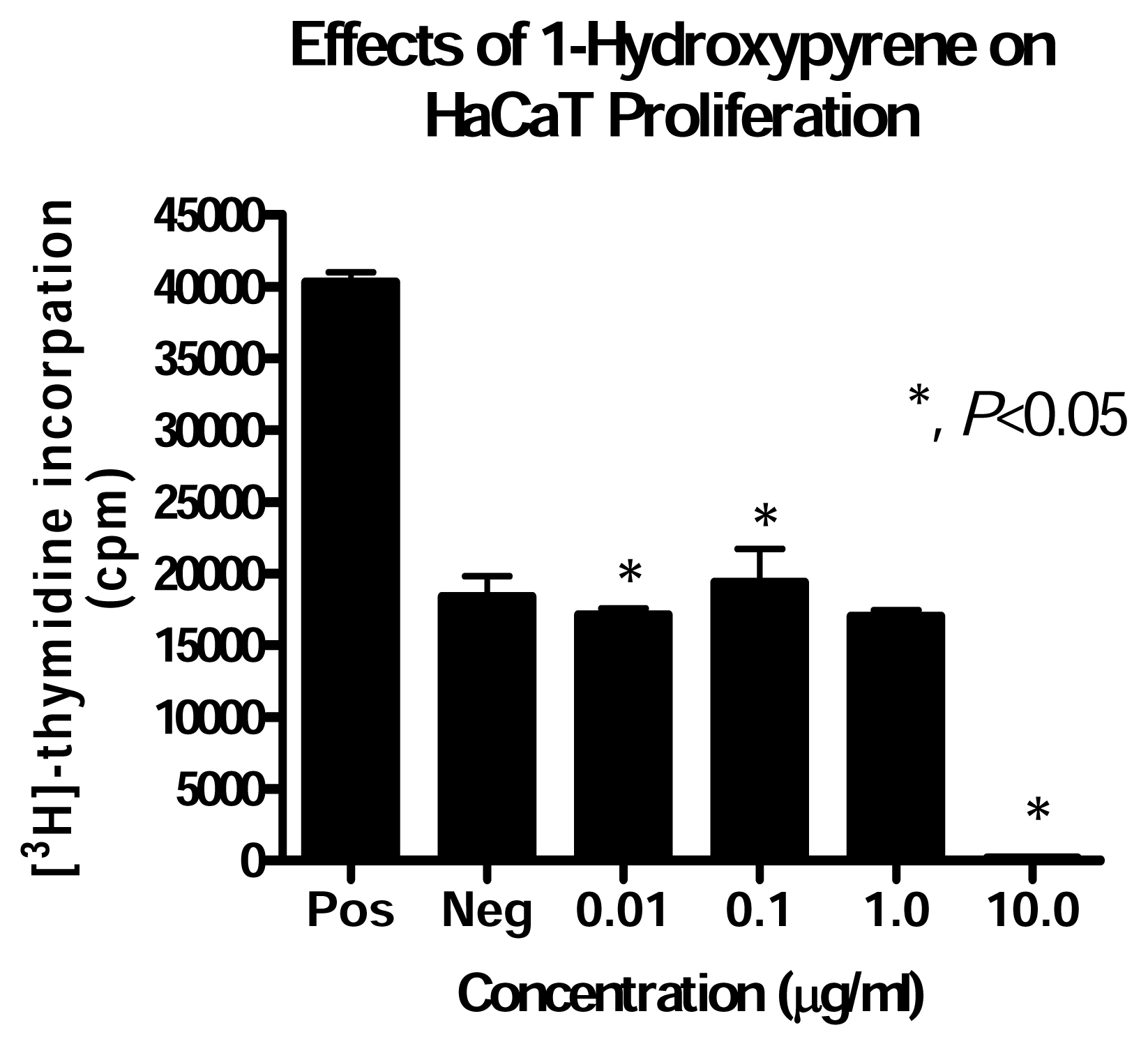
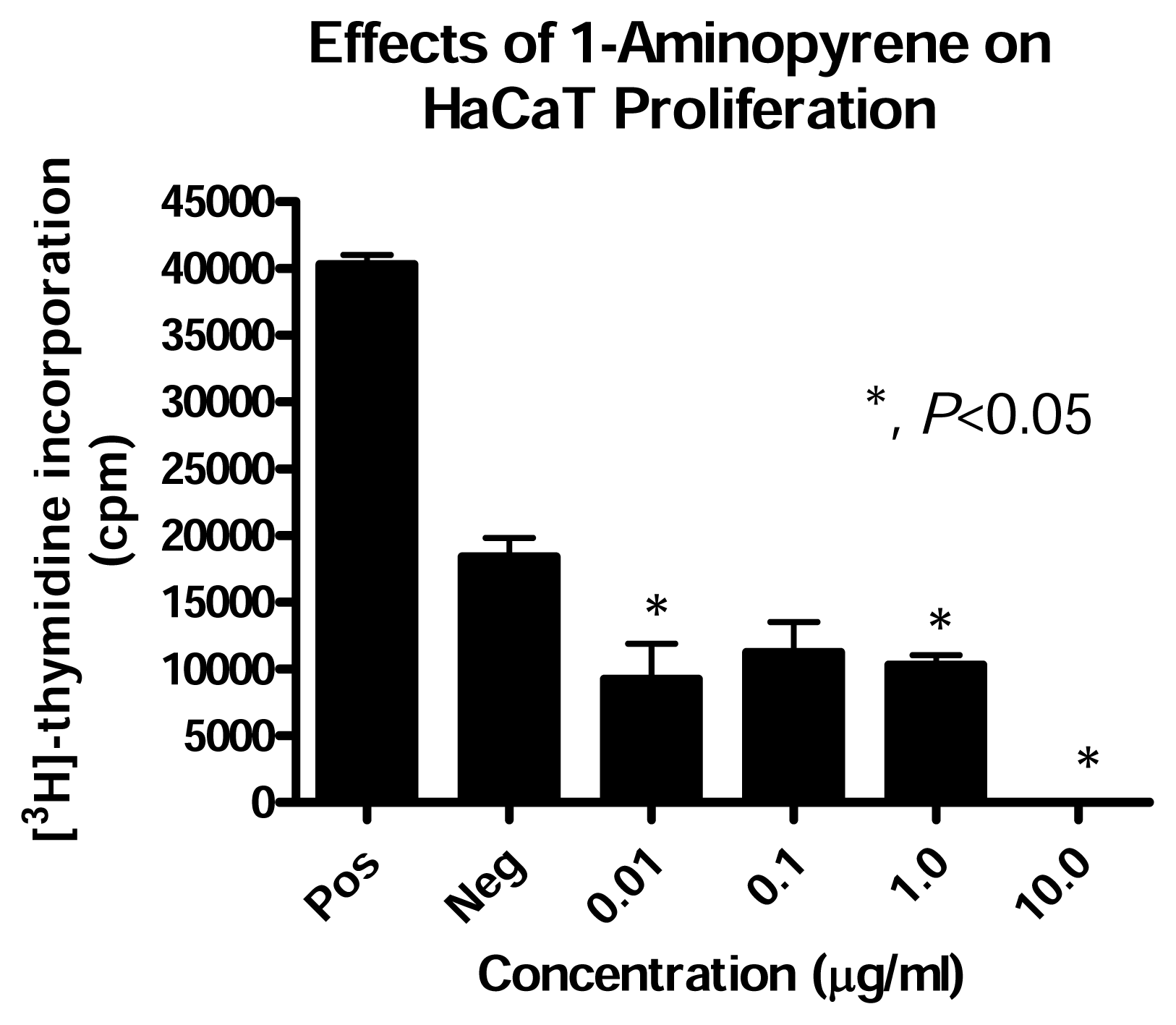
Effect of PAH Treatment on HaCaT Cell Proliferation

PAHs are also known to be toxic to human cells, causing lung cancer when inhaled and skin cancer when exposed by skin contact [9]. Results obtained in this set of experiment suggest a biphasic response to PAH exposure. At low concentrations, pyrene caused stimulation of HaCaT proliferation while 1-HP and 1-AP inhibit cell proliferation; and at 10μg/mL, pyrene, 1-AP and 1-HP significantly inhibit HaCaT proliferation (Figures 24). It is an established fact that substituted PAH derivatives are more soluble than their parent compounds, and are therefore more accessible and toxic to the cells [10]. This could explain why, at low concentrations, the substituted PAHs were more toxic to the HaCaT cells than the parent compound pyrene.
Other reports have also demonstrated PAH toxicity to eukaryotic cells. Comet assay profiles of blood cells collected from Ctenomys torquatus (rodent) captured from a coal strip mine, an area highly contaminated with PAHs, verify considerable DNA damage when compared to blood cells from the same type of rodent captured from coal free sites [11].

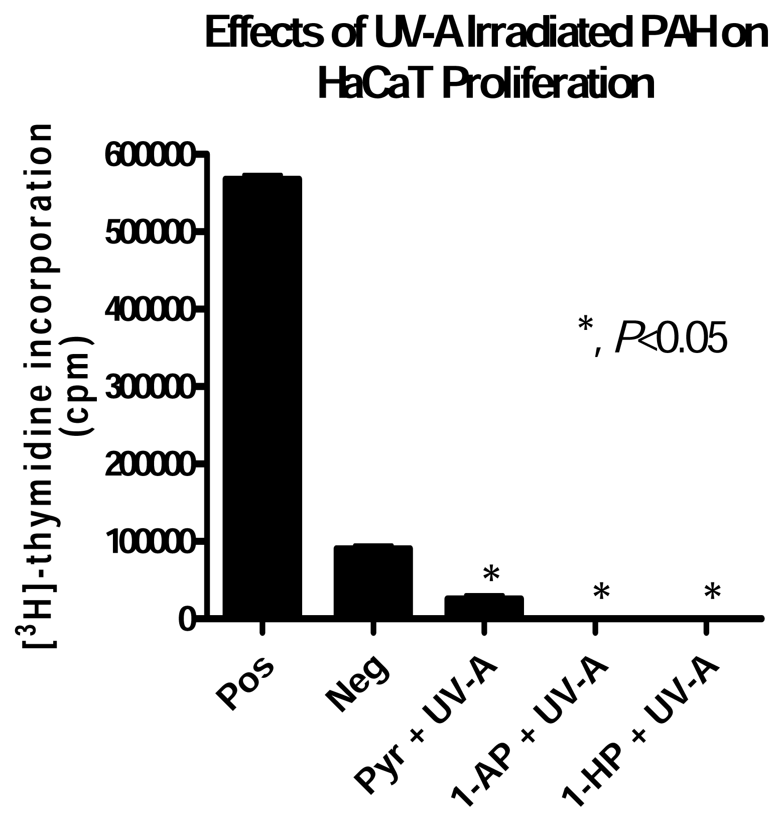
HaCaT Cell Response to UV-A irradiated PAH Exposure

To assess the response of human keratinocytes to UV-A irradiated PAHs, HaCaT cells were exposed to UV-A irradiated pyrene and two of its substituted forms, 1-AP and 1-HP. The exposure time (60 minutes) and PAH concentration (1.0μg/ml) were selected because in the single factor experiments, proliferation of HaCaT cells in either case was not significantly different from that of the unexposed control. In this group of experiments our findings suggest that pyrene, 1-AP and 1-HP are considerably more toxic to HaCaT cells when irradiated with UV-A light. Cell growth was almost completely prevented in the case of UV-A irradiated 1-HP and 1-AP, while in the case of UV-irradiated pyrene, cell growth was inhibited by more than 70% (Figure 5).

Discussion

Our findings indicate that simultaneous treatment of human keratinocyte cell line, HaCaT, with 1.0μg/ml pyrene, 1-AP or 1-HP and 3.9J/cm2/min UV-A light resulted in significant inhibition of cell proliferation. Approximately 100% of the cells died in the case of UV-A irradiated 1-AP and 1-HP. In the case of UV-A irradiated pyrene, more than 70% of the cells died, indicating that UV-A is able to transform these PAHs into more harmful intermediates. These results are consistent with other reports that discuss cell death caused by the interaction between UV light and PAHs. The photo-modification of PAHs results in the formation of diones and quinones, which can bind to cellular macromolecules and cause problems in signalling and proliferative pathways, which ultimately lead to cell death. With the binding of large macromolecules to DNA, the cell becomes severely challenged to replicate such DNA molecules with these large adduct. The DNA damage repair system could be ineffective in repairing such DNA damage. The end result is cell death. Cytotoxic effect, such as lipid peroxidation of the cell membrane leads to compromise of cell membrane integrity. Once more, the end point is cell death, either by necrosis or apoptosis. It has been found that UV-A irradiated benzo[a]pyrene causes cellular reactions to occur that heighten DNA damage [10]. Such exposure to human epidermoid carcinoma cells and human keratinocytes results in a 5-fold increase in the production of H2O2, which is known to cause significant DNA damage [1213]. The classic Haber-Weiss-Fenton reaction involving H2O2 in the presence of Fe+2 leads to double strand-break of DNA strands. Either could be a possible reason for the cell death observed in this study. These results are significant because in the South and South-eastern US, exposure of the population to UV-A is considerably higher than in the North. Exposure to combination of UV-A and PAHs from petrochemicals, creosote use in wood treatment facilities and tractor exhaust gases in the cotton fields of the Delta, makes this a vexing environmental health concern. The results obtained in this study do not only increase our understanding of the problem but also provide us the opportunity to seek appropriate solutions for the problem.

Conclusions

From the results in this study, the following conclusions can be made: (1) UV-A light inhibits growth and proliferation of HaCaT cells (2) UV-A irradiated PAHs are highly toxic to HaCaT cells (3) UV-A irradiated substituted pyrenes are more toxic than their parent compound. This agrees with what is already known about UV-A light causing damage to cellular DNA, resulting in cell death [78]. The toxic effects of UV-A irradiated PAHs to human cells, is established. Drastically reduced exposure of humans to this combination of UV-A and PAHs is necessary to avoid lung and skin cancer induced by exposure to UV-A irradiated PAHs [9].
Figure 1. Effects of UV-A light on HaCaT proliferation. HaCaT cells were grown in 35mm plates to subconfluent in CGM. Cells were serum starved for 24 hours and then exposed to UV-A (3.9J/cm2/min) for 20, 40, 60, 80, 100, 120, 140, 160, 180, and 200 minute intervals. Thymidine incorporation assay was then performed as described in materials and methods. Results represent the mean +/− SD values of experiment performed in triplicate. [* indicates that treatment mean is significantly different from negative control according to Dunett test (p<0.05)].
Ijerph 02 00058f1
Figure 2. Effects of pyrene on HaCaT proliferation. HaCaT cells were grown in 35mm plates to subconfluent in CGM. Cells were serum starved for 24 hours and then treated with pyrene at 0.01, 0.1, 1.0 and 10.0μg/ml concentrations for 18 hours. Thymidine incorporation assay was then performed as described in materials and methods. Results represent the mean +/− SD values of experiment performed in triplicate. [* indicates that treatment mean is significantly different from negative control according to Dunett test (p<0.05)].
Ijerph 02 00058f2
Figure 3. Effects of 1-hydroxypyrene on HaCaT proliferation. HaCaT cells were grown in 35mm plates to subconfluent in CGM. Cells were serum starved for 24 hours and then treated with pyrene at 0.01, 0.1, 1.0 and 10.0μg/ml concentrations for 18 hours. Thymidine incorporation assay was then performed as described in materials and methods. Results represent the mean +/− SD values of experiment performed in triplicate. [* indicates that treatment mean is significantly different from negative control according to Dunett test (p<0.05)].
Ijerph 02 00058f3
Figure 4. Effects of 1-aminopyrene on HaCaT proliferation. HaCaT cells were grown in 35mm plates to subconfluent in CGM. Cells were serum starved for 24 hours and then treated with pyrene at 0.01, 0.1, 1.0 and 10.0μg/ml concentrations for 18 hours. Thymidine incorporation assay was then performed as described in materials and methods. Results represent the mean +/− SD values of experiment performed in triplicate. [* indicates that treatment mean is significantly different from negative control according to Dunett test (p<0.05)].
Ijerph 02 00058f4
Figure 5. Effects of UV-A irradiated pyrene, 1-aminopyrene and 1-hydroxypyrene on HaCaT proliferation. HaCaT cells were grown in 35mm plates to sub-confluent in CGM. Cells were serum starved for 24 hours and then treated with each PAH at 1.0μg/ml and immediately irradiated with UV-A (3.9J/cm2/min). PAH treatment remained on cells for a total of 18 hours, including time during UV-A exposure. Thymidine incorporation assay was then performed as described in materials and methods. Results represent the mean +/− SD values of experiment performed in triplicate. [* indicates that treatment mean is significantly different from negative control according to Dunett test (p<0.05)].
Ijerph 02 00058f5

Acknowledgements

This research was supported in part by a grant from the Army Research Office (Grant# DAAD 19-01-1-0733), awarded to Jackson State University, and in part by a grant from the National Institutes of Health Research Centers in Minority Institutions, NIH-RCMI (Grant #1G12RR13459) awarded to Jackson State University. Part of this work was used by Rochelle Hunter to satisfy the Masters Degree thesis requirement at Jackson State University.

References

  1. Villemur, R.; Deziel, E.; Benachenhou, A.; Marcoux, J.; Gauthier, E.; Lepine, F.; Beaudet, R.; Commeau, Y. Two-liquid-phase slurry bioreactors to enhance the degradation of high-molecular-weight polycyclic aromatic hydrocarbons in soil. Biotech. Prog 2000, 16, 966–972. [Google Scholar]
  2. Guerin, T. Bioremediation of phenols and polycyclic aromatic hydrocarbons in creosote contaminated soil using ex-situ land treatment. J Haz Mat 1999, B65, 305–315. [Google Scholar]
  3. Dagher, F.; Deziel, E.; Lirette, P.; Paquette, G.; Bisaillon, J. G.; Villemur, R. Comparative study of five polycyclic aromatic hydrocarbon degrading bacterial strains isolated from contaminated soils. Can J. Micro 1997, 43, 368–377. [Google Scholar]
  4. Dong, S.; Hwang, H-M.; Shi, X.; Holloway, L.; Yu, H. UVA induced single strand cleavage by 1-hydroxypyrene and formation of covalent adducts between DNA and 1-hydroxypyrene. Chem. Res. Toxicol 2000, 13, 585–593. [Google Scholar]
  5. Schirmer, K.; Xhan, A. G. J.; Greenberg, B. M.; Dixon, D. G.; Bols, N. C. Ability of 16 priority PAHs to be phototoxic to a cell line from rainbow trout gill. Toxicol 1998, 127, 143–155. [Google Scholar]
  6. Boukamp, P.; Petrussevska, R. T.; Breitkreutz, D.; Hornung, J.; Markham, A.; Fusenig, N. E. Normal keratinization in a spontaneously immortalized aneuploid human keratinocyte cell line. J. Cell Biol 1988, 106(3), 761–771. [Google Scholar]
  7. Quiec, D.; Maziere, C.; Auclair, M.; Santus, R.; Gardette, J.; Redziniak, G.; Franchi, J.; Dubertret, L.; Maziere, J. C. Lovastatin enhances the photocytotoxicity of UVA radiation towards cultured N.C.T.C.2544 human keratinocytes: prevention by cholesterol supplementation and by a cathepsin inhibitor. Biochem J 1995, 310, 305–309. [Google Scholar]
  8. Niggli, H.; Applegate, L. Glutathione response after UVA irradiation in mitotic and postmitotic human skin fibroblasts and keratinocytes. Photochem Photobiol 1997, 65(4), 680–684. [Google Scholar]
  9. Heudorf, U.; Angerer, J. Internal exposure to PAHs of children and adults living in homes with parquet flooring containing high levels of PAHs in the parquet glue. Internat Arch. Occup Environ Health 2001, 74, 91–101. [Google Scholar]
  10. Shyong, E.; Lu, Y.; Goldstein, A.; Lebwohl, M.; Wei, H. Synergistic enhancement of H2O2 production in human epidermoid carcinoma cells by benxo[a]pyrene and ultraviolet A radiation. Toxicol Appl Pharmacol 2003, 188, 104–109. [Google Scholar]
  11. Da Silva, J.; Da Freitas, T.; Marinho, J.; Speit, G.; Erdtmann, B. An alkaline single-cell gel electrophoresis (comet) assay for environmental bio-monitoring with native rodents. Genet. Mol. Biol 2000, 23(1), 241–245. [Google Scholar]
  12. Daroui, P.; Desai, S. D.; Li, T. K.; Liu, A. A.; Liu, L. F. Hydrogen peroxide induces topoisomerase I-mediated DNA damage and cell death. J. Biol. Chem 2004, 279(15), 14587–14594. [Google Scholar]
  13. Slupphaug, G.; Kavli, B.; Krokan, H. E. The interacting pathways for prevention and repair of oxidative DNA damage. Mut. Res 2003, 531(1–2), 231–251. [Google Scholar]

