Article

Comparison of Commensal Escherichia coli Isolates from Adults and Young Children in Lubuskie Province, Poland: Virulence Potential, Phylogeny and Antimicrobial Resistance

1
Department of Microbiology and Genetics, Faculty of Biological Sciences, University of Zielona Góra, 65-561 Zielona Góra, Poland
2
Laboratory of Virology, Department of Immunology of Infectious Diseases, Ludwik Hirszfeld Institute of Immunology and Experimental Therapy, Polish Academy of Sciences, 53-114 Wroclaw, Poland
3
Michigan Nanotechnology Institute for Medicine and Biological Sciences, University of Michigan Medical School, Ann Arbor, MI 48109-5648, USA
4
Department of Mathematical Statistics and Econometrics, Faculty of Mathematics, Computer Science and Econometrics, University of Zielona Góra, 65-516 Zielona Góra, Poland
*
Author to whom correspondence should be addressed.
Int. J. Environ. Res. Public Health 2018, 15(4), 617; https://doi.org/10.3390/ijerph15040617
Submission received: 6 March 2018 / Revised: 22 March 2018 / Accepted: 23 March 2018 / Published: 28 March 2018
(This article belongs to the Section Environmental Health)

Abstract

:
Commensal Escherichia coli population is a dynamic structure which may be important in the pathogenesis of extraintestinal infections. The aim of this study was the comparison of genetic diversity of commensal E. coli isolates from two age group—adults and young children. E. coli strains were isolated on MacConkey agar and identified by biochemical tests. Determination of four major phylogenetic groups, identification of virulence genes and antimicrobial resistance determinants were performed by using multiplex or simplex PCR. Phenotypic analysis of resistance was based on disc-diffusion method. The prevalence of virulence genes was significantly higher among isolates from adults than from young children. Phylogroup B2 predominated among E. coli from adults, whereas phylogroup A was the most common in isolates from young children. The analyses of antimicrobial resistance revealed that resistance to at least one antimicrobial agent and multidrug-resistance were detected significantly more frequent in the isolates from adults than from young children. This study documented that the commensal E. coli isolates from adults showed greater genetic diversity than from young children and constitutes a substantial reservoir of the virulence genes typical for extraintestinal pathogenic E. coli.

1. Introduction

Escherichia coli bacteria is one of the most explored model organisms, partially due to its versatile nature. These bacteria constitute a component of the natural microbiota of warm-blooded animals including humans [1]. Some strains of the species are pathogens associated with intestinal or extraintestinal diseases in some cases leading to morbidity and mortality [2,3,4,5]. The vast diversity of this species results from the high recombination frequency and acquisition or loss of genetic information by horizontal gene transfer [1]. Such great plasticity of the genome speeds up the adaptation to new niches [6]. The origin of pathogenicity of E. coli remains unclear and it is unknown whether transition will go from commensal to pathogen or the other way around. Intestinal pathogenic E. coli strains (IPEC) are obligatory pathogens and can be easily distinguished from commensal or extraintestinal pathogenic E. coli (ExPEC) based on the presence of the selected virulence genes and phenotypic traits. ExPEC strains are facultative pathogens. They are part of healthy intestinal microflora, however they may become pathogenic in immune-deficient patients or during adaptation to other niches [3,7,8,9]. Various environmental factors may influence the emergence of virulence genes and antimicrobial resistance among commensal E. coli strains, and such strains constitute a reservoir of virulence and antimicrobial resistance [10,11,12].
E. coli strains colonize the infant’s intestine within a few hours after birth, likely deriving from the mother’s fecal microflora but also from maternity ward staff contact [13,14,15]. It was reported that each individual carries a single predominant strain. It is well known that the extraintestinal pathogenic strains that cause urinary tract infection, neonatal septicemia or meningitis belong mainly to phylogenetic group B2 and, to a lesser extent, group D [16,17]. Moreover, the strains of these phylogroups harbor more virulence factors than the strains of the A and B1 groups [17,18].
E. coli strains inhabiting the intestine of an individual can be divided into two groups: the resident strains that are present for months or years and the transient strains that appear and then disappear within a few weeks [19]. The resident E. coli strains, persisting in the intestinal microflora, more frequently carry the virulence genes encoding P fimbriae, K1 and K5 capsules, hemolysin, and other compounds responsible for iron acquisition such as aerobactin, compared with colonic transient strains in the same hosts [15,20]. These virulence genes are considered as encoding fitness factors to maintain E. coli in the human intestinal tract, not always associated with virulence [21].
The use of antibiotics in medicine and in the environment has caused intestinal microorganisms to become a reservoir of antibiotic resistance factors. Many recent studies revealed high, or increasing, incidence of antibiotic resistance in commensal E. coli from healthy children and adults coming from many countries [12,22,23]. Naturally, without the pressure of antibiotics in the gut, susceptible E. coli strains should have an advantage over the resistant ones. However it has been reported that the resistant E. coli are able to persist among intestinal microflora as well as susceptible strains [24].
Previous studies have reported the characteristics of commensal E. coli strains derived from adults [25,26], schoolgirls aged 7–16 [20,21] and from infants [15,24,27], whereas there is limited data in the literature regarding E. coli derived from young children aged 0.5–3. Moreover, it was also reported that the diversity and composition of microorganisms found in the intestine changes dramatically between birth and the age of 3 years [28]. It is important to ask if the pattern with dominance of phylogroup B2 in E. coli from infants, will still persists among young children. The age of 0.5–3 years is an interesting time period because the diet of young children is rather simple and consists of organic, less processed food as compared to adult diet. Besides, young children aged 0.5–3 are very active and interested in the surrounding world but not interested in hygiene practices, therefore they can acquire many E. coli strains from the environment. This is the first report on the comparison of genetic diversity of commensal E. coli isolates from two age group—adults and young children.
The aim of the present study was to characterize and compare commensal E. coli isolates derived from healthy adults and young children. The investigation encompassed genotypic identification of the virulence genes typically found in intestinal and extraintestinal pathogens, and the determination of the main phylogenetic structure of E. coli. Additionally, the prevalence of the antimicrobial resistance and resistance genes were determined. This study give the better insight into understanding of the relations between virulence genes, phylogenetic structure and the resistance genes of E. coli from adults and young children.

2. Materials and Methods

2.1. Sample Collection and E. coli Identification

A total of 382 fecal samples were collected from two groups of healthy humans living in Lubuskie Province (Western Poland). The first group consisted of 296 unrelated adult volunteers (from separate households) of both sexes (234 females and 62 males) aged 18–56 years (Figure S1), recruited mainly from students and university staff. The second group consisted of 86 unrelated young children (aged between 6 months and 3 years) of university staff and students attending the nursery. Freshly voided fecal samples were collected at home by the participants or by the participants’ parents, in sterile polystyrene containers. Samples from young children were collected in 2009–2010 and samples from adults were collected in 2012–2015. The fecal sampling procedure followed laboratory standards used in sampling for inoculation. The procedure ensured proper hygiene and posed no threat to volunteers. The volunteers themselves transported the samples to the laboratory. The samples were refrigerated upon arrival at the laboratory and processed the same day. After processing, samples were properly discarded. Every volunteer was adequately informed of the research aims. This study was approved by the bioethics committee at the District Medical Council in Zielona Góra (No. 03/85/2018). The volunteers completed a short questionnaire concerning: health, age, sex, diet and antibiotics usage within the period prior to sampling. The adults and young children used a regular diet appropriate to their age. The volunteers had not been treated with any antimicrobial agents within the three-month period prior to sampling. The stool samples were streaked on MacConkey agar and incubated at 37 °C for 24 h. The bacterial colonies showing a typical E. coli morphology were randomly selected and identified by biochemical IMVC tests (indole production, methyl red reaction, Voges-Proskauer test and citrate utilization). A single isolate representing each sample was randomly picked for further analysis. A single isolate was selected because it has been reported that individuals usually carry a predominant E. coli strain that constitutes more than half of the colonies isolated from stool sample [1,29]. It was also reported that the number of isolates sampled per individual (1, 5, or 10) did not affect the general pattern of phylogenetic distribution among the analysed populations [25]. Our pilot studies (data not shown) also confirm that most of the strains isolated from one stool samples were identical. All 382 E. coli isolates were stored frozen as a glycerol stock, at −80 °C, until the investigation. The DNA extraction was carried out using the thermal cell lysis method; 1.5–3 μL of the boiled bacterial supernatant was used as a template in all the PCR reactions.

2.2. Virulence Genotyping

The isolates were examined for the presence of 30 different intestinal and extraintestinal virulence associated genes, representing five functional categories presented in Table S1: List of virulence genes tested in this study. Multiplex PCR-based genotyping was performed with primers and conditions previously described [16,30,31,32,33,34,35,36]. The PCR amplification mixture in a volume of 25 μL contained: buffer solution (Thermo Scientific, Waltham, MA, USA); 2.5 mM MgCl2 (Promega, Madison, WI, USA); 0.5 mM of each dNTP (Promega); 0.2 μM of each primer (IDT, Coralville, IA, USA); 1 U of DyNAzyme II polymerase (Thermo Scientific) and 3 μL of DNA template. All PCR reactions included a negative control containing no DNA template and a positive control, containing DNA template from an E. coli isolates known to carry the identified gene (validated by sequencing). The PCR products were separated in 1.5% or 2% agarose gel electrophoresis and stained with ethidium bromide.

2.3. Phylogenetic Typing

The E. coli isolates were assigned to four major phylogenetic groups—A, B1, B2 or D based on the triplex PCR amplification method described by Clermont et al. [37].

2.4. Antimicrobial Susceptibility Testing

All the E. coli isolates were examined for their antimicrobial susceptibilities to 17 antimicrobial agents using the disc-diffusion method on Mueller Hinton agar (Merck, Darmstadt, Germany). The tests were performed following the European Committee on Antimicrobial Susceptibility Testing (EUCAST) standard methods [38]. The growth inhibition zone diameters were measured and interpreted according to the recommendations of the EUCAST [39] (for most antimicrobials) and the Clinical and Laboratory Standards Institute (CLSI) [40] (for cephalothin, cefotaxime, streptomycin, tetracycline, doxycycline, nalidixic acid and nitrofurantoin). The following antimicrobial agents were tested in this study: ampicillin (AMP, 10 µg), amoxicillin-clavulanic acid (AMC, 30 µg), piperacillin (PIP, 30 µg), cephalothin (CF, 30 µg), cefuroxime (CXM, 30 µg), cefotaxime (CTX, 30 µg), streptomycin (S, 10 µg), gentamicin (GM, 10 µg), amikacin (AN, 30 µg), tetracycline (TE, 30 µg), doxycycline (D, 30 µg), trimethoprim-sulfamethoxazole (SXT, 1.25/23.75 µg), chloramphenicol (C, 30 µg), nalidixic acid (NA, 30 µg), norfloxacin (NOR, 10 µg), ciprofloxacin (CIP, 5 µg), and nitrofurantoin (FM, 300 µg). The E. coli strains ATCC 25922 and ATCC 35218—a β-lactamase-producing strain were used as a negative and positive quality controls, respectively. The susceptible or intermediately susceptible isolates were considered to be susceptible. Multi-drug resistance (MDR) was defined as the non-susceptible profile to ≥1 agent in ≥3 antimicrobial categories. The antibiotic disks were purchased from Becton Dickinson (Franklin Lakes, NJ, USA).

2.5. Identification of Resistance Genes

The E. coli isolates that showed resistance to ampicillin, cefotaxime, streptomycin, tetracycline, trimethoprim/sulfamethoxazole and nalidixic acid were screened for the presence of genes encoding resistance to these antimicrobials. The following genes associated with resistance to beta lactams (blaTEM, blaSHV, blaCTX-M), streptomycin (strA/strB, aadA1), tetracycline (tetA, tetB, tetC), trimethoprim (dfrA1, dfrA7), sulfonamides (sul1, sul2, sul3), or quinolones (qnrA, qnrB, qnrS) were identified by PCR. The PCR reactions were carried out using previously published primers and conditions [41,42,43,44,45]. The PCR amplification mixture was prepared in a volume 25 μL as described above.

2.6. Statistical Methods

The presence of the virulence genes and antimicrobial resistance were categorized as 1 = present and 0 = absent. In order to determine whether there was a significant association between the prevalence of the virulence genes (as well as the antimicrobial agent) and the host (adults vs. young children) Pearson’s chi-squared test for independence or Fisher’s exact test were used. The Fisher’s exact test was used if more than 20% of the cells in the contingency table have expected frequencies below five. The evaluations of the frequency of the virulence genes among E. coli isolates from young children and adults within each phylogenetic group were tested using the chi-squared test for proportions or Fisher’s exact test for proportions, as appropriate. The null hypothesis assumes that the proportions in isolates from adults and young children are equal. The alternative hypothesis is one-sided and assumes that the proportion in one group (adults or young children) was lower or higher than in the other, as appropriate. For all the statistical tests the level of statistical significance was defined as 0.05.
The relations between the presence of a virulence gene and the age of individual persons (adults) were described using univariate logistic regression models. Given each model, the odds ratios (OR) and their 95% confidence intervals for the population were estimated. This relation is deemed statistically significant at the 0.05 level, if the confidence interval does not contain the value of 1.
In order to measure the strength of the associations for the cross tabulation of virulence genes and virulence genes with antimicrobial resistance of the E. coli isolates from both adults and young children, the Goodman and Kruskal tau coefficient was calculated.
Multiple correspondence analyses (MCA) were conducted to determine the overall relationship between the group of the host (adults, young children) and the characteristic of the isolates (the phylogenetic group and different profiles of the iron acquisition or protectins genes). The MCA were performed on the Burt table using the host’s age as the supplementary categorical variable. The statistical analyses were performed using the program R (R Core Team, R Foundation for Statistical Computing, Vienna, Austria).

3. Results

3.1. Frequency of Virulence Associated Genes

Two virulence determinants typical for IPEC pathotypes escV (EPEC, EHEC) and east1 (EAEC), and 15 typical for ExPEC—fimH, papA, sfaS, fyuA, iutA, iroN, ireA, kpsMT II, kpsMT III, ompT, traT, iss, cnf1, hlyA, agn43—were identified among the analyzed isolates. The genes characteristic for the IPEC pathotypes—bfpB (EPEC), ehxA (EPEC, EHEC), stx1, stx2 (EHEC), eltA, estI, estII (ETEC)—were not found in any of the tested isolates. Table 1 presents the distribution of the virulence genes in two groups of isolates, derived from adults and young children. Three of the detected virulence genes—sfaS, escV, kpsMT III—occurred only among the E. coli isolates from adults. The most common detected genes among tested functional category for the isolates from adults were: fimH (96.6%)—adhesins; fyuA (76%)—iron acquisition; kpsMT II (67.9%)—protectins, with the most frequent variant K2 (64.9%); and cnf1 (21.6%)—toxins. In the biofilm formation category allele K12 of the agn43 gene showed the highest frequency of 59.5%. For the isolates from young children the following genes prevailed: fimH (88.4%)—adhesins; fyuA (41.9%)—iron acquisition; traT (43%)—protectins; and east1 (25.6%)—toxins.
The most frequent variant in the group of agn43 genes was K12, involving 61.6% of young children’s isolates. The comparison of the frequency of the isolates positive for particular virulence genes between the two tested groups showed statistically significant differences (p < 0.02) for most of the analyzed genes. The E. coli isolates derived from adults carried the virulence genes more frequently than isolates from young children, with the exception of east1 and agn43 K12 genes. There are no statistically significant differences between the two tested groups of E. coli for the papA, sfaS, escV and agn43 K12 genes (Table 1).

3.2. Distribution of Virulence Genes among E. coli Isolates According to the Age of Adults

An interesting question was whether there were any associations between the age of individuals and prevalence of virulence genes. The age range in the group of adults was high (from 18 to 56 years). Figure 1 presents the influence of adults’ age on the distribution of virulence genes in the E. coli isolates. The age of adults had a statistically significant effect on the prevalence of some genes (Figure 1). The chance of the presence of ireA, variants K1 and K5 of the kpsMT II, cnf1 and hlyA genes increased with age and was 2.8% (95% CI: 1.004–1.052), 2.9% (95% CI: 1.005–1.054), 2.7% (95% CI: 1.004–1.05), 5% (95% CI: 1.024–1.076) and 5.6% (95% CI: 1.029–1.084) respectively, while for the traT gene the chance decreased with age and reached 5% (95% CI: 0.931–0.976). There was no statistically significant influence of the age of young children (the age ranged from 0.5 to 3 years) on the prevalence of virulence genes (data not shown).

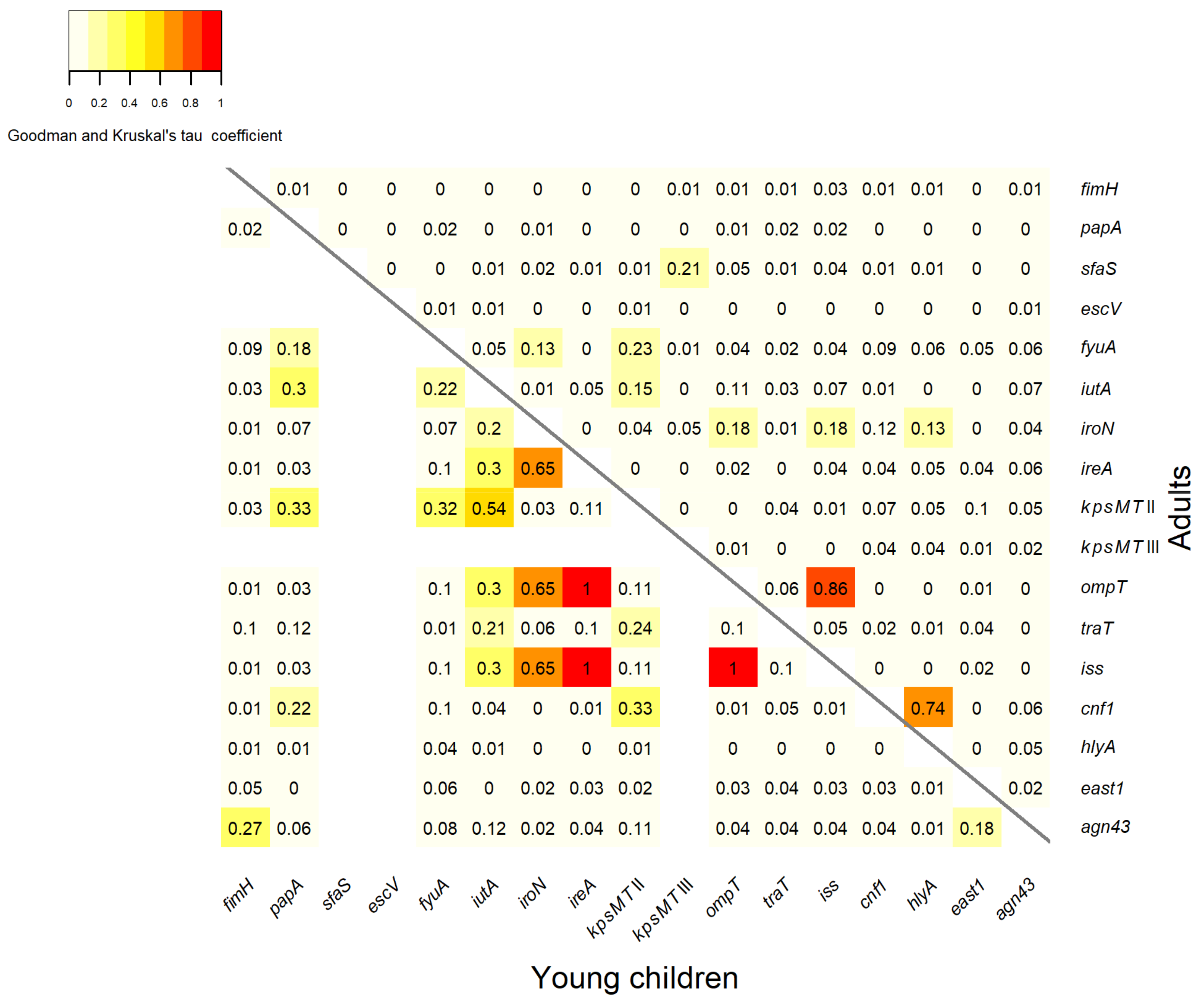
3.3. Association between Virulence Genes

The statistical analysis of the association between the virulence genes of the E. coli isolates is shown in Figure 2. A strong association occurred between the genes cnf1 and hlyA, (association coefficient of 0.74), among the isolates from adults (Figure 2, the part above the diagonal). In the group of isolates from young children, very strong positive associations were found between the gene ireA and both genes iss and ompT, with association coefficients of 1. Strong associations were observed between the gene iroN and genes iss, ompT, ireA, with a coefficient of 0.65. The genes iutA and kpsMT II were also positively associated, with a coefficient of 0.54. There were moderate association between the gene kpsMT II and genes cnf1, fyuA, papA, with association coefficients of 0.33, 0.32 and 0.33, respectively, in the isolates from young children (Figure 2, the part under the diagonal). There is good evidence of a strong association between the genes ompT and iss among E. coli isolates from both adults and from young children, with coefficients of 0.86 and 1 respectively. Weak association, with coefficients ≤0.3, in the isolates from both young children and adults occurred between the remaining virulence genes.

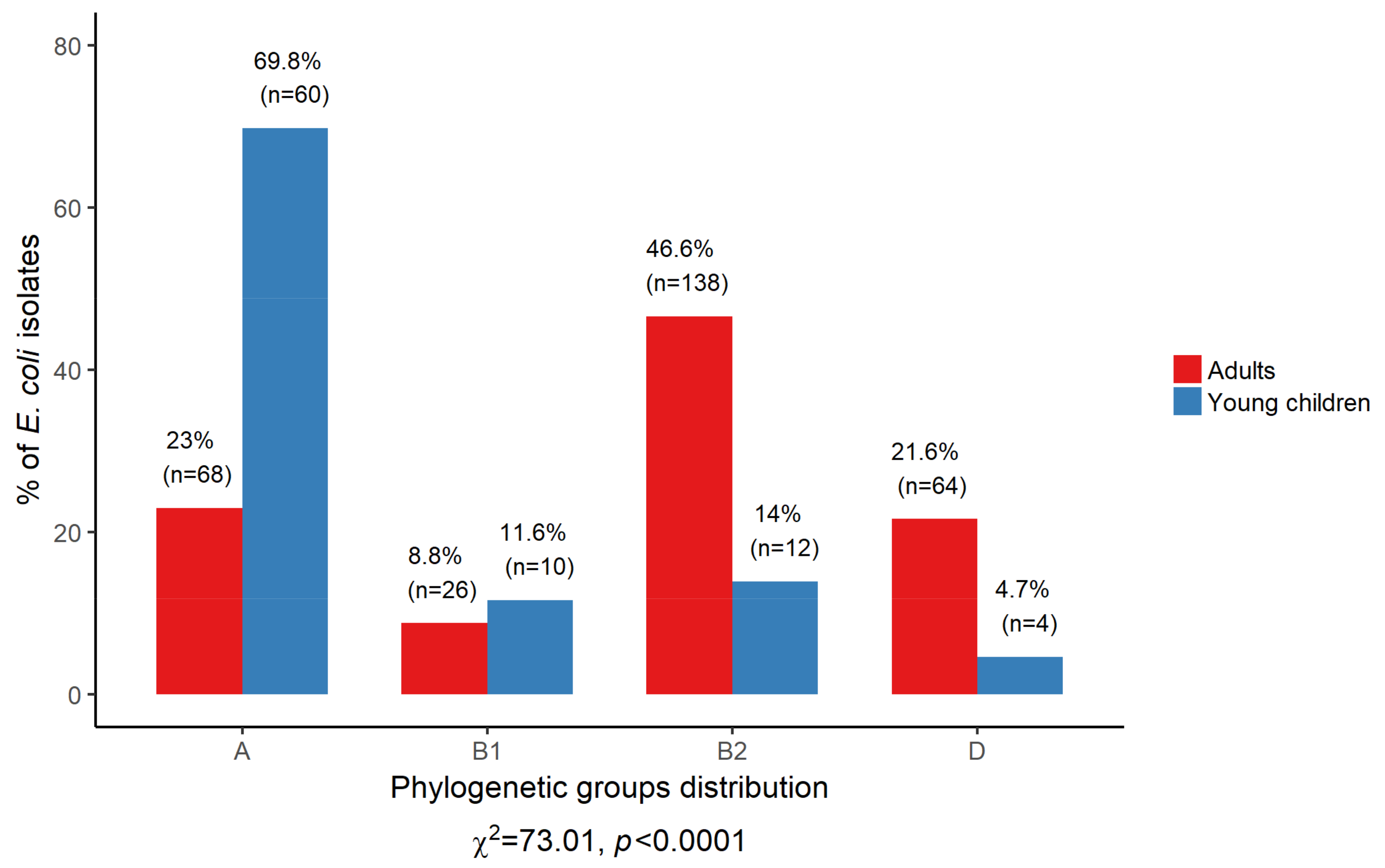
3.4. Phylogenetic Structure of Commensal E. coli

The E. coli isolates derived from adults and young children differed significantly in their assignments to the phylogenetic groups (p < 0.0001) (Figure 3). The phylogroups B2 and D significantly prevailed among the isolates from adults compared to young children, p < 0.0001 and p = 0.0005, respectively. Conversely, phylogenetic group A was significantly more frequent in the isolates from young children as compared to adults (p < 0.0001).

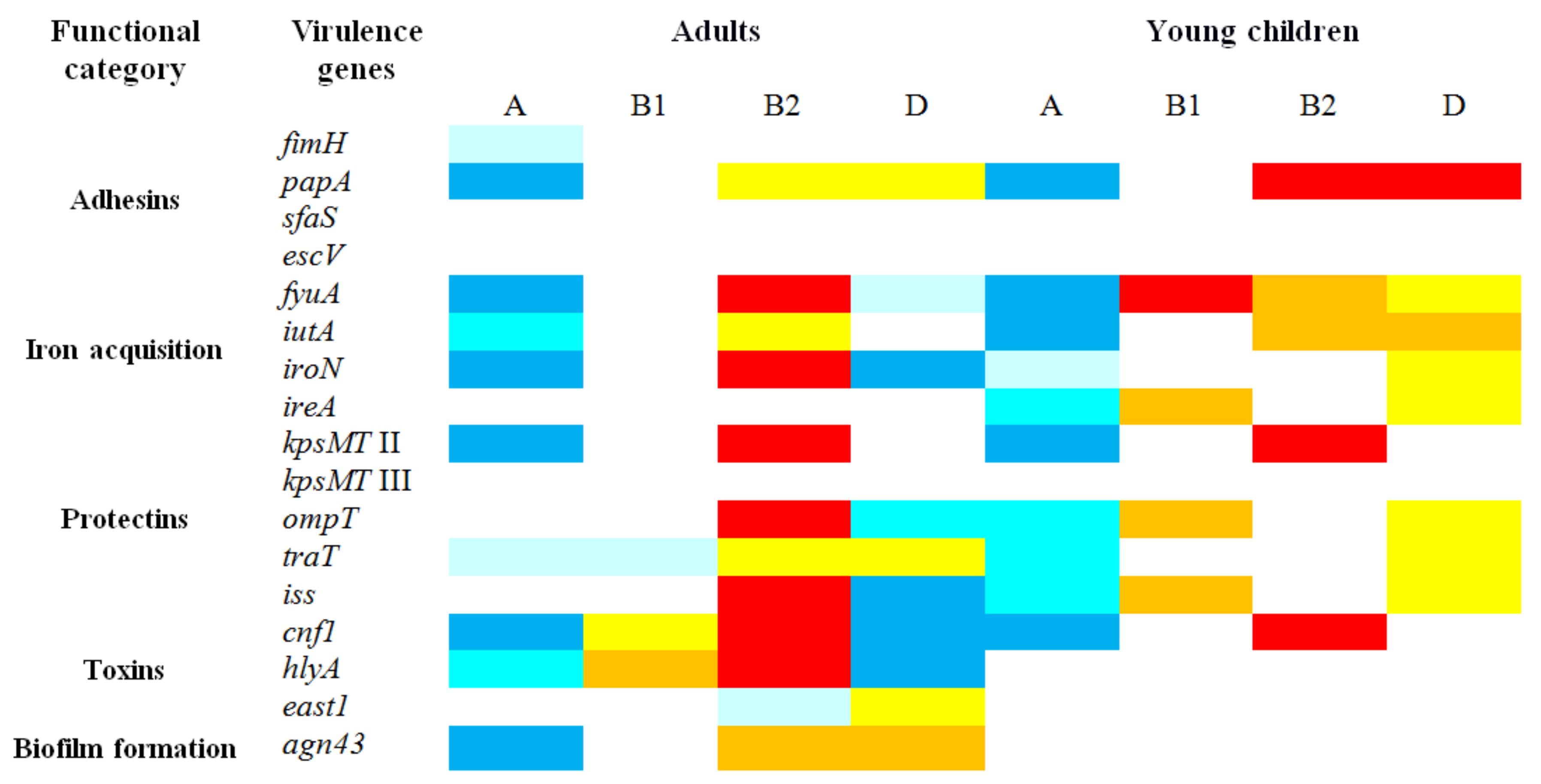
3.5. Distribution of Virulence Genes in Relation to Phylogenetic Groups

Figure 4 shows that the majority of the tested virulence genes were not randomly distributed among the four main phylogenetic groups of isolates. Generally, among the isolates derived from both adults and young children the frequencies of the particular genes were significantly lower (p < 0.05) in the isolates of group A, whereas the isolates of groups B2 harbored the virulence determinants significantly more frequently (p < 0.05).
The further analysis encompassed comparing the prevalence of different virulence genes between the isolates from adults and young children within each phylogenetic group. The results indicated that group A and B2 isolates and to a lesser extent group B1 from adults carried frequently virulence factors (p < 0.05) than the isolates from young children. The reverse situation was characteristic for the isolates of group D; several virulence genes were detected less frequently (p < 0.05) in the isolates from adults than from young children (Table 2). The fyuA, iutA, ireA, kpsMT II, ompT, traT and iss genes occurred more frequently in the isolates of group A from adults than from young children, while the east1 gene was more frequent detected among the isolates from young children. The three genes cnf1, hlyA and agn43 occurred more frequently in the isolates of group B1 from adults. The genes for iroN, ireA, ompT, iss and hlyA were found more frequently among the isolates of group B2 from adults than from young children; the one exception was the papA gene detected more often in the E. coli from young children. The papA, iroN, ompT and iss genes occurred less frequently in the isolates of group D from adults than from young children (Table 2).

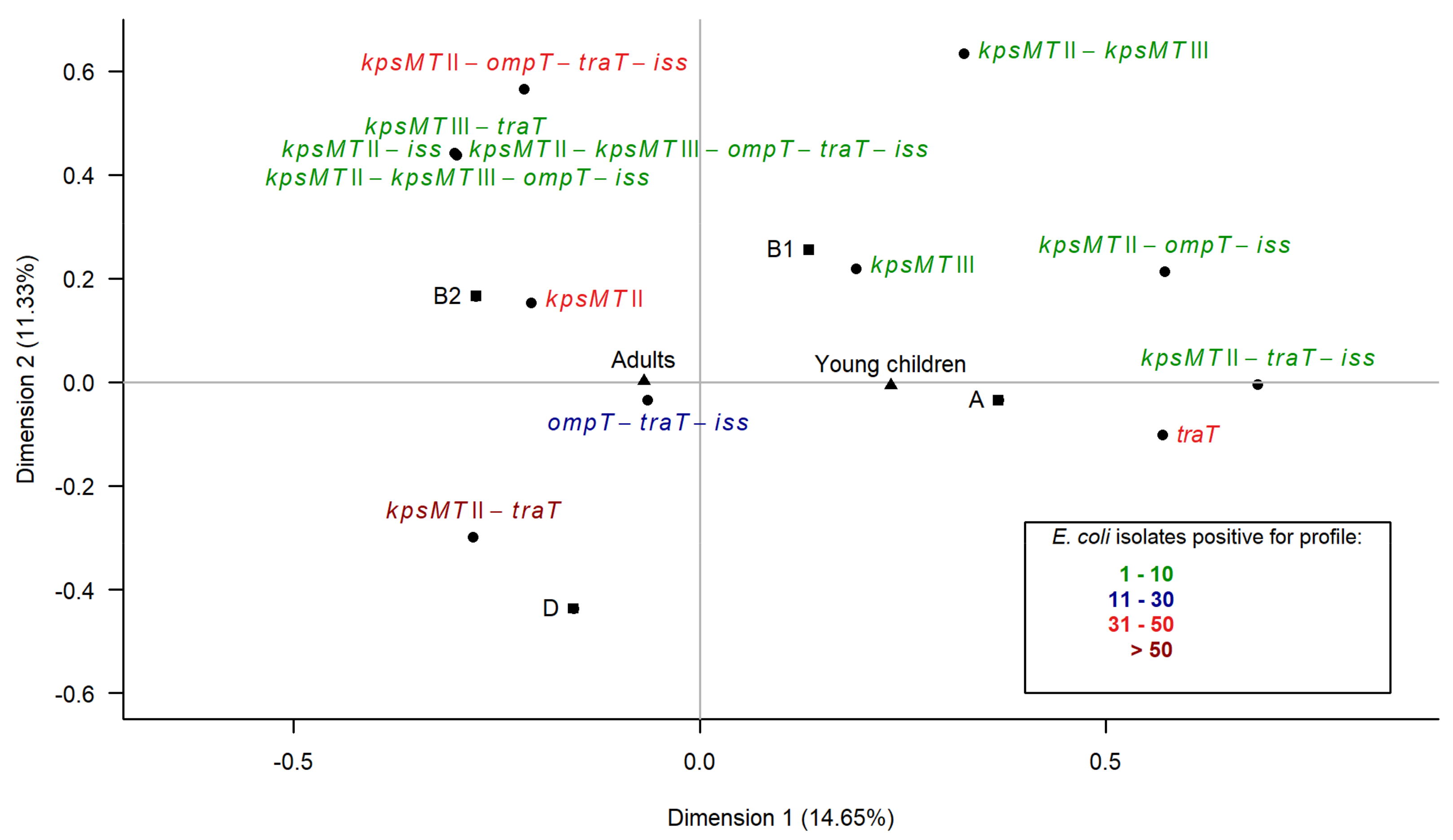
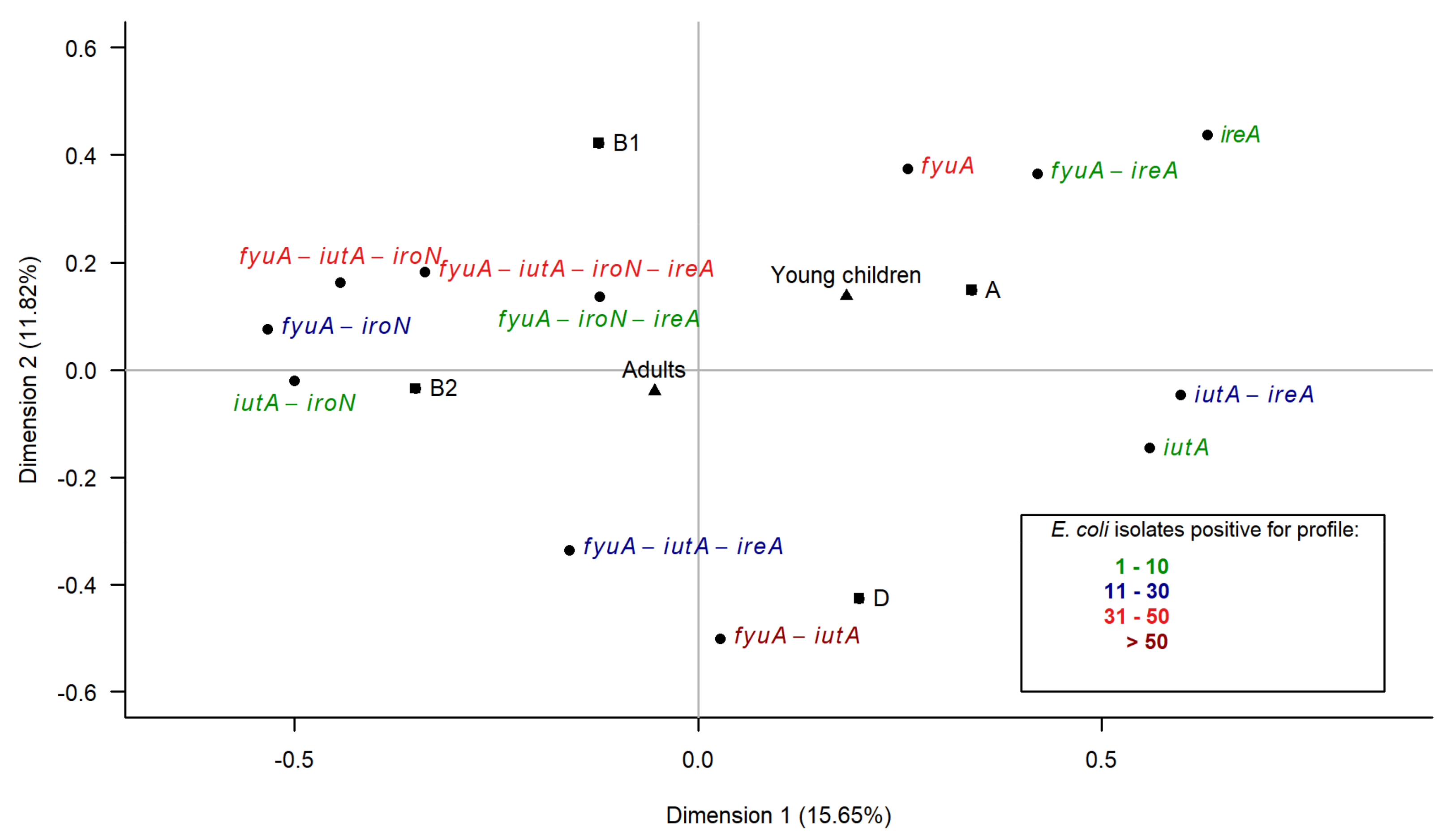
3.6. Multiple Correspondence Analysis

The virulence genes within two categories—iron acquisition and protectins—were particularly prevalent among the tested E. coli isolates. To determine the overall relationships between two groups of the host (adults, young children) and the characteristics of the isolates, multiple correspondence analysis (MCA) was performed. The first analysis concerned the iron acquisition category. The projection of the variables on the dimension1–dimension2 plane accounted for 27.47% of the total variance and distinguished two major associations (Figure 5). The first association was positioned on the negative values of the first dimension and the positive and negative values of the second dimension and encompassed the isolates from adults, phylogenetic groups B1 and B2 variables and five complex iron acquisition profiles, consisting of two, three or four genes. The isolates from young children, the phylogenetic group A variables and three simple iron acquisition profiles consisting of one or two genes formed the second association and were located on the positive values of both dimensions. This analysis clearly distinguished adults, phylogroups B1 and B2 and the complex iron acquisition profile variables on the negative values of the first dimension from the young children, phylogroups A and D and the simple iron acquisition profile variables on the positive values of the first dimension.
The second analysis concerned the protectin category. The projection of the variables on the dimension1–dimension2 plane accounted for 25.98% of the total variance (Figure 6). The first association encompassed the isolates from adults, phylogroup B2 and D variables and eight protectin profiles consisting of one to five genes, located on the negative values of the first dimension. The second association was formed by the isolates from young children, phylogroup A and B1 variables and five profiles consisting of one to three protectin genes, positioned on the positive values of the first dimension. The most complex protectin profiles were located close to phylogroup B2 and the isolates from adults’ variables. The analysis clearly distinguished adults, phylogroups B2 and D and the complex protectin profile variables from the young children, phylogroups A and B1 and the simple protectin profile variables.

3.7. Prevalence of Antimicrobial Resistance

Resistance rates to 17 antimicrobial agents were compared between the E. coli isolates from adults and young children and presented in Table 3. The most frequent resistance was detected for ampicillin (38.2% vs. 31.4%), cephalothin (29.1% vs. 27.9%), streptomycin (46.3% vs. 22.1%) and tetracycline (21.3% vs. 19.8%) in the isolates from adults and young children respectively. The resistance rates for norfloxacin and ciprofloxacin (2.7% vs. 2.3%) and cefotaxime (1.4% vs. 2.7%) were the lowest among the isolates from adults and young children respectively. Significantly higher frequency (p < 0.01) in resistance to amoxicillin/clavulanic acid, piperacillin and streptomycin were observed in isolates from adults as compared with young children. Moreover, resistance to at least one antimicrobial agent (75.7% vs. 54.7%) and multidrug-resistance (30.4% vs. 14%) were detected significantly more frequent (p = 0.0002 and p = 0.0024 respectively) in the isolates from adults than from young children (Table 3).

3.8. Detection of Resistance Determinants

The isolates were screened according to their resistance phenotypes for the carriage of a set of commonly encountered resistance genes. Genetic determinants associated with resistance to ampicillin, cefotaxime, streptomycin, tetracycline, sulfamethoxazole, trimethoprim and nalidixic acid were tested in this study. There were no statistically significant differences between the frequencies of the resistance determinants among the resistant E. coli isolates from adults and young children (Table S2). The most frequently observed gene encoding resistance to ampicillin was blaTEM with 47.8% vs. 48.1% for adults’ and young children’s isolates, respectively. The gene encoding the extended spectrum β-lactamase blaCTX-M was detected in all the isolates resistant to cefotaxime. Regarding streptomycin resistant E. coli the aadA1 gene occurred most often (8.8% vs. 21.1%, in the isolates from adults and young children, respectively). The predominant gene among the tetracycline resistant isolates was tetB with the frequencies 50.8% vs. 41.2% for adults’ and young children’s isolates, respectively. The genes dfrA7 (54.5% vs. 33.3%) and sul2 (60.6% vs. 25%) prevailed among the trimethoprim/sulfamethoxazole resistant isolates from adults and young children, respectively. The sul3 gene was not detected in any isolates. The qnrS gene was predominant among the E. coli resistant to nalidixic acid (17.9% vs. 25%, in the isolates from adults and young children, respectively). The qnrA gene was not detected in any isolates.

3.9. Correlations between Virulence Genes and Antimicrobial Resistance

The results revealed that the associations between the virulence genes and antimicrobial resistance were weak (association coefficients < 0.2) for E. coli isolates from adults and young children.

4. Discussion

To the best of our knowledge, this is the first study to compare the genetic diversity of commensal E. coli isolates collected from two different age groups (adults and young children). E. coli isolates from fecal samples were characterized in terms of the presence of virulence genes, phylogenetic structure, and antimicrobial resistance. Our results indicate that E. coli isolated from adults differ in their phylogenetic structure, harbour a greater variety of virulence genes, and multidrug-resistance phenotypes, compared to E. coli from young children.
Our results have shown that commensal E. coli isolates from healthy humans constitute a substantial reservoir of genes related to the extraintestinal pathotypes. All of the 15 tested virulence genes typical for ExPEC were detected and, what is important, the prevalence of these genes was significantly higher among the isolates from adults than from young children. In contrast, only two of the nine tested virulence genes associated with the intestinal pathotypes were found: escV and east1. The escV gene was identified only in one isolate from adults, while the east1 gene was more common and was detected more frequent among the E. coli isolates from young children than adults. The previous studies also reported that the presence of the intestinal virulence markers actually was associated with pathogenesis [2,3,46,47]. The extraintestinal virulence genes such as those encoding the fimbrial adhesins, iron acquisition systems, protectins or toxins occur not only in pathogens, but also in commensal microflora of healthy people [3,9,21,47,48]. Previous reports indicated that virulence genes associated with extraintestinal pathogenesis in fact help the E. coli strains to colonize the human gut; therefore they may be considered as a fitness factor and the virulence is a coincidental side effect [8,21,48].
The question arises whether the adult’s age has also an influence on the prevalence of virulence genes. The analysis revealed that there are no statistically significant differences in the distribution of virulence genes in the E. coli isolates in relation to the age of the adult host. The only exception was some genes from the categories of iron acquisition (ireA), protectins (variants K1 and K5 of the kpsMT II) and toxins (cnf1, hlyA), which tend to accumulate in E. coli over the life time of the host. The opposite situation occurred only for the traT gene; the chance of accumulation decreases with age. Overall, the results suggest that the difference in prevalence of virulence genes is not correlated with age of adults, however, there is difference in prevalence of virulence genes between adults and young children.
The phylogenetic analysis revealed that the distribution of four main E. coli phylogroups differ significantly (p < 0.0001) between the isolates derived from adults and from young children. E. coli assigned to phylogroup B2 dominated among adults, whereas phylogenetic group A isolates prevailed among young children. The distribution of the phylogroups among the human population represents two patterns, the first with predominance of group A typical for European population (in 1980s), Africa, South America and Asia [1,25,26,49], and the second pattern that the isolates of group B2 prevailed is characteristic for Europe in the 2000s, North America, Japan and Australia [1,25]. The changes in the phylogenetic structure of the E. coli isolates from Europe during the period of 20 years may be the result of the increasing of food processing and change in dietary habits [1,25]. The results of the present study with the dominance of phylogroup B2 among E. coli isolates from adults are consistent with the previous reports presenting the second pattern of phylogenetic structure, whereas the predominance of isolates of phylogroup A among young children is in accordance with the first pattern [1,25,26,49]. The diet of young children aged 0.5–3 years is rather simple and in the most part consists of organic, less processed food as compared to adult diet. The host diet may be a key factor which had an influence on the phylogenetic structure of the E. coli isolates.
The analysis of the distribution of the virulence genes in relation to the phylogenetic groups revealed that the E. coli group B2 isolates were an extensive reservoir of virulence genes while group A was not.
Our results suggest that the higher frequencies of the virulence genes in the E. coli isolates from adults is due to predomination of phylogroup B2, therefore the comparison between the E. coli isolates from adults and young children with respect to the frequency of the virulence genes in each phylogroup was performed. The results showed that the E. coli isolates from adults assigned to either phylogroup A or B2 carried statistically significant (p < 0.05) more virulence determinants, particularly associated with iron acquisition and protection, than the isolates from young children assigned to phylogroup A and B2. Three genes from the toxin and biofilm formation categories occurred more frequently among phylogroup B1 isolates from adults than from young children. Our results documented that the genetic diversity of the isolates of phylogroups A, B2 and to a lesser extent B1 differs between adults and young children. The isolates may acquire genetic diversity through horizontal gene transfer. Possibly the resident strains that persist longer time in the intestinal tract have a greater chance to undergo numerous horizontal transfer events resulting in accumulation of the virulence determinants. Our studies may suggest that the strains of phylogroup D from adults are less diverse regarding the virulent determinants than E. coli strains from young children, but these results should be confirmed on a larger number of subjects.
Virulence genes are often clustered in the chromosomal genomic regions acquired by horizontal gene transfer called pathogenicity islands [10,50], or in large plasmidic regions [51,52]. One such region typical for the ExPEC plasmids has been recently described and called the conserved virulence plasmidic (CVP) region. This region harbors eight virulence genes, including iroN, ompT and iss [52,53]. The results regarding the associations between the virulence genes revealed that there were fewer statistically significant associations among the E. coli from adults than from young children. Strong associations were found between the iroN, ompT and iss genes among the isolates from young children. In the E. coli isolates from adults there remained only associations between the ompT and iss genes. The prevalence of these genes was much higher among the E. coli isolates from adults than among the isolates from young children. These results suggested that there was a tendency for accumulation of these genes in different combinations in the isolates from adults. This observation indicated that in commensal E. coli strains from adults there is low probability of presence of a specific pathogenic plasmidic CVP region. Two genes from the toxin category—cnf1 and hlyA—were strongly associated among the isolates from adults. What it was previously reported, these genes may be concealed together on pathogenicity island PAI IIJ96 in pathogenic strains [10]. The existence of PAI IIJ96 in our set of E. coli isolates from adults will be confirmed in near future.
Previous studies revealed that the ability of some E. coli strains to persist in the colonic microbiota may be related to the carriage of the virulence genes for P fimbriae, aerobactin (iutA), yersiniabactin (fyuA) and capsular antigen (kpsMT II variants K1 and K5) and that these genes may play a key role as fitness factors [15,20,21,48]. In our studies the results of MCA analyses showed that the isolates from adults correspond primarily to phylogroup B2 and these variables were positioned close to the isolates positive for the more complex and also more numerous profiles both the iron acquisition and protectin categories. The E. coli isolates from young children correspond strongly to phylogroup A and to the isolates with simpler profiles. These results confirmed that the E. coli isolates derived from adults exhibited more genetic diversity than isolates from young children. There were isolates carrying three, four and even five genes of the same functional category. This indicated that the E. coli isolates derived from adults, particularly of phylogroup B2, constitute a significant reservoir of extraintestinal virulence genes and have a tendency to collect them.
The comparison of the resistance rates to 17 antimicrobial agents revealed that the E. coli isolates showed significantly higher (p < 0.01) resistance to three antibiotics: amoxicillin/clavulanic acid, piperacillin and streptomycin in adults as compared to young children. Likewise, significantly more isolates from the adults than from the young children were classified as strains resistant to at least one antimicrobial agent (p = 0.0002) and to multidrug-resistant strains (p = 0.0024). These results suggested that the older the host, the higher accumulation of resistance determinants in E. coli strains occurs but to a lesser degree as in the case of the virulence genes.
Our results indicated that the prevalence of resistance to ampicillin, cephalothin, streptomycin and tetracycline among the E. coli isolates from both adults and young children was rather moderate in the region of Western Poland, prevalence of resistance to trimethoprim/sulfamethoxazole was low/moderate, whereas the rates of resistance to the third-generation cephalosporin cefotaxime and fluoroquinolones were low. Depending on the country and different regions of the world, higher [22,23,49,54,55,56], similar [23,54,55] or lower [24,49,54,57] rates of resistance to the antibiotics mentioned above were observed among E. coli isolates from adults and children. The fact that the E. coli isolates from young children exhibited moderate prevalence of resistance to tetracycline, a drug which should not be administered to children, may be a consequence of the previous exposure of food animals to growth-promoting antibiotics and the dissemination of these agents in the environment [58,59,60]. Some previous studies have revealed that resistant bacteria can be found even in conventional retail foods and ready-to-consume items, which suggests that humans are often exposed to such bacteria through daily food intake [61,62,63].
All the resistant E. coli isolates were screened for the carriage of a set of commonly encountered resistance determinants. The results revealed no statistically significant differences between the frequencies of the tested resistance genes in the resistant E. coli isolates from adults and young children. It was probably due to the similar rates of resistance to most of the tested antibiotics in these two groups of isolates. The blaTEM, aadA1, tetB, dfrA7, sul2 and qnrS genes related to ampicillin, streptomycin, tetracycline, trimethoprim/sulfamethoxazole and quinolone resistance respectively were the most common. The resistant isolates negative for the tested resistance determinants may have carried one of the other resistance genes.
Previous studies have reported that strains carrying the papC or iutA gene were more often resistant to at least one of the tested antibiotics than were strains without these traits [24,64]. Virulence genes and resistance genes may occur together on mobile genetic elements and be co-selected. The results of these studies revealed that the association between the virulence genes and antimicrobial resistance were weak in both groups of E. coli isolates.

5. Conclusions

This study analyzed the relationships between the distribution of virulence genes, phylogenetic structure and prevalence of antimicrobial resistance among E. coli isolated from adults and young children in Lubuskie province, Poland. This is the first such comparison regarding only commensal strains. The results revealed significant difference in the distribution of the virulence genes, phylogenetic groups as well as the multidrug-resistance among E. coli from adults and young children. This study confirmed that the commensal E. coli isolates, particularly that derived from adults, constitutes a considerable reservoir of the virulence genes typical for ExPEC. The comparison of the frequency of the virulence genes between the E. coli isolates from adults and young children in relation to each phylogroup showed that greater genetic diversity exists in isolates from adults. This may suggest accumulation of the virulence determinants among commensal E. coli strains through horizontal gene transfer over the years. Further research will be needed to better understand the relationships between environmental factors and the genetic diversity of E. coli commensal strains. Plasticity of E. coli genome provides the ability to adapt to different nutrient conditions and niches in the host’s body [1,6]. An interesting question remains how the environmental conditions (diet, travel, cultural background, etc.) effect the shift of E. coli from commensal to pathogen strain responsible for extraintestinal infection.

Supplementary Materials

The following are available online at https://www.mdpi.com/1660-4601/15/4/617/s1, Figure S1: Age distribution among adults. Table S1: List of virulence genes tested in this study. Table S2: Prevalence of the resistance genes among the antimicrobial-resistant E. coli isolates from healthy adults and young children.

Acknowledgments

We would like to thank Wanda Tomala, Katarzyna Janecka and Paweł Pusz for skillful technical assistance in our work. This study was supported by Ministry of Science and Higher Education grant N N304 176 340.

Author Contributions

E.B. and K.B.-C. conceived and designed the experiments. E.B. and J.M. performed the experiments. E.B., K.B.-C., J.M., A.M., M.S. analyzed the data. M.W. performed all statistical analysis. E.B. wrote the paper. All authors read and approved the final manuscript.

Conflicts of Interest

The authors declare no conflict of interest.

References

  1. Tenaillon, O.; Skurnik, D.; Picard, B.; Denamur, E. The population genetics of commensal Escherichia coli. Nat. Rev. Microbiol. 2010, 8, 207–217. [Google Scholar] [CrossRef] [PubMed]
  2. Kaper, J.B.; Nataro, J.P.; Mobley, H.L. Pathogenic Escherichia coli. Nat. Rev. Microbiol. 2004, 2, 123–140. [Google Scholar] [CrossRef] [PubMed]
  3. Köhler, C.D.; Dobrindt, U. What defines extraintestinal pathogenic Escherichia coli? Int. J. Med. Microbiol. 2011, 301, 642–647. [Google Scholar] [CrossRef] [PubMed]
  4. Kosek, M.; Bern, C.; Guerrant, R.L. The global burden of diarrhoeal disease, as estimated from studies published between 1992 and 2000. Bull. World Health Organ. 2003, 81, 197–204. [Google Scholar] [PubMed]
  5. Russo, T.A.; Johnson, J.R. Medical and economic impact of extraintestinal infections due to Escherichia coli: Focus on an increasingly important endemic problem. Microbes Infect. 2003, 5, 449–456. [Google Scholar] [CrossRef]
  6. Touchon, M.; Hoede, C.; Tenaillon, O.; Barbe, V.; Baeriswyl, S.; Bidet, P.; Bingen, E.; Bonacorsi, S. Organised genome dynamics in the Escherichia coli species results in highly diverse adaptive paths. PLoS Genet. 2009, 5. [Google Scholar] [CrossRef] [PubMed] [Green Version]
  7. Alteri, C.J.; Mobley, H.L. Escherichia coli physiology and metabolism dictates adaptation to diverse host microenvironments. Curr. Opin. Microbiol. 2012, 15, 3–9. [Google Scholar] [CrossRef] [PubMed]
  8. Le Gall, T.; Clermont, O.; Gouriou, S.; Picard, B.; Nassif, X.; Denamur, E.; Tenaillon, O. Extraintestinal virulence is a coincidental by-product of commensalism in B2 phylogenetic group Escherichia coli strains. Mol. Biol. Evol. 2007, 24, 2373–2384. [Google Scholar] [CrossRef] [PubMed]
  9. Starčič, E.M.; Žgur-Bertok, D. Virulence potential for extraintestinal infections among commensal Escherichia coli isolated from healthy humans—The Trojan horse within our gut. FEMS Microbiol. Lett. 2015, 362. [Google Scholar] [CrossRef]
  10. Sabaté, M.; Moreno, E.; Pérez, T.; Andreu, A.; Prats, G. Pathogenicity island markers in commensal and uropathogenic Escherichia coli isolates. Clin. Microbiol. Infect. 2006, 12, 880–886. [Google Scholar] [CrossRef] [PubMed]
  11. Koga, V.L.; Tomazetto, G.; Cyoia, P.S.; Neves, M.S.; Vidotto, M.C.; Nakazato, G.; Kobayashi, R.K.T. Molecular screening of virulence genes in extraintestinal pathogenic Escherichia coli isolated from human blood culture in Brazil. BioMed Res. Int. 2014. [Google Scholar] [CrossRef] [PubMed]
  12. Bailey, J.K.; Pinyon, J.L.; Anantham, S.; Hall, R.M. Commensal Escherichia coli of healthy humans: A reservoir for antibiotic-resistance determinants. J. Med. Microbiol. 2010, 59, 1331–1339. [Google Scholar] [CrossRef] [PubMed]
  13. Bettelheim, K.A.; Lennox-King, S.M. The acquisition of Escherichia coli by new-born babies. Infection 1976, 4, 174–179. [Google Scholar] [CrossRef] [PubMed]
  14. Fryklund, B.; Tullus, K.; Berglund, B.; Burman, L.G. Importance of the environmental and the faecal flora of infants, nursing staff and parents as sources of Gramnegative bacteria colonizing newborns in three neonatal wards. Infection 1992, 20, 253–257. [Google Scholar] [CrossRef] [PubMed]
  15. Nowrouzian, F.; Hesselmar, B.; Saalman, R.; Strannegard, I.L.; Aberg, N.; Wold, A.E.; Adlerberth, I. Escherichia coli in infants’ intestinal microflora: Colonization rate, strain turnover, and virulence gene carriage. Pediatr. Res. 2003, 54, 8–14. [Google Scholar] [CrossRef] [PubMed]
  16. Johnson, J.R.; Stell, A.L. Extended virulence genotypes of Escherichia coli strains from patients with urosepsis in relation to phylogeny and host compromise. J. Infect. Dis. 2000, 181, 261–272. [Google Scholar] [CrossRef] [PubMed]
  17. Picard, B.; Garcia, J.S.; Gouriou, S.; Duriez, P.; Brahimi, N.; Bingen, E.; Elion, J.; Denamur, E. The link between phylogeny and virulence in Escherichia coli extraintestinal infection. Infect. Immun. 1999, 67, 546–553. [Google Scholar] [PubMed]
  18. Johnson, J.R.; Oswald, E.; O’Bryan, T.T.; Kuskowski, M.A.; Spanjaard, L. Phylogenetic distribution of virulence-associated genes among Escherichia coli isolates associated with neonatal bacterial meningitis in the Netherlands. J. Infect. Dis. 2002, 185, 774–784. [Google Scholar] [CrossRef] [PubMed]
  19. Caugant, D.A.; Levin, B.R.; Selander, R.K. Genetic diversity and temporal variation in the E. coli population of a human host. Genetics 1981, 98, 467–490. [Google Scholar] [PubMed]
  20. Nowrouzian, F.; Adlerberth, I.; Wold, A.E. P fimbriae, capsule and aerobactin characterize colonic resident Escherichia coli. Epidemiol. Infect. 2001, 126, 11–18. [Google Scholar] [CrossRef] [PubMed]
  21. Nowrouzian, F.L.; Adlerberth, I.; Wold, A.E. Enhanced persistence in the colonic microbiota of Escherichia coli strains belonging to phylogenetic group B2: Role of virulence factors and adherence to colonic cells. Microbes Infect. 2006, 8, 834–840. [Google Scholar] [CrossRef] [PubMed]
  22. Domínguez, E.; Zarazaga, M.; Sáenz, Y.; Briñas, L.; Torres, C. Mechanisms of antibiotic resistance in Escherichia coli isolates obtained from healthy children in Spain. Microb. Drug Resist. 2002, 8, 321–327. [Google Scholar] [CrossRef] [PubMed]
  23. Li, B.; Zhao, Z.C.; Wang, M.H.; Huang, X.H.; Pan, Y.H.; Cao, Y.P. Antimicrobial resistance and integrons of commensal Escherichia coli strains from healthy humans in China. J. Chemother. 2014, 26, 190–192. [Google Scholar] [CrossRef] [PubMed]
  24. Karami, N.; Hannoun, C.; Adlerberth, I.; Wold, A.E. Colonization dynamics of ampicillin-resistant Escherichia coli in the infantile colonic microbiota. J. Antimicrob. Chemother. 2008, 62, 703–708. [Google Scholar] [CrossRef] [PubMed]
  25. Escobar-Páramo, P.; Grenet, K.; Le Menac’h, A.; Rode, L.; Salgado, E.; Amorin, C.; Gouriou, S.; Picard, B.; Rahimy, M.C.; Andremont, A.; et al. Large-scale population structure of human commensal Escherichia coli isolates. Appl. Environ. Microbiol. 2004, 70, 5698–5700. [Google Scholar] [CrossRef] [PubMed]
  26. Al-Mayahie, S.M.G.; Al-Khafajy, A.A.M.; Dosh, N.A.S.; Al-Rekabi, A.R.K.; Al-Atabie, A.G.N. Phylogenetic grouping of dominant fecal escherichia coli isolates from healthy males and females in Al-Kut/Wasit Province/Iraq. J. Bacteriol. Parasitol. 2015, 6. [Google Scholar] [CrossRef]
  27. Ostblom, A.; Adlerberth, I.; Wold, A.E.; Nowrouzian, F.L. Pathogenicity island markers, virulence determinants malX and usp, and the capacity of Escherichia coli to persist in infants’ commensal microbiotas. Appl. Environ. Microbiol. 2011, 77, 2303–2308. [Google Scholar] [CrossRef] [PubMed]
  28. Song, S.J.; Lauber, C.; Costello, E.K.; Lozupone, C.A.; Humphrey, G.; Berg-Lyons, D.; Caporaso, J.G.; Knights, D.; Clemente, J.C.; Nakielny, S.; et al. Cohabiting family members share microbiota with one another and with their dogs. Elife 2013, 16. [Google Scholar] [CrossRef] [PubMed]
  29. Sears, H.J.; Brownlee, I.; Uchiyama, J.K. Persistence of individual strains of Escherichia coli in the intestinal tract of man. J. Bacteriol. 1950, 59, 293–301. [Google Scholar] [PubMed]
  30. Müller, D.; Hagedorn, P.; Brast, S.; Heusipp, G.; Bielaszewska, M.; Friedrich, A.W.; Karch, H.; Schmidt, M.A. Rapid identification and differentiation of clinical isolates of enteropathogenic Escherichia coli (EPEC), atypical EPEC, and Shiga toxin-producing Escherichia coli by a one-step multiplex PCR method. J. Clin. Microbiol. 2006, 44, 2626–2629. [Google Scholar] [CrossRef] [PubMed]
  31. Russo, T.A.; Carlino, U.B.; Johnson, J.R. Identification of a new iron-regulated virulence gene, ireA, in an extraintestinal pathogenic isolate of Escherichia coli. Infect. Immun. 2001, 69, 6209–6216. [Google Scholar] [CrossRef] [PubMed]
  32. Johnson, J.R.; O’Bryan, T.T. Detection of the Escherichia coli group 2 polysaccharide capsule synthesis gene kpsM by a rapid and specific PCR-based assay. J. Clin. Microbiol. 2004, 42, 1773–1776. [Google Scholar] [CrossRef] [PubMed]
  33. Mellata, M.; Ameiss, K.; Mo, H.; Curtiss, R., 3rd. Characterization of the contribution to virulence of three large plasmids of avian pathogenic Escherichia coli chi7122 (O78:K80:H9). Infect. Immun. 2010, 78, 1528–1541. [Google Scholar] [CrossRef] [PubMed]
  34. Yamamoto, S.; Terai, A.; Yuri, K.; Kurazono, H.; Takeda, Y.; Yoshida, O. Detection of urovirulence factors in Escherichia coli by multiplex polymerase chain reaction. FEMS Immunol. Med. Microbiol. 1995, 12, 85–90. [Google Scholar] [CrossRef] [PubMed]
  35. Chapman, T.A.; Wu, X.Y.; Barchia, I.; Bettelheim, K.A.; Driesen, S.; Trott, D.; Wilson, M.; Chin, J.J. Comparison of virulence gene profiles of Escherichia coli strains isolated from healthy and diarrheic swine. Appl. Environ. Microbiol. 2006, 72, 4782–4795. [Google Scholar] [CrossRef] [PubMed] [Green Version]
  36. Restieri, C.; Garriss, G.; Locas, M.C.; Dozois, C.M. Autotransporter-encoding sequences are phylogenetically distributed among Escherichia coli clinical isolates and reference strains. Appl. Environ. Microbiol. 2007, 73, 1553–1562. [Google Scholar] [CrossRef] [PubMed]
  37. Clermont, O.; Bonacorsi, S.; Bingen, E. Rapid and simple determination of the Escherichia coli phylogenetic group. Appl. Environ. Microbiol. 2000, 66, 4555–4558. [Google Scholar] [CrossRef] [PubMed]
  38. EUCAST Disk Diffusion Method for Antimicrobial Susceptibility Testing Version 5.0 (January 2015). Available online: http://www.eucast.org/fileadmin/src/media/PDFs/EUCAST_files/Disk_test_documents/Manual_v_5.0_EUCAST_Disk_Test.pdf (accessed on 27 March 2018).
  39. The European Committee on Antimicrobial Susceptibility Testing (EUCAST). Breakpoint Tables for Interpretation of MICs and Zone Diameters; Version 4.0; EUCAST: Basel, Switzerland, 2014. [Google Scholar]
  40. Clinical Laboratory Standards Institute (CLSI). Performance Standards for Antimicrobial Susceptibility Testing; Twenty-Third Informational Supplement; Document M100-S23; CLSI: Wayne, PA, USA, 2013. [Google Scholar]
  41. Ng, L.K.; Martin, I.; Alfa, M.; Mulvey, M. Multiplex PCR for the detection of tetracycline resistant genes. Mol. Cell. Probes 2001, 15, 209–215. [Google Scholar] [CrossRef] [PubMed]
  42. Frech, G.; Kehrenberg, C.; Schwarz, S. Resistance phenotypes and genotypes of multiresistant Salmonella enterica subsp. enterica serovar Typhimurium var. Copenhagen isolates from animal sources. J. Antimicrob. Chemother. 2003, 51, 180–182. [Google Scholar] [CrossRef] [PubMed]
  43. Kozak, G.K.; Boerlin, P.; Janecko, N.; Reid-Smith, R.J.; Jardine, C. Antimicrobial resistance in Escherichia coli isolates from swine and wild small mammals in the proximity of swine farms and in natural environments in Ontario, Canada. Appl. Environ. Microbiol. 2009, 75, 559–566. [Google Scholar] [CrossRef] [PubMed]
  44. Cattoir, V.; Poirel, L.; Rotimi, V.; Soussy, C.J.; Nordmann, P. Multiplex PCR for detection of plasmid-mediated quinolone resistance qnr genes in ESBL-producing enterobacterial isolates. J. Antimicrob. Chemother. 2007, 60, 394–397. [Google Scholar] [CrossRef] [PubMed]
  45. Cao, V.; Lambert, T.; Nhu, D.Q.; Loan, H.K.; Hoang, N.K.; Arlet, G.; Courvalin, P. Distribution of extended-spectrum beta-lactamases in clinical isolates of Enterobacteriaceae in Vietnam. Antimicrob. Agents Chemother. 2002, 46, 3739–3743. [Google Scholar] [CrossRef] [PubMed]
  46. Nataro, J.P.; Kaper, J.B. Diarrheagenic Escherichia coli. Clin. Microbiol. Rev. 1998, 11, 142–201. [Google Scholar] [PubMed]
  47. Leimbach, A.; Hacker, J.; Dobrindt, U. E. coli as an all-rounder: the thin line between commensalism and pathogenicity. Curr. Top. Microbiol. Immunol. 2013, 358, 3–32. [Google Scholar] [PubMed]
  48. Nowrouzian, F.L.; Wold, A.E.; Adlerberth, I. Escherichia coli strains belonging to phylogenetic group B2 have superior capacity to persist in the intestinal microflora of infants. J. Infect. Dis. 2005, 191, 1078–1083. [Google Scholar] [CrossRef] [PubMed]
  49. Dureja, C.; Mahajan, S.; Raychaudhuri, S. Phylogenetic distribution and prevalence of genes encoding class I Integrons and CTX-M-15 extended-spectrum β-lactamases in Escherichia coli isolates from healthy humans in Chandigarh, India. PLoS ONE 2014, 9, e112551. [Google Scholar] [CrossRef] [PubMed]
  50. Hacker, J.; Kaper, J.B. Pathogenicity islands and the evolution of microbes. Ann. Rev. Microbiol. 2000, 54, 641–679. [Google Scholar] [CrossRef] [PubMed]
  51. Johnson, T.J.; Nolan, L.K. Pathogenomics of the virulence plasmids of Escherichia coli. Microbiol. Mol. Biol. Rev. 2009, 73, 750–774. [Google Scholar] [CrossRef] [PubMed]
  52. Lemaître, C.; Mahjoub-Messai, F.; Dupont, D.; Caro, V.; Diancourt, L.; Bingen, E.; Bidet, P.; Bonacorsi, S. A conserved virulence plasmidic region contributes to the virulence of the multiresistant Escherichia coli meningitis strain S286 belonging to phylogenetic group C. PLoS ONE 2013, 8. [Google Scholar] [CrossRef] [PubMed]
  53. Tivendale, K.A.; Noormohammadi, A.H.; Allen, J.L.; Browning, G.F. The conserved portion of the putative virulence region contributes to virulence of avian pathogenic Escherichia coli. Microbiology 2009, 155, 450–460. [Google Scholar] [CrossRef] [PubMed]
  54. Garcia, P.G.; Silva, V.L.; Diniz, C.G. Occurrence and antimicrobial drug susceptibility patterns of commensal and diarrheagenic Escherichia coli in fecal microbiota from children with and without acute diarrhea. J. Microbiol. 2011, 49, 46–52. [Google Scholar] [CrossRef] [PubMed]
  55. Cho, S.H.; Lim, Y.S.; Park, M.S.; Kim, S.H.; Kang, Y.H. Prevalence of antibiotic resistance in Escherichia coli fecal isolates from healthy persons and patients with diarrhea. Osong Public Health Res. Perspect. 2011, 2, 41–45. [Google Scholar] [CrossRef] [PubMed]
  56. Dyar, O.J.; Hoa, N.Q.; Trung, N.V.; Phuc, H.D.; Larsson, M.; Chuc, N.T.; Lundborg, C.S. High prevalence of antibiotic resistance in commensal Escherichia coli among children in rural Vietnam. BMC Infect. Dis. 2012, 12, 92. [Google Scholar] [CrossRef] [PubMed]
  57. Johnson, T.J.; Logue, C.M.; Johnson, J.R.; Kuskowski, M.A.; Sherwood, J.S.; Barnes, H.J.; DebRoy, C.; Wannemuehler, Y.M.; Obata-Yasuoka, M.; Spanjaard, L.; et al. Associations between multidrug resistance, plasmid content, and virulence potential among extraintestinal pathogenic and commensal Escherichia coli from humans and poultry. Foodborne Pathog. Dis. 2012, 9, 37–46. [Google Scholar] [CrossRef] [PubMed]
  58. Leite-Martins, L.R.; Mahú, M.I.; Costa, A.L.; Mendes, A.; Lopes, E.; Mendonça, D.M.; Niza-Ribeiro, J.J.; de Matos, A.J.; da Costa, P.M. Prevalence of antimicrobial resistance in enteric Escherichia coli from domestic pets and assessment of associated risk markers using a generalized linear mixed model. Prev. Vet. Med. 2014, 117, 28–39. [Google Scholar] [CrossRef] [PubMed]
  59. Koczura, R.; Semkowska, A.; Mokracka, J. Integron-bearing Gram-negative bacteria in lake waters. Lett. Appl. Microbiol. 2014, 59, 514–519. [Google Scholar] [CrossRef] [PubMed]
  60. Proia, L.; Anzil, A.; Subirats, J.; Borrego, C.; Farrè, M.; Llorca, M.; Balcázar, J.L.; Servais, P. Antibiotic resistance along an urban river impacted by treated wastewaters. Sci. Total Environ. 2018, 628–629, 453–466. [Google Scholar] [CrossRef] [PubMed]
  61. Zhang, L.; Kinkelaar, D.; Huang, Y.; Li, Y.; Li, X.; Wang, H.H. Acquired antibiotic resistance: Are we born with it? Appl. Environ. Microbiol. 2011, 77, 7134–7141. [Google Scholar] [CrossRef] [PubMed]
  62. Wang, H.H.; Manuzon, M.; Lehman, M.; Wan, K.; Luo, H.; Wittum, T.E.; Yousef, A.; Bakaletz, L.O. Food commensal microbes as a potentially important avenue in transmitting antibiotic resistance genes. FEMS Microbiol. Lett. 2006, 254, 226–231. [Google Scholar] [CrossRef] [PubMed]
  63. Li, X.; Wang, H.H. Tetracycline resistance associated with commensal bacteria from representative ready-to-consume deli and restaurant foods. J. Food Prot. 2010, 73, 1841–1848. [Google Scholar] [CrossRef] [PubMed]
  64. Karami, N.; Wold, A.E.; Adlerberth, I. Antibiotic resistance is linked to carriage of papC and iutA virulence genes and phylogenetic group D background in commensal and uropathogenic Escherichia coli from infants and young children. Eur. J. Clin. Microbiol. Infect. Dis. 2017, 36, 721–729. [Google Scholar] [CrossRef] [PubMed]
Figure 1. Odds ratios (the dots) and their 95% confidence intervals for the association of the occurrence of particular virulence genes in the E. coli isolates with the age of adults (18–56 years). The relations between the virulence gene and age of adults were described by logistic models. If the 95% CI is above the value of 1, this means that the ratio of the odds of the virulence gene’s occurrence increases with age; otherwise this ratio decreases with age. A narrow confidence interval indicates higher precision of the odds ratio.
Ijerph 15 00617 g001
Figure 2. Statistical association between virulence genes of the E. coli isolates derived from adults (the part above the diagonal) and young children (the part under the diagonal). No values were introduced in the case of undetected genes.
Ijerph 15 00617 g002
Figure 3. Phylogenetic structure of the E. coli isolates derived from two groups: adults and young children.
Ijerph 15 00617 g003
Figure 4. The frequency distribution of virulence genes among E. coli phylogroups was compared by Fisher’s exact test to its prevalence in a group consisted of the isolates of all the other phylogenetic groups. The values significantly higher than among the other groups are indicated as follows: Ijerph 15 00617 i001 p < 0.001, Ijerph 15 00617 i002 p < 0.01, Ijerph 15 00617 i003 p < 0.05. The values significantly lower than among the other groups are indicated as follows: Ijerph 15 00617 i004 p < 0.001, Ijerph 15 00617 i005 p < 0.01, Ijerph 15 00617 i006 p < 0.05.
Ijerph 15 00617 g004
Figure 5. Multiple correspondence analysis (MCA) of the characteristics of the isolates (the phylogenetic group and different profiles of the iron acquisition genes) regarding two groups of the host (adults, young children) among 382 E. coli isolates from humans. The projection of the variables on the dimension1–dimension2 plane: the age of the host (adults, young children) is denoted by triangles, the phylogenetic group (A, B1, B2, D) by squares, the profiles (combinations of the iron acquisition genes fyuA, iutA, iroN, ireA) by circles. The number of isolates positive for a particular profile is distinguished by color.
Ijerph 15 00617 g005
Figure 6. Multiple correspondence analysis (MCA) of the characteristics of the isolates (the phylogenetic group and different profiles of the protectin genes) regarding two groups of the host (adults, young children) among 382 E. coli isolates from humans. The projection of the variables on the dimension1–dimension2 plane: the age of the host (adults, young children) is denoted by triangles, the phylogenetic group (A, B1, B2, D) by squares, the profiles (combinations of the protectin genes: kpsMT II kpsMT III, ompT, traT, iss) by circles. The number of isolates positive for a particular profile is distinguished by color.
Ijerph 15 00617 g006
Table 1. Prevalence of virulence genes by functional categories among E. coli isolates from healthy adults and young children.
Functional Category Virulence GeneE. coli PathotypeNumber (%) of E. coli Isolates with Virulence GenesTest of Independence
p-Value
AdultsYoung Children
n = 296n = 86
Adhesins
fimHExPEC286 (96.6)76 (88.4)0.005 *
papAExPEC54 (18.2)10 (11.6)0.1482
sfaSExPEC4 (1.4)00.5787
escVEPEC, EHEC1 (0.3)01
bfpBEPEC00-
Iron acquisition
fyuAExPEC225 (76)36 (41.9)<0.0001 *
iutAExPEC184 (62.2)17 (19.8)<0.0001 *
iroNExPEC110 (37.2)4 (4.7)<0.0001 *
ireAExPEC87 (29.4)6 (7)<0.0001 *
Protectins
kpsMT IIExPEC201 (67.9)16 (18.6)<0.0001 *
 -K1ExPEC158 (53.4)10 (11.6)<0.0001 *
 -K2ExPEC192 (64.9)15 (17.4)<0.0001 *
 -K5ExPEC125 (42.2)10 (11.6)<0.0001 *
kpsMT IIIExPEC18 (6.1)00.02 *
ompTExPEC62 (21)6 (7)0.003 *
traTExPEC190 (64.2)37 (43)0.0004 *
issExPEC70 (23.6)6 (7)0.0007 *
Toxins
cnf1ExPEC, NTEC64 (21.6)6 (7)0.002 *
hlyAExPEC50 (16.2)4 (2.3)0.004 *
east1EAEC28 (9.5)23 (25.6)<0.0001 *
ehxAEPEC, EHEC00-
stx1EHEC00-
stx2EHEC00-
eltAETEC00-
estIETEC00-
estIIETEC00-
Biofilm formation
agn43ExPEC240 (81.1)58 (67.4)0.007 *
 -aExPEC106 (35.8)12 (14)0.0001 *
 -bExPEC73 (24.7)4 (4.7)<0.0001 *
 -K12ExPEC176 (59.5)53 (61.6)0.7179
* Statistically significant.
Table 2. Comparison of the frequency of virulence genes among the E. coli isolates from adults and young children within each phylogenetic group using Pearson’s chi-squared test or Fisher’s exact test for proportions as appropriate.
Functional CategoryVirulence GenesNumber (%) of E. coli Isolates with Virulence Genes within Phylogenetic Groups
AB1B2D
AdultsYoung ChildrenAdultsYoung ChildrenAdultsYoung ChildrenAdultsYoung Children
n = 68n = 60n = 26n = 10n = 138n = 12n = 64n = 4
AdhesinsfimH62 (91.2)50 (83.3)26 (100)10 (100)134 (97.1)12 (100)64 (100)4 (100)
papA004 (15.4)032 (23.2)6 (50) *18 (28.1)4 (100) ***
sfaS00004 (2.9)000
escV001 (0.3)00000
Iron acquisitionfyuA32 (47.1) **12 (20)20 (76.9)10 (100)131 (94.9)10 (83.3)42 (65.6)4 (100)
iutA30 (44.1) ***2 (3.3)14 (53.8)4 (40)96 (69.6)7 (58.3)44 (68.8)4 (100)
iroN4 (5.9)014 (53.8)2 (20)88 (63.8) ***04 (6.2)2 (50) *
ireA24 (35.3) ***06 (23.1)4 (40)40 (29) *017 (26.6)2 (50)
ProtectinskpsMT II22 (32.4) ***014 (53.8)4 (40)117 (84.8)10 (83.3)48 (75)2 (50)
kpsMT III2 (2.9)02 (7.7)012 (8.7)02 (3.1)0
ompT8 (11.8) **04 (15.4)4 (40)46 (33.3) **04 (6.2)2 (50) *
traT34 (50) *18 (30)10 (38.5)4 (40)98 (71)11 (91.7)48 (75)4 (100)
iss12 (17.6) ***04 (15.4)4 (40)50 (36.2) **04 (6.2)2 (50) *
Toxinscnf12 (2.9)010 (38.5) *050 (36.2)6 (50)2 (3.1)0
hlyA2 (2.9)4 (6.7)10 (38.5) *038 (27.5) *000
east18 (11.8)21 (35) **2 (7.7)06 (4.3)2 (16.7)12 (18.8)0
Biofilm formationagn4336 (52.9)40 (66.7)20 (76.9) *4 (40)124 (89.9)10 (83.3)60 (93.8)4 (100)
p-value: *** 0–0.001, ** 0.001–0.01, * 0.01–0.05.
Table 3. Prevalence of resistance among the E. coli isolates from groups of adults and young children to 17 antimicrobials.
Antimicrobial AgentNumber (%) of E. coli IsolatesTest of Independence
p-Value *
AdultsYoung Children
n = 296n = 86
Ampicillin113 (38.2)27 (31.4)0.2506
Amoxicillin/Clavulanic acid40 (13.5)3 (3.5)0.0096 *
Piperacillin72 (24.3)5 (5.8)0.0002 *
Cephalothin86 (29.1)24 (27.9)0.8362
Cefuroxime20 (6.8)3 (3.5)0.3152
Cefotaxime4 (1.4)2 (2.7)0.6206
Streptomycin137 (46.3)19 (22.1)<0.0001 *
Gentamicin32 (10.8)9 (10.5)0.9273
Amikacin30 (10.1)7 (8.1)0.5818
Tetracycline63 (21.3)17 (19.8)0.7610
Doxycycline46 (15.5)11 (12.8)0.5287
Trimethoprim/sulfamethoxazole33 (11.1)12 (14)0.4776
Chloramphenicol18 (6.1)00.0171
Nalidixic acid39 (13.2)8 (9.3)0.3358
Norfloxacin8 (2.7)2 (2.3)1
Ciprofloxacin8 (2.7)2 (2.3)1
Nitrofurantoin12 (4.1)3 (3.5)1
Antimicrobial susceptibility characteristic
R224 (75.7)47 (54.7)0.0002 *
MDR90 (30.4)12 (14)0.0024 *
R—resistant to at least one agent; MDR—multidrug-resistant. * Statistically significant.

