Article

Genetic Approach for the Fast Discovery of Phenazine Producing Bacteria

Kieler Wirkstoff-Zentrum (KiWiZ) am Leibniz-Institut für Meereswissenschaften (IFM-GEOMAR), Am Kiel-Kanal 44, 24106 Kiel, Germany
*
Author to whom correspondence should be addressed.
Mar. Drugs 2011, 9(5), 772-789; https://doi.org/10.3390/md9050772
Submission received: 17 February 2011 / Revised: 1 April 2011 / Accepted: 29 April 2011 / Published: 9 May 2011

Abstract

:
A fast and efficient approach was established to identify bacteria possessing the potential to biosynthesize phenazines, which are of special interest regarding their antimicrobial activities. Sequences of phzE genes, which are part of the phenazine biosynthetic pathway, were used to design one universal primer system and to analyze the ability of bacteria to produce phenazine. Diverse bacteria from different marine habitats and belonging to six major phylogenetic lines were investigated. Bacteria exhibiting phzE gene fragments affiliated to Firmicutes, Alpha- and Gammaproteobacteria, and Actinobacteria. Thus, these are the first primers for amplifying gene fragments from Firmicutes and Alphaproteobacteria. The genetic potential for phenazine production was shown for four type strains belonging to the genera Streptomyces and Pseudomonas as well as for 13 environmental isolates from marine habitats. For the first time, the genetic ability of phenazine biosynthesis was verified by analyzing the metabolite pattern of all PCR-positive strains via HPLC-UV/MS. Phenazine production was demonstrated for the type strains known to produce endophenazines, 2-hydroxy-phenazine, phenazine-1-carboxylic acid, phenazine-1,6-dicarboxylic acid, and chlororaphin as well as for members of marine Actinobacteria. Interestingly, a number of unidentified phenazines possibly represent new phenazine structures.

1. Introduction

Nature is a profitable source of pharmaceutically active substances covering the whole range of biological activities such as antimicrobial, antitumoral, antiparasitic or immunosuppressive [1,2]. Various natural products or natural product-derived compounds have been approved as drugs or are undergoing clinical evaluation and registration [2,3]. Because the rediscovery rate of already known substances in standard screening approaches is high, new strategies are urgently needed. Genetic approaches for the detection of secondary metabolite pathways are promising tools for the selection of biosynthetically talented microorganisms. So far, main targets in this respect were genes encoding for polyketide synthases (PKSs) or non-ribosomal peptide synthetases (NRPSs) [4,5]. By focusing on polyketides and non-ribosomal peptides other interesting compound classes such as phenazines have been neglected so far. Phenazines of natural or synthetic origin turned out to be good drug candidates and therefore are promising secondary metabolites [6]. They are heterocyclic, nitrogenous compounds that are substituted at different sites of the core ring system and therefore display a wide range of structural derivatives and biological activities. More than 100 biologically active (antibacterial, antifungal, antiviral, antitumor) phenazines from natural origin are known to date, synthesized mainly by Pseudomonas and Streptomyces species [7]. Phenazines produced by the root-colonizing Pseudomonas fluorescens 2–79 and Pseudomonas aureofaciens 30–84 are good examples for congeners with promising biological activity, in this case inhibiting several fungal plant pathogens [8]. An economically successful phenazine is clofazimine (Lamprene®, Novartis), first described in 1957 which was established as an antileprosy agent and exerts anti-inflammatory properties in controlling erythema nodosum leprosum reactions (Official FDA Drug label) [6] as well as other antimicrobial activities [911]. Clofazimine is a synthetic phenazine analogue belonging to the riminophenazines, a group of compounds which was originally discovered in lichens [12,13]. Another phenazine, bis(phenazine-1-carboxamide), acts as a potent cytotoxin and represents an interesting class of dual topoisomerase I/II directed anticancer drugs [14].
Although principal parts of the phenazine biosynthesis (Figure 1) and their genetics are known [15,16], there are still gaps in our knowledge and the phenazine pathway is still hypothetical [17]. The function and gene products of phzC, phzE and phzD have been experimentally proven. These enzymes catalyze the conversion into 3-deoxy-d-arabino-heptulosonic acid 7-phosphate (DAHP), 2-amino-2-deoxyisochorismic acid (ADIC) and trans-2,3-dihydro-3-hydroxyanthranilic acid (DHHA) [1720]. The phenazine core structure is synthesized subsequently by condensation of two molecules of 6-amino-5-oxocyclohex-2-ene-1-carboxylic acid [17]. Presumably, PhzF is responsible for the isomerization of DHHA [18]. By now, the function of the enzymes PhzA, PhzB and PhzG are almost enlightened [19] and their role in the condensation and rearrangement reactions to form PCA is demonstrated [17]. According to this information, the design of appropriate oligonucleotide probes and primers was possible. Mavrodi et al. (2010) [21] quite recently designed four different pairs of oligonucleotides on the basis of the phzF gene of the phenazine biosynthesis and investigated the diversity of phenazine producers mainly in soil-dwelling and plant-associated bacteria.
In particular marine bacteria have attracted our interest to search for promising natural products [1,2226]. Therefore, we have investigated the possible application of a genetic approach for the detection of new marine isolates using phzE gene fragments as markers for the ability to produce bioactive phenazines. In contrast to Mavrodi et al. (2010) [21], we wanted to design a more universal primer system able to study a wide range of bacterial groups including actinomycetes and pseudomonades, in order to detect genes of phenazine production in unidentified new isolates without prior performance of a phylogenetic classification.

2. Results and Discussion

The high diversity of microbes and their manifold biosynthetic capabilities offer a great potential for novel, bioactive secondary products. Genetic approaches such as the application of primer systems for NRPS, PKS or halogenases are efficient methods to select appropriate strains for further analyses [27,28]. The screening of bacteria for the presence of phenazine genes significantly adds to these selection filters. We report here on a rapid universal genetic system for the discovery of bacteria that are able to produce phenazines. The suitability of this PCR based screening approach for the identification of phenazine producing bacteria was demonstrated. A gene fragment indispensable for phenazine biosynthesis (phzE) served as a template for primer generation. As positive controls the type strain of S. cinnamonensis, and three subspecies of Pseudomonas chlororaphis, which are all well known producers of phenazines, were included in this study. For control strains several phenazine biosynthesis gene sequences were available at the NCBI (e.g., AM384985, HM594285, AF007801). Altogether 168 bacterial strains (including the four reference organisms) were examined for the presence of phzE. The PCR analysis was complemented by a chemical investigation in order to demonstrate the expression of the biosynthesis genes and to confirm the production of phenazines as proof of concept.

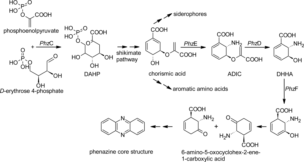
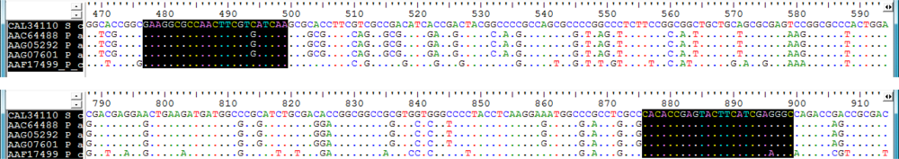
2.1. Design of Oligonucleotides to Search for phzE Phenazine Gene Fragments

Former investigations were able to associate different steps of the phenazine biosynthetic pathway with the corresponding genes, e.g., phzC, phzD, phzE and phzF. The transformation from chorismate to 2-amino-2-deoxyisochorismic acid (ADIC) is necessary for the formation of the core structure of phenazines and is catalyzed by the enzyme PhzE. Thus, PhzE is a key enzyme in phenazine biosynthesis and the corresponding gene phzE is suitable for primer design. Sequences from the phenazine biosynthetic pathway for Alpha-, Beta- and Gammaproteobacteria, Actinomycetes and Firmicutes are available at the homepage of the National Centre for Biotechnology Information (NCBI) and known from literature [29,30]. To ensure the inclusion of only true phenazine sequences, oligonucleotide primers were constructed only from those genes known to be involved in the biosynthesis of corresponding chemical substances. Two conserved sites occurred within the alignment of phzE sequences (Figure 2), which had a distance to each other to produce fragments of an appropriate length. The degenerated primers phzEf (5′-GAA GGC GCC AAC TTC GTY ATC AA-3′) and phzEr (5′-GCC YTC GAT GAA GTA CTC GGT GTG-3′) were designed to amplify a highly conserved stretch of the phzE gene of approximately 450 bp. The comparison of the oligonucleotide sequences from designed phzEf and phzEr primers with known phenazine genes verified this stretch as highly specific for phzE genes. Because the basic phenazine gene cluster including the phzE gene is highly conserved and derivatization of the basic phenazine structure are made at a later stage in the biosynthesis, the constructed phzE primers are expected to detect genes of a large variety of different phenazine structures and are appropriate to search for unknown bacteria producing novel phenazines.

2.2. Screening for phzE Gene Fragments with the Constructed Primers

Genes belonging to the phenazine biosynthetic pathway were present in approximately 10% of the bacterial strains analyzed. PCR results of 13 (8%) out of 164 bacterial strains and four reference organisms were positive in regard to the presence of phzE gene fragments (Figure 3, Tables 1 and 2). The investigated bacteria comprised different bacterial phyla, namely Actinobacteria (76), Bacteroidetes (2), Firmicutes (28) and Proteobacteria (62) (Table 1).
Corresponding gene fragments were detected in 11 strains of Actinobacteria, one strain of Firmicutes and two strains of the Alphaproteobacteria. All sequences were similar to known phzE gene sequences in a range from 65% similarity (phzE of strain LB151 to phzE of P. chlororaphis, AAF17499) to 95% similarity (phzE of strain AB108 to phzE of gene from S. cinnamonensis, CAL34110) (Table 2). Regarding the environmental isolates none of the strains within the Bacteroidetes, Beta- as well as Gammaproteobacteria could be shown to contain phzE in PCR amplification. This was unexpected, because among the 36 gammaproteobacterial isolates 18 Pseudomonas strains were examined and our PCR approach was performed with primer sequences largely based on sequences from Pseudomonas strains known as producers of phenazines [16,31,32]. The suitability of our primer set to detect phenazine genes in Pseudomonas species was further demonstrated by performing a database search that matched perfectly several phenazine genes, e.g., P. chlororaphis (L48339), Pseudomonas sp. M18 (FJ494909), P. aeruginosa (FM209186, CP000744, CP000438, AE004091, AF005404). Anyhow, a study based on phzF sequences exhibited a hit ratio of 100% including 51 Pseudomonas strains [21]. Therefore, all 18 pseudomonads from our study exhibiting negative results using phzE primers were subjected to a genetic approach with phzF primers. While the PCR-amplification of phzE and phzF gene fragments of the control type strains was positive (Table 2), amplification of the investigated isolates failed. As an additional control experiment, crude extracts of six Pseudomonas strains were analyzed by HPLC-UV/MS. Because of the distinctive properties of phenazine UV-absorption spectra the presence of phenazine metabolites was out of question. Additionally, for another study all natural products from two of the investigated Pseudomonas strains were isolated and chemically identified. No phenazines were detected. We conclude that the Pseudomonas strains analyzed in this study lack genes for phenazine production and are unable to produce phenazines. In agreement with this, the only known marine phenazine producing Pseudomonas species is P. aeruginosa [3335], synthesizing almost always pyocyanin. In contrast, different marine streptomycetes are known for production of variable phenazine structures [9]. Streptomyces strains in this study are the most productive group as well. While Brevibacterium, Bacillus and Pelagibacter were known as marine phenazine producers [3638], this is the first time that representatives of the genera Micromonospora, Kiloniella and Pseudovibrio were identified as marine phenazine producers as well.

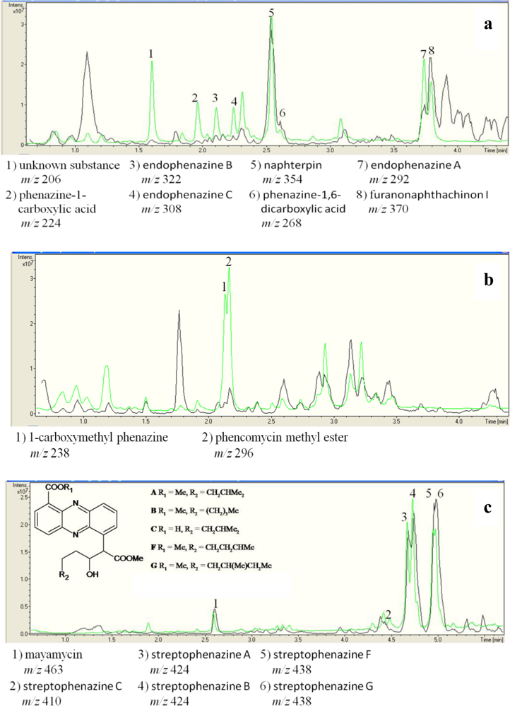
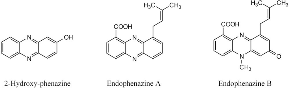
2.3. Detection of Phenazines in the phzE Positive Strains

To demonstrate the synthesis of phenazines in all phzE positive strains, cultures of these strains were extracted and analyzed by HPLC-UV/MS analyses. 14 out of 17 of these strains were able to produce one or more substances with molecular masses and UV-spectra similar to known phenazines (Table 3, Figure 4a–c). In S. cinnamonensis DSM 1042T the production of endophenazines A–C (Figure 5) and phenazine-1,6-dicarboxylic acid [15] could be demonstrated (Figure 3a). The metabolite chlororaphin was discovered from Pseudomonas chlororaphis subsp. chlororaphis DSM 50083T. 2-hydroxy-phenazine (Figure 5) and phenazine-1-carboxylic acid were produced by Pseudomonas chlororaphis subsp. aureofaciens DSM 6698T and Pseudomonas chlororaphis subsp. aurantiaca DSM 19603T. In addition, the presence of senacarcin A (strain Streptomyces sp. HB117), saphenyl ester D, aestivophoenin C and a derivative thereof (strains Streptomyces sp. HB122 and HB291) as well as phencomycin methyl ester and 1-carboxymethyl phenazine from strain Streptomyces sp. LB129 (Figure 3b) were identified.
All environmental isolates producing phenazines (6%) were marine Streptomyces sp. or Micromonospora sp. strains. Most of these strains produced both known phenazines and phenazines which did not show any accordance to a database entry. In total, 22 known phenazines were identified. In the case of strain Streptomyces sp. HB202 (Figure 4c), the production of streptophenazines A-H was verified using NMR spectroscopic analyses [39]. The large number of Streptomyces strains containing phzE genes is in good agreement with previous reports describing streptomycetes as a rich source for phenazines [9,15,39,40].
In nine of the culture extracts a total of 13 different substances showed typical phenazine like UV-absorption spectra, but gave no hit in the databases concerning UV and mass data. This indicates the presence of unidentified and possibly new natural phenazine products which warrant further investigation.
For some of the identified phenazines interesting biological activities were reported. Senacarcin A is known for its activity against Gram-positive bacteria and tumor cell lines [45] and aestivophoenin C has antioxidative activity and acts as a neuronal cell protecting substance [44]. Interesting bioactivities of phenazines are also known from the marine Streptomyces sp. strain HB202, which produced several streptophenazines with activity against Gram-positive bacteria [39].
We expect that investigation of other so far unidentified phenazines from marine Actinobacteria is a remunerative challenge. Interestingly, phenazines were not detected in culture extracts of phzE positive strains of Alphaproteobacteria and Firmicutes. Though, all bacteria containing a phzE phenazine gene fragment have the capability to synthesize the phenazine core structure, proof of gene fragments from a biosynthetic pathway does not give evidence of the integrity of corresponding gene cluster. Additionally, the expression of a gene cluster under conditions used is not warranted. Therefore, it is most likely that the cultivation conditions used were not appropriate for the production of some of the phenazines and have to be modified for the selected strains by our genetic approach in further studies.

3. Experimental Section

3.1. Bacterial Strains and Their Phylogenetic Affiliation

166 bacterial strains used in this study were of diverse phylogenetic affiliation and were isolated from Halichondria panicea (HB strains) [46] and Saccharina latissima (synonym Laminaria saccharina) (LB strains) [47] collected at the Kiel Fjord, Germany, and also from different sponges collected from the Adriatic Sea near Rovinj, Croatia (AB strains). The strains belong to six different phylogenetic groups (Table 1). Additionally, type strains known to produce phenazines were used as positive controls: Streptomyces cinnamonensis DSM 1042T, Pseudomonas chlororaphis subsp. chlororaphis DSM 50083T, Pseudomonas chlororaphis subsp. aureofaciens DSM 6698T, and Pseudomonas chlororaphis subsp. aurantiaca DSM 19603T. For S. cinnamonensis DSM 1042T phenazine gene sequences and the production of different endophenazines and PCA (phenazine-1-carboxylic acid) have been demonstrated [15]. P. chlororaphis subsp. chlororaphis produced chlororaphin [48], P. chlororaphis subsp. aureofaciens and P. chlororaphis subsp. aurantiaca produces 2-hydroxy-phenazine [42] and phenazine-1-carboxylic acid [41], respectively.
For identification of the strains 16S rRNA gene sequence analyses were carried out according to Thiel et al. 2007 [49]. Comparison of the 16S rDNA sequences was performed using the EMBL nucleotide database available at the European Bioinformatics Institute homepage using the Basic Local Alignment Search Tool (nucleotide blast) [50] and the Ribosomal Database Project (RDP) database [51].

3.2. Design of Oligonucleotides for Molecular Detection of phzE Phenazine Gene Fragments

For the primer construction, amino acid sequences and nucleotide sequences of different phzE genes were retrieved from the European Bioinformatics Institute homepage and aligned using the program CLUSTAL_X [52]. Nucleotide sequences were deduced from amino acid sequences. The alignment was analyzed manually. The following phzE sequences were used for primer design: Streptomyces cinnamonensis (AM384985/CAL34110/68793…70757; putative 2-amino-2-desoxy-isochorismate synthase), Pseudomonas chlororaphis PCL1391 (AF195615/AAF17499/4873…6786; phenazine-1-carboxamide), Pseudomonas aeruginosa PAO1 (AF005404/AAC64488/3294…5177; pyocyanin), and Pseudomonas aeruginosa PAO1 (AE004091/AAG07601/4716660…4718543/AAG05292/2073555…2075438; phenazine biosynthesis protein PhzE). Primers (Table 4) were synthesized by MWG (Ebersbach, Germany). In order to check the specificity of the primers, the sequences of phzEf and phzEr were compared with sequences from the EMBL database using the Basic Local Alignment Search Tool ( http://blast.ncbi.nlm.nih.gov/Blast.cgi). This comparison revealed 100% identity of the primers with corresponding sites of phenazine biosynthesis genes. Since Ashenafi et al. (2008) [53] reported that the anthranilate synthase (SvTrpEG) of Streptomyces venezuelae has a high degree of amino acid sequence similarity to the phenazine biosynthetic enzyme PhzE, the corresponding nucleotide sequence (AF01267) was compared with the phzE primers using the bl2seq tool ( http://blast.ncbi.nlm.nih.gov/Blast.cgi). No significant similarity was found indicating that false positive results are excluded.

3.3. Amplification and Identification of the Phenazine Gene Fragments

The amplification reactions were carried out in a final volume of 25 μL. Taq DNA Polymerase (New England BioLabs, Ipswich, UK; MA, 5 U reaction−1) with the ThermoPol Buffer Kit (New England BioLabs, Ipswich, UK; MA, USA) was applied. Primers phzEf and phzEr were deployed in a 10 μM concentration. 1 μL of a preparation containing each deoxynucleoside triphosphate at a concentration of 2.5 mM was used. 10 to 50 ng DNA of all strains used in this study was employed as template.
The amplification of the phzF gene sequence of the pseudomonads used in this study was performed using puReTaq Ready-To-Go polymerase chain reaction Beads (Amersham Biosciences, Uppsala, Sweden) with the primers Ps_up1 and Ps_low1 [21]. Cycler conditions for both PCR experiments were as follows: Initial denaturation: 94 °C for 120 s followed by 36 cycles of primer annealing at 54.7 °C (phzE) and 57 °C (phzF), respectively, for 60 s; primer extension at 72 °C for 120 s and denaturation at 94 °C for 60 s. A final extension of 72 °C for 420 s was performed. All PCR reactions were conducted in a T1 thermocycler (Whatman Biometra®, Göttingen, Germany). Results of the amplifications were checked on a 1.5% agarose gel stained with ethidium bromide. DNA sequencing was done according to Wiese et al. [47]. The comparison of the phzE and phzF fragments, respectively, was done in the EMBL nucleotide database available at the European Bioinformatics Institute homepage using the Basic Local Alignment Search Tool (blastx) [50].

3.4. Cultivation of phzE Strains

3.4.1. Cultivation of phzE Positive Strain

All strains with a positive result for phenazine gene fragments were cultivated for subsequent chemical analysis of their cell extracts. They were grown on GYM agar plate (4 g glucose, 4 g yeast extract, 4 g malt extract, 2 g CaCO3, 15 g agar, 1 L water, pH 7.2) or MB agar plate (37.4 g Difco marine broth, 15 g agar, 1 L water, pH 7.2) at 28 °C for 17 days as well as in 100 mL and 1000 mL GYM (4 g glucose, 4 g yeast extract, 4 g malt extract, 1 L water, pH 7.2) or MB medium (37.4 g Difco marine broth, 1 L water, pH 7.2) at 28 °C and 120 rpm for seven days. The three P. chlororaphis strains were cultivated in 1000 mL King B medium [56] at 28 °C and 120 rpm for 24 h.

3.4.2. Cultivation of phzE Negative Strains

All strains with a negative result for phenazine gene fragments were cultivated for subsequent chemical analysis of their cell extracts. The precultures were grown on TSB medium agar plates (tryptic soy broth[Difco], 12 g/L; NaCl 20 g/L; agar 15 g/L) at 28 °C for 1 day. A 1 cm2 piece of the agar plate was used for inoculation of the main culture. Main cultures were grown in 100 mL TSB medium (with four baffles) and KingB medium [56] at 28 °C and 120 rpm for one, three and six days.

3.5. Culture Extracts of phzE Positive and Negative Strains

For 1 L cultures the supernatants were separated from the cell mass pellets by centrifugation at 4.700 × g for 20 min and extracted separately. Cells were homogenized by addition of 150 mL 96% EtOH and using Ultra-Turrax (IKA, Staufen, Germany) at 13,000 rpm for 30 s. The extracts were dried in vacuo and redissolved in MeOH for further analyses. Supernatants and the other cultures were extracted with EtOAc by homogenization with the help of Ultra-Turrax at 16,000 rpm for 30 s, also dried in vacuo and redissolved in MeOH for further analyses.

3.6. Chemical Analysis of phzE Positive and Negative Strains

Reversed phase HPLC experiments were performed using a C18 column (Phenomenex Onyx Monolithic C18, 100 × 3.00 mm) applying an H2O (A)/MeCN (B) gradient with 0.1% HCOOH added to both solvents (gradient 0 min 5% B, 4 min 60% B, 6 min 100% B; flow 2 mL/min) on a VWR Hitachi Elite LaChrom system coupled to an ESI-ion trap detector (Esquire 4000, Bruker Daltonics). Dereplication of substances was realized by comparison of MS and UV data obtained by HPLC-UV/MS analyses used data from the Antibase [57] and the Chapman & Hall/CRC Dictionary of Natural Products databases [58]. For endophenazines A and B, 2-hydroxy-phenazine and phenazine-1-carboxylic acid structure was confirmed by 1H NMR analysis.

3.7. Nucleotide Sequence Accession Numbers

The nucleotide sequence data reported in the present study were deposited in the GenBank nucleotide sequence database under the accession numbers HM460698 (AB049), HM460699 (AB108), AJ849545 (YIM 90018), AM231308 (YIM 36723), GQ863906 (HB117), GQ863907 (HB122), GQ863918 (HB202), GQ863921 (HB253), GQ863922 (HB254), GQ863926 (HB291), AM749667 (LB066), AM913982 (LB114), AM913952 (LB129), AM913970 (LB150) and AM913971 (LB151) for 16S rRNA and HM460700-HM460715 for phzE gene fragments.

4. Conclusions

In conclusion, the application of the phzE primer system is a useful tool to indicate the presence of the phenazine biosynthetic pathway in various groups of bacteria. The proof of concept was shown for well known producers of phenazines, but also for marine Streptomyces sp. strain. This approach is particularly relevant, because many marine Actinobacteria turned out to be active phenazine producers and Streptomyces strains are known to synthesize phenazines with anticancer and/or anti-infective activities [59]. The method used in this study offers a promising method to test the ability of producing phenazines in new isolates of all kinds of bacteria including marine Actinobacteria.

Acknowledgments

The authors gratefully thank B. Ohlendorf and N. Jansen for the NMR-analyses, A. Labes and I. Kajahn for fruitful discussions and K. Schumann for help with the cultivation experiments This study was supported by the Ministerium für Wissenschaft, Wirtschaft und Verkehr in the frame of the program “Zukunft Wirtschaft”, which is co-financed by the European Union (EFRE).
  • Samples Availability: Available from the authors.

References

  1. Blunt, JW; Copp, BR; Munro, MHG; Northcote, PT; Prinsep, MR. Marine natural products. Nat Prod Rep 2011, 28, 196–268. [Google Scholar]
  2. Newman, DJ; Cragg, GM. Natural products as sources of new drugs over the last 25 years. J Nat Prod 2007, 70, 461–447. [Google Scholar]
  3. Butler, MS. Natural products to drugs: Natural product-derived compounds in clinical trials. Nat Prod Rep 2008, 25, 475–516. [Google Scholar]
  4. Donadio, S; Monciardini, P; Sosio, M. Polyketide synthases and nonribosomal peptide synthetases: The emerging view from bacterial genomics. Nat Prod Rep 2007, 24, 1073–1109. [Google Scholar]
  5. Willey, JM; van der Donk, WA. Lantibiotics: Peptides of diverse structure and function. Annu Rev Microbiol 2007, 61, 477–501. [Google Scholar]
  6. Arbiser, JL; Moschella, SL. Clofazimine: A review of its medical uses and mechanisms of action. J Am Acad Dermatol 1995, 32, 241–247. [Google Scholar]
  7. McDonald, M; Mavrodi, DV; Thomashow, LS; Floss, HG. Phenazine biosynthesis in Pseudomonas fluorescens: Branchpoint from the primary shikimate biosynthetic pathway and role of phenazine-1,6-dicarboxylic acid. J Am Chem Soc 2001, 123, 9459–9460. [Google Scholar]
  8. Pierson, LS, III; Gaffney, T; Lam, S; Gong, F. Molecular analysis of genes encoding phenazine biosynthesis in the biological control bacterium Pseudomonas aureofaciens 30–84. FEMS Microbiol Lett 1995, 134, 299–307. [Google Scholar]
  9. Pusecker, K; Laatsch, H; Helmke, E; Weyland, H. Dihydrophencomycin methyl ester, a new phenazine derivative from a marine Streptomycete. J Antibiot 1997, 50, 479–483. [Google Scholar]
  10. Van Niekerk, S; Huygens, F; van Rensburg, CEJ. A time-kill study to evaluate the in vitro activity of clofazimine in combination with cefotaxime against a penicillin- and cefotaxime-resistant strain of Streptococcus pneumoniae. J Antimicrob Chemother 1997, 40, 602–604. [Google Scholar]
  11. Van Rensburg, CEJ; Joone, GK; O’Sullivan, JF; Anderson, R. Antimicrobial activities of clofazimine and B669 are mediated by lysophospholipids. Antimicrob Agents Chemother 1992, 36, 2729–2735. [Google Scholar]
  12. Mavrodi, DV; Blankenfeldt, W; Thomashow, LS. Phenazine compounds in fluorescent Pseudomonas spp. biosynthesis and regulation. Annu Rev Phytopathol 2006, 44, 417–445. [Google Scholar]
  13. Reddy, VM; O’Sullivan, JF; Gangadharam, PR. Antimycobacterial activities of riminophenazines. J Antimicrob Chemother 1999, 43, 615–623. [Google Scholar]
  14. Spicer, JA; Gamage, SA; Rewcastle, GW; Finlay, GJ; Bridewell, DJ; Baguley, BC; Denny, WA. Bis(phenazine-1-carboxamides): Structure-activity relationships for a new class of dual topoisomerase I/II-directed anticancer drugs. J Med Chem 2000, 43, 1350–1358. [Google Scholar]
  15. Haagen, Y; Glück, K; Fay, K; Kammerer, B; Gust, B; Heide, L. A gene cluster for prenylated naphthoquinone and prenylated phenazine biosynthesis in Streptomyces cinnamonensis DSM 1042. Chembiochem 2006, 7, 2016–2027. [Google Scholar]
  16. Mavrodi, DV; Ksenzenko, VN; Bonsall, RF; Cook, RJ; Boronin, AM; Thomashow, LS. A seven-gene locus for synthesis of phenazine-1-carboxylic acid by Pseudomonas fluorescens 2-79. J Bacteriol 1998, 180, 2541–2548. [Google Scholar]
  17. Mentel, M; Ahuja, EG; Mavrodi, DV; Breinbauer, R; Thomashow, LS; Blankenfeldt, W. Of two make one: The biosynthesis of phenazines. Chembiochem 2009, 10, 2295–2304. [Google Scholar]
  18. McDonald, M; Mavrodi, DV; Thomashow, LS; Floss, HG. Phenazine biosynthesis in Pseudomonas fluorescens: Branchpoint from the primary shikimate biosynthetic pathway and role of phenazine-1,6-dicarboxylic acid. J Am Chem Soc 2001, 123, 9459–9460. [Google Scholar]
  19. Pierson, LS, III; Pierson, EA. Metabolism and function of phenazines in bacteria: Impacts on the behavior of bacteria in the environment and biotechnological processes. Appl Microbiol Biotech 2010, 86, 1659–1670. [Google Scholar]
  20. Calhoun, DH; Carson, M; Jensen, RA. The branch point metabolite for pyocyanine biosynthesis in Pseudomonas aeruginosa. J Gen Microbiol 1972, 72, 581–583. [Google Scholar]
  21. Mavrodi, DV; Peever, TL; Mavrodi, OV; Parejko, JA; Raaijmakers, JM; lemanceau, P; Mazurier, S; Heide, L; Blankenfeldt, W; Weller, DM; et al. Diversity and evolution of the phenazine biosynthesis pathway. Appl Environ Microbiol 2010, 76, 866–879. [Google Scholar]
  22. Fiedler, HP; Bruntner, C; Bull, AT; Ward, AC; Goodfellow, M; Potterat, O; Puder, C; Mihm, G. Marine actinomycetes as a source of novel secondary metabolites. Anton Leeuwenhoek 2005, 87, 37–42. [Google Scholar]
  23. Jensen, PR; Mincer, TJ; Williams, PG; Fenical, W. Marine actinomycete diversity and natural product discovery. Anton Leeuwenhoek 2005, 87, 43–48. [Google Scholar]
  24. Salomon, CE; Magarvey, NA; Sherman, DH. Merging the potential of microbial genetics with biological and chemical diversity: An even brighter future for marine natural product drug discovery. Nat Prod Rep 2004, 21, 105–121. [Google Scholar]
  25. Williams, PG. Panning for chemical gold: Marine bacteria as a source of new therapeutics. Trends Biotech 2009, 27, 45–52. [Google Scholar]
  26. Gulder, TAM; Moore, BS. Chasing the treasures of the sea—Bacterial marine natural products. Curr Opin Microbiol 2009, 12, 252–260. [Google Scholar]
  27. Metsä-Ketelä, M; Salo, V; Halo, L; Hautala, A; Hakala, J; Mäntsälä, P; Ylihonko, K. An efficient approach for screening minimal PKS genes from Streptomyces. FEMS Microbiol Lett 1999, 180, 1–6. [Google Scholar]
  28. Piel, J. A polyketide synthase-peptide synthetases gene cluster from an uncultured bacterial symbiont of Paederus beetles. Proc Natl Acad Sci USA 2002, 99, 14002–14007. [Google Scholar]
  29. Laursen, JB; de Visser, PC; Nielsen, HK; Jensen, KJ; Nielsen, J. Solid-phase synthesis of new saphenamycin analogues with antimicrobial activity. Bioorg Med Chem Lett 2002, 12, 171–175. [Google Scholar]
  30. Price-Whelan, A; Dietrich, LE; Newman, DK. Rethinking ‘secondary’ metabolism: Physiological roles for phenazine antibiotics. Nat Chem Biol 2006, 2, 71–78. [Google Scholar]
  31. Thomashow, LS; Weller, DM; Bonsall, RF; Pierson, LS. Production of the antibiotic phenazine-1-carboxylic acid by fluorescent Pseudomonas species in the rhizosphere of wheat. Appl Environ Microbiol 1990, 56, 908–912. [Google Scholar]
  32. Wilson, R; Sykes, DA; Watson, D; Rutman, A; Taylor, GW; Cole, PJ. Measurement of Pseudomonas aeruginosa phenazine pigments in sputum and assessment of their contribution to sputum sol toxicity for respiratory epithelium. Infect Immun 1988, 56, 2515–2517. [Google Scholar]
  33. Angell, S; Bench, BJ; Williams, H; Watanabe, CMH. Pyocyanin isolated from a marine microbial population: Synergistic production between two distinct bacterial species and mode of action. Chem Biol 2006, 13, 1349–1359. [Google Scholar]
  34. Isnansetyo, A; Kamei, Y. Bioactive substances produced by marine isolates of Pseudomonas. J Ind Microbiol Biotech 2009, 36, 1239–1248. [Google Scholar]
  35. Jayatilake, GS; Thornton, MP; Leonard, AC; Grimwade, JE; Baker, BJ. Metabolites from an Antarctic sponge-associated bacterium, Pseudomonas aeruginosa. J Nat Prod 1996, 59, 293–296. [Google Scholar]
  36. Imamura, N; Nishijima, M; Takadera, T; Adachi, K; Sakai, M; Sano, H. New anticancer antibiotics pelagiomicins, produced by a new marine bacterium Pelagiobacter variabilis. J Antibiot 1997, 50, 8–12. [Google Scholar]
  37. Choi, EJ; Kwon, HC; Ham, J; Yang, HO. 6-Hydroxymethyl-1-phenazine-carboxamide and 1,6-phenazinedimethanol from a marine bacterium, Brevibacterium sp. KMD 003, associated with marine purple vase sponge. J Antibiot 2009, 62, 621–624. [Google Scholar]
  38. Li, D; Wang, F; Xiao, X; Zeng, X; Gu, QQ; Zhu, W. A new cytotoxic phenazine derivative from a deep sea bacterium Bacillus sp. Arch Pharm Res 2007, 30, 552–555. [Google Scholar]
  39. Mitova, MI; Lang, G; Wiese, J; Imhoff, JF. Subinhibitory concentrations of antibiotics induce phenazine production in a marine Streptomyces sp. J Nat Prod 2008, 71, 824–827. [Google Scholar]
  40. Turner, JM; Messenger, AJ. Occurrence, biochemistry and physiology of phenazine pigment production. Adv Microb Physiol 1986, 27, 211–275. [Google Scholar]
  41. Brisbane, PG; Janik, LJ; Tate, ME; Warren, RF. Revised structure for the phenazine antibiotic from Pseudomonas fluorescens 2–79 (NRRL B-15132). Antimicrob Agents Chemother 1987, 31, 1967–1971. [Google Scholar]
  42. Levitch, ME; Rietz, P. The isolation and characterization of 2-hydroxyphenazine from Pseudomonas aureofaciens. Biochemistry 1966, 5, 689–692. [Google Scholar]
  43. Kanner, D; Gerber, NN; Bartha, R. Pattern of phenazine pigment production by a strain of Pseudomonas aeruginosa. J Bacteriol 1978, 134, 690–692. [Google Scholar]
  44. Kunigami, T; Shin-Ya, K; Furihata, K; Furihata, K; Hayakawa, Y; Seto, H. A novel neuronal cell protecting substance, aestivophoenin C, produced by Streptomyces purpeofuscus. J Antibiot 1998, 51, 880–882. [Google Scholar]
  45. Nakano, H; Yoshida, M; Shirahata, K; Ishii, S; Arai, Y; Morimoto, M; Tomita, F. Senacarcin A, a new antitumor antibiotic produced by Streptomyces endus subsp aureus. J Antibiot 1982, 35, 760–762. [Google Scholar]
  46. Schneemann, I; Nagel, K; Kajahn, I; Labes, A; Wiese, J; Imhoff, JF. Comprehensive investigations of marine Actinobacteria associated with the sponge Halichondria panicea. Appl Environ Microbiol 2010, 76, 3702–3714. [Google Scholar]
  47. Wiese, J; Thiel, V; Nagel, K; Staufenberger, T; Imhoff, JF. Diversity of antibiotic-active bacteria associated with the brown alga Laminaria saccharina from the Baltic Sea. Mar Biotech 2009, 11, 287–300. [Google Scholar]
  48. Peix, A; Valverde, A; Rivas, R; Igual, JM; Ramírez-Bahena, MH; Mateos, PF; Santa-Regina, I; Rodríguez-Barrueco, C; Martínez-Molina, E; Velázquez, E. Reclassification of Pseudomonas aurantiaca as a synonym of Pseudomonas chlororaphis and proposal of three subspecies, P. chlororaphis subsp. chlororaphis subsp. nov., P. chlororaphis subsp. aureofaciens subsp. nov., comb. nov. and P. chlororaphis subsp. aurantiaca subsp. nov., comb. nov. Int J Syst Evol Microbiol 2007, 57, 1286–1290. [Google Scholar]
  49. Thiel, V; Neulinger, SC; Staufenberger, T; Schmaljohann, R; Imhoff, JF. Spatial distribution of sponge-associated bacteria in the Mediterranean sponge Tethya aurantium. FEMS Microbiol Ecol 2007, 59, 47–63. [Google Scholar]
  50. Altschul, SF; Gish, W; Miller, W; Myers, EW; Lipman, DJ. Basic Local alignment search tool. J Mol Biol 1990, 215, 403–410. [Google Scholar]
  51. Cole, JR; Wang, Q; Cardenas, E; Fish, J; Chai, B; Farris, RJ; Klam-Syed-Mohideen, AS; McCarell, DM; Marsh, T; Garrity, GM; et al. The Ribosoaml Database Project: Improved alignments and new tools for rRNA analysis. Nucl Acids Res 2009, 37, D141–D145. [Google Scholar]
  52. Thompson, JD; Gibson, TJ; Plewniak, F; Jeanmougin, F; Higgins, DG. The CLUSTAL_X windows interface: Flexible strategies for multiple sequence alignment aided by quality tools. Nucl Acid Res 1997, 25, 4876–4882. [Google Scholar]
  53. Ashenafi, M; Carrington, R; Collins, AC; Byrnes, WM. The fused TrpEG from Streptomyces venezuelae is an anthranilate synthase, not an 2-amino-4-deoxyisochorismate (ADIC) synthase. Ethn Dis 2008, 18(Suppl. 2), S2–S9–13. [Google Scholar]
  54. Weisburg, WG; Barns, SM; Pelletier, DA; Lane, DJ. 16S ribosomal DNA amplification for phylogenetic study. J Bacteriol 1991, 173, 697–703. [Google Scholar]
  55. Muyzer, G; de Waal, EC; Uitterlinden, AG. Profiling of complex microbial populations by denaturing gradient gel electrophoresis analysis of polymerase chain reaction-amplified genes coding for 16S rRNA. J Mol Biol 1993, 59, 695–700. [Google Scholar]
  56. King, EO; Ward, M; Raney, DE. Two simple media for the demonstration of pyocyanin and fluorescein. J Lab Clin Med 1954, 44, 301–307. [Google Scholar]
  57. Laatsch, H. Antibase 2007 SciDex: The Natural Products Identifier, 1st ed; Wiley-VCH: Weinheim, Germany, 2007. [Google Scholar]
  58. Buckingham, J. Dictionary of Natural Products on CD-ROM, 16.2 ed; Chapman and Hall: London, UK, 2008. [Google Scholar]
  59. Laursen, JB; Nielsen, J. Phenazine natural products: Biosynthesis, synthetic analogues, and biological activity. Chem Rev 2004, 104, 1663–1686. [Google Scholar]
Figure 1. Schematic representation of the phenazine biosynthesis pathway.
Marinedrugs 09 00772f1
Figure 2. Alignment of known phzE gene sequences. Marked blocks served as the basis for primer construction.
Marinedrugs 09 00772f2
Figure 3. PhzE gene fragment amplification of selected strains. Lane 1 and 10: 1 kb DNA-ladder; 2: positive control Pseudomonas chlororaphis subsp. chlororaphis DSM 50083T; 3: negative control (without template DNA); 4, 5, and 6: isolates H253, HB117, and LB129, respectively, exhibiting phzE fragments; 7, 8, and 9: isolates HB290, HB147, and LB164, respectively, exhibiting nor phzE fragments.
Marinedrugs 09 00772f3
Figure 4. (a) UV/MS-chromatogram (black line: MS; green line: UV at 250 nm) of a 17 day-old GYM agar plate of strain Streptomyces cinnamonensis DSM 1042T. Endophenazine A-C, phenazine-1-carboxylic acid, phenazine-1,6-dicarboxylic acid and an unknown phenazine substance were detected as well as naphterpin and furanonaphthachinon I; (b) UV/MS-chromatogram (black line: MS; green line: UV at 250 nm) of a 17 day-old GYM agar plate of Streptomyces strain LB129. 1-carboxymethyl phenazine and phencomycin methyl ester were detected; (c) UV/MS-chromatogram (black line: MS; green line: UV at 250 nm) of a 17 day-old GYM agar plate of strain HB202. Different streptophenazines and the aromatic polyketide mayamycin were detected.
Marinedrugs 09 00772f4
Figure 5. Structures of selected phenazines produced by Pseudomonas chlororaphis subsp. chlororaphis DSM 50083T and Streptomyces cinnamonensis DSM 1042T.
Marinedrugs 09 00772f5
Table 1. Distribution of phzE phenazine genes among major phylogenetic groups and phenazine production of cultures (number of strains).
Phylogenetic groupNumber of strains
AnalyzedPCR amplification phzE gene positiveProducing phenazines in culture
Actinobacteriaa761111
Bacteroidetes200
Firmicutes2810
Alphaproteobacteria2120
Betaproteobacteria200
Gammaproteobacteriab3933
In total1681714
aincluding control strain S. cinnamonensis.
bincluding 3 control P. chlororaphis strains.
Table 2. Presence of phenazine biosynthesis genes in the strains investigated. Information on related type strains (according to 16S rRNA sequences) and sequence similarity of phzE genes to known phenazine genes is displayed. Length, similarity and original bearer of the genetic information of the phzE PCR products are also included.
Strain no.Next relative type strain and acc.-no.aPhylumbSequence lengthcRelated phenazine gene, acc.-no.d, similarity and producer
Strains used as DSM 1042TPositive control
Streptomyces cinnamonensis DSM 1042T; DQ462657

A

127

phzE; CAL34110; 100%; S. cinnamonenis
DSM 6698TPseudomonas chlororaphis subsp. aureofaciens DSM 6698T; AY509898GP139phzE: ADP21173; 100% P. chlororaphis
phzF: ADP21174; 100% P. chlororaphis
DSM 19603TPseudomonas chlororaphis subsp. aurantiaca DSM 19603T; DQ682655GP137phzE: ADP21173; 98% P. chlororaphis
phzF: ADP21174 49% P. chlororaphis
DSM 50083TPseudomonas chlororaphis subsp. chlororaphis DSM 50083T; Z76673GP125phzE; AAF17499; 92% P. chlororaphis
phzF: AAF17500; 99% P. chlororaphis
EnvironmentalIsolates
AB108Pseudovibrio ascidiaceicola F423T; AB175663AP144phzE; CAL34110; 95%; S. cinnamonenis
HB117Streptomyces fulvorobeus LMG 19901T; AJ781331A141phzE; AAF17499; 73%; P. chlororaphis
HB122Streptomyces luridiscabiei S63T AF361784A141phzE; AAF17499; 74%; P. chlororaphis
HB202Streptomyces mediolani LMG 20093T; AJ781354A91phzE; NP_252903; 84%; P. aeruginosa
HB253Micromonospora matsumotoense IMSNU 22003T; AF152109A144phzB; AAF17496; 67%; P. chlororaphis
HB254Micromonospora matsumotoense IMSNU 22003T; AF152109A140phzE; AAF17499; 73%; P. chlororaphis
HB291Streptomyces fulvorobeus LMG 19901T; AJ781331A140phzE; AAF17499; 73%; P. chlororaphis
LB066Kiloniella laminariae LD81T; AM749667AL132phzE; CAL34110; 91%; S. cinnamonenis
LB114Streptomyces flavogriseus DSM 40323T; AJ494864A141phzE; AAF17499; 79%; P. chlororaphis
LB129Streptomyces fimicarius ISP 5322T; AY999784A145phzB; AAF17496; 75%; P. chlororaphis
LB150Streptomyces luridiscabiei S63T; AF361784A132phzB; AAF17496; 74%; P. chlororaphis
LB151Streptomyces griseus ATCC51928T; AF112160A133phzE; AAF17499; 65%; P. chlororaphis
a NCBI accession number. (all sequences were at least 98.5% similar to the corresponding type strain).
bA = Actinobacteria, AP = Alphaproteobacteria, GP = Gammaproteobacteria, F = Firmicutes.
cGiven is the number of amino acids.
dNCBI accession number.
Table 3. Known and putative novel phenazines from the strains studied.
Strain no.Next relative type strain[M+]UV absorption maxima (nm)aDereplication of phenazines
Strains used as DSM 1042Tpositive control
Streptomyces cinnamonensis DSM 1042T

206

327, 249, 212

no hit in database
224371, 249, 215phenazine-1-carboxylic acid [41]
268375, 256, 223phenazine-1,6-dicarboxylic acid [15]
292371, 254, 214endophenazine A [15] c
306387, 269, 211no hit in database
308372, 249, 212endophenazine C [15]
322375, 256, 223endophenazine B [15]c
336372, 249, 212no hit in database
DSM 6698TPseudomonas chlororaphis subsp. aureofaciens DSM 6698T196368, 257, 2192-hydroxy-phenazine [42]
224371, 249, 215phenazine-1-carboxylic acid [41]
DSM 19603 TPseudomonas chlororaphis subsp. aurantiaca DSM 19603T196368, 257, 2192-hydroxy-phenazine [42]c
224371, 249, 215phenazine-1-carboxylic acid [41]c
DSM 50083 TPseudomonas chlororaphis subsp. chlororaphis DSM 50083T223370, 248, 213chlororaphin [43]
EnvironmentalIsolates
HB117Streptomyces fulvorobeus LMG 19901T494370(br), 274, 224Senacarcin A
512370(br), 275, 230saphenyl ester D [29]
HB122Streptomyces luridiscabiei S63T492376, 275, 235shsaphenyl ester D [29]
496438sh, 383(br), 276, 227no hit in database
498419sh, 393-325, 289, 253sh, 220no hit in database
508376, 275, 235shno hit in database
510430(br), 325, 224derivative of aestivophoenin C [44]
512432(br), 327, 226aestivophoenin C [44]
HB202Streptomyces mediolani LMG 20093T396368, 364sh, 351sh, 252, 218streptophenazines E [39]c
410371, 364sh, 354sh, 252, 213streptophenazines C [39]
410368, 364sh, 351sh. 252, 218streptophenazines D [39]c
424367, 363sh, 350sh, 252, 215streptophenazines A [39]
424368, 364sh, 351sh, 252, 218streptophenazines B [39]c
438368, 364sh, 353sh, 252, 215streptophenazines F [39]c
438368, 363sh, 351sh, 252, 214streptophenazines G [39]
440368, 363sh, 352sh, 252, 215streptophenazines H [39]
HB253Micromonospora matsumotoense IMSNU 22003T260458, 302sh, 261, 232no hit in database
465362sh, 345, 299, 221no hit in database
566362sh, 345, 299, 221no hit in database
HB254Micromonospora matsumotoense IMSNU 22003T451361, 343, 352, 301, 223no hit in database
HB291Streptomyces fulvorobeus LMG 19901T492376, 275, 235shsaphenyl ester D [29]
496438sh, 383(br), 276, 227no hit in database
498419sh, 393-325, 289, 253sh, 220no hit in database
508376, 275, 235shno hit in database
510430(br), 325, 224derivative of aestivophoenin C [44]
512432(br), 327, 226aestivophoenin C [44]
LB114Streptomyces flavogriseus DSM 40323Tn.d.b370, 270, 244no hit in database
n.d.b419, 367, 305, 228no hit in database
LB129Streptomyces fimicarius ISP 5322T296366, 249, 214phencomycin methyl ester [9]
238366, 249, 2141-carboxymethyl phenazine
LB150Streptomyces luridiscabiei S63T510sh401, 378, 274, 227no hit in database
LB151Streptomyces griseus ATCC 51928T510sh401, 378, 274, 227no hit in database
ash, shoulder, br, broad.
bn.d., not detectable.
csubstance was isolated by prepHPLC and structure was identified by UV-MS and 1H NMR analysis (data not shown).
Table 4. Primers used in this study.
PrimerSequenceFunctionRef.
27f5′-GAGTTTGATCCTGGCTCAG-3′PCR of the 16S rRNA gene[54]
1492r5′-GGTTACCTTGTTACGACTT-3′PCR of the 16S rRNA gene[54]
534r5′-ATTACCGCGGCTGCTGG-3′Sequencing of the 16S rRNA gene[55]
342f5′-TACGGGAGGCAGCAG-3′sequencing of the 16S rRNA gene[55]
790f5′-GATACCCTGGTAGTCC-3′sequencing of the 16S rRNA gene[50]
phzEf5′-GAAGGCGCCAACTTCGTYATCAA-3′PCR and sequencing of phzE genethis study
phzEr5′-GCCYTCGATGAAGTACTCGGTGTG-3′PCR and sequencing of phzE genethis study
Ps_up15′-ATCTTCACCCCGGTCAACG-3′PCR and sequencing of phzF gene[21]
Ps_low15′-CCRTAGGCCGGTGAGAAC-3′PCR and sequencing of phzF gene[21]

