Phage Therapy and Photodynamic Therapy: Low Environmental Impact Approaches to Inactivate Microorganisms in Fish Farming Plants

 ,
, 
Abstract
:
1. Introduction
1.1. Fish Farming Diseases
1.2. Preventive Measures in Fish Farming Plants
1.2.1. Vaccination
1.2.2. Chemotherapy
2. Phage Therapy: A Low Environmental Impact Technology as an Alternative to Antibiotics
2.1. Bacteriophages in the Marine Environment
2.2. Phage Therapy and Its Clinical Applications
2.3. Phage Therapy and Its Fish Farm Application
2.4. Advantages of Phage Therapy over Chemotherapy in the Environment
3. Photodynamic Therapy: A New Antimicrobial Approach to Infectious Disease
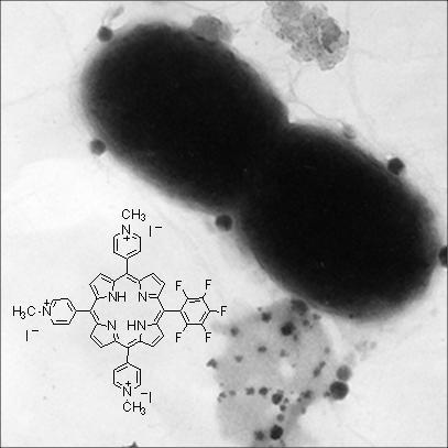
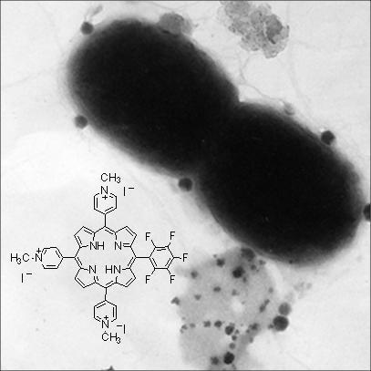
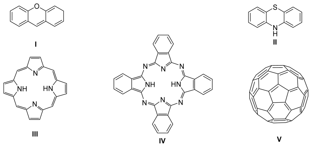
3.1. Photosensitizers
3.2. Photodymamic Therapy and Its Clinic Applications
3.3. Photodymamic Antimicrobial Therapy Application in the Environment
3.4. Photodynamic Antimicrobial Therapy Application in Fish Farm Plants
3.5. Advantages of Photodynamic Antimicrobial Chemotherapy over other Treatments in the Environment
Conclusions
References and Notes
- Pillay, TVR; Kutty, MN. Aquaculture: Principles and Practices; Wiley-Blackwell: Oxford, UK, 2005. [Google Scholar]
- FAO, The State of World Fisheries and Aquaculture-2008; FAO Fisheries and Aquaculture Department: Rome, Italy, 2009.
- FAO, The State of World Fisheries and Aquaculture; FAO Fisheries and Aquaculture Department: Rome, Italy, 1998.
- Flegel, TW. Detection of major penaeid shrimp viruses in Asia, a historical perspective with emphasis on Thailand. Aquaculture 2006, 258, 1–33. [Google Scholar]
- Subasinghe, RP; Bondad-Reantaso, MG; McGladdery, SE. Aquaculture development, health and wealth. In Aquaculture in the Millennium. Technical Proceedings of the Conference on Aquaculture in the Third Millennium, Bangkok, Thailand; Subasinghe, RP, Bueno, P, Phillips, MJ, Hough, C, McGladdery, SE, Arthur, JR, Eds.; 2001. [Google Scholar]
- Saksida, S; Constantine, J; Karreman, GA; Neville, C; Sweeting, R; Beamish, R. Evaluation of sea lice, Lepeophtheirus salmonis, abundance levels on farmed salmon in British Columbia, Canada. The Proceedings from the International Symposium on Veterinary Epidemiology and Economics XI, Cairns, Australia; 2006. [Google Scholar]
- Alderman, DJ. Geographical spread of bacterial and fungal diseases of crustaceans. Rev Sci Tech OIE 1996, 603–632. [Google Scholar]
- Shao, ZJ. Aquaculture pharmaceuticals and biologicals: current perspectives and future possibilities. Adv Drug Delivery Rev 2001, 50, 229–243. [Google Scholar]
- Wahli, T; Knuesel, R; Bernet, D; Segner, H; Pugovkin, D; Burkhardt-Holm, P; Escher, M; Schmidt-Posthaus, H. Proliferative kidney disease in Switzerland: current state of knowledge. J Fish Dis 2002, 25, 491–500. [Google Scholar]
- Muroga, K; Iida, M; Matsumoto, H; Nakai, T. Detection of Vibrio anguillarum from waters. Nippon Suisan Gakk 1986, 52, 641–647. [Google Scholar]
- Muroga, K; Tanasomwang, V; Momoyama, K. Vibrio anguillarum infection in juveniles of tiger puffer (Takifugu rubripes). Fish Pathol 1987, 22, 29–30. [Google Scholar]
- Nakai, T; Park, SC. Bacteriophage therapy of infectious diseases in aquaculture. Res Microbiol 2002, 153, 13–18. [Google Scholar]
- Fukuda, Y; Nguyen, HD; Furuhashi, M; Nakai, T. Mass mortality of cultured sevenband grouper, Epinephelus septemfasciatus, associated with viral nervous necrosis. Fish Pathol 1996, 31, 165–170. [Google Scholar]
- Iida, H; Tanaka, S; Shibata, Y. Small GTP-binding protein, Rab6, is associated with secretory granules in atrial myocytes. Am J Physiol Cell Physiol 1997, 272, 1594–1601. [Google Scholar]
- Nakai, T; Sugimoto, R; Park, K; Matsuoka, S; Mori, K; Nishioka, T; Maruyama, K. Protective effects of bacteriophage on experimental Lactococcus garvieae infection in yellowtail. Dis Aquat Org 1999, 37, 33–41. [Google Scholar]
- Huss, BCR. Gershom Scholem's major trends in Jewish Mysticism: 50 years after. Proceedings of the Sixth International Conference on the History of Jewish Mysticism; 1994; pp. 1265–1267. [Google Scholar]
- Noya, M; Magarinos, B; Lamas, J. La administracion intraperitoneal y oral de glucano afecta al sistema immune no especifico y a la resistencia de la dorada, Sparus aurata, a la pasteurelosis. Proceedings of the Fifth National Congress of Aquaculture, Sant Carles de la Rapita, Spain; 1995; pp. 734–738. [Google Scholar]
- Toranzo, AE; Barreiro, S; Casal, JF; Figueras, A; Magariños, B; Barja, JL. Pasteurellosis in cultured gilthead seabream, Sparus aurata: first report in Spain. Aquaculture 1991, 99, 1–15. [Google Scholar]
- Thyssen, A; Ollevier, F. In vitro antimicrobial susceptibility of Photobacterium damselae subsp. piscicida to 15 different antimicrobial agents. Aquaculture 2001, 200, 259–269. [Google Scholar]
- Fryer, JL; Lannan, CN. Rickettsial infections of fish. Annu Rev Fish Dis 1996, 6, 3–13. [Google Scholar]
- Bernoth, EM. Furunculosis: multidisciplinary fish disease research; Elsevier Academic Press: San Diego, CA, USA, 1997. [Google Scholar]
- Nakatsugawa, T. Edwardsiella tarda isolated from cultured young flounder. Fish Pathol 1983, 18, 99–101. [Google Scholar]
- Mekuchi, T; Kiyokawa, T; Honda, K; Nakai, T; Muroga, K. Vaccination trials in the Japanese flounder against Edwardsiellosis. Fish Pathol 1995, 30, 251–256. [Google Scholar]
- Kusuda, R; Kimura, H. Characteristics of gliding bacterium isolated from cultured yellowtail Seriola quinqueradiata. Bull Japan Soc Sci Fish 1982, 48, 1107–1112. [Google Scholar]
- Wakabayashi, H; Hikida, M; Masumura, K. Flexibacter infection in cultured marine fish in Japan. Helgolander Meeresun 1984, 37, 587–593. [Google Scholar]
- Hamaguchi, M; Kawahara, I; Usuki, H. Mass mortality of Pseudocentrotus depressus caused by a bacterial infection in summer. Suisan Zoshoku 1993, 41, 189–193. [Google Scholar]
- Park, SC; Shimamura, I; Fukunaga, M; Mori, K-I; Nakai, T. Isolation of bacteriophages specific to a fish pathogen, Pseudomonas plecoglossicida, as a candidate for disease control. Appl Environ Microbiol 2000, 66, 1416–1422. [Google Scholar]
- Munn, CB. Viruses as pathogens of marine organisms-from bacteria to whales. J Mar Biol Ass UK 2006, 86, 453–467. [Google Scholar]
- Sorimachi, M; Hara, T. Characteristics and pathogenicity of a virus isolated from yellowtail fingerlings showing ascites. Fish Pathol 1985, 19, 231–238. [Google Scholar]
- Iida, Y; Masumura, K; Nakai, T; Sorimachi, M; Matsuda, H. A viral disease in larvae and juveniles of the Japanese flounder Paralichthys olivaceus. J Aquat Anim Health 1989, 1, 7–12. [Google Scholar]
- Kusuda, R; Kawai, K. Bacterial diseases of cultured marine fish in Japan. Fish Pathol 1998, 33, 221–227. [Google Scholar]
- Miyazaki, T; Fujiwara, K; Kobara, J; Matsumoto, N; Abe, M; Nagano, T. Histopathology associated with two viral diseases of larval and juvenile fishes: epidermal necrosis of the Japanese flounder Paralichthys olivaceus and epithelial necrosis of black sea bream Acanthopagrus schlegeli. J Aquat Anim Health 1989, 1, 85–93. [Google Scholar]
- Nakatsugawa, T. Studies on amyotrophia of Japanese black abalone, Nordotis discus discus. Kyoto Inst Ocean Fish Sci 2000, (Spec. Rep. 5), 1–61. [Google Scholar]
- Vlak, JM; Bonami, JR; Flegel, TW; Kou, GH; Lightner, DV; Loh, CF; Loh, PC; Walker, PW. Fauquet, CM, Mayo, MA, Maniloff, J, Desselberger, U, Ball, LA, Eds.; Nimaviridae-Virus Taxonomy. In 8th Reports of the International Committee on Taxonomy of Viruses; Academic Press, Elsevier: New York, NY, USA, 2005. [Google Scholar]
- Muroga, K. Viral and bacterial diseases of marine fish and shellfish in Japanese hatcheries. Aquaculture 2001, 202, 23–44. [Google Scholar]
- Suttle, CA. Marine viruses-major players in the global ecosystem. Nat Rev Microbiol 2007, 5, 801–812. [Google Scholar]
- Meyers, T; Short, S; Lipson, K. Isolation of the North American strain of viral hemorrhagic septicemia virus (VHSV) associated with epizootic mortality in two new host species of Alaskan marine fish. Dis Aquat Org 1999, 38, 81–86. [Google Scholar]
- Kurath, G; Garver, KA; Troyer, RM; Emmenegger, EJ; Einer-Jensen, K; Anderson, ED. Phylogeography of infectious haematopoietic necrosis virus in North America. J Gen Virol 2003, 84, 803–814. [Google Scholar]
- Skall, HF; Olesen, NJ; Mellergaard, S. Viral haemorrhagic septicaemia virus in marine fish and its implications for fish farming-a review. J Fish Dis 2005, 28, 509–529. [Google Scholar]
- World Health Organization, The World Health Report 1995-Bridging the Gaps; WHO: Geneva, Switzerland, 1995.
- Ogbonna, CIC; Alabi, RO. Studies on species of fungi associated with mycotic infections of fish in a Nigerian freshwater fish pond. Hydrobiologia 1991, 220, 131–135. [Google Scholar]
- Gieseker, CM; Serfling, SG; Reimschuessel, R. Formalin treatment to reduce mortality associated with Saprolegnia parasitica in rainbow trout, Oncorhynchus mykiss. Aquaculture 2006, 253, 120–129. [Google Scholar]
- Leaño, EM; Vrijmoed, LLP; Jones, EBG. Saprolegnia diclina isolated from pond cultured red drum (Sciaenops ocellatus) in Hong Kong. Mycol Res 1999, 103, 701–706. [Google Scholar]
- Jori, G; Fabris, C; Soncin, M; Ferro, S; Coppellotti, O; Dei, D; Fantetti, L; Chiti, G; Roncucci, G. Photodynamic therapy in the treatment of microbial infections: basic principles and perspective applications. Lasers Surg Med 2006, 38, 468–481. [Google Scholar]
- Beakes, GW; Wood, SE; Burr, AW. Mueller, J, Ed.; Salmon Saprolegniasis; US Department of Energy, Bonneville Power Administration: Portland, OR, USA, 1994. [Google Scholar]
- Hatai, K; Hoshiai, G. Mass mortality in cultured coho salmon (Oncorhynchus kisutch) due to Saprolegnia parasitica coker. J Wildl Dis 1992, 28, 532–536. [Google Scholar]
- Bruno, DW; Wood, BP. Woo, PTK, Bruno, DW, Eds.; Fish Diseases and Disorders, Viral, Bacterial and Fungal Infections; CABI Publishing: Wallingford, Oxon, UK, 1999; Volume 3. [Google Scholar]
- Willoughby, LG; Roberts, RJ; Chinabut, S. Aphanomyces invaderis sp. nov., the fungal pathogen of freshwater tropical fish affected by epizootic ulcerative syndrome. J Fish Dis 1995, 18, 273–276. [Google Scholar]
- Burton, TO; Meyers, TR; Starkey, NS; Follett, JE. Experimental transmission of the fungus phoma herbarum to Chinook salmon. J Aquat Anim Health 2004, 16, 251–257. [Google Scholar]
- Mohamed, F; Ehab, E; Scott, F; Silva, V; Mendoza, L. Outbreaks of phaeohyphomycosis in the chinook salmon (Oncorhynchus tshawytscha) caused by Phoma herbarum. Mycopathologia 2007, 163, 41–48. [Google Scholar]
- Weiss, KR. Fish farms become feedlots of the sea. Los Angeles Times 2002. [Google Scholar]
- Karlsson, L; Bjerselius, R; Aune, M; Darnerud, PO; Glynn, A; Tysklind, M; Wichardt, U-P. Impacts of the fishery, disease, and contaminants on Baltic salmon (Salmo salar L.) in the sea. North Pacific Anadromous Fish Comission, 2002. [Google Scholar]
- Dalton, R. Aquaculture: Fishing for trouble. Nature 2004, 431, 502–504. [Google Scholar]
- Defoirdt, T; Boon, N; Sorgeloos, P; Verstraete, W; Bossier, P. Alternatives to antibiotics to control bacterial infections: luminescent vibriosis in aquaculture as an example. Trends Biotechnol 2007, 25, 472–479. [Google Scholar]
- Mladineo, I. Parasite communities of Adriatic cage-reared fish. Dis Aquat Organ 2005, 64, 77–83. [Google Scholar]
- Kennedy, CR; Di Cave, D; Berrilli, F; Orecchia, P. Composition and structure of helminth communities in eels Anguilla anguilla from Italian coastal lagoons. J Helminthol 1997, 71, 35–40. [Google Scholar]
- Plarre, H; Devold, M; Snow, M; Nylund, A. Prevalence of infectious salmon anaemia virus (ISAV) in wild salmonids in western Norway. Dis Aquat Organ 2005, 66, 71–79. [Google Scholar]
- Rimstad, E; Mjaaland, S; Snow, M; Mikalsen, AB; Cunningham, CO. Characterization of the infectious salmon anemia virus genomic segment that encodes the putative hemagglutinin. J Virol 2001, 75, 5352–5356. [Google Scholar]
- Nylund, A; Devold, M; Plarre, H; Isdal, E; Aarseth, M. Emergence and maintenance of infectious salmon anaemia virus (ISAV) in Europe: a new hypothesis. Dis Aquat Organ 2003, 56, 11–24. [Google Scholar]
- Vike, S; Nylund, S; Nylund, A. ISA virus in Chile: evidence of vertical transmission. Arch Virol 2009, 154, 1–8. [Google Scholar]
- Raynard, RS; Murray, AG; Gregory, A. Infectious salmon anaemia virus in wild fish from Scotland. Dis Aquat Org 2001, 46, 93–100. [Google Scholar]
- Pike, AW; Wadsworth, SL. Sealice on salmonids: Their biology and control. Adv Parasitol 1999, 44, 233–337. [Google Scholar]
- Anon. Sea lice as a population regulating factor in Norwegian salmon: status, effects of measures and future management; Institute of Marine Research: Bergen, Norway, 2005. [Google Scholar]
- Howgate, PC; Lima dos Santos, CA; Shehadeh, ZK. Safety of food products from aquaculture; FAO: Rome, Italy, 1997. [Google Scholar]
- Almeida, A; Cunha, Â; Santos, L; Salvador, S; Gomes, A. Mendez-Vilas, A, Ed.; Current Research Topics in Applied Microbiology and Microbial Biotechnology; World Scientific Publishing Co. Pte. Ltd: Singapore, 2009. [Google Scholar]
- Alderman, DJ. De Pauw, N, Joyce, J, Eds.; Aquaculture and environment. In European Aquaculture Society Special Publication 16; EAS: Bruxelles, Belgium, 1999. [Google Scholar]
- Commision Regulation (EC) 1911/2005. Amending Annex I to Council Regulation (EEC) No 2377/90 laying down a Community procedure for the establishment of maximum residue limits of veterinary medicinal products in foodstuffs of animal origin, as regards flugestone acetate. Official Journal of the European Union. 2005. Available online: http://eurlex.europa.eu/LexUriServ/LexUriServ.do?uri=OJ:L:2005:305:0030:0031:EN:PDF.
- Romalde, J. Photobacterium damselae subsp. piscicida: an integrated view of a bacterial fish pathogen. Int Microbiol 2002, 1, 3–9. [Google Scholar]
- Arijo, S; Rico, R; Chabrillon, M; Diaz-Rosales, P; Martínez-Manzanares, E; Balebona, MC; Magariños, B; Toranzo, AE; Moriñigo, MA. Effectiveness of a divalent vaccine for sole, Solea senegalensis (Kaup), against Vibrio harveyi and Photobacterium damselae subsp. piscicida. J Fish Dis 2005, 28, 33–38. [Google Scholar]
- Lin, X; Huang, JC; Mitchell, TG; Heitman, J. Virulence attributes and hyphal growth of C. neoformans are quantitative traits and the MATα allele enhances filamentation. PLoS Genet 2006, 2, e187. [Google Scholar]
- Reed, PA; Francis-Floyd, R. Vibrio infections of fish; Institute of Food and Agricultural Sciences, University of Florida: Gainesville, FL, USA, 1996. [Google Scholar]
- Press, CM; Lillehaug, A. Vaccination in European salmonid aquaculture: a review of practices and prospects. Br Vet J 1995, 151, 45–69. [Google Scholar]
- Vadstein, O. The use of immunostimulation in marine larviculture: possibilities and challenges. Aquaculture 1997, 155, 401–417. [Google Scholar]
- Christie, KE. Gudding, R, Lillehaug, A, Midtlyng, P, Brown, F, Eds.; Fish Vaccinology; Karger: Basel, Switzerland, 1997; Volume 90. [Google Scholar]
- Fijan, N. Ellis, AE, Ed.; Fish Vaccination; Academis Press: London, UK, 1988. [Google Scholar]
- Leong, JC; Fryer, JL; Winton, JR. Ellis, AE, Ed.; Fish Vaccination; Academic Press: London, UK, 1988. [Google Scholar]
- Martinez, JL. Fingerman, N, Ed.; Recent Advances in Marine Biotechnology. Molecular Genetics of Marine Organisms; Science Publishers, Inc: Enfield, New Hampshire, USA, 2003; Volume 10. [Google Scholar]
- Cabello, FC. Heavy use of prophylactic antibiotics in aquaculture: a growing problem for human and animal health and for the environment. Environ Microbiol 2006, 8, 1137–1144. [Google Scholar]
- Baquero, F; Martínez, J-L; Cantón, R. Antibiotics and antibiotic resistance in water environments. Curr Opin Biotechnol 2008, 19, 260–265. [Google Scholar]
- Lorch, A. Bacteriophages: an alternative to antibiotics? Biotechnol Dev Monit 1999, 14–17. [Google Scholar]
- Moriarty, DJW. Microbial Biosynthesis: New Frontiers. Proceedings of the 8th International Symposium on Microbial Ecologytlantic; Canada Society for Microbial Ecology: Halifax, NS, Canada, 1999; pp. 237–243. [Google Scholar]
- Holmström, K; Gräslund, S; Wahlström, A; Poungshompoo, S; Bengtsson, B-E; Kautsky, N. Antibiotic use in shrimp farming and implications for environmental impacts and human health. Int J Food Sci Technol 2003, 38, 255–266. [Google Scholar]
- Tendencia, EA; de la Peña, LD. Antibiotic resistance of bacteria from shrimp ponds. Aquaculture 2001, 195, 193–204. [Google Scholar]
- Roque, A; Molina-Aja, A; Bolán-Mejía, C; Gomez-Gil, B. In vitro susceptibility to 15 antibiotics of vibrios isolated from penaeid shrimps in Northwestern Mexico. Int J Antimicrob Agents 2001, 17, 383–387. [Google Scholar]
- Bakopoulos, V; Adams, A; Richards, RH. Some biochemical properties and antibiotic sensitivities of Pasteurella piscicida isolated in Greece and comparison with strains from Japan, France and Italy. J Fish Dis 1995, 18, 1–7. [Google Scholar]
- Sano, T. Control of fish disease, and the use of drugs and vaccines in Japan. J Appl Ichthyol 1998, 14, 131–137. [Google Scholar]
- Alderman, DJ. Malachite green: A review. J Fish Dis 1985, 8, 289–298. [Google Scholar]
- Schnick, RA. The impetus to register new therapeutants for aquaculture. Prog Fish-Cult 1988, 50, 190–196. [Google Scholar]
- Hoffman, GL; Meyer, FP. Parasites of Freshwater Fishes: A Review of Their Control and Treatment; TFH Publications: Jersey City, NJ, USA, 1974. [Google Scholar]
- Srivastava, S; Sinha, R; Roy, D. Toxicological effects of malachite green. Aquat Toxicol 2004, 66, 319–329. [Google Scholar]
- Alderman, DJ; Clifton-Hadley, RS. Malachite green: a pharmacokinetic study in rainbow trout, Oncorhynchus mykiss (Walbaum). J Fish Dis 1993, 16, 297–311. [Google Scholar]
- Burchmore, S; Wilkinson, M. Proposed environmental quality standards for malachite green in water (DWE 9026); Water Research Center: Marlow, Buckinghamshire, UK, 1993. [Google Scholar]
- Prasenjit, D; Katoch, RC; Subhas, V; Kumar, R. Drug sensitivity of gram positive bacteria from fish fauna of Himachal Pradesh. Indian Vet J 2001, 78, 576–578. [Google Scholar]
- Hardwick, J. Pyceze an alternative to malachite green-TRT01. Trout News (CEFAS) 2000, 28–29. [Google Scholar]
- Kaijser, B; Torud, B; Sorgaard, M. Replacing malachite green. Fish Farm Int 2001, 28, 25. [Google Scholar]
- Chen, CR; Meng, CM; Chen, CF. Eradication of stable chlorine dioxide for adherent bacteria on fish zygotes. J Huazhong Agric Univ 2001, 20, 568–570. [Google Scholar]
- Yamamoto, A; Toyomura, S; Saneyoshi, M; Hatai, K. Control of fungal infection of salmonid eggs by hydrogen peroxide. Fish Pathol 2001, 4, 241–246. [Google Scholar]
- Heidrich, S; Herms, J; Schneider, J. Chromatography with humic acids in fish culture. EAFP 1999, 157–163. [Google Scholar]
- Khodabandeh, S; Abtahi, B. Effects of sodium chloride, formalin and iodine on the hatching success of common carp, Cyprinus carpio, eggs. J Appl Ichthyol 2006, 22, 54–56. [Google Scholar]
- Magaraggia, M; Faccenda, F; Gandolfi, A; Jori, G. Treatment of microbiologically polluted aquaculture waters by a novel photochemical technique of potentially low environmental impact. J Environ Monit 2006, 8, 923–931. [Google Scholar]
- Raoult, D; Forterre, P. Redefining viruses: lessons from Mimivirus. Nat Rev Microbiol 2008, 6, 315–319. [Google Scholar]
- Comeau, AM; Hatfull, GF; Krisch, HM; Lindell, D; Mann, NH; Prangishvili, D. Exploring the prokaryotic virosphere. Res Microbiol 2008, 159, 306–313. [Google Scholar]
- Angly, FE; Felts, B; Breitbart, M; Salamon, P; Edwards, RA; Carlson, C; Chan, AM; Haynes, M; Kelley, S; Liu, H; Mahaffy, JM; Mueller, JE; Nulton, J; Olson, R; Parsons, R; Rayhawk, S; Suttle, CA; Rohwer, F. The marine viromes of four oceanic regions. PLoS Biol 2006, 4, e368. [Google Scholar]
- Culley, AI; Lang, AS; Suttle, CA. Metagenomic analysis of coastal RNA virus communities. Science 2006, 312, 1795–1798. [Google Scholar]
- Allen, MJ; Wilson, WH. Aquatic virus diversity accessed through omic techniques: A route map to function. Curr Opin Microbiol 2008, 11, 226–232. [Google Scholar]
- Fuhrman, JA. Marine viruses and their biogeochemical and ecological effects. Nature 1999, 399, 541–548. [Google Scholar]
- Wilhelm, SW; Suttle, CA. Viruses and nutrient cycles in the sea. Bioscience 1999, 49, 781–788. [Google Scholar]
- Wommack, KE; Colwell, RR. Virioplankton: viruses in aquatic ecosystems. Microbiol Mol Biol Rev 2000, 64, 69–114. [Google Scholar]
- Fischer, UR; Velimirov, B. High control of bacterial production by viruses in a eutrophic oxbow lake. Aquat Microb Ecol 2002, 27, 1–12. [Google Scholar]
- Suttle, CA. Hurst, CJ, Ed.; Viral Ecology; Academic Press: New York, NY, USA, 2000. [Google Scholar]
- Almeida, MA; Cunha, MA; Alcântara, F. Loss of estuarine bacteria by viral infection and predation in microcosm conditions. Microb Ecol 2001, 42, 562–571. [Google Scholar]
- Weinbauer, M. Ecology of prokaryotic viruses. FEMS Microbiol Rev 2004, 28, 127–181. [Google Scholar]
- Suttle, CA. Viruses in the sea. Nature 2005, 437, 356–361. [Google Scholar]
- Pedulla, ML; Ford, ME; Houtz, JM; Karthikeyan, T; Wadsworth, C; Lewis, JA; Jacobs-Sera, D; Falbo, J; Gross, J; Pannunzio, NR; Brucker, W; Kumar, V; Kandasamy, J; Keenan, L; Bardarov, S; Kriakov, J; Lawrence, JG; Jacobs, WR, Jr; Hendrix, RW; Hatfull, GF. Origins of highly mosaic mycobacteriophage genomes. Cell 2003, 113, 171–182. [Google Scholar]
- Suttle, CA. The significance of viruses to mortality in aquatic microbial communities. Microb Ecol 1994, 28, 237–243. [Google Scholar]
- Fuhrman, JA; Noble, RT. Viruses and protists cause similar bacterial mortality in coastal seawater. Limnol Oceanogr 1995, 40, 1236–1242. [Google Scholar]
- Yoshida, T; Takashima, Y; Tomaru, Y; Shirai, Y; Takao, Y; Hiroishi, S; Nagasaki, K. Isolation and characterization of a cyanophage infecting the toxic cyanobacterium Microcystis aeruginosa. Appl Environ Microbiol 2006, 72, 1239–1247. [Google Scholar]
- Breitbart, M; Hewson, I; Felts, B; Mahaffy, JM; Nulton, J; Salamon, P; Rohwer, F. Metagenomic analyses of an uncultured viral community from human feces. J Bacteriol 2003, 185, 6220–6223. [Google Scholar]
- Breitbart, M; Miyake, JH; Rohwer, F. Global distribution of nearly identical phage-encoded DNA sequences. FEMS Microbiol Lett 2004, 236, 249–256. [Google Scholar]
- Breitbart, M; Salamon, P; Andresen, B; Mahaffy, JM; Segall, AM; Mead, D; Azam, F; Rohwer, F. Genomic analysis of uncultured marine viral communities. Proc Natl Acad Sci USA 2002, 99, 14250–14255. [Google Scholar]
- B⊘rsheim, KY. Native marine bacteriophages. FEMS Microbiol Lett 1993, 102, 141–159. [Google Scholar]
- Alcântara, F; Almeida, MA. Virological quality of the Ria de Aveiro: validity of potential microbial indicators. Aquat Ecol 1995, 29, 419–425. [Google Scholar]
- Dore, WJ; Henshilwood, K; Lees, DN. Evaluation of F-specific RNA bacteriophage as a candidate human enteric virus indicator for bivalue molluscan shellfish. Appl Environ Microbiol 2000, 1280–1295. [Google Scholar]
- Grabow, WOK. Bacteriophages: update on application as models for viruses in water. Water SA 2001, 2, 251–268. [Google Scholar]
- Cole, D; Long, SC; Sobsey, MD. Evaluation of F+ RNA and DNA coliphages as sources-specific indicators of fecal contamination in surface waters. Appl Environ Microbiol 2003, 69, 6507–6514. [Google Scholar]
- Griffin, DW; Stokes, R; Rose, JB; Paul, JH. Bacterial indicator occurrence and the use of an F+ specific RNA coliphage assay to identify fecal sources in Homosassa Springs, Florida. Microb Ecol 2000, 39, 56–64. [Google Scholar]
- Culley, AI; Lang, AS; Suttle, CA. High diversity of unknown picorna-like viruses in the sea. Nature 2003, 424, 1054–1057. [Google Scholar]
- Shivu, MM; Rajeeva, BC; Girisha, SK; Karunasagar, I; Krohne, G; Karunasagar, I. Molecular characterization of Vibrio harveyi bacteriophages isolated from aquaculture environments along the coast of India. Environ Microbiol 2007, 9, 322–331. [Google Scholar]
- Vinod, MG; Shivu, MM; Umesha, KR; Rajeeva, BC; Krohne, G; Karunasagar, I; Karunasagar, I. Isolation of Vibrio harveyi bacteriophage with a potential for biocontrol of luminous vibriosis in hatchery environments. Aquaculture 2006, 255, 117–124. [Google Scholar]
- Karunasagar, I; Shivu, MM; Girisha, SK; Krohne, G; Karunasagar, I. Biocontrol of pathogens in shrimp hatcheries using bacteriophages. Aquaculture 2007, 268, 288–292. [Google Scholar]
- Drake, JW; Holland, JJ. Mutation rates among RNA viruses. Proc Natl Acad Sci USA 1999, 96, 13910–13913. [Google Scholar]
- Cissoko, M; Desnues, A; Bouvy, M; Sime-Ngando, T; Verling, E; Bettarel, Y. Effects of freshwater and seawater mixing on virio- and bacterioplankton in a tropical estuary. Freshwater Biol 2008, 53, 1154–1162. [Google Scholar]
- Cochran, PK; Kellog, CA; Paul, JH. Prophage induction of indigenous marine lysogenic bacteria by environmental pollutants. Mar Ecol-Prog Ser 1998, 164, 125–133. [Google Scholar]
- McDaniel, L; Paul, JH. Effect of nutrient addition and environmental factors on prophage induction in natural populations of marine Synechococcus species. Appl Environ Microbiol 2005, 71, 842–850. [Google Scholar]
- Weinbauer, MG; Suttle, CA. Lysogeny and prophage induction in coastal and offshore bacterial communities. Aquat Microb Ecol 1999, 18, 217–225. [Google Scholar]
- Ramírez, E; Villaverde, A. Viral spread within ageing bacterial populations. Gene 1997, 202, 147–149. [Google Scholar]
- Wilson, WH; Mann, NH. Lysogenic and lytic viral production in marine microbial communities. Aquat Microb Ecol 1997, 13, 95–100. [Google Scholar]
- Freifelder, D. Molecular Biology: A Comprehensive Introduction to Prokaryotes and Eukaryotes; Science Books International: Boston, MA, USA, 1983. [Google Scholar]
- Levin, BR; Lenski, RE. Futuyma, DJSM, Ed.; Coevolution; Sinauer: Sunderland, UK, 1983. [Google Scholar]
- Edlin, G; Lin, L; Kudrna, R. Lambda lysogens of E. coli reproduce more rapidly than non-lysogens. Nature 1975, 255, 735–737. [Google Scholar]
- Edlin, G; Lin, L; Bitner, R. Reproductive fitness of P1, P2, and Mu lysogens of Escherichia coli. J Virol 1977, 21, 560–564. [Google Scholar]
- Ackermann, H-W; DuBow, MS. Viruses of prokaryotes; CRC Press: Boca Raton, FL, USA, 1987. [Google Scholar]
- Ripp, S; Miller, RV. The role of pseudolysogeny in bacteriophage-host interactions in a natural freshwater environment. Microbiology (Reading, Engl) 1997, 143, 2065–2070. [Google Scholar]
- Ripp, S; Miller, RV. Dynamics of the pseudolysogenic response in slowly growing cells of Pseudomonas aeruginosa. Microbiology (Reading, Engl) 1998, 144, 2225–2232. [Google Scholar]
- Jiang, SC; Paul, JH. Seasonal and diel abundance of viruses and occurrence of lysogeny/bacteriogenicity in the marine environment. Mar Ecol-Prog Ser 1994, 104, 163–172. [Google Scholar]
- Alisky, J; Iczkowski, K; Rapoport, A; Troitsky, N. Bacteriophages show promise as antimicrobial agents. J Infect 1998, 36, 5–15. [Google Scholar]
- Barrow, P; Lovell, M; Berchieri, A, Jr. Use of lytic bacteriophage for control of experimental Escherichia coli septicemia and meningitis in chickens and calves. Clin Diagn Lab Immunol 1998, 5, 294–298. [Google Scholar]
- Smith, HW; Huggins, MB. Treatment of experimental Escherichia coli infection in mice with colicine V. J Med Microbiol 1977, 10, 479–482. [Google Scholar]
- Hagens, S; Habel, A; von Ahsen, U; von Gabain, A; Blasi, U. Therapy of experimental pseudomonas infections with a nonreplicating genetically modified phage. Antimicrob Agents Chemother 2004, 48, 3817–3822. [Google Scholar]
- Watanabe, R; Matsumoto, T; Sano, G; Ishii, Y; Tateda, K; Sumiyama, Y; Uchiyama, J; Sakurai, S; Matsuzaki, S; Imai, S; Yamaguchi, K. Efficacy of bacteriophage therapy against gut-derived sepsis caused by Pseudomonas aeruginosa in mice. Antimicrob Agents Chemother 2007, 51, 446–452. [Google Scholar]
- Matsuzaki, S; Yasuda, M; Nishikawa, H; Kuroda, M; Ujihara, T; Shuin, T; Shen, Y; Jin, Z; Fujimoto, S; Nasimuzzaman, MÂD; Wakiguchi, H; Sugihara, S; Sugiura, T; Koda, S; Muraoka, A; Imai, S. Experimental protection of mice against lethal Staphylococcus aureus infection by novel bacteriophage phi MR11. J Infect Dis 2003, 187, 613–624. [Google Scholar]
- Wills, QF; Kerrigan, C; Soothill, JS. Experimental bacteriophage protection against Staphylococcus aureus abscesses in a rabbit model. Antimicrob Agents Chemother 2005, 49, 1220–1221. [Google Scholar]
- Biswas, B; Adhya, S; Washart, P; Paul, B; Trostel, AN; Powell, B; Carlton, R; Merril, CR. Bacteriophage therapy rescues mice bacteremic from a clinical isolate of vancomycin-resistant Enterococcus faecium. Infect Immun 2002, 70, 204–210. [Google Scholar]
- Jado, I; Lopez, R; Garcia, E; Fenoll, A; Casal, J; Garcia, P. on behalf of the Spanish Pneumococcal Infection Study, N. Phage lytic enzymes as therapy for antibiotic-resistant Streptococcus pneumoniae infection in a murine sepsis model. J Antimicrob Chemother 2003, 52, 967–973. [Google Scholar]
- Cao, J; Sun, Y-Q; Berglindh, T; Mellgard, B; Li, Z-Q; Mardh, B; Mardh, S. Helicobacter pylori-antigen-binding fragments expressed on the filamentous M13 phage prevent bacterial growth. Biochim Biophys Acta 2000, 1474, 107–113. [Google Scholar]
- Bogovazova, GG; Voroshilova, NN; Bondarenko, VM. The efficacy of Klebsiella pneumoniae bacteriophage in the therapy of experimental Klebsiella infection. Zh Mikrobiol Epidemiol Immunobiol 1991, 4, 5–8. [Google Scholar]
- Fiorentin, L; Vieira, ND; Barioni Júnior, W. Use of lytic bacteriophages to reduce Salmonella enteritidis in experimentally contaminated chicken cuts. Rev Bras Cienc Avic 2005, 7, 255–260. [Google Scholar]
- Toro, H; Price, SB; McKee, AS; Hoerr, FJ; Krehling, J; Perdue, M; Bauermeister, L. Use of bacteriophages in combination with competitive exclusion to reduce Salmonella from infected chickens. Avian Dis 2005, 9, 118–124. [Google Scholar]
- Sulakvelidze, A; Kutter, E. Kutter, E, Sulakvelidze, A, Eds.; Bacteriophages: Biology and Applications; CRC Press: Boca Ratan, FL, USA, 2005. [Google Scholar]
- Housby, JN; Mann, NH. Phage therapy. Drug Discov Today 2009, 14, 536–540. [Google Scholar]
- Brussow, H. Phage therapy: the Escherichia coli experience. Microbiology 2005, 151, 2133–2140. [Google Scholar]
- Leverentz, B; Conway, WS; Alavidze, Z; Janisiewicz, WJ; Fuchs, Y; Camp, MJ; Chighladze, E; Sulakvelidze, A. Examination of bacteriophage as a biocontrol method for salmonella on fresh-cut fruit: a model study. J Food Protect 2001, 64, 1116–1121. [Google Scholar]
- Goode, D; Allen, VM; Barrow, PA. Reduction of experimental Salmonella and Campylobacter contamination of chicken skin by application of lytic bacteriophages. Appl Environ Microbiol 2003, 69, 5032–5036. [Google Scholar]
- Huff, WE; Huff, GR; Rath, NC; Balog, JM; Donoghue, AM. Alternatives to antibiotics: utilization of bacteriophage to treat colibacillosis and prevent foodborne pathogens. Poult Sci 2005, 84, 655–659. [Google Scholar]
- Flaherty, JE; Jones, JB; Harbaugh, BK; Somodi, GC; Jackson, LE. Control of bacterial spot on tomato in the greenhouse and field with h-mutant bacteriophages. HortScience 2000, 35, 882–884. [Google Scholar]
- Withey, S; Cartmell, E; Avery, LM; Stephenson, T. Bacteriophages-potential for application in wastewater treatment processes. Sci Total Environ 2005, 339, 1–18. [Google Scholar]
- Stevenson, RMW; Airdrie, DW. Isolation of Yersinia ruckeri bacteriophages. Appl Environ Microbiol 1984, 47, 1201–1205. [Google Scholar]
- Rodgers, CJ; Pringle, JH; McCarthy, DH; Austin, B. Quantitative and qualitative studies of Aeromonas salmonicida bacteriophage. J Gen Microbiol 1981, 125, 335–345. [Google Scholar]
- Wu, JL; Chao, WJ. The epizootic of milkfish vibriosis and its biological control by bacteriophage AS10, COA Fisheries No. 10. Fish Disease Research VI 1984, 34–46. [Google Scholar]
- Wu, JL; Lin, HM; Jan, L; Hsu, YL; Chang, LH. Biological control of fish pathogen, Aeromonas hydrophila, by bacteriophage AH1. Fish Pathol 1981, 15, 271–276. [Google Scholar]
- Merino, S; Camprubi, S; Tomas, JM. Isolation and characterization of bacteriophage PM2 from Aeromonas hydrophila. FEMS Microbiol Lett 1990, 68, 239–244. [Google Scholar]
- Yuksel, S; Thompson, K; Ellis, A; Adams, A. Purification of Piscirickettsia salmonis and associated phage particles. Dis Aquat Org 2001, 44, 231–235. [Google Scholar]
- Munro, J; Oakey, J; Bromage, E; Owens, L. Experimental bacteriophage-mediated virulence in strains of Vibrio harveyi. Dis Aquat Org 2003, 54, 187–194. [Google Scholar]
- Park, S; Nakai, T. Bacteriophage control of Pseudomonas plecoglossicida infection in ayu Plecoglossus altivelis. Dis Aquat Org 2003, 53, 33–39. [Google Scholar]
- Imbeault, S; Parent, S; Lagacé, M; Uhland, CF; Blais, J-F. Using bacteriophages to prevent furunculosis caused by Aeromonas salmonicida in farmed brook trout. J Aquat Anim Health 2006, 18, 203–214. [Google Scholar]
- Verner-Jeffreys, DW; Algoet, M; Pond, MJ; Virdee, HK; Bagwell, NJ; Roberts, EG. Furunculosis in Atlantic salmon (Salmo salar L.) is not readily controllable by bacteriophage therapy. Aquaculture 2007, 270, 475–484. [Google Scholar]
- Walakira, JK; Carrias, AA; Hossain, MJ; Jones, E; Terhune, JS; Liles, MR. Identification and characterization of bacteriophages specific to the catfish pathogen, Edwardsiella ictaluri. J Appl Microbiol 2008, 105, 2133–2142. [Google Scholar]
- Stenholm, AR; Dalsgaard, I; Middelboe, M. Isolation and characterization of bacteriophages infecting the fish pathogen Flavobacterium psychrophilum. Appl Environ Microbiol 2008, 74, 4070–4078. [Google Scholar]
- Chrisolite, B; Thiyagarajan, S; Alavandi, SV; Abhilash, EC; Kalaimani, N; Vijayan, KK; Santiago, TC. Distribution of luminescent Vibrio harveyi and their bacteriophages in a commercial shrimp hatchery in South India. Aquaculture 2008, 275, 13–19. [Google Scholar]
- Smith, HW; Huggins, RB. Effectiveness of phages in treating experimental Escherichia coli diarrhoea in calves, piglets and lambs. J Gen Microbiol 1983, 129, 2659–2675. [Google Scholar]
- Smith, HW; Huggins, MB; Shaw, KM. The control of experimental Escherichia coli diarrhea in calves by means of bacteriophages. J Gen Microbiol 1987, 133, 1111–1126. [Google Scholar]
- Smith, HW; Huggins, RB. Successful treatment of experimental E. coli infections in mice using phage: Its general superiority over antibiotics. J Gen Microbiol 1982, 128, 307–318. [Google Scholar]
- Durán, AE; Muniesa, M; Méndez, X; Valero, F; Lucena, F; Jofre, J. Removal and inactivation of indicator bacteriophages in fresh waters. J Appl Microbiol 2002, 92, 338–347. [Google Scholar]
- Lucena, F; Duran, AE; Morón, A; Calderón, E; Campos, C; Gantzer, C; Skraber, S; Jofre, J. Reduction of bacterial indicators and bacteriophages infecting faecal bacteria in primary and secondary wastewater treatments. J Appl Microbiol 2004, 97, 1069–1076. [Google Scholar]
- Duckworth, DH; Gulig, PA. Bacteriophages: potential treatment for bacterial infections. BioDrugs 2002, 16, 57–62. [Google Scholar]
- Merril, CR; Biswas, B; Carlton, R; Jensen, NC; Creed, GJ; Zullo, S; Adhya, S. Long-circulating bacteriophage as antibacterial agents. Proc Natl Acad Sci USA 1996, 93, 3188–3192. [Google Scholar]
- Oakey, HJ; Owens, L. A new bacteriophage, VHML, isolated from a toxin-producing strain of Vibrio harveyi in tropical Australia. J Appl Microbiol 2000, 89, 702–709. [Google Scholar]
- Oakey, HJ; Cullen, BR; Owens, L. The complete nucleotide sequence of the Vibrio harveyi bacteriophage VHML. J Appl Microbiol 2002, 93, 1089–1098. [Google Scholar]
- Austin, B; Pride, AC; Rhodie, GA. Association of a bacteriophage with virulence in Vibrio harveyi. J Fish Dis 2003, 26, 55–58. [Google Scholar]
- Ruangpan, L; Yaowanit, D; Sataporn, D; Siriporn, S; Flegel, TW. Lethal toxicity of Vibrio harveyi to cultivated Penaeus monodon induced by a bacteriophage. Dis Aquat Org 1999, 35, 195–201. [Google Scholar]
- Wagner, PL; Waldor, MK. Bacteriophage control of bacterial virulence. Infect Immun 2002, 70, 3985–3993. [Google Scholar]
- Brussow, H; Canchaya, C; Hardt, W-D. Phages and the evolution of bacterial pathogens: from genomic rearrangements to lysogenic conversion. Microbiol Mol Biol Rev 2004, 68, 560–602. [Google Scholar]
- Anonymous Phage therapy. Lancet 1983, 2, 1287–1288.
- Raab, O. Uber die wirkung fluoreszierender stoffe auf Infusorien. Z Biol 1900, 524–546. [Google Scholar]
- Moan, J; Peng, Q. An outline of the hundred-year history of PDT. Anticancer Res 2003, 3591–3600. [Google Scholar]
- Bonnett, R. Chemical Aspects of Photodynamic Therapy; Gordon and Breach Science: Amsterdam, 2000. [Google Scholar]
- Juarranz, A; Jaen, P; Sanz-Rodriguez, F; Cuevas, J; Gonzalez, S. Photodynamic therapy of cancer. Basic principles and applications. Clin Transl Oncol 2008, 10, 148–154. [Google Scholar]
- Kadish, KM; Smith, KM; Guilard, R. The Porphyrin Handbook, 1st ed; Academic Press: San Diego, CA, USA, 2000. [Google Scholar]
- Via, LD; Magno, SM. Photochemotherapy in the treatment of cancer. Curr Med Chem 2001, 8, 1405–1418. [Google Scholar]
- Dougherty, TJ; Gomer, CJ; Henderson, BW; Jori, G; Kessel, D; Korbelik, M; Moan, J; Peng, Q. Photodynamic therapy. J Natl Cancer Inst 1998, 90, 889–905. [Google Scholar]
- Shackley, D; Whitehurst, C; Clarke, N; Betts, C; Moore, J. Photodynamic therapy. J R Soc Med 1999, 29, 562–565. [Google Scholar]
- Hamblin, MR; Hasan, T. Photodynamic therapy: A new antimicrobial approach to infectious disease? Photochem Photobiol Sci 2004, 3, 436–450. [Google Scholar]
- Wainwright, M. Photodynamic antimicrobial chemotherapy (PACT). J Antimicrob Chemother 1998, 42, 13–28. [Google Scholar]
- Wainwright, M; Phoenix, D; Laycock, S; Wareing, D; Wright, P. Photobactericidal activity of phenothiazinium dyes against methicillin-resistant strains of Staphylococcus aureus. FEMS Microbiol Lett 1998, 177–181. [Google Scholar]
- De Rosa, MC; Crutchley, RJ. Photosensitized singlet oxygen and its applications. Coord Chem Rev 2002, 233, 351–371. [Google Scholar]
- Jori, G; Brown, SB. Photosensitized inactivation of microorganisms. Photochem Photobiol Sci 2004, 3, 403–405. [Google Scholar]
- Wainwright, M. Methylene blue derivatives-suitable photoantimicrobials for blood product disinfection? Int J Antimicrob Agents 2000, 16, 381–394. [Google Scholar]
- Mroz, P; Tegos, GP; Gali, H; Wharton, T; Sarna, T; Hamblin, MR. Photodynamic therapy with fullerenes. Photochem Photobiol Sci 2007, 6, 1139–1149. [Google Scholar]
- Montanari, F; Casella, L. Metalloporphyrins Catalyzed Oxidations; Kluwer Academic: Dordrecht, The Netherlands, 1994. [Google Scholar]
- Feiters, MC; Rowan, AE; Nolte, RJM. From simple to supramolecular cytochrome P450 mimics. Chem Soc Rev 2000, 29, 375–384. [Google Scholar]
- Kropf, M; van Loyen, D; Schwarz, O; Durr, H. Biomimetic models of the photosynthetic reaction center based on ruthenium-polypyridine complexes. J Phys Chem A 1998, 102, 5499–5505. [Google Scholar]
- Choi, MS; Yamazaki, T; Yamazaki, I; Aida, T. Bioinspired molecular design of light-harvesting multiporphyrin arrays. Angew Chem Int Ed Engl 2004, 43, 150–158. [Google Scholar]
- James, TD; Shinkai, S. Host-Guest Chemistry; Springer-Verlag Berlin: Berlin, 2002; Volume 218. [Google Scholar]
- Maiya, BG. New porphyrin architectures and host-guest chemistry. J Porphyrins Phthalocyanines 2004, 8, 1118–1128. [Google Scholar]
- Wada, K; Mizutani, T; Matsuoka, H; Kitagawa, S. A new strategy for the design of water-soluble synthetic receptors: specific recognition of DNA intercalators and diamines. Chem-Eur J 2003, 9, 2368–2380. [Google Scholar]
- Guldi, DM. Fullerene-porphyrin architectures; photosynthetic antenna and reaction center models. Chem Soc Rev 2002, 31, 22–36. [Google Scholar]
- Guldi, DM; Rahman, GMA; Prato, M; Jux, N; Qin, S; Ford, W. Single-wall carbon nanotubes as integrative building blocks for solar-energy conversion. Angew Chem Int Ed Engl 2005, 44, 2015–2018. [Google Scholar]
- Tagmatarchis, N; Prato, M; Guldi, DM. Soluble carbon nanotube ensembles for light-induced electron transfer interactions. Physica E 2005, 29, 546–550. [Google Scholar]
- Tomé, JPC; Pereira, AMVM; Alonso, CMA; Neves, MGPMS; Tomé, AC; Silva, AMS; Cavaleiro, JAS; Martínez-Díaz, MV; Torres, T; Rahman, GMA; Ramey, J; Guldi, DM. Synthesis and photophysical studies of new porphyrin-phthalocyanine dyads with hindered rotation. Eur J Org Chem 2005, 257–267. [Google Scholar]
- Ehli, C; Aminur Rahman, GM; Jux, N; Balbinot, D; Guldi, DM; Paolucci, F; Marcaccio, M; Paolucci, D; Melle-Franco, M; Zerbetto, F; Campidelli, S; Prato, M. Interactions in single wall carbon nanotubes/pyrene/porphyrin nanohybrids. J Am Chem Soc 2006, 128, 11222–11231. [Google Scholar]
- Guldi, DM; Rahman, GMA; Jux, N; Tagmatarchis, N; Prato, M. Integrating single-wall carbon nanotubes into donor-acceptor nanohybrids. Angew Chem Int Ed Engl 2004, 43, 5526–5530. [Google Scholar]
- Bautista-Sanchez, A; Kasselouri, A; Desroches, MC; Blais, J; Maillard, P; de Oliveira, DM; Tedesco, AC; Prognon, P; Delaire, J. Photophysical properties of glucoconjugated chlorins and porphyrins and their associations with cyclodextrins. J Photochem Photobiol B Biol 2005, 81, 154–162. [Google Scholar]
- Derycke, ASL; de Witte, PAM. Liposomes for photodynamic therapy. Adv Drug Delivery Rev 2004, 56, 17–30. [Google Scholar]
- Lang, K; Mosinger, J; Wagnerova, DM. Photophysical properties of porphyrinoid sensitizers non-covalently bound to host molecules; models for photodynamic therapy. Coord Chem Rev 2004, 248, 321–350. [Google Scholar]
- Cavaleiro, JAS; Tomé, JPC; Faustino, MAF. El-Ashry, ESH, Ed.; Heterocycles from Carbohydrate Precursors; Springer-Verlag: Berlin Heidelberg, Germany, 2007; Volume 7. [Google Scholar]
- Mikata, Y; Onchi, Y; Tabata, K; Ogura, SI; Okura, I; Ono, H; Yano, S. Sugar-dependent photocytotoxic property of tetra- and octa-glycoconjugated tetraphenylporphyrins. Tetrahedron Lett 1998, 39, 4505–4508. [Google Scholar]
- Jemli, M; Alouini, Z; Sabbahi, S; Gueddari, M. Destruction of fecal bacteria in wastewater by three photosensitizers. J Environ Monit 2002, 4, 511–516. [Google Scholar]
- Nyman, ES; Hynninen, PH. Research advances in the use of tetrapyrrolic photosensitizers for photodynamic therapy. J Photochem Photobiol B, Biol 2004, 73, 1–28. [Google Scholar]
- Ando, A; Kumadaki, I. Progress on the syntheses of fluorine analogs of natural porphyrins potentially useful for the diagnosis and therapy of certain cancers. J Fluor Chem 1999, 100, 135–146. [Google Scholar]
- Banfi, S; Caruso, E; Buccafurni, L; Battini, V; Zazzaron, S; Barbieri, P; Orlandi, V. Antibacterial activity of tetraaryl-porphyrin photosensitizers: An in vitro study on Gram negative and Gram positive bacteria. J Photochem Photobiol B, Biol 2006, 85, 28–38. [Google Scholar]
- Boyle, RW; Dolphin, D. Structure and biodistribution relationships of photodynamic sensitizers. Photochem Photobiol 1996, 64, 469–485. [Google Scholar]
- Caminos, D; Durantini, E. Photodynamic inactivation of Escherichia coli immobilized on agar surfaces by a tricationic porphyrin. Bioorg Med Chem 2006, 14, 4253–4259. [Google Scholar]
- Caminos, DA; Spesia, MB; Durantini, EN. Photodynamic inactivation of Escherichia coli by novel meso-substituted porphyrins by 4-(3-N,N,N-trimethylammoniumpropoxy)phenyl and 4-(trifluoromethyl)phenyl groups. Photochem Photobiol Sci 2005, 5, 56–65. [Google Scholar]
- Grancho, JCP; Pereira, MM; Miguel, MDG; Gonçalves, AMR; Burrows, HD. Synthesis, spectra and photophysics of some free base tetrafluoroalkyl and tetrafluoroaryl porphyrins with potential applications in imaging. Photochem Photobiol 2002, 75, 249–256. [Google Scholar]
- Spesia, MB; Lazzeri, D; Pascual, L; Rovera, M; Durantini, EN. Photoinactivation of Escherichia coli using porphyrin derivatives with different number of cationic charges. FEMS Immunol Med Microbiol 2005, 44, 289–295. [Google Scholar]
- Lazzeri, D; Rovera, M; Pascual, L; Durantini, EN. Photodynamic studies and photoinactivation of Escherichia coli using meso-substituted cationic porphyrin derivatives with asymmetric charge distribution. Photochem Photobiol 2004, 80, 286–293. [Google Scholar]
- Maisch, T; Szeimies, RM; Jori, G; Abels, C. Antibacterial photodynamic therapy in dermatology. Photochem Photobiol Sci 2004, 3, 907–917. [Google Scholar]
- von Tappeiner, H; Jesionek, A. Therapeutische versuche mit fluoreszierenden stoffen. Münch Med Wochenschr 1903, 2042–2044. [Google Scholar]
- Luksiene, Z. Photodynamic therapy: mechanism of action and ways to improve the efficiency of treatment. Medicina (Kaunas) 2003, 39, 1137–1150. [Google Scholar]
- Triesscheijn, M; Baas, P; Schellens, JHM; Stewart, FA. Photodynamic therapy in oncology. Oncologist 2006, 11, 1034–1044. [Google Scholar]
- Ackroyd, R; Kelty, C; Brown, N; Reed, M. The history of photodetection and photodynamic therapy. Photochem Photobiol 2001, 74, 656. [Google Scholar]
- Bressler, NM; Bressler, SB. Photodynamic therapy with verteporfin (Visudyne): impact on ophthalmology and visual sciences. Invest Ophthalmol Vis Sci 2000, 624–628. [Google Scholar]
- Cassell, G; Mekalanos, J. Development of antimicrobial agents in the era of new and reemerging infectious diseases and increasing antibiotic resistance. J Am Med Assoc 2001, 601–605. [Google Scholar]
- Wilson, M; Yianni, C. Killing of methicillin-resistant Staphylococcus aureus by low-power laser light. J Med Microbiol 1995, 42, 62–66. [Google Scholar]
- O'Riordan, K; Akilov, OE; Hasan, T. The potential for photodynamic therapy in the treatment of localized infections. Photodiagnosis Photodyn Ther 2005, 2, 247–262. [Google Scholar]
- Ben-Hur, E; Chan, WS; Yim, Z; Zuk, MM; Dayal, V; Roth, N; Heldman, E; Lazlo, A; Valeri, CR; Horowitz, B. Optical Methods for Tumor Treatment and Detection: Mechanisms and Techniques in Photodynamic Therapy IX; 2000; pp. 34–39. [Google Scholar]
- Floyd, RA; Schneider, JE; Dittmer, DR. Methylene blue photoinactivation of RNA viruses. Antiviral Res 2004, 61, 141–151. [Google Scholar]
- Grandadam, M; Ingrand, D; Huraux, JM; Aveline, B; Delgado, O; VeverBizet, C; Brault, D. Photodynamic inactivation of cell-free HIV strains by a red-absorbing chlorin-type photosensitizer. J Photochem Photobiol B, Biol 1995, 31, 171–177. [Google Scholar]
- Hirayama, J; Ikebuchi, K; Abe, H; Kwon, KW; Ohnishi, Y; Horiuchi, M; Shinagawa, M; Ikuta, K; Kamo, N; Sekiguchi, S. Photoinactivation of virus infectivity by hypocrellin A. Photochem Photobiol 1997, 66, 697–700. [Google Scholar]
- Wagner, SJ; Robinette, D; Storry, J; Chen, XY; Shumaker, J; Benade, L. Differential sensitivities of viruses in red-cell suspensions to methylene-blue photosensitization. Transfusion 1994, 34, 521–526. [Google Scholar]
- Maisch, T. Anti-microbial photodynamic therapy: useful in the future? Lasers Med Sci 2007, 22, 83–91. [Google Scholar]
- Carre, V; Gaud, O; Sylvain, I; Bourdon, O; Spiro, M; Biais, J; Granet, R; Krausz, P; Guilloton, M. Fungicidal properties of meso-arylglycosylporphyrins: influence of sugar substituents on photoinduced damage in the yeast Saccharomyces cerevisiae. J Photochem Photobiol B Biol 1999, 48, 57–62. [Google Scholar]
- Carvalho, CMB; Gomes, A; Fernandes, SCD; Prata, ACB; Almeida, MA; Cunha, MA; Tome, JPC; Faustino, MAF; Neves, M; Tome, AC. Photoinactivation of bacteria in wastewater by porphyrins: bacterial beta-galactosidase activity and leucine-uptake as methods to monitor the process. J Photochem Photobiol B, Biol 2007, 88, 112–118. [Google Scholar]
- Cormick, MP; Alvarez, MG; Rovera, M; Durantini, EN. Photodynamic inactivation of Candida albicans sensitized by tri- and tetra-cationic porphyrin derivatives. Eur J Med Chem 2009, 44, 1592–1599. [Google Scholar]
- Demidova, T; Hamblin, M. Effect of cell-photosensitizer binding and cell density on microbial photoinactivation. Antimicrob Agents Chemother 2005, 49, 2329–2335. [Google Scholar]
- Demidova, TN; Hamblin, MR. Photodynamic inactivation of Bacillus spores, mediated by phenothiazinium dyes. Appl Environ Microbiol 2005, 71, 6918–6925. [Google Scholar]
- Drábková, M; Marálek, B; Admiraal, W. Photodynamic therapy against cyanobacteria. Environ Toxicol 2007, 22, 112–115. [Google Scholar]
- Ehrenberg, B; Malik, Z; Nitzan, Y; Ladan, H; Johnson, F; Hemmi, G; Sessler, J. The binding and photosensitization effects of tetrabenzoporphyrins and texaphyrin in bacterial cells. Lasers Med Sci 1993, 8, 197–203. [Google Scholar]
- Foschi, F; Fontana, CR; Ruggiero, K; Riahi, R; Vera, A; Doukas, AG; Pagonis, TC; Kent, R; Stashenko, PP; Soukos, NS. Photodynamic inactivation of Enterococcus faecalis in dental root canals in vitro. Lasers Surg Med 2007, 39, 782–787. [Google Scholar]
- Gábor, F; Szocs, K; Maillard, P; Csík, G. Photobiological activity of exogenous and endogenous porphyrin derivatives in Escherichia coli and Enterococcus hirae cells. Radiat Environ Biophys 2001, V40, 145–151. [Google Scholar]
- Grinholc, M; Szramka, B; Kurlenda, J; Graczyk, A; Bielawski, KP. Bactericidal effect of photodynamic inactivation against methicillin-resistant and methicillin-susceptible Staphylococcus aureus is strain-dependent. J Photochem Photobiol B, Biol 2008, 90, 57–63. [Google Scholar]
- Maisch, T; Bosl, C; Szeimies, RM; Lehn, N; Abels, C. Photodynamic effects of novel XF porphyrin derivatives on prokaryotic and eukaryotic cells. Antimicrob Agents Chemother 2005, 49, 1542–1552. [Google Scholar]
- Merchat, M; Bertolini, G; Giacomini, P; Villanueva, A; Jori, G. Meso-substituted cationic porphyrins as efficient photosensitizers of gram-positive and gram-negative bacteria. J Photochem Photobiol B, Biol 1996, 32, 153–157. [Google Scholar]
- Merchat, M; Spikes, J; Bertoloni, G; Jori, G. Studies on the mechanism of bacteria photosensitization by meso-substituted cationic porphyrins. J Photochem Photobiol B Biol 1996, 35, 149–157. [Google Scholar]
- Minnock, A; Vernon, DI; Schofield, J; Griffiths, J; Parish, JH; Brown, SB. Mechanism of uptake of a cationic water-soluble pyridinium zinc phthalocyanine across the outer membrane of Escherichia coli. Antimicrob Agents Chemother 2000, 44, 522–527. [Google Scholar]
- Nitzan, Y; Ashkenazi, H. Photoinactivation of Acinetobacter baumannii and Escherichia coli B by a cationic hydrophilic porphyrin at various light wavelengths. Curr Microbiol 2001, V42, 408–414. [Google Scholar]
- Oliveira, A; Almeida, A; Carvalho, CMB; Tomé, JPC; Faustino, MAF; Neves, MGPMS; Tomé, AC; Cavaleiro, JAS; Cunha, Â. Porphyrin derivatives as photosensitizers for the inactivation of Bacillus cereus endospores. J Appl Microbiol 2009. In press. [Google Scholar]
- Tang, HM; Hamblin, MR; Yow, CMN. A comparative in vitro photoinactivation study of clinical isolates of multidrug-resistant pathogens. J Infect Chemother 2007, 13, 87–91. [Google Scholar]
- Nitzan, Y; Balzam-Sudakevitz, A; Ashkenazi, H. Eradication of Acinetobacter baumannii by photosensitized agents in vitro. J Photochem Photobiol B, Biol 1998, 42, 211–218. [Google Scholar]
- Rojas-Valencia, M; Orta-de-Velásquez, M; Vaca-Mier, M; Franco, V. Ozonation by-products issued from the destruction of microorganisms present in wastewaters treated for reuse. Water Sci Technol 2004, 50, 187–193. [Google Scholar]
- Mendes, B; Oliveira, J. Qualidade da água para consumo humano; Lidel: Lisboa, 2004. [Google Scholar]
- Richardson, SD; Thruston, AD; Caughran, TV; Chen, PH; Collette, TW; Schenck, KM; Lykins, BW; Rav-Acha, C; Glezer, V. Identification of new drinking water disinfection by-products from ozone, chlorine dioxide, chloramine, and chlorine. Water Air Soil Pollut 2000, 123, 95–102. [Google Scholar]
- Alves, E; Carvalho, CMB; Tome, JPC; Faustino, MAF; Neves, M; Tome, AC; Cavaleiro, JAS; Cunha, A; Mendo, S; Adelaide, A. Photodynamic inactivation of recombinant bioluminescent Escherichia coli by cationic porphyrins under artificial and solar irradiation. J Ind Microbiol Biotechnol 2008, 35, 1447–1454. [Google Scholar]
- Alves, E; Costa, L; Carvalho, C; Tome, J; Faustino, M; Neves, M; Tome, A; Cavaleiro, J; Cunha, A; Almeida, A. Charge effect on the photoinactivation of Gram-negative and Gram-positive bacteria by cationic meso-substituted porphyrins. BMC Microbiol 2009, 9, 70. [Google Scholar]
- Bonnett, R; Evans, RL; Galia, AB. Photochemotherapy: Photodynamic Therapy and Other Modalities III. Proc SPIE 1997, 3191, 79–88. [Google Scholar]
- Cerveny, KE; DePaola, A; Duckworth, DH; Gulig, PA. Phage therapy of local and systemic disease caused by Vibrio vulnificus in iron-dextran-treated mice. Infect Immun 2002, 70, 6251–6262. [Google Scholar]
- Jiménez-Hernández, ME; Manjón, F; Garcia-Fresnadillo, D; Orellana, G. Solar water disinfection by singlet oxygen photogenerated with polymer-supported Ru(II) sensitizers. Sol Energy 2006, 80, 1382–1387. [Google Scholar]
- Casteel, MJ; Jayaraj, K; Gold, A; Ball, LM; Sobsey, MD. Photoinactivation of hepatitis A virus by synthetic porphyrins. Photochem Photobiol 2004, 80, 294–300. [Google Scholar]
- Costa, L; Alves, E; Carvalho, C; Tomé, J; Faustino, M; Neves, M; Tomé, A; Cavaleiro, J; Cunha, Â; Almeida, A. Sewage bacteriophage photoinactivation by cationic porphyrins: a study of charge effect. Photochem Photobiol Sci 2008, 7, 415–422. [Google Scholar]
- Alouini, Z; Jemli, M. Destruction of helminth eggs by photosensitized porphyrin. J Environ Monit 2001, 3, 548–551. [Google Scholar]
- Bonnett, R; Krysteva, MA; Lalov, IG; Artarsky, SV. Water disinfection using photosensitizers immobilized on chitosan. Water Res 2006, 40, 1269–1275. [Google Scholar]
- Artarsky, S; Dimitrova, S; Bonnett, R; Krysteva, M. Immobilization of zinc phthalocyanines in silicate matrices and investigation of their photobactericidal effect on E.coli. Sci World J 2006, 6, 374–382. [Google Scholar]
- Faust, D; Funken, KH; Horneck, G; Milow, B; Ortner, J; Sattlegger, M; Schafer, M; Schmitz, C. Immobilized photosensitizers for solar photochemical applications. Sol Energy 1999, 65, 71–74. [Google Scholar]
- Krouit, M; Bras, J; Belgacem, MN. Cellulose surface grafting with polycaprolactone by heterogeneous click-chemistry. Eur Polym J 2008, 44, 4074–4081. [Google Scholar]
- Krouit, M; Granet, R; Branland, P; Verneuil, B; Krausz, P. New photoantimicrobial films composed of porphyrinated lipophilic cellulose esters. Bioorg Med Chem Lett 2006, 16, 1651–1655. [Google Scholar]
- Krouit, M; Granet, R; Krausz, P. Photobactericidal plastic films based on cellulose esterified by chloroacetate and a cationic porphyrin. Bioorg Med Chem 2008, 16, 10091–10097. [Google Scholar]
- Cavaleiro, JAS; Almeida, A; Tomé, JPC; Faustino, MAF; Neves, MGPMS; Tomé, AC; Carvalho, CMB; Costa, L; Alves, E; Oliveira, A; Cunha, Â. Antimicrobial photodynamic activity of porphyrin derivatives: Potential application on medical and water disinfection. J Porphyrins Phthalocyanines 2009. In press. [Google Scholar]
- Almeida, M.A.; Cavaleiro, J.A.S.; Rocha, J.; Carvalho, C.M.B.; Costa, L.A.S.; Alves, E.S.C.F.; Cunha, M.A.S.D.A.; Tomé, J.P.C.; Faustino, M.A.F.; Neves, M.G.P.M.S.; Tomé, A.C.; Lin, Z.; Rainho, J.P.J. Materiais híbridos nanomagnete-porfirina, processo para a sua síntese e respectiva aplicação em formulações para desinfecção de águas. C07D 487/22 (2006.01) ed.; Portuguese Patent 103, 828, 2009.
- Costa, L; Carvalho, CMB; Tomé, JPC; Faustino, MAF; Neves, MGPMS; Tomé, AC; Cavaleiro, JAS; Lin, Z; Rainho, JPJ; Rocha, J; Cunha, Â; Almeida, A. Mendez-Vilas, A, Ed.; Sewage bacteriophage photoinactivation by porphyrins immobilized in solid matrixes. II International Conference on Environmental Industrial and Applied Microbiology (BioMicroworld2007), World Scientific Publishing Co. Pte. Ltd: Toh Tuck Link, Singapore, 2007. [Google Scholar]
- Jori, G; Coppellotti, O. Inactivation of pathogenic microorganisms by photodynamic techniques: mechanistic aspects and perspective applications. Anti-Infect Agents Med Chem 2007, 6, 119–131. [Google Scholar]
- Wong, P-N; Mak, S-K; Lo, M-W; Lo, K-Y; Tong, GM-W; Wong, Y; Wong, AK-M. Vibrio vulnificus peritonitis after handling of seafood in a patient receiving CAPD. Am J Kidney Dis 2005, 46, e87–e90. [Google Scholar]
- Arrojado, C. Evaluation of antibacterial activity of free-porphyrins to inactive fish pathogenic bacteria; MSc Thesis; Department of Biology, University of Aveiro: Aveiro, Portugal, 2009. [Google Scholar]
- Baker, KS; Smith, RC. Calkins, J, Ed.; The Role of Solar Ultraviolet Radiation in Marine Ecosystems; Plenum Press: New York, NY, USA, 1982. [Google Scholar]
- Capella, M; Coelho, A; Menezes, S. Effect of glucose on photodynamic action of methylene blue in Escherichia coli cells. Photochem Photobiol 1996, 205–210. [Google Scholar]
- Contag, C; Contag, P; Mullins, J; Spilman, S; Stevenson, D; Benaron, D. Photonic detection of bacterial pathogens in living hosts. Mol Microbiol 1995, 4, 593–603. [Google Scholar]
- Contag, CH; Jenkins, D; Contag, PR; Negrin, RS; Christopher, H. Use of reporter genes for optical measurements of neoplastic disease in vivo. Neoplasia 2000, 2, 41–52. [Google Scholar]
- Cunha, B. Antibiotic resistance. Control strategies. Crit Care Clin 1998, 309–327. [Google Scholar]
- Demidova, T; Gad, F; Zahra, T; Francis, K; Hamblin, M. Monitoring photodynamic therapy of localized infections by bioluminescence imaging of genetically engineered bacteria. J Photochem Photobiol B, Biol 2005, 81, 25. [Google Scholar]
- Tome, JPC; Neves, MGPMS; Tome, AC; Cavaleiro, JAS; Soncin, M; Magaraggia, M; Ferro, S; Jori, G. Synthesis and antibacterial activity of new poly-S-lysine-porphyrin conjugates. J Med Chem 2004, 47, 6649–6652. [Google Scholar]
- Malik, Z; Ladan, H; Nitzan, Y. Photodynamic inactivation of Gram-negative bacteria: Problems and possible solutions. J Photochem Photobiol B Biol 1992, 262–266. [Google Scholar]


| Reference | Phage | Bacteria | Treated fish/shellfish | Phage application | Effects |
|---|---|---|---|---|---|
| Barrow et al., 1998 [147] | Bacteriophage R isolated from domestic sewage | E. coli H247 (O18:K1:H7) (bacteremic) | Chickens and calves | Intramuscular (102–106 PFU) and intracranial (106 PFU) inoculation of chickens; oral and intramuscular inoculation of calves with 3 × 10¹⁰ PFU) | Protection against morbidity and mortality |
| Biswas et al., 2002 [153] | Enterococcus phages ENB6 and C33 isolated from raw sewage | Vancomycin-resistant Enterococcus faecium (agent of VRE bacteremia) | BALB/c mice | Intraperitoneal injection of 3 × 10⁸ PFU | Complete rescue of bacteraemia in 48 hours |
| Bogovazova et al., 1991 [156] | Klebsiella pneumoniae bacteriophage | Klebsiella | BALB/c mice | Intraperitoneal, intravenous or intranasal administration | Rescue of generalized Klebsiella infection |
| Cao et al., 2000 [155] | Helicobacter pylori M13 recombinant phage | Helicobacter pylori | BALB/c mice | Oral administration of 107 PFU | Reduction of stomach colonization by Helicobacter pylori |
| Fiorentin et al., 2005 [157] | Salmonella enteritidis lytic phages CNPSA 1, CNPSA 3, CNPSA 4 | Salmonella enteritidis | Chicken cuts (thighs and drumsticks) | Immersion in 109 CFU mL−1 bacteriophage suspensions | Reduction of Salmonella enteritidis counts in treated chicken cuts |
| Flaherty et al., 2000 [165] | Xanthomonas campestris pv. vesicatoria specific H-mutant bacteriophages | Xanthomonas campestris pv. vesicatoria | “Sunbeam” tomato Lycopersicon esculentum | Foliar applications of 108 PFU mL−1 phage suspensions | Reduction of bacterial spots and increase in fruit weight |
| Goode et al., 2003 [163] | Salmonella enteritidis phage types P125589, phage 29C and transducing lambdoid phage P22, HTint, isolated on S. enterica serovar Enteritidis from sewage; Campylobacter jejuni phage 12673 | Salmonella enterica serovar Enteritidis and Campylobacter jejuni | Chicken skin | Surface spreading with 103 PFU cm−2 | Reduction by 2 log units in bacterial abundance over 48 hours |
| Hagens et al., 2004 [149] | Genetically engineered non-replicating, non-lytic filamentous phage Pf3R obtained from phage Pt1 isolated from river water using PAO1 as the host | Pseudomonas aeruginosa | BALB/c mice | Intraperitoneal inoculation with 106–108 PFU | Higher survival rate and reduced inflammatory response after 12–24 hours |
| Huff et al., 2005 [164] | Escherichia coli phages SPR02 and DAF6 | Escherichia coli isolated from poultry | Broiler chickens | Injection in the air sac with 104 or 108 PFU mL−1 phage suspensions and bird spraying with phage suspensions | Decreased bird mortality |
| Imbeault et al., 2006 [175] | HER 110 | Aeromonas salmonicida HER 1107 | Brook trout Salvelinus fontinalis | Addition to aquarium water of stock bacteriophage suspensions 109 PFU mL−1 | The onset of furunculosis in brook trout was delayed by 7 days |
| Jado et al., 2003 [154] | Phage-coded lysins (enzybiotic): Pal amidase and/or Cpl-1 lysozyme | Antibiotic-resistant Streptococcus pneumoniae 541, serotype 6B | BALB/c mice | Intraperitoneal injection of 1 mg mL−1 (110 000 U mg−1) enzyme solutions | Rescue of bacteraemia and prevention of death in 72 hours |
| Karunasa gar et al., 2007 [130] | Siphoviridae isolated from from oyster tissue and from shrimp hatchery water | Vibrio harveyi | Shrimp larvae Penaeus monodon | Ammendment of water in hatchery tanks with bacteriophage suspension (106 PFU mL−1) | Improved larval survival |
| Leverentez et al., 2001 [162] | Salmonella – specific phages | Salmonella enteritidis | Fresh-cut fruit (melons and apples) | Direct application of 5 × 10⁷ PFU mL−1 phage suspension on fruit slices | Reduction of Salmonella concentration by 2.5–3.5 logs on melon but not on apple |
| Matsuzaki et al., 2003 [151] | Bacteriophage fMR11 | Staphylococcus aureus | BALB/c mice | Intraperitoneal inoculation with phage suspension | Higher survival rate and bacterial eradication in 1 and 7 days |
| Nakai and Park, 2002 [12] | Siphoviridae isolated from diseased fish and sea water in fish culture cages. | Lactococcus garvieae, formerly Enterococcus seriolicida | Yellowtail Seliora quinqueradiata and Ayu Plecoglossus altivelis | Oral administration of phage-impregnated feed or intraperitoneal injection | Protective/curative effect (increase in the survival rate) |
| Nakai et al., 1999 [15] | Siphoviridae isolated from diseased fish and sea water in fish culture cages. | Lactococcus garvieae, formerly Enterococcus seriolicida | Yellowtail Seliora quinqueradiata | Oral administration of phage-impregnated feed or intraperitoneal injection | Protective/curative effect (increase in the survival rate) |
| Park and Nakai, 2003 [174] | PPp-W4 (Podoviridae) PPpW-3 (Myoviridae) | Pseudomonas plecoglossicida | Ayu Plecoglossus altivelis | Oral administration of phage-impregnated feed (107 PFU/fish). | Reduced infection and increased fish survival |
| Park et al., 2000 [27] | Myoviridae and Podoviridae isolated from diseased ayu and the rearing pond water | Pseudomonas plecoglossicida | Ayu Plecoglossus altivelis | Oral administration of phage-impregnated feed | Protection against experimental infection |
| Toro et al., 2005 [158] | Salmonella – specific bacteriophages | Salmonella typhimurium | Chicken | Oral administration | Reduction in Salmonella counts in cecum and ileum treated chickens |
| Verner-Jeffreys et al., 2007 [176] | Aeromonas salmonicida phages O, R and B | Aeromonas salmonicida subsp. salmonicida | Atlantic salmon Salmo salar | Injection (1.9 × 10⁸ PFU/fish), oral administration (1.88 × 10⁵ PFU g−1) and bath (1.04 × 10⁵ PFU mL−1) | Lower rate mortality but similar absolute mortality. No protection was offered by any of the bacteriophage treatments. |
| Vinod et al., 2006 [129] | Siphoviridae | Vibrio harveyi | Shrimp larvae Penaeus monodon | In vitro ammendment with bacteriophage suspension (109 PFU mL−1) | Improved larval survival |
| Watanabe et al., 2007 [150] | Phage strain KPP10 isolated from a highly polluted river using P. aeruginosa strain PA20 as the host. | Pseudomonas aeruginosa strain D4 (agent of gut-derived sepsis) | ICR mice | Intraperitoneal inoculation with 1010 PFU | Higher survival rate and reduced inflammatory response after 24 hours |
| Wills et al., 2005 [152] | Bacteriophage LS2a | Staphylococcus aureus strain 2698 (abscess forming) | New Zealand White rabbits | Subcutaneous injection with 2 × 10⁹ PFU | Prevention of abscess formation |
| Reference | Microorganisms | Photosensitizer (PS) | Concentration of PS | Irradiation time | Type of light | Light dose | Fluence rate | Cell concentration |
|---|---|---|---|---|---|---|---|---|
| Alouini et al., 2001 [280] | Helminth eggs | Cationic meso-substituted porphyrin, tetra-(4-N-methylpyridyl) porphin tetratosylate (T4MPYP) | 10 μM | 30 minutes | Visible light | 0.5 W cm−2 | 15–20 cell mL−1 | |
| Alves et al., 2008 [273] | Recombinant bioluminescent Escherichia coli (E. coli) | Three cationic meso-substituted porphyrin derivatives | 0.5 μM, 1μM and 5 μM | 0–270 minutes | Artificial white light/sunlight | 64.8 J cm−2/1004.4 J cm−2 | 40 W cm−2/~620 W cm−2 | 107 CFU mL−1 |
| Alves et al., 2009 [274] | E. coli and Enterococcus faecalis (E. faecalis) | Seven cationic porphyrins differing in meso-substituent groups, charge number and charge distribution | 0.5 μM, 1 μM and 5 μM | 0–270 minutes | White light | 64.8 J cm−2 | 40 W cm−2 | 107 CFU mL−1 |
| Banfi et al., 2006 [230] | E. coli, Pseudomonas aeruginosa and Staphylococcus aureus (S. aureus) | Three tetracationic porphyrins, a dicationic porphyrin and a neutral porphyrin | 0.4–60 μM | 30–60 minutes | Visible light | 266 J cm−2 | 108 CFU mL−1 | |
| Caminos and Durantini, 2006 [232] | E. coli immobilized on agar surfaces | 5,10,15-tris[4-(3-N,N,N-trimethylammoniumpropoxy) phenyl]-20-(4-trifluoromethylphenyl)-porphyrin iodide and 5,10,15,20-tetra(4-N,N,N-trimethylammoniumphenyl) porphyrin p-tosylate | 0–14 nmol | 0–180 minutes | Visible light (a projector or midday sun) | 90 mW cm−2 | small colonies on agar surfaces | |
| Carre et al., 1999 [252] | Saccharomyces cerevisiae | Neutral meso-arylglycosylporphyrins | 10−5 M | 10–120 minutes | Visible light (150 W tungsten lamps) | 500 W m−2 | 107 CFU mL−1 | |
| Carvalho et al., 2007 [253] | Faecal coliforms | Two sets of neutral and cationic porphyrins | 5 μM | 270 minutes | White light (9 mW cm−2) | |||
| Cormick et al., 2009 [254] | Candida albicans (C. albicans)strain PC31 | 5-(4-trifluorophenyl)-10,15,20-tris(4-trimethylammoniumphenyl) porphyrin iodide (TFAP3+); 5,10,15,20-tetra(4-N,N,N-trimethylammoniumphenyl) porphyrin p-tosylate (TMAP4+); 5,10,15,20-tetra(4-sulphonatophenyl) porphyrin (TPPS4 ) sodium salt | 1 μM–5 μM | 240 minutes | Visible light (350–800 nm) | 90 mW cm−2 | 106–108 CFU mL−1 | |
| Demidova and Hamblin, 2005 [255] | E. coli, S. aureus, C. albicans | Rose bengal, toluidine blue O, and a poly-L-lysine chlorin(e6) conjugate (pL-ce6) | 2–3.3 mM | Visible light (noncoherent light source with interchangeable fiber bundles) | 0 to 200 J cm−2 | 50 to 400 mW cm−2 | 107–108 CFU mL−1 | |
| Demidova and Hamblin, 2005 [256] | Bacillus atrophaeus (ATCC 9372), B. cereus (ATCC14579), B. megaterium (ATCC14581), B. thuringiensis (ATCC 33740) and B. subtilis (ATCC 6051) | Rose bengal, toluidine blue O, methylene blue, new methylene blue N (zinc chloride double salt; NMBN), 1,9-dimethylmethylene blue chloride (DMMB), 5-phenyl-10,15,20-tris(N-methyl-4-pyridyl-)porphyrin chloride [TriP(4)], poly-L-lysine chlorin(e6) conjugate, benzoporphyrin derivative | 5μM–1.600 μM | 180 minutes | Visible light (noncoherent light source with interchangeable fiber bundles) | 0 to 200 J cm−2 | 200 to 400 mW cm−2 | 107 CFU mL−1 |
| Drábková et al., 2007 [257] | Cyanobacteria | Phthalocyanines, tetraphenol porphyrine, methylene blue | 0.001–5 mg L−1 | 60 minutes | White ligh (5000 lx under fluorescent tubes) | 5000 lx under fluorescent tubes | 105–106 CFU mL−1 | |
| Ehrenberg et al., 1993 [258] | S. aureus and E. coli | Mg and Zn-tetrabenzoporphyrin | ||||||
| Foschi et al., 2007 [259] | E. faecalis (ATCC 29212) | Methylene blue | 16.75 mM | 10 minutes | Diode laser | 60 J cm−2 | 100 mW cm−2 | 109 CFU mL−1 |
| Gábor et al., 2001 [260] | E. coli and Enterococcus hirae | Exogenous and endogenous porphyrin derivatives | 1.2 × 10⁻⁶–4 × 10⁻³ M | 120 minutes | White ligh (halogen lamp) | 0.08–0.25 W cm−2 | 107 CFU mL−1 | |
| Grinholc et al., 2008 [261] | 40 methicillinresistant S. aureus (MRSA) and 40 methicillin sensitive S. aureus (MSSA) strains; and also one reference strain of S. aureus (ATCC 25904) | Protoporphyrin diarginate (PPArg2) | 25 μM | White ligh (624 nm) | 12 J cm−2 | |||
| Jemli et al., 2002 [227] | Fecal coliforms | Rose Bengal, methylene blue, meso-substituted cationic porphyrin | 1μM, 5μM, 10μM | 60 minutes | Sunlight | 234 μM m−2 s−1 | 1235 mW cm−2 | |
| Lazzeri et al., 2004 [236] | E. coli | Asymmetric meso-substituted cationic porphyrins: 5,10-di(4-methylphenyl)-15,20-di(4-trimethylammoniumphenyl)porphyrin iodide 1 and 5-(4-trifluorophenyl)-10,15,20-tris(4-trimethylammoniumphenyl)porphyrin iodide 2 and its metal complex with Pd(II) 3 and a non cationic sensitizer 5-(4-carboxyphenyl)-10,15,20-tris(4-methylphenyl)porphyrin 4 | 10 μM | 30 minutes | Withe ligh (slide projector equipped with a 150 W lamp) | 90 mW cm−2 | 106 CFU mL−1 | |
| Maisch et al., 2005 [262] | Two MRSA strains, one MSSA strain, one methicillin-resistant Staphylococcus epidermidis strain, one E. coli strain | Porphyrin-based photosensitizers (CTP1, XF70, and XF73) | 0–10 μM | 15 minutes | Visible light (incoherent light source, UV236; 380 to 480 nm) | 13.7 J cm−2 | 15.2 mW cm−2 | 108–109 CFU mL−1 |
| Merchat et al., 1996 [263] | Vibrio anguillarum E. coli Enterococcus seriolicida | Two meso-substituted cationic porphyrins: tetra(4N-methyl-pyridyl) porphine tetraiodide and tetra(4N,N,N-trimethyl-anilinium) porphine, and negatively charged meso-substituted porphyrin, tetra(4-sulphonatophenyl)porphine | 10 μg mL−1 | 0–30 minutes | White light (four 250 W tungsten lamps) | 6 mW cm−2 | 108 CFU mL−1 | |
| Merchat et al. (b), 1996 [264] | Vibrio anguillarum E. coli Enterococcus seriolicida | meso-tetra (4-N-methyl-pyridyl) porphine tetraiodide, T4(4-N-MePy)P; meso-tetra (3-N-methyl-pyridyl)porphine tetrachloride, 1"4(3-N-MePy) P; tri(4-N-methyl-pyridyl) monophenylporphine Iritosylate, T3(4-N-MePy) PhP; di(N-methyl-4-pyridyl)diphenylporphine dichloride (D(4-N-MePy) Ph2P | 8.4 μM | 0–30 minutes | White ligh (250 W quartztungsten lamps) | 6 mW cm−2 | ||
| Nitzan and Ashkenazi, 2001 [266] | Acinetobacter baumannii and Escherichia coli B | Cationic TMPyP | 29.4 mmol L−1 (p), 3.7 mmol L−1 (}), 1.83 mmol L−1 (F), and 0.73 mmol L−1 (h) | Blue, green and red light | 140–150 mW cm−2 | 109 CFU mL−1 | ||
| Nitzan et al., 1998 [269] | Acinetobacter baumannii | Deuteroporphyrin (Dp) and polymyxin nonapeptide (PMNP) ; Cd-texaphyrin (Cd-Tx) in the presence of PMNP; cationic photosensitizer tetramethylpyridyl porphine (TMPyP); anionic photosensitizer tetra-sulfonatophenyl porphine (TPPS4) | Deuteroporphyrin (Dp) at a concentration of 34 Ixmoles I ~ and polymyxin nonapeptide (PMNP) at a concentration of 200 txmoles 1 ~ | 0–210 minutes | White ligh (unfiltered tungsten lamps) | 140 W m−2 | 108 CFU mL−1 | |
| Oliveira et al., 2009 [267] | Bacillus cereus endospores and vegetative cells | Neutral and cationic porphyrin derivatives, and phenothiazinium dye toluidine blue O and 10,15,20-tris(1-methylpyridinium-4-yl)-5-(phenyl)porphyrin tri-iodide (Tri-Py+-Me-Ph, tricationic | 10 and 60 μM | 4 and 10 minutes for endospores and for 15 minutes for vegetative cells | White light (400–800 nm) | 152.1 J cm−2 (maximum dosis) | 1690 W m−2 | 106–107 CFU mL−1 |
| Spesia et al., 2005 [235] | E. coli | Meso-substituted cationic porphyrins, 5-[4-(trimethylammonium)phenyl]-10,15,20-tris(2,4,6-trimethoxyphenyl)porphyrin iodide 1, 5,10-di(4-methylphenyl)-15,20-di(4-trimethylammoniumphenyl)porphyrin iodide 2 and 5-(4-trifluorophenyl)-10,15,20-tris(4-trimethylammoniumphenyl)porphyrin iodide 3 | 10 μM | 5 minutes | Visible light | 0.68, 2.60 and 90 mW cm−2 | 106 CFU mL−1 | |
| Tang et al., 2007 [268] | S. aureus (ATCC 25923), E. coli (ATCC 25922), a clinical isolate of MRSA, and a clinical isolate of ESBL-producing E. coli | Toluidine blue O and poly-l-lysine chlorin(e6) conjugate (pL-ce6) | 4–8 μM | 30 minutes | Red light | 10–30 J cm−2 | 400 W | 108 CFU mL−1 |
| Wainwright et al., 1998 [204] | MRSA strains | Phenothiazinium dyes | ||||||
| Wilson and Yianni, 1995 [244] | MRSA strain | Toluidine blue | 1.6–12.5 μg mL−1 | 1 minute | Low power helium/neon laser | 0.5–2.1 J cm−2 | 35 mW | 1010 CFU mL−1 |
© 2009 by the authors; licensee Molecular Diversity Preservation International, Basel, Switzerland This article is an open-access article distributed under the terms and conditions of the Creative Commons Attribution license (http://creativecommons.org/licenses/by/3.0/).
Share and Cite
Almeida, A.; Cunha, Â.; Gomes, N.C.M.; Alves, E.; Costa, L.; Faustino, M.A.F. Phage Therapy and Photodynamic Therapy: Low Environmental Impact Approaches to Inactivate Microorganisms in Fish Farming Plants. Mar. Drugs 2009, 7, 268-313. https://doi.org/10.3390/md7030268
Almeida A, Cunha Â, Gomes NCM, Alves E, Costa L, Faustino MAF. Phage Therapy and Photodynamic Therapy: Low Environmental Impact Approaches to Inactivate Microorganisms in Fish Farming Plants. Marine Drugs. 2009; 7(3):268-313. https://doi.org/10.3390/md7030268
Chicago/Turabian StyleAlmeida, Adelaide, Ângela Cunha, Newton C. M. Gomes, Eliana Alves, Liliana Costa, and Maria A. F. Faustino. 2009. "Phage Therapy and Photodynamic Therapy: Low Environmental Impact Approaches to Inactivate Microorganisms in Fish Farming Plants" Marine Drugs 7, no. 3: 268-313. https://doi.org/10.3390/md7030268