Article

Zoanthamine Alkaloids from the Zoantharian Zoanthus cf. pulchellus and Their Effects in Neuroinflammation

1
ESPOL Escuela Superior Politécnica del Litoral, ESPOL, Centro Nacional de Acuacultura e Investigaciones Marinas, Campus Gustavo Galindo km. 30.5 vía Perimetral, P.O. Box 09-01-5863 Guayaquil, Ecuador
2
Marine Biodiscovery, School of Chemistry and Ryan Institute, National University of Ireland Galway (NUI Galway), University Road, H91 TK33 Galway, Ireland
3
Departamento de Farmacología, Facultad de Veterinaria, Universidade de Santiago de Compostela, 27002 Lugo, Spain
4
Zoology, School of Natural Sciences and Ryan Institute, National University of Ireland Galway (NUI Galway), University Road, H91 TK33 Galway, Ireland
*
Authors to whom correspondence should be addressed.
Mar. Drugs 2018, 16(7), 242; https://doi.org/10.3390/md16070242
Submission received: 1 July 2018 / Revised: 15 July 2018 / Accepted: 19 July 2018 / Published: 20 July 2018
(This article belongs to the Special Issue Marine Anti-inflammatory Agents)

Abstract

:
Two new zoanthamine alkaloids, namely 3-acetoxynorzoanthamine (1) and 3-acetoxyzoanthamine (2), have been isolated from the zoantharian Zoanthus cf. pulchellus collected off the coast of the Santa Elena Peninsula, Ecuador, together with three known derivatives: zoanthamine, norzoanthamine, and 3-hydroxynorzoanthamine. The chemical structures of 1 and 2 were determined by interpretation of their 1D and 2D NMR data and comparison with literature data. This is the first report of zoanthamine-type alkaloids from Zoanthus cf. pulchellus collected in the Tropical Eastern Pacific. The neuroinflammatory activity of all the isolated compounds was evaluated in microglia BV-2 cells and high inhibitory effects were observed in reactive oxygen species (ROS) and nitric oxide (NO) generation.

Graphical Abstract

1. Introduction

Zoanthamines are a bioactive family of marine alkaloids featuring a unique chemical architecture of fused cycles culminating in an unusual azepane ring. They have been isolated essentially from marine zoantharians, particularly from the genus Zoanthus. The first alkaloid of this group was isolated in 1984 from an unidentified species of Zoanthus, collected off the coast of India by Faulkner et al. [1]. Following this first description, several studies on the chemical diversity of species of the genus Zoanthus have led to the discovery of additional zoanthamine-type alkaloids, including zoanthenamine, zoanthenamide [2], norzoanthamine, oxyzoanthamine, norzoanthaminone, cyclozoanthamine, epinorzoanthamine [3], zoanthaminone [4], zoaramine [5], kuroshines [6], epioxyzoanthamine [7], zoanthenol [8], hydroxylated zoanthamines and norzoanthamines [9], and two halogenated zoanthamines [10]. This interesting family of alkaloids has been structurally classified in two different groups based on the presence of a methyl at C-19 (Type I) or its absence (Type II), also called norzoanthamines [10]. Due to the structural complexity of these natural products, the first total synthesis of norzoanthamine was accomplished by Miyashita et al. in 2004 [11], who also synthesized other analogues [12,13]. Other research groups are now addressing this synthetic challenge through alternative approaches [14,15,16]. Up to date, 38 zoanthamine-type alkaloids have been reported from zoantharian species essentially inhabiting the Central Indo-Pacific and these polycyclic alkaloids seem to be chemical markers of zoantharians from the genus Zoanthus. In addition, some members of this family have displayed a wide range of biological activities against P388 murine leukemia cells [3] as well as anti-osteoporosis, anti-inflammatory, and anti-bacterial activity, and have also been found to inhibit human platelet aggregation [9,17]. The most promising therapeutic application is associated with norzoanthamine in the treatment of osteoporosis, as it inhibits interleukin-6, a primary mediator of bone resorption. Furthermore, an interesting study by Tachibana et al. suggested that the principal function of norzoanthamine in Zoanthus sp. is collagen strengthening [18].
In our continuous investigation of the bio- and chemodiversity of marine invertebrates present in the understudied Marine Protected Area El Pelado, Santa Elena, Ecuador, located in the Tropical Eastern Pacific [19,20], we came across a massive substrate cover of the intertidal region by undescribed fluorescent green zoantharians. A first taxonomic assessment of these zoantharian species led to the identification of the main species as being closely related to Zoanthus cf. pulchellus, previously described in the Caribbean [21]. No chemical study of this species has been reported so far, and our first chemical screening by UHPLC-HRMS revealed unknown masses related to the zoanthamine family as major compounds of the extract. In this paper, we describe the isolation and structure elucidation of two new zoanthamine alkaloids, namely 3-acetoxynorzoanthamine (1) and 3-acetoxyzoanthamine (2) (Figure 1), along with the known zoanthamine [1], norzoanthamine [3], and 3-hydroxynorzoanthamine [8] from the Eastern Pacific zoantharian Zoanthus cf. pulchellus, as well as their biological activity in cellular pathways related to oxidative stress and neuroinflammation.

2. Results

Colonies of the zoantharian Zoanthus cf. pulchellus were collected by hand in the intertidal coast of San Pedro, Santa Elena, Ecuador. The sample was freeze-dried and extracted with a mixture of solvents CH3OH:CH2Cl2 (v/v; 1:1). The extract was then fractionated through reversed-phase C18 Vacuum Liquid Chromatography (VLC) using a mixture of solvents of decreasing polarity. The aqueous methanolic fractions were analyzed by UPLC-DAD-ELSD, combined, and then subjected to semipreparative RP-HPLC using a C18 column to yield two new zoanthamine-type alkaloids: 3-acetoxynorzoanthamine (1) and 3-acetoxyzoanthamine (2), along with the known zoanthamine [1], norzoanthamine [18], and 3-hydroxynorzoanthamine [8].
Compound 1 was obtained as a brown amorphous powder and (+)-HRESIMS analyses revealed a major molecular peak at m/z 540.2956 [M + H]+, consistent with the molecular formula C31H41NO7 for the neutral molecule. A preliminary inspection of the 1H and 13C NMR data revealed characteristic signals of the zoanthamine family, as already speculated on the basis of the HRMS data: an olefinic proton at δH 5.90 (H-16) along with four methyl singlets at δH 0.97 (H-28), 0.99 (H-25), 1.15 (H-29), and 2.00 (H-27), and a methyl doublet at δH 0.87 (H-30) together with two ketone signals at δC 198.5 (C-17) and δC 209.0 (C-20), one ester signal at δC 172.3 (C-24), and two olefinic carbons at δC 125.6 (C-16) and 160.0 (C-15) (Table 1). The absence of a second doublet of a methyl present in zoanthamines was indicative of a loss of the methyl CH3-26 at C-19; therefore, the compound belonged to the norzoanthamine-type. Unlike most studies on norzoanthamines, in order to make the NMR table more homogeneous, we decided to keep the numbering of the zoanthamines especially for the methyls 27, 28, 29, and 30. Comparing with analogues of this type, we observed the presence of an additional methyl singlet signal at δH 2.11 corresponding to an acetyl moiety (Table 1). The presence of the acetyl group on an oxygen at C-3 was evidenced by the deshielding of the signal corresponding to the methine H-3 with δH 4.62 and key H-3/C-1′ and H3-2′/C-1′ HMBC correlations.
We then addressed the question of the relative configurations of the different chiral centers. To the best of our knowledge, this is the first occurrence of an acetoxy group at position C-3 for zoanthamines; however, other analogues oxygenated at this position have already been described. First, 3-hydroxynorzoanthamine was isolated from an undescribed species of Zoanthus from the Canary Islands in the Atlantic Ocean [8]. Later, kuroshines C and F as well as 3β-hydroxyzoanthenamide also possess an hydroxyl group at this position [6]. All these four derivatives were shown to have a hydroxyl group on the β-side of the polycyclic compound and this position was deduced from nOes between H-3 and other protons of the azepane ring. In our case, and because both H-3/H-4a and H-3/H-4b coupling constant values were not fully conclusive, we relied on the key H-3/H-1b nOe correlation to place H-3 on the opposite side of the bridged oxygen (α-side). Subsequently, the acetoxy group was located on the β-side like for the other four 3-hydroxylated analogues. The very low coupling constant values of H-3 with H-2 and H-4 were similar to those observed for all 3-hydroxylated compounds and in perfect agreement with this relative configuration. Additionally, a previous study by Uemura et al. assigned the absolute configuration of norzoanthamine as 2R, 4S, 6S, 9S, 10R, 12R, 13R, 18S, 21S, and 22S and suggest the same absolute configuration for all norzoanthamine-type alkaloids [22]. In our case, the positive specific rotation obtained for 1 was in accordance with that obtained for 3-hydroxyzoanthamine and therefore confirmed the same absolute configuration [8].
Compound 2 was isolated as an amorphous yellowish powder and the molecular formula C32H43NO7 was deduced from HRESIMS revealing a major peak at m/z 554.3115 [M + H]+; therefore, 2 is an homologue of 1. A quick inspection of the 1H NMR spectrum evidenced the presence of the acetoxy group at C-3 as in 1. An additional methyl signal at δH 1.17 (d, J = 7.0 Hz, H3-26) suggested that 2 is a member of the zoanthamine-type alkaloids. The presence of the methyl at C-19 was confirmed by the key H-19/C-26 and H3-26/C-18/C-19 HMBC correlations. The β-position of the methyl 26 was then inferred from the coupling constant value JH-18/H-19 of 6.0 Hz, reminiscent of an axial/equatorial coupling. Because H-18 is placed in an axial position, H-19 should be placed in an equatorial position; therefore, the methyl 26 occupies the corresponding axial β-position at C-19. The β-position of the acetoxy at C-3 was deduced from the same coupling constant values of H-3 as for 1, and the absolute configuration was supposed to be the same as that of 1, again because of similar positive specific rotations.
The compounds were tested for biological activity in the BV-2 microglia cell line, a cellular model often used in neuroinflammation studies. The first step was to determine the effect of compounds on cell viability. Five concentrations (from 0.001 to 10 µM) were investigated and after 24 h of incubation no effects on cell viability were observed, which suggested non-toxic compounds. Microglia-mediated inflammation is known to produce reactive oxygen species (ROS) and release nitric oxide (NO), and thus induce oxidative damage [23]. Therefore, zoanthamines were checked as modulators within these processes. BV-2 cells were activated with lipolysaccharide (LPS) to simulate neuroinflammatory conditions. As shown in Figure 2, when cells were pre-treated with the same concentrations of compounds for 1 h and then incubated for 24 h with LPS (500 ng/mL), a significant reduction in ROS production was observed. As expected, the stimulation of BV-2 cells with LPS significantly increased the ROS production, 50% (p < 0.001), while the compounds alone did not induce any effect. However, when cells were pre-treated with norzoanthamine and 1, a dose-dependent inhibitory effect was observed, while 3-hydroxynorzoanthamine, zoanthamine, or 2 were effective at all concentrations tested, with 2 being the most potent ROS inhibitor. From these results, 0.1 and 1 µM were chosen to investigate the effect on NO release (Figure 3). Zoanthamine alkaloids alone did not produce any effect on NO production, while LPS treatment increased it by three times. In the presence of this family of compounds, NO release was significantly inhibited. The anti-inflammatory effect of zoanthamines was previously investigated in neutrophils [10]. From our results in the BV-2 cellular model, zoanthamine and derivatives show effective properties as protective drugs in neuroinflammation processes.

3. Discussion

The isolation of two 3-acetoxy derivatives of zoanthamine and norzoanthamine in Zoanthus cf. pulchellus strengthens the hypothesis that zoanthamines are markers of the genus Zoanthus. However, another species identified as Zoanthus cf. sociatus was found in the same area and did not present any zoanthamine derivatives [21]. Nevertheless, even if these compounds should not be considered as taxonomic markers of the genus Zoanthus, they are clear and characteristic features of some species of Zoanthus and could facilitate a more precise classification of this group.
Interestingly, we first ran the NMR analyses of 1 in a different solvent, CD3OD, and observed clear changes for the signals surrounding the nitrogen atom. Especially, the signals corresponding to H-11 disappeared. This observation reinforced the conclusions on zoanthamine analogues reached by the group of Norte [8]. In a highly polar and protic solvent, the opening of the lactone ring would give rise to an iminium ion at C-11 in equilibrium with its enamine base that can be trapped by exchangeable deuterium atoms provided by the protic deuterated solvent. This behavior signals the high reactivity of this family of compounds at this particular position.
Because these compounds were isolated after a purification step involving acetic acid in the eluent of the HPLC, we wanted to ascertain the presence of these compounds in the collected specimen. For this purpose, we inspected the chemical profiles obtained before any contact with acetic acid and were able to observe the masses corresponding to the new compounds 1 and 2. These analyses rule out the possibility of a transformation during the purification process.
Finally, the activity observed for all compounds highlights the potential of zoanthamine derivatives as new ROS and NO modulators in neuronal processes, and we will continue our efforts in the study of their mode of action in neuroinflammatory related diseases.

4. Materials and Methods

4.1. General Experimental Procedures

Optical rotation measurements were obtained at the sodium D line (589.3 nm) with a 10-cm cell at 20 °C on a UniPol L1000 polarimeter (Schmidt + Haensch, Berlin, Germany). The UV measurements were obtained on a Cary 300 UV-Visible spectrophotometer (Agilent, Santa-Clara, CA, USA). NMR spectra were recorded on a Inova 500 MHz spectrometer (500 and 125 MHz for 1H and 13C, respectively) (Varian, Palo Alto, CA, USA), and signals were referenced in ppm to the residual solvent signals (CDCl3, at δH 7.26 and δC 77.16 ppm). HRESIMS data were obtained with a UHPLC-qTOF 6540 mass spectrometer (Agilent, Santa Clara, CA, USA). Purification was carried out on a HPLC equipped with a PU4087 pump (JASCO, Tokyo, Japan) and a UV4070 UV/Vis detector (JASCO, UV, Tokyo, Japan).

4.2. Biological Material

Specimens of Zoanthus cf. pulchellus were collected by hand on rocks of the shoreline of San Pedro located in the Santa Elena Peninsula, Ecuador. A sample with a voucher 161125SP-01 is stored at CENAIM-ESPOL (San Pedro, Santa Elena, Ecuador). This species has been previously identified using morphological and molecular data [21].

4.3. Extraction and Isolation

The freeze-dried sample of Z. cf. pulchellus (200 g) was extracted with a mixture of solvents DCM/MeOH (1:1) three times (500 mL) at room temperature. The collected extract was concentrated under reduced pressure to obtain the extract (10 g). The extract was subjected to C18 reversed-phase VLC (LiChroprep® (Merck KGaA, Darmstadt, Germany) RP-18, 40–63 µm, 1:25 ratio for the weight of C18 used, funnel of 10 cm × 10 cm) using a mixture of solvents of decreasing polarity (1) H2O; (2) H2O/MeOH (1:1); (3) H2O/MeOH (1:3); (4) MeOH; (5) MeOH/DCM (3:1); (6) MeOH/DCM (1:1); and (7) DCM using 500 mL of each solvent. The aqueous-methanolic fraction F3 was purified by reversed-phase HPLC (Ultra AQ C18, 10 × 250 mm, 5 µm) using an isocratic method CH3CN:H2O:Acetic acid (30:70:0.1) as a mobile phase with a flow rate of 3 mL/min with detection at λ 254 nm for 20 min yielding compound 1 (52.7 mg) and the known compounds norzoanthamine (6.3 mg) [3] and zoanthamine (6.6 mg) [1]. The methanolic fraction F4 was purified by reversed-phase HPLC (Ultra AQ C18, 10 × 250 mm, 5 µm) using the following mobile phases: (A) CH3CN/Acetic acid 0.1%; (B) H2O/Acetic acid 0.1%; starting with an isocratic 0–25 min with A 22, B 78; linear gradient for 25–30 min until A 100; then isocratic for 30–60 min at a flow rate of 3 mL/min with UV detection at λ 254 nm to yield compounds 2 (12.3 mg) and the known 3-hydroxynorzoanthamine (2.7 mg) [8].

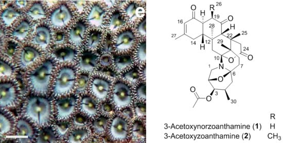
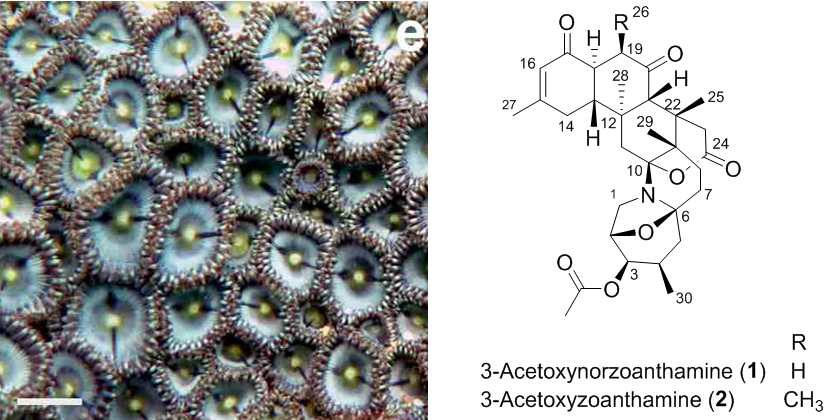
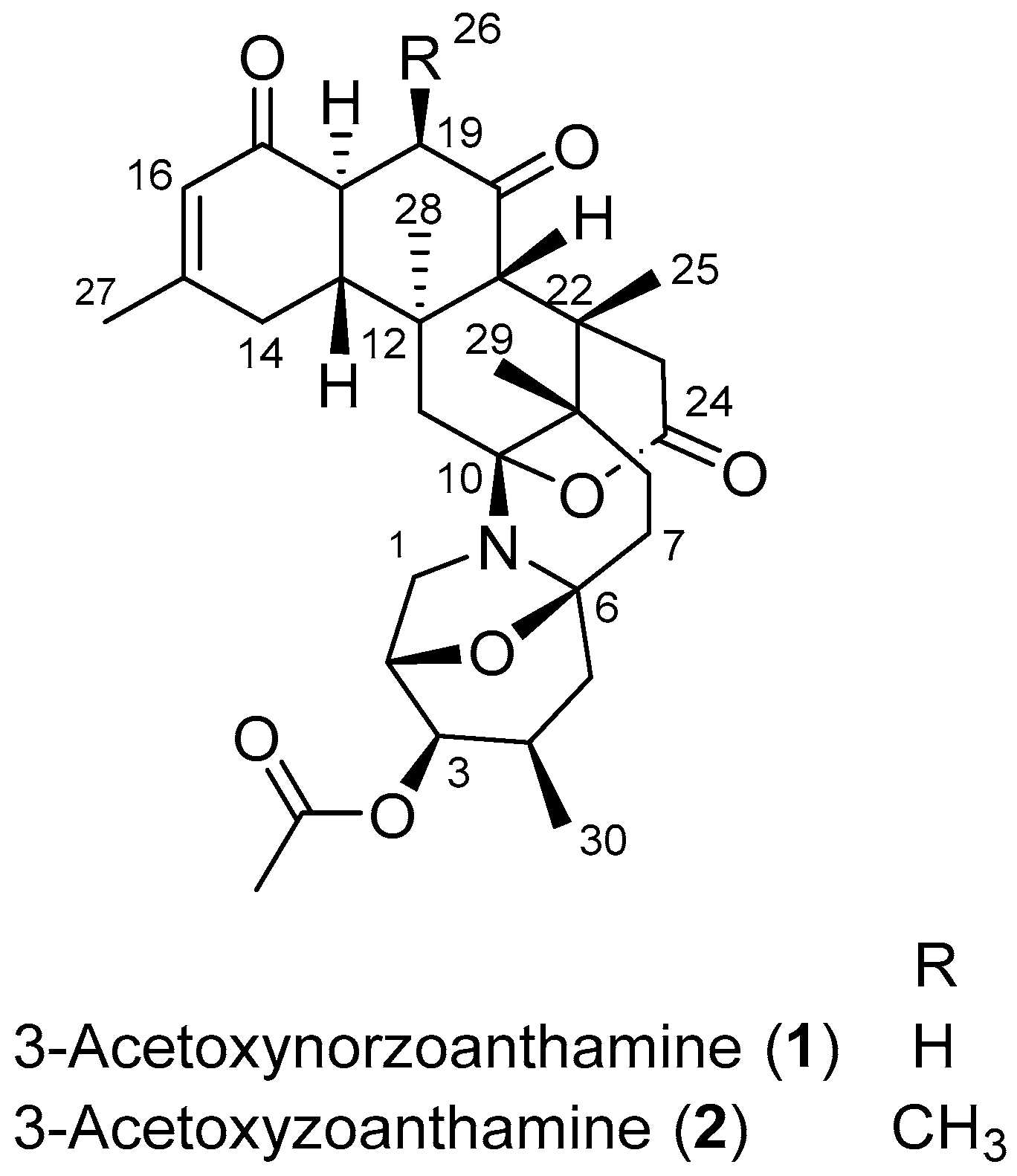
4.4. 3-Acetoxynorzoanthamine (1)

Amorphous yellow powder; [ α ] D 20 +10 (c 0.45, CH3OH); UV (CH3OH) λmax (log ε) 237 (4.1) nm; 1H NMR and 13C NMR data see Table 1; HRESIMS (+) m/z [M + H]+ 540.2956 (calc. for C31H42NO7 540.2956Δ + 0.0 ppm) (Spectra in the Supplementary Materials).

4.5. 3-Acetoxyzoanthamine (2)

Amorphous yellowish powder; [ α ] D 20 + 6.7 (c 0.12, CH3OH); UV (CH3OH) λmax (log ε) 238 (4.0) nm; 1H NMR and 13C NMR data see Table 1; HRESIMS (+) m/z [M + H]+ 554.3115 (calc. for C32H44NO7 554.3112Δ + 0.5 ppm) (Spectra in the Supplementary Materials).

4.6. Biological Assays

4.6.1. Cell Culture

The microglia BV-2 cell line was obtained from InterLab Cell Line Collection (ICLC) (Genova, Italy), number ATL03001. Cells were maintained in Roswell Park Memorial Institute Medium (RPMI) supplemented with 10% fetal bovine serum (FBS), penicillin (100 U/mL), and 100 µg/mL streptomycin at 37 °C in a humidified atmosphere of 5% CO2 and 95% air. Cells were dissociated twice a week using 0.05% trypsin/EDTA.

4.6.2. Cell Viability

The 3-(4,5-dimethyl thiazol-2-yl)-2,5-diphenyl tetrazolium bromide (MTT) assay was used to analyzed cell viability as previously described [24]. Briefly, the microglia BV-2 cell line was grown in a 96-well plate at a density of 4 × 10⁴ cells per well. Cells were exposed to different compounds concentration (0.001, 0.01, 0.1, 1 and 10 µM) for 24 h. Then, cells were rinsed and incubated with MTT (500 µg/mL) diluted in a saline buffer for 1 h at 37 °C. The resulting formazan crystals were dissolved with 5% sodium dodecyl sulfate, (SDS) and the absorbance values were obtained using a spectrophotometer plate reader (595 nm). Saponin was used for cellular death control and its absorbance was substrate from the other data.

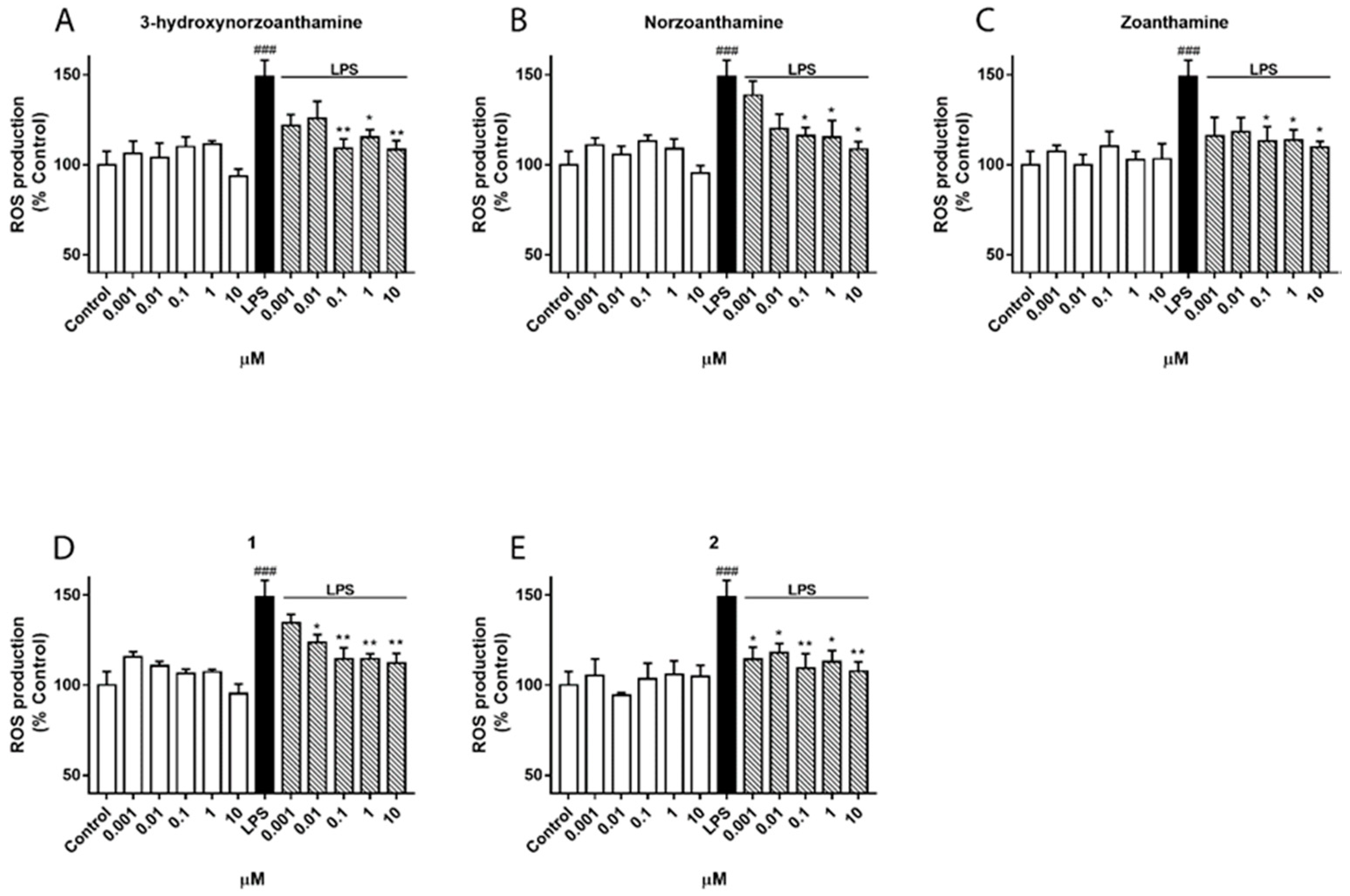
4.6.3. Measurement of Intracellular ROS Production

The intracellular ROS levels in microglia activation were performed using 7′,2′-dichlorofluorescein diacetate (DCFH-DA), as previously described [25]. Cells were pre-treated with different compounds concentration (0.001, 0.01, 0.1, 1, and 10 µM) 1 h prior to the stimulation with LPS (500 ng/mL) for 24 h. Afterwards, cells were rinsed twice with saline solution and incubated 1 h at 37 °C with 20 µM DCFH-DA. Then, cells were washed and kept in saline solution for 30 min at 37 °C. Intracellular production of ROS was measured by fluorescence detection of dichlorofluorescein (DCF) as the oxidized product of DCFH-DA on a spectrophotometer plate reader (495 nm excitation and 527 nm emission).

4.6.4. NO Determination

The NO concentration in the culture media was established by measuring nitrite formed by the oxidation of NO, using the Griess reagent kit, according to manufacturer instructions. The detection limit of this method is 1 µM. Briefly, microglia cells were seeded in a 12-well plate at a density of 1 × 10⁶ cells per well and pre-incubated with compounds (0.1 and 1 µM) for 1 h and then stimulated with LPS (500 ng/mL) for 24 h. Thereafter, the following were mixed in a microplate: 150 µL of cells supernatant, 130 µL of deionized water, and 20 µL of Griess Reagent, which was incubated for 30 min at room temperature. The absorbance was measured on a spectrophotometer plate reader at a wavelength of 548 nm.

4.6.5. Statistical Analysis

Results were expressed as mean ± SEM of a minimum of three experiments, repeated twice or three times. Comparisons were performed using Student’s t-test or one-way ANOVA with Dunnett’s post hoc analysis. p values < 0.05 were considered statistically significant.

Supplementary Materials

The following are available online at https://www.mdpi.com/1660-3397/16/7/242/s1: HRMS and NMR data for compounds 1 and 2.

Author Contributions

Methodology and Formal Analysis, P.O.G., S.G., K.B.J., K.C.; Validation, E.A., A.A., K.C.; Writing-Original Draft Preparation, P.O.G.; Writing-Review & Editing, A.A., O.P.T.; Supervision, E.A., O.P.T.; Project Administration, J.R., O.P.T.; Funding Acquisition, J.R., L.M.B., O.P.T.

Funding

The project is originally funded by the Secretaria de Educación Superior, Ciencia, Tecnología e Innovación (SENESCYT) in the framework of the PIC-14-CENAIM-001 Project Caracterización de la Biodiversidad Microbiológica y de Invertebrados de la Reserva Marina “El Pelado” a Escala Taxonómica, Metabolómica y Metagenómica para su Uso en Salud Humana y Animal. Part of this project (Grant-Aid Agreement No. PBA/MB/16/01) is carried out with the support of the Marine Institute and is funded under the Marine Research Programme by the Irish Government. P.O.G. and K.B.J. acknowledge NUI Galway for supporting part of their Ph.D. scholarship. The research leading to the results of the biological assays has received funding from the following FEDER cofunded-grants: Consellería de Cultura, Educación e Ordenación Universitaria, Xunta de Galicia, 2017 GRC GI-1682 (ED431C 2017/01); CDTI and Technological Funds, supported by Ministerio de Economía, Industria y Competitividad, AGL2014-58210-R, AGL2016-78728-R (AEI/FEDER, UE), ISCIII/PI16/01830 and RTC-2016-5507-2, ITC-20161072; European Union POCTEP 0161-Nanoeaters-1-E-1, Interreg AlertoxNet EAPA-317-2016, and H2020 778069-EMERTOX.

Acknowledgments

We acknowledge the support of Cristobal Dominguez (CENAIM-ESPOL, Ecuador) in the collection of the sample and Frederic Sinniger (University of the Ryukyus, Japan) for his help with the taxonomic identification of this species through the training of K.B.J.

Conflicts of Interest

The authors declare no conflict of interest. The funders had no role in the design of the study; in the collection, analyses, or interpretation of data; in the writing of the manuscript; or in the decision to publish the results.

References

  1. Rao, C.B.; Anjaneyula, A.S.R.; Sarma, S.S.; Venkatateswarlu, Y.; Chen, M.; Clardy, J.; Rosser, R.; Faulkner, J. Zoanthamine: A novel alkaloid from a marine zoanthid. J. Am. Chem. Soc. 1984, 106, 7984–7985. [Google Scholar] [CrossRef]
  2. Rao, C.B.; Anjaneyulu, A.S.R.; Sarma, N.S.; Venkateswarlu, Y.; Rosser, R.M.; Faulkner, J. Alkaloids from a marine zoanthid. J. Org. Chem. 1985, 50, 3757–3760. [Google Scholar] [CrossRef]
  3. Fukuzawa, S.; Hayashi, Y.; Uemura, D.; Nagatsu, A.; Yamada, K.; Ijuin, Y. The isolation and structures of five new alkaloids, norzoanthamine, oxyzoanthamine, norzoanthamine, cyclozoanthamine and epinorzoanthamine. Heterocycl. Commun. 1995, 1, 207–214. [Google Scholar] [CrossRef]
  4. Atta-ur-Rahman; Alvi, K.A.; Abbas, S.A.; Choudhary, M.I.; Clardy, J. Zoanthaminone, a new alkaloid from a marine zoanthid. Tetrahedron Lett. 1989, 30, 6825–6828. [Google Scholar] [CrossRef]
  5. Cen-Pacheco, F.; Norte, M.; Fernández, J.J.; Daranas, A.H. Zoaramine, a zoanthamine-like alkaloid with a new skeleton. Org. Lett. 2014, 16, 2880–2883. [Google Scholar] [CrossRef] [PubMed]
  6. Cheng, Y.-B.; Lo, I.-W.; Shyur, L.-F.; Yang, C.-C.; Hsu, Y.-M.; Su, J.-H.; Lu, M.-C.; Chiou, S.-F.; Lan, C.-C.; Wu, Y.-C.; et al. New alkaloids from Formosan zoanthid Zoanthus kuroshio. Tetrahedron 2015, 71, 8001–8006. [Google Scholar] [CrossRef]
  7. Daranas, A.H.; Fernández, J.J.; Gavin, J.A.; Norte, M. Epioxyzoanthamine, a new zoanthamine-type alkaloid and the unusual deuterium exchange in this series. Tetrahedron 1998, 54, 7891–7896. [Google Scholar] [CrossRef]
  8. Daranas, A.H.; Fernandez, J.J.; Gavin, J.A.; Norte, M. New alkaloids from a marine zoanthid. Tetrahedron 1999, 55, 5539–5546. [Google Scholar] [CrossRef]
  9. Behenna, D.C.; Stockdill, J.L.; Stoltz, B.M. The biology and chemistry of the zoanthamine alkaloids. Angew. Chem. Int. Ed. 2008, 47, 2365–2386. [Google Scholar] [CrossRef] [PubMed]
  10. Hsu, Y.-M.; Chang, F.-R.; Lo, I.W.; Lai, K.-H.; El-Shazly, M.; Wu, T.-Y.; Du, Y.-C.; Hwang, T.-L.; Cheng, Y.-B.; Wu, Y.-C. Zoanthamine-type alkaloids from the zoanthid Zoanthus kuroshio collected in Taiwan and their effects on inflammation. J. Nat. Prod. 2016, 79, 2674–2680. [Google Scholar] [CrossRef] [PubMed]
  11. Miyashita, M.; Sasaki, M.; Hattori, I.; Sakai, M.; Tanino, K. Total synthesis of norzoanthamine. Science 2004, 305, 495–499. [Google Scholar] [CrossRef] [PubMed]
  12. Takahashi, Y.; Yoshimura, F.; Tanino, K.; Miyashita, M. Total synthesis of zoanthenol. Angew. Chem. Int. Ed. 2009, 48, 8905–8908. [Google Scholar] [CrossRef] [PubMed]
  13. Yoshimura, F.; Sasaki, M.; Hattori, I.; Komatsu, K.; Sakai, M.; Tanino, K.; Miyashita, M. Synthetic studies of the zoanthamine alkaloids: The total syntheses of norzoanthamine and zoanthamine. Chem. Eur. J. 2009, 15, 6626–6644. [Google Scholar] [CrossRef] [PubMed]
  14. Yoshimura, F.; Tanino, K.; Miyashita, M. Total synthesis of zoanthamine alkaloids. Acc. Chem. Res. 2012, 45, 746–755. [Google Scholar] [CrossRef] [PubMed]
  15. Fischer, D.; Nguyen, T.X.; Trzoss, L.; Dakanali, M.; Theodorakis, E.A. Intramolecular cyclization strategies toward the synthesis of zoanthamine alkaloids. Tetrahedron Lett. 2011, 52, 4920–4923. [Google Scholar] [CrossRef] [PubMed] [Green Version]
  16. Nakajima, T.; Yamashita, D.; Suzuki, K.; Nakazaki, A.; Suzuki, T.; Kobayashi, S. Different modes of cyclization in zoanthamine alkaloid system, bisaminal versus spiroketal formation. Org. Lett. 2011, 13, 2980–2983. [Google Scholar] [CrossRef] [PubMed]
  17. Villar, R.M.; Gil-Longo, J.; Daranas, A.H.; Souto, M.L.; Fernández, J.J.; Peixinho, S.; Barral, M.A.; Santafé, G.; Rodríguez, J.; Jiménez, C. Evaluation of the effect of several zoanthamine-type alkaloids on the aggregation of human platelets. Bioorg. Med. Chem. 2003, 11, 2301–2306. [Google Scholar] [CrossRef]
  18. Genji, T.; Fukuzawa, S.; Tachibana, K. Distribution and possible function of the marine alkaloid, norzoanthamine, in the zoanthid Zoanthus sp. using MALDI imaging mass spectrometry. Mar. Biotechnol. 2010, 81–87. [Google Scholar] [CrossRef] [PubMed]
  19. Guillen, P.O.; Calabro, K.; Jaramillo, K.B.; Dominguez, C.; Genta-Jouve, G.; Rodriguez, J.; Thomas, O.P. Ecdysonelactones, ecdysteroids from the Tropical Eastern Pacific zoantharian Antipathozoanthus hickmani. Mar. Drugs 2018, 16, 58. [Google Scholar] [CrossRef] [PubMed]
  20. Guillen, P.O.; Jaramillo, K.B.; Genta-Jouve, G.; Sinniger, F.; Rodriguez, J.; Thomas, O.P. Terrazoanthines, 2-aminoimidazole alkaloids from the Tropical Eastern Pacific zoantharian Terrazoanthus onoi. Org. Lett. 2017, 19, 1558–1561. [Google Scholar] [CrossRef] [PubMed]
  21. Jaramillo, K.B.; Reverter, M.; Guillen, P.O.; McCormack, G.; Rodriguez, J.; Sinniger, F.; Thomas, O.P. Assessing the zoantharian diversity of the Tropical Eastern Pacific through an integrative approach. Sci. Rep. 2018, 8, 7138. [Google Scholar] [CrossRef] [PubMed]
  22. Kuramoto, M.; Hayashi, K.; Fujitani, Y.; Yamaguchi, K.; Tsuji, T.; Yamada, K.; Ijuin, Y.; Uemura, D. Absolute configuration of norzoanthamine, a promising candidate for an osteoporotic drug. Tetrahedron Lett. 1997, 38, 5683–5686. [Google Scholar] [CrossRef]
  23. Dumont, M.; Beal, M.F. Neuroprotective strategies involving ROS in Alzheimer disease. Free Radic. Biol. Med. 2011, 51, 1014–1026. [Google Scholar] [CrossRef] [PubMed] [Green Version]
  24. Sanchez, J.A.; Alfonso, A.; Leiros, M.; Alonso, E.; Rateb, M.E.; Jaspars, M.; Houssen, W.E.; Ebel, R.; Tabudravu, J.; Botana, L.M. Identification of Spongionella compounds as cyclosporine A mimics. Pharmacol. Res. 2016, 107, 407–414. [Google Scholar] [CrossRef] [PubMed]
  25. Leiros, M.; Alonso, E.; Rateb, M.E.; Houssen, W.E.; Ebel, R.; Jaspars, M.; Alfonso, A.; Botana, L.M. Gracilins: Spongionella-derived promising compounds for Alzheimer disease. Neuropharmacology 2015, 93, 285–293. [Google Scholar] [CrossRef] [PubMed]
Figure 1. Structures of 3-acetoxynorzoanthamine (1) and 3-acetoxyzoanthamine (2), isolated from Zoanthus cf. pulchellus.
Marinedrugs 16 00242 g001
Figure 2. Effect of zoanthamines on intracellular reactive oxygen species (ROS) production in microglia BV-2 cell line. Cells were pre-treated with 3-hydroxynorzoanthamine (A); norzoanthamine (B); zoanthamine (C); 1 (D); and 2 (E) at different concentrations (0.001, 0.01, 0.1, 1, and 10 µM) 1 h and then stimulated with lipolysaccharide (LPS) (1 µg/mL) for 24 h. ROS production is presented as a percentage of cells control, being the result of mean fluorescence intensity ± SEM of three independent experiments. The values are shown as the difference between cells treated with LPS alone versus cells treated with zoanthamines in presence of LPS by ANOVA followed by post hoc Dunnett’s test. * p < 0.05 and ** p < 0.01, and LPS-treated cells versus control cells ### p < 0.001.
Marinedrugs 16 00242 g002
Figure 3. Effect of zoanthamines on nitric oxide (NO) production in BV-2 microglia cell line. Cells were pre-treated with 3-hydroxynorzoanthamine (3-HNZ), norzoanthamine (NZ), zoanthamine (Z), 1, and 2 (0.1 or 1 µM) for 1 h and then stimulated with lipolysaccharide (LPS) (500 ng/mL) for 24 h. The values are presented in percentage of cells control, being the result of mean ± SEM of a minimum of three independent experiments. The cells treated only with LPS were compared to cells treated with compounds in presence of LPS by ANOVA followed by post hoc Dunnett’s test. * p < 0.05 and ** p < 0.01, and LPS-treated cells versus control cells ### p < 0.001.
Marinedrugs 16 00242 g003
Table 1. 1H and 13C NMR data in ppm for compounds 1 and 2 in CDCl3 (500 MHz for 1H NMR and 125 MHz for 13C NMR data).
12
No.δH, mult. (J in Hz)δCδH, mult. (J in Hz)δC
13.24, t (7.0) 45.33.24, t (7.5)45.5
3.19, d (7.0)3.20, d (7.0)
24.58, br d (6.5)75.64.59, d (7.0)75.7
34.62, br t (3.0)72.54.63, t (3.0)72.6
42.44, br sext (5.5)26.02.43, br sext (6.0)26.1
51.92, dd (12.0, 6.0)40.31.95, dd (12.5, 6.0)40.4
1.36, t (12.5)1.37, t (13.0)
6-90.1-90.2
71.88, dd (12.5, 4.5)29.81.90, dd (12.5, 4.5)29.9
1.80, dt (12.5, 3.5)1.80, dt (12.5, 3.5)
81.66, td (13.5, 3.5)23.71.67, td (14.0, 3.5)23.8
1.57, dt (13.5, 4.0)1.57, dt (14.0, 4.0)
9-40.0-40.5
10-100.9-101.0
112.08, d (13.0)41.82.11, d (13.0)42.0
1.94, d (13.0)1.93, d (13.0)
12-39.9-39.8
132.20, td (12.0, 4.5)53.12.41, td (12.0, 4.5)48.1
142.26, br s32.02.24, br s30.7
2.24, br s2.22, br s
15-160.0-160.1
165.90, s125.65.92, s127.0
17-198.5-197.3
182.69, td (12.0, 6.5)46.42.66, dd (12.5, 6.5)48.2
192.62, dd (14.5, 6.5)42.43.02, dq (7.0, 6.5)45.9
2.50, dd (14.5, 12.0)
20-209.0-212.2
212.83, s59.13.23, s53.9
22-36.5-40.3
233.65, d (20.0)35.93.68, d (20.0)36.1
2.36, d (20.0)2.37, d (20.0)
24-172.3-172.4
250.99, s21.10.98, s20.8
26--1.17, d (7.0)13.9
272.00, s24.42.01, s24.6
280.97, s18.50.99, s18.5
291.15, s18.41.21, s18.4
300.87, d (7.0)16.30.89, d (7.0)16.4
Ac-171.2-171.4
2.11, s21.12.14, s21.2

