Article

Lipophilic Fraction of Cultivated Bifurcaria bifurcata R. Ross: Detailed Composition and In Vitro Prospection of Current Challenging Bioactive Properties

1
CICECO—Aveiro Institute of Materials and Department of Chemistry, University of Aveiro, 3810-193 Aveiro, Portugal
2
Centro de Biotecnologia Agrícola e Agro-Alimentar do Alentejo (CEBAL)/Instituto Politécnico de Beja (IPBeja), 7801-908 Beja, Portugal
3
ICAAM—Instituto de Ciências Agrárias e Ambientais Mediterrânicas, Universidade de Évora, Pólo da Mitra, 7006-554 Évora, Portugal
4
CNC—Center for Neuroscience and Cell Biology, University of Coimbra, 3004-504 Coimbra, Portugal
5
FFUC—Faculty of Pharmacy, University of Coimbra, 3000-548 Coimbra, Portugal
6
ALGAplus—Prod. e Comerc. De Algas e Seus Derivados, Lda., 3830-196 Ílhavo, Portugal
7
QOPNA and Department of Chemistry, University of Aveiro, 3810-193 Aveiro, Portugal
*
Author to whom correspondence should be addressed.
Mar. Drugs 2017, 15(11), 340; https://doi.org/10.3390/md15110340
Submission received: 20 September 2017 / Revised: 19 October 2017 / Accepted: 23 October 2017 / Published: 1 November 2017
(This article belongs to the Special Issue Marine Lipids 2017)

Abstract

:
Macroalgae have been seen as an alternative source of molecules with promising bioactivities to use in the prevention and treatment of current lifestyle diseases. In this vein, the lipophilic fraction of short-term (three weeks) cultivated Bifurcaria bifurcata was characterized in detail by gas chromatography–mass spectrometry (GC-MS). B. bifurcata dichloromethane extract was composed mainly by diterpenes (1892.78 ± 133.97 mg kg−1 dry weight (DW)), followed by fatty acids, both saturated (550.35 ± 15.67 mg kg−1 DW) and unsaturated (397.06 ± 18.44 mg kg−1 DW). Considerable amounts of sterols, namely fucosterol (317.68 ± 26.11 mg kg−1 DW) were also found. In vitro tests demonstrated that the B. bifurcata lipophilic extract show antioxidant, anti-inflammatory and antibacterial activities (against both Gram-positive and Gram-negative bacteria), using low extract concentrations (in the order of µg mL−1). Enhancement of antibiotic activity of drug families of major clinical importance was observed by the use of B. bifurcata extract. This enhancement of antibiotic activity depends on the microbial strain and on the antibiotic. This work represents the first detailed phytochemical study of the lipophilic extract of B. bifurcata and is, therefore, an important contribution for the valorization of B. bifurcata macroalgae, with promising applications in functional foods, nutraceutical, cosmetic and biomedical fields.

1. Introduction

Currently there is a remarkable demand for new bioactive molecules to treat diseases, caused by the modern lifestyle and the growing exposure to industrial pollutants and environmental toxins. Antioxidant compounds have been widely researched, due to their protective action against damage induced by oxidative stress, which is the major cause of diseases like cancer, diabetes, asthma, cardiovascular, or neurodegenerative problems [1], and of skin damage. Current lifestyle diseases have been also associated with inflammatory processes [2], which have also encouraged the scientific community to search for new molecules with anti-inflammatory capacity. At the same time, the emergence and spread of antibiotic-resistant bacteria strains is a growing public health problem [3], which has led the scientific community to search for new antibacterial substitutes or combination of drugs that might overcome this resistance [4].
Nature has always been an important source of molecules with biological properties as such, or after chemical modification [5]. In recent years, marine resources have become alternative sources of several value-added compounds [6,7]. Macroalgae, due to their high biological and chemical diversity and fast-growing properties, are one of the most explored marine resources [8,9]. In fact, approximately 28.5 million tons of macroalgae (brown, red or green) were produced worldwide in 2014, from both capture and aquaculture [10], with an estimated increase of 15%/year. This resource has been mainly explored for food consumption or for the production of specialty products, such as alginic acid, carrageenan, agar, or colorants [7]. In this context, the exploitation of macroalgae has been gaining increasing attention from several research groups around the world [11,12,13] and notably in Portugal [14,15,16,17,18]. In fact, the exploitation of marine resources is also one of the critical sectors for Portuguese development, given the extent and richness of its Exclusive Economic Zone (EEZ), being included in the Portuguese Research and Innovation Strategy for Smart Specialization for 2014–2020 [19].
Amongst the macroalgae species (close to 10,000), only a limited number have been the object of extensive studies due to their unique composition and consequent diversity of biological activities or health benefits [20]. Species belonging to the brown Dictyotaceae and Sargassaceae families have been studied in detail due to the presence of diterpenes, which have a wide range of biological properties, such as antimicrobial or antitumoral [21]. Among those families, Bifurcaria bifurcata, which can only be found in the northeastern Atlantic coasts, from Morocco to northwestern Ireland, is known to biosynthesize components rarely found in other macroalgae species, namely linear diterpenes [22]. In fact, the presence of these components in B. bifurcata has been widely reported [23,24,25,26,27]. In addition, several in vitro effects of B. bifurcata extracts, such as antimicrobial [28], antimitotic [29], or antiproliferative activity [30], have been attributed to the presence of diterpenes, although this assumption has never been adequately complemented by a detailed study of the composition of the tested extracts. In fact, the relationship between the chemical composition (limited to the fatty acids profile) and the biological properties (namely, antimicrobial and antioxidant activity) of B. bifurcata extracts has been addressed in only one study [31].
It has been suggested that the diterpenic composition of B. bifurcata is largely dependent on abiotic factors and especially on its geographic origin [32]. Furthermore, there are no studies regarding the lipophilic composition (and the diterpenic fraction in particular) of B. bifurcata cultivated in nutrient-rich waters (Integrated Multi-Trophic Aquaculture systems (IMTA)) (or even from the Portuguese coast), where, due to the totally different biotic and abiotic growth conditions, substantial differences in composition might be anticipated.
In addition, most of the published studies using wild B. bifurcata have neglected the abundance of diterpenic components, and the few studies that reported their quantification have focused on specific fractions, not considering the lipophilic fraction as a whole [29,33,34]. The abundance of other important classes of lipophilic compounds are also unknown, namely sterols, long-chain aliphatic alcohols and even fatty acids, for which only a single study can be found [31]. The limited number of compounds identified in these studies can be overcome using gas chromatography coupled with mass spectrometry (GC-MS), which allows the detailed identification and quantification of complex mixtures of compounds found in the lipophilic extracts [35,36].
In this study the lipophilic fraction of B. bifurcata cultivated for a short period (three weeks) in a land-based IMTA system was detailed characterized by GC-MS analysis. In addition, in vitro assays were performed to evaluate the antioxidant, antibacterial and anti-inflammatory activities of this fraction. Experiments were also conducted aiming to access the synergistic antibiotic–B. bifurcata lipophilic extract bacterial growth inhibition by determining the minimum inhibitory concentration (MIC—the lowest concentration where no bacterial growth was detected).

2. Results and Discussion

2.1. Lipophilic Composition

The dichloromethane extraction yield of B. bifurcata accounted for 3.92 ± 0.09% (w/w), which is quite similar to the n-hexane extraction yield obtained for B. bifurcata from both aquaculture and wild collected in Portuguese coast [37]. Additionally, the dichloromethane extraction yield was considerably higher than those observed previously for other Phaeophyta species [15] or for Chlorophyta or Rhodophyta macroalgae [14].
The chemical composition of the B. bifurcata dichloromethane extract was studied in detail by GC-MS analysis. With the exception of the diterpenes family, all the lipophilic components were identified as their trimethylsilyl derivatives, after derivatization of the dichloromethane extract. The identification of the main lipophilic extractives and the corresponding quantification is summarized in Table 1. In general, the extract was mainly composed of diterpenes, free fatty acids (C14–C22), long-chain aliphatic alcohols (C14–C28), and sterols, among other components. To the best of our knowledge this is the first study reporting the analysis of the lipophilic fraction of B. bifurcata by GC-MS. Recently, Alves et al. [31] analyzed the dichloromethane extract of B. bifurcata by GC with flame ionization detector; however, only the presence of fatty acids was reported.

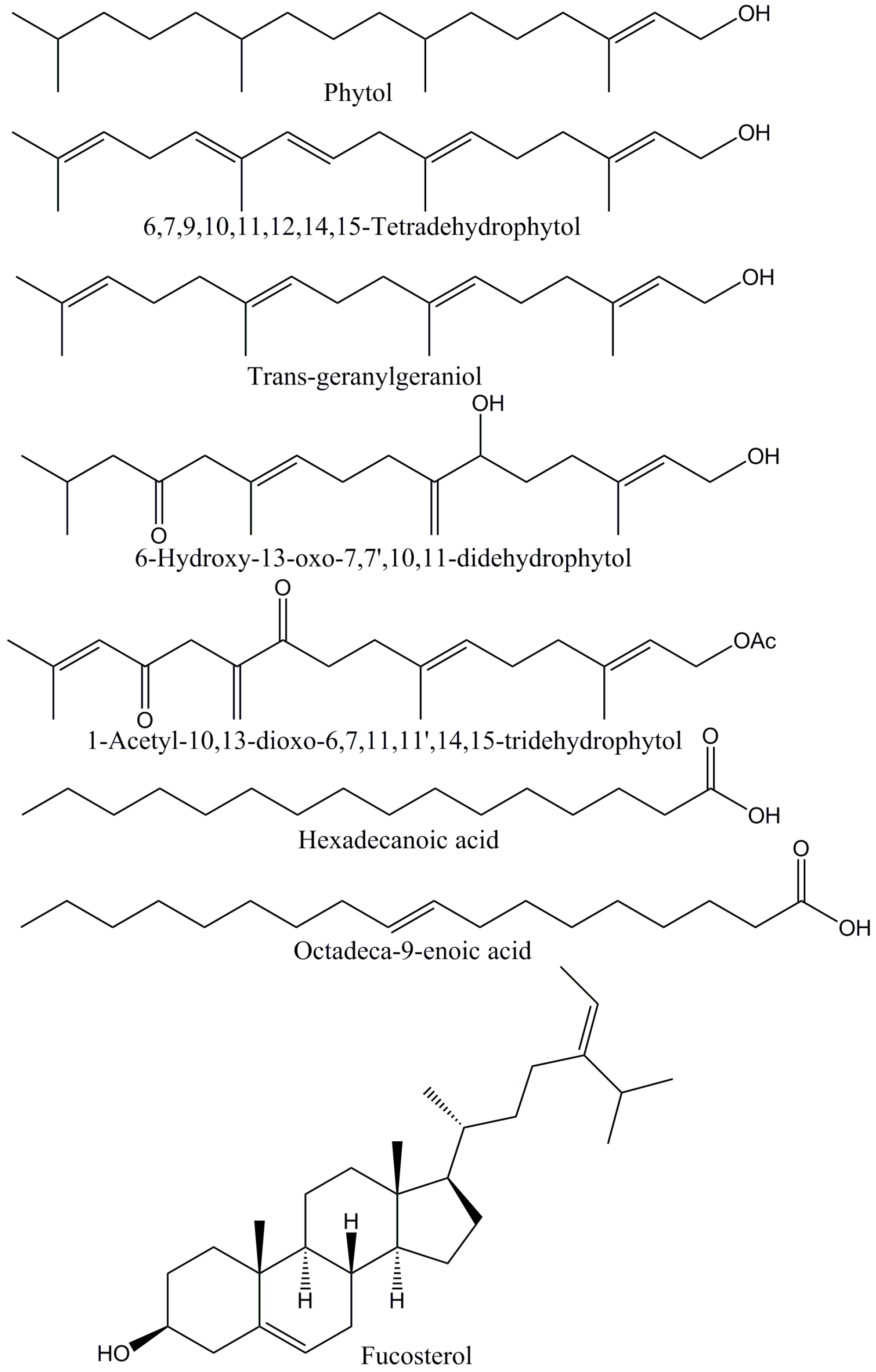
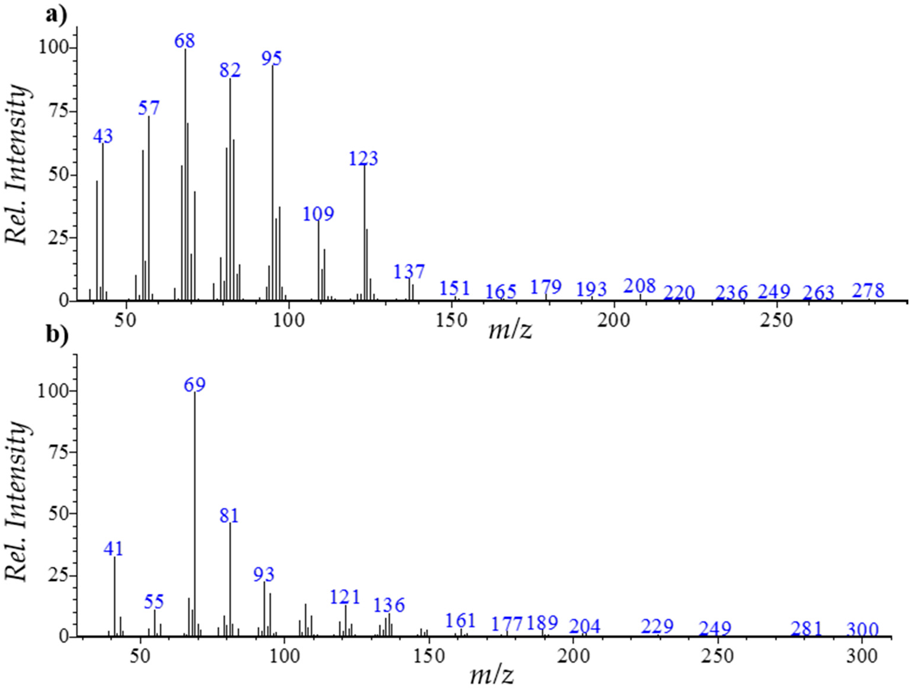
2.1.1. Diterpenes and Other Terpenoids

Several diterpenes (Figure 1) and other terpenoids were identified in B. bifurcata dichloromethane extract, namely neophytadiene, phytol, trans-geranylgeraniol, 6,7,9,10,11,12,14,15-tetradehydrophytol, 6-hydroxy-13-oxo-7,7′,10,11-didehydrophytol, 1-acetyl-10,13-dioxo-6,7,11,11′,14,15-tridehydrophytol and 6,10,14-trimethyl-2-pentadecanone.
The fragmentation patterns of these components depend on the number and position of the double bonds, and on the nature of the substituent groups [23,25,26]. As an example, the mass spectra of neophytadiene and trans-geranylgeraniol are presented in Figure 2.
The mass spectrum of neophytadiene presents a molecular ion [M]+ at m/z 278 and major product ions at m/z 43 ([C3H7]+), 57 ([C4H9]+), 68, 82, 95 ([C7H11]+), 109 and 123 ([C9H15]+). Similarly, trans-geranylgeraniol presents a molecular ion [M]+ at m/z 290, and characteristic product ions at m/z 69 ([C5H9]+), 81 ([C6H9]+), 121 ([C11H19]+) and 272 (M-H2O]+).
Diterpenes, accounting for 1892.78 ± 133.97 mg kg−1 DW, correspond to about 57% of the total amount of lipophilic compounds detected in B. bifurcata extract, with 1-acetyl-10,13-dioxo-6,7,11,11′,14,15-tridehydrophytol and 6-hydroxy-13-oxo-7,7′,10,11-didehydrophytol as the major components of this family, accounting for 1000.17 ± 94.47 and 637.84 ± 34.41 mg kg−1 DW, respectively. 1-acetyl-10,13-dioxo-6,7,11,11′,14,15-tridehydrophytol and 6-hydroxy-13-oxo-7,7′,10,11-didehydrophytol were already described as constituent of B. bifurcata collected in Brittany (France) [25,26], but no information was reported regarding its quantification. Considerable amounts of 6,7,9,10,11,12,14,15-tetradehydrophytol, trans-geranylgeraniol and neophytadiene were also found, and minor amounts of phytol were also detected. 6,7,9,10,11,12,14,15-Tetradehydrophytol was previously identified in B. bifurcata collected in Morocco [23], though no information has been provided concerning its abundance, while, to the best of our knowledge, phytol and neophytadiene are reported, for the first time, as constituents of B. bifurcata. In opposition, geranylgeraniol has been one of the most reported constituents of this macroalga species [26,29,38]. Additionally, the abundance of this component in the B. bifurcata dichloromethane extract is in the range of those (0.0–1.5 mg g−1 DW) reported in a seasonal variation study of the same species collected in Brittany (France) [39]. Notwithstanding, only traces of this diterpene were found in a geographical variation study of the diterpene composition of B. bifurcata collected in Spain [33].
Despite the high abundance verified for all the detected diterpenes, these were not reported in some of the studies regarding the seasonal and geographical variation of diterpenes in B. bifurcata [29,33,34].
Diterpenes have been associated with a wide number of health benefits [40]. In particular, those from macroalgae have been reported to have antioxidant, antimicrobial [41], antifungal [42], anti-viral [43], or antitumoral activities [44].
Minor amounts (10.79 ± 0.18 mg kg−1 DW) of 6,10,14-trimethyl-2-pentadecanone were found in B. bifurcata dichloromethane extract. This terpenoid and its derivatives have been described to be produced by macroalgae from Cystoseiraceae family, namely Cystophora moniliformis [45,46].

2.1.2. Fatty Acids

Fatty acids were the second most abundant family of compounds detected in the lipophilic fraction of B. bifurcata, corresponding to 947.88 ± 21.94 mg kg−1 DW. Hexadecanoic acid (Figure 1) was the most abundant fatty acid (396.43 ± 17.01 mg kg−1 DW), followed by tetradecanoic (76.23 ± 1.69 mg kg−1 DW) and octadecanoic (41.01 ± 3.49 mg kg−1 DW) acids. The high abundance of hexadecanoic and tetradecanoic acid in B. bifurcata was already reported [31], however, in considerably higher amounts than those found in this study.
Considerable amounts of unsaturated fatty acids were found in B. bifurcata (397.06 ± 18.44 mg kg−1 DW), representing about 42% of the total fatty acids. Octadec-9-enoic acid (Figure 1) was the most abundant unsaturated fatty acid, accounting for 200.95 ± 10.32 mg kg−1 DW, followed by the ω-3 octadeca-9,12,15-trienoic acid (57.81 ± 2.70 mg kg−1 DW) and the hexadec-9-enoic acid (42.24 ± 2.79 mg kg−1 DW). Significant amounts of the ω-3 fatty acid eicosa-5,8,11,14,17-pentaenoic acid were also found (35.86 ± 2.57 mg kg−1 DW), despite being 10 times lower than those reported previously by Alves et al. [31] for B. bifurcata collected in Peniche coast, Portugal. Notwithstanding, the high abundance of this component contributes to the low ω-6/ω-3 ratio verified for this macroalga (~0.46), which is considerably lower than the maximum (4:1) recommended for a diet in order to prevent the development of inflammatory processes [47]. Additionally, this ratio is significantly lower than those verified for other macroalgae species [15,48], which were considered promising to be incorporated in healthy diets. The high abundance of such components, widely associated with the prevention or delay of chronic diseases, such as cancer, cardiovascular, or coronary problems [49], highlights the potential of valorization of B. bifurcata also in food and/or nutraceutical industry. However, such exploitation requires that more detailed studies should be performed, namely evaluating their side effects and toxicity.

2.1.3. Long-Chain Aliphatic Alcohols

Minor amounts of long-chain aliphatic alcohols (LCAA) were found in B. bifurcata dichloromethane extract, accounting for 17.03 ± 1.29 mg kg−1 DW. To the best of our knowledge this is the first study reporting the presence of these components in B. bifurcata. Hexadecan-1-ol was the most abundant LCAA detected, followed by octadecan-1-ol and tetradecan-1-ol.

2.1.4. Sterols

Significant amounts of sterols were found in the dichloromethane extract of B. bifurcata (406.45 ± 26.19 mg kg−1 DW). Other studies reported the total sterols content in this macroalga collected in different countries [33,39]; however, only fucosterol has been described, with no further detailed characterization. In fact, fucosterol (Figure 1) was the major sterol detected in B. bifurcata dichloromethane extract, accounting for 317.68 ± 26.11 mg kg−1 DW. Notwithstanding, this value was lower than the previous reported contents (1400–5900 mg kg−1 DW) [33,39]. The bioactivities assigned to this phytosterol have been widely described in literature, such as its capacity to modulate cholesterol levels [50], its anti-inflammatory potential [51], or its dermo-protective effect [52]. Considerable amounts of desmosterol (43.56 ± 1.10 mg kg−1 DW) and campesterol (37.92 ± 1.61 mg kg−1 DW) were also found, which is in accordance with results observed for other Phaeophyta macroalgae [15]. Minor amounts of cholesterol were also detected.

2.1.5. Monoglycerides

Monoglycerides were also detected in B. bifurcata dichloromethane extract, representing only a small fraction of the total lipophilic compounds identified (34.99 ± 1.10 mg kg−1 DW). To our knowledge, this is also the first study reporting the monoglycerides profile of B. bifurcata. 1-Monohexadecanoin was the most abundant compound detected from this family, representing almost 74% of the total monoglycerides. Minor amounts of 1-monooctadecenoin (5.17 ± 0.17 mg kg−1 DW) and 1-monoeicosa-tetraenoin (4.00 ± 0.31 mg kg−1 DW) were also found.

2.2. Antioxidant Activity

The antioxidant activity of B. bifurcata dichloromethane extract was evaluated by both DPPH and ABTS in vitro assays. Table 2 presents the obtained results, expressed as the amount of extract needed to decrease the DPPH and ABTS+ concentrations by 50% (IC50), as well as in mg of ascorbic acid (AAE) and trolox (TE) equivalents per g of dry weight. The IC50 values for ascorbic acid and BHT (for DPPH assay) and for trolox (for ABTS assay) were also estimated and are reported in Table 2 for comparative purposes. B. bifurcata lipophilic extract showed antioxidant activity against both radicals, although a higher activity was observed against ABTS+, with the respective IC50 accounting for 116.25 ± 2.54 µg mL−1, representing 23.10 ± 0.51 mg of trolox equivalents g−1 DW.
To our knowledge, this is the first study reporting the antioxidant activity of a B. bifurcata extract against ABTS+. In addition, the IC50 value determined for DPPH assay (365.57 ± 10.04 µg mL−1) was quite similar to that described before for a dichloromethane extract of B. bifurcata collected from rock pools in Portuguese coast [31]. Notwithstanding, B. bifurcata lipophilic extracts showed IC50 values against both radicals significantly lower than those determined for ascorbic acid or trolox (Table 2).

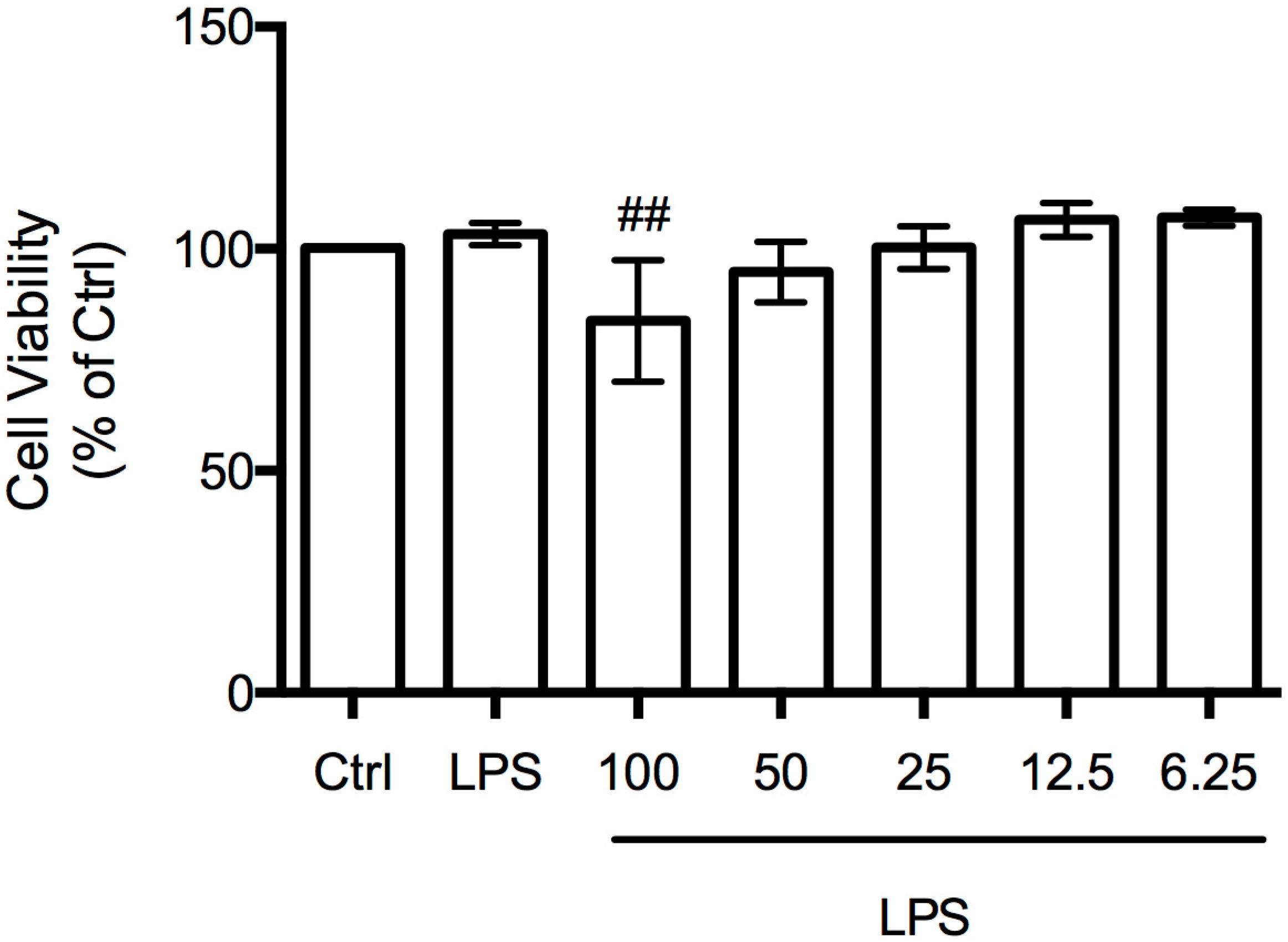
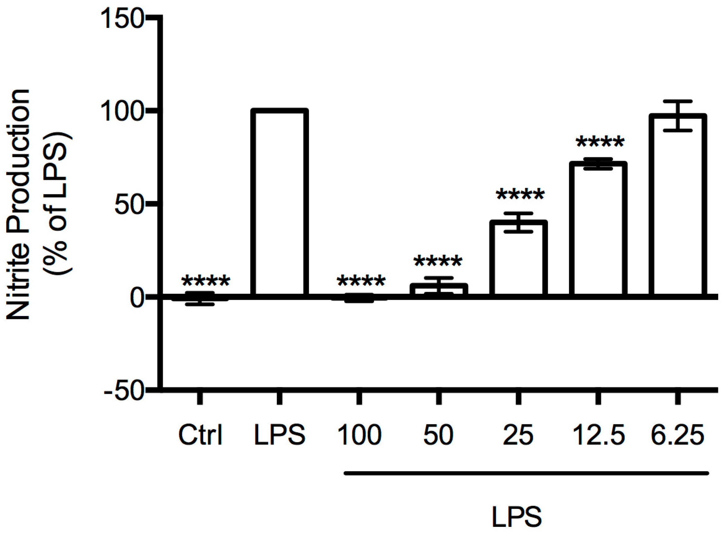
2.3. Anti-Inflammatory Activity

In order to study the capacity of B. bifurcata dichloromethane extract to modulate nitric oxide production, an in vitro model of inflammation consisting of macrophages stimulated with LPS was performed. Concomitantly, cell viability was evaluated by the resazurin-based assay (Figure 3) in order to select concentrations with bioactivity and without cytotoxicity. B. bifurcata lipophilic extract showed a slightly but statistically significant cytotoxic effect at 100 µg mL−1, when compared to LPS-treated cells.
The effect of B. bifurcata extract on NO production was analyzed by measuring the accumulation of nitrites in the culture medium, using the Griess assay. RAW 264.7 cells were stimulated with LPS in the presence or absence of B. bifurcata extract for 24 h. B. bifurcata extract inhibited LPS-induced NO production in a concentration-dependent manner (Figure 4). This extract markedly inhibited LPS-induced NO production to 6% and 40% at 50 and 25 µg mL−1, respectively.
The decrease in LPS-induced NO production at 50 µg mL−1 to levels similar to those observed in untreated cells reveals the remarkable anti-inflammatory potential of this extract. In fact, the anti-inflammatory activity of the extract is considerable higher than reported for other macroalgae extracts, namely from Saccharina japonica [53] or Undaria pinnatifida [54]. To our knowledge, this is the first study reporting the anti-inflammatory activity of B. bifurcata.

2.4. Antibacterial Activity

The antibacterial activity of B. bifurcata lipophilic extract against Staphylococcus aureus, Escherichiacoli, Pseudomonas aeruginosa, and Staphylococcus epidermidis were evaluated. Therapeutic strategies to counteract infections caused by these bacterial strains have been one of the main concerns, particularly in health care settings. In fact, S. aureus, E. coli, and P. aeruginosa present the higher rates of antibiotic resistance, while S. epidermidis has been considered an opportunistic pathogen, being the most common source of infections on indwelling medical devices [55]. Thus, in this study, the antimicrobial efficacy of four antibiotics representing drug families of major clinical importance, such as aminoglycosides (Gent: Gentamicin), tetracyclines (Tetra: Tetracycline), macrocyclics (Rif: Rifampicin), and β-lactams antibiotics, such as aminopenicillins, (Amp: Ampicillin) were evaluated in combination with the B. bifurcata lipophilic extract.
Results regarding the antibacterial activity of the B. bifurcata dichloromethane extract are presented in Table 3. Activity against S. aureus ATCC®6538 (MIC = 1024 µg mL−1), S. aureus ATCC®43300 (MIC = 2048 µg mL−1) and E. coli (MIC = 2048 µg mL−1) was observed. No growth inhibition of S. epidermidis and P. aeruginosa PAO1 was verified in the range of concentrations tested (MIC > 2048 μg mL−1). B. bifurcata extract showed inhibition against both Gram-negative and Gram-positive bacteria, in opposition to that observed by Alves et al. [31], which only verified activity of a B. bifurcata dichloromethane extract against Gram-negative bacteria. Additionally, these authors have not reported growth inhibition against the same E. coli strain. Notwithstanding the fact that the extraction conditions in both studies were not the same, the macroalgae origins are distinct, which may alter the metabolite composition and therefore their bioactivities, highlighting the importance of controlling the growth conditions of macroalgae in order to maximize their valorization.
The synergism of B. bifurcata dichloromethane extract with antibiotics was also evaluated against the same bacterial panel. This approach has been suggested as a promising tool to overcome the bacterial resistance that has been developed to most of the antibiotic classes recommended for their treatment [3,4,56]; to our knowledge, no similar study has previously been reported for this macroalga. The synergetic activity (antibiotic + extract) screening was performed against the bacterial strains for which a new antibiotic MIC was possible to determine, namely for E. coli ATCC®25922, S. aureus ATCC®43300 and S. aureus ATCC®6538 (Table 3). Interestingly, the combination of the extract with gentamicin or tetracycline drastically decreased the antibiotic MIC against the three strains under study, enabling it to be effective at considerably lower concentrations. Similar behavior was observed for both E. coli ATCC®25922 and S. aureus ATCC®43300 when the extract was combined with rifampicin. These observations suggest that the use of B. bifurcata lipophilic fraction may be considered within a coadjutant/synergistic approach to conventional antibiotherapy, enabling a superior therapeutic potential against the above-mentioned bacteria.
Concerning the interaction between B. bifurcata dichloromethane extract and ampicillin, an antagonism effect was observed, with MIC values increasing considerably. This may be linked to alterations in the antibiotic structure induced by the presence of the extract in solution, leading to its partial inactivation.

3. Material and Methods

3.1. Sample

Bifurcaria bifurcata R. Ross was cultivated in the land-based aquaculture system of ALGAplus, Lda, at Ria de Aveiro (Portugal, 40°36′43″ N, 8°40′43″ W). The company operates under the IMTA concept, using solely filtered nutrient-rich water from a fishpond to produce seaweed biomass. The culture starting material was obtained at Aguda beach (Portugal, 41°2′38″ N, 8°39′10″ W) in May 2014 and grown by vegetative propagation during three weeks at constant conditions. The biomass was washed with seawater to remove salts, epiphytes, and/or microorganisms and dried at 25 °C until it reached a total moisture content of 12%. The samples were transformed into flakes (1–2 mm), packed, and stored in hermetic bags in the company’s storage room.

3.2. Lipophilic Compounds Extraction

Three aliquots (20 g) of lyophilized macroalgae samples were Soxhlet extracted with dichloromethane (Sigma Chemical Co., Madrid, Spain) for 9 h. Solvent was evaporated to dryness, lipophilic extracts weighted and the results were expressed in percent of dry weight material (% DW). Dichloromethane was selected as a fairly specific solvent for lipophilic extractives isolation for analytical purposes only.

3.3. GC-MS Analysis

The analysis of the lipophilic extracts followed two distinct methodologies: the first suitable for trimethylsilyl (TMS) derivatizable compounds and the second for diterpenic compounds, as described below.

3.3.1. Analysis of Trimethylsilyl Derivatizable Compounds

Before GC-MS analysis, two aliquots of each dried extract (20 mg each) and an accurate amount of internal standard (tetracosane, 0.25–0.50 mg, Sigma Chemical Co., Madrid, Spain) were dissolved in 250 μL of pyridine (Sigma Chemical Co.).
The compounds containing hydroxyl and carboxyl groups were converted into TMS ethers and esters, respectively, by adding 250 μL of N,O-bis(trimethylsilyl)trifluoroacetamide (Sigma Chemical Co.) and 50 μL of trimethylchlorosilane (Sigma Chemical Co.), standing the mixture at 70 °C for 30 min [57]. The derivatized extracts were analyzed by GC-MS following previously described methodologies [15,35] on a GCMS-QP2010 Ultra (Shimadzu, Kyoto, Japan), equipped with a DB–1 J&W capillary column (30 m × 0.32 mm inner diameter, 0.25 μm film thickness). The chromatographic conditions were as follows: initial temperature, 80 °C for 5 min; temperature gradient, 4 °C min−1; final temperature, 260 °C; temperature gradient, 2 °C min−1; final temperature, 285 °C for 8 min; injector temperature, 250 °C; transfer-line temperature, 290 °C; split ratio, 1:33.
Compounds were identified as TMS derivatives by comparing their mass spectra with two commercial GC-MS spectral libraries (Wiley 275 and U.S. National Institute of Science and Technology (NIST14) ), their characteristic retention times obtained under the described experimental conditions [15,35], and by comparing their mass spectra fragmentation profiles with published data or by injection of standards.
For semi-quantitative analysis, GC-MS was calibrated with pure reference compounds, representative of the major lipophilic extractive families (cholesterol, hexadecanoic acid and nonadecan-1-ol (Sigma Chemical Co.)) relative to tetracosane. The respective response factors were calculated as an average of six GC-MS runs. Each one of the three extracts prepared from macroalgae was injected in duplicate (n = 6). The presented results are the average of the concordant values obtained (less than 5% variation between injections of the same aliquot and between triplicated extracts of the same macroalgae).

3.3.2. Analysis of diterpenes

Diterpenes were identified and quantified by the analysis of the B. bifurcata extract without derivatization. About 10 mg of extract were dissolved in 1100 µL of dichloromethane with 0.8 mg of internal standard (n-hexadecane (Supelco, Bellefonte, PA, USA)).
The extracts were injected in the same GC-MS equipment as described above and the chromatographic conditions were as follows: initial temperature, 80 °C for 5 min; temperature gradient, 5 °C min−1, final temperature 200 °C, temperature gradient, 2 °C min−1, final temperature 240 °C; temperature gradient, 5 °C min−1, final temperature 285 °C for 8 min; injector temperature, 250 °C; transfer-line temperature, 290 °C; split ratio, 1:40. All other conditions were the same as described above. Diterpenes were identified by comparing their mass spectra fragmentation profile with library (Wiley 275 and U.S. National Institute of Science and Technology (NIST14)) and with the characteristic fragmentation pathway described in literature for these components [23,25,26].
Semi-quantitative analysis was carried out determining the response factor (an average of six GC-MS runs) of a representative standard, namely phytol (Sigma Chemical Co.), relative to n-hexadecane. Each one of the three aliquots were injected in duplicate (n = 6).

3.4. Antioxidant Activity

3.4.1. DPPH Assay

The antioxidant activity of the B. bifurcata lipophilic extract was measured by their hydrogen-donating or radical scavenging ability using the stable free radical 2,2-diphenyl-1-picrylhydrazyl (DPPH) (Sigma Chemical Co.), following a previously described procedure [35]: 0.25 mL of DPPH (0.8 mM in methanol (Fluka Chemie, Madrid, Spain)) were added to 1.00 mL of the aqueous solution of the B. bifurcata dichloromethane extract and 2.75 mL of methanol. The extract concentration ranged between 61.9 and 556.9 μg mL−1. After 30 min of incubation in the dark, at room temperature, the absorbance was determined at 517 nm, using a Shimadzu UV-1800 spectrophotometer (Kyoto, Japan). Standards of ascorbic acid (Fluka Chemie) and 3,5-di-tert-4-butylhydroxytoluene (BHT) (Sigma Chemical Co. were used with concentrations ranging from 0.7 to 5.6 μg mL−1 and 5.0 to 75.8 μg mL−1. Duplicate measurements of three extracts were carried out (n = 6). The antioxidant activity was expressed in IC50 values, defined as the inhibitory concentration of the extract required to decrease the initial DPPH radical concentration by 50%, as well as in g of ascorbic acid equivalents per kg of dry weight (g AAE kg−1 DW).

3.4.2. ABTS Assay

The ABTS assay is based on the scavenging of the 2,2′-azino-bis(3-ethylbenzothiazoline-6-sulphonic acid) radical cation, ABTS+ (Sigma Chemical Co.) converting it into a colorless product. In this test, ABTS+ cation was generated by reacting 50 mL of ABTS 7 mM solution with 25 mL of potassium persulfate (Fluka Chemie) 2.45 mM, following a methodology described before with minor modifications [35]. This mixture was then incubated in the dark, at room temperature, for 16 h. Before usage, the ABTS+ solution was diluted with methanol to obtain an absorbance of 0.700 ± 0.02 at 734 nm. 30 μL of B. bifurcata dichloromethane extract or trolox (Aldrich Chemical Co., Madrid, Spain), used as reference, were mixed with 3 mL of ABTS+ solution, obtaining final concentrations of 47.9–239.5 µg mL−1 and 1.0–5.0 µg mL−1, respectively. The absorbance was measured at 734 nm using a Shimadzu UV-1800 spectrophotometer (Kyoto, Japan). Duplicate measurements of three extracts were performed.
The antioxidant activity was expressed as IC50 values (extract or trolox concentration providing 50% of ABTS+ inhibition) as well as in mg of trolox equivalents per g of dry weight (mg TE g−1 DW).

3.5. Anti-Inflammatory Activity

Test solutions of B. bifurcata dichloromethane extract (100 mg mL−1) were prepared in ethanol and diluted in a culture medium (prepared as described below). Ethanol concentrations ranged from 0.007 to 0.1% (v/v), corresponding to extract concentrations between 6.25 and 100 µg mL−1.

3.5.1. Cell Culture

Raw 264.7, a mouse leukaemic monocyte macrophage cell line from American Type Culture Collection (ATCC TIB-71), kindly supplied by Dr. Otília Vieira (Centro de Neurociências e Biologia Celular, Universidade de Coimbra, Coimbra, Portugal), was cultured in Dulbecco’s Modified Eagle Medium supplemented with 10% non-inactivated fetal bovine serum, 100 U mL−1 penicillin, and 100 μg mL−1 streptomycin at 37 °C in a humidified atmosphere of 95% air and 5% CO2. During the experiments, cells were monitored by microscope observation in order to detect any morphological changes.

3.5.2. Determination of Cell Viability

Assessment of metabolically active cells was performed using a resazurin (a nonfluorescent blue dye) based assay [58]. Briefly, cell duplicates were plated at a density of 0.1 × 10⁶/well, in a 96 well plate and allowed to stabilize overnight. Following this period, cells were either maintained in culture medium (control) or pre-incubated with B. bifurcata extract diluted in culture medium for 1 h, and later activated with 50 ng mL−1 of the Toll-like receptor 4 agonist lipopolysaccharide (LPS) for 24 h. After the treatments, resazurin solution (50 μM in culture medium) was added to each well and incubated at 37 °C for 1 h, in a humidified atmosphere of 95% air and 5% CO2. Viable cells are able to reduce resazurin into resorufin (fluorescent pink) and, hence, their number correlates with the magnitude of dye reduction. Quantification of resorufin was performed on a Biotek Synergy HT plate reader (Biotek, Winooski, VT, USA) at 570 nm, with a reference wavelength of 620 nm.

3.5.3. Measurement of Nitrite Production by Griess Reagent

The production of nitric oxide (NO) was measured by the accumulation of nitrite in the culture supernatants of cells treated with or without B. bifurcata dichloromethane extract (6.25–100 µg mL−1) in the presence or absence of LPS, using a colorimetric reaction with the Griess reagent [59]. Briefly, 170 μL of culture supernatants were diluted with equal volumes of the Griess reagent [0.1% (w/v) N-(1-naphthyl)-ethylenediamine dihydrochloride and 1% (w/v) sulphanilamide containing 5% (w/v) H3PO4] and maintained during 30 min, in the dark. The absorbance at 550 nm was measured in a Biotek Synergy HT plate reader. Culture medium was used as blank and nitrite concentration was determined from a regression analysis using serial dilutions of sodium nitrite as standard (0.5–50 µM).
• Statistical Analysis
Statistical analysis was performed using GraphPad Prism 6.0 for Mac OS X (GraphPad Software, San Diego, CA, USA). For each experiment, the results are expressed as mean ± SD of, at least, three independent experiments. Evaluation of statistical significance was performed using one-way ANOVA with Dunnett’s multiple comparison test. Values of p < 0.05 were considered statistically significant.

3.6. Antibacterial Activity

3.6.1. Bacterial Strains

Bacterial stocks of Gram-positive, Staphylococcus aureus ATCC®6538, S. aureus ATCC®43300 and S. epidermidis and Gram-negative Escherichia coli ATCC®25922 and Pseudomonas aeruginosa PAOI were kept in Brucella Broth (Fluka Chemie, Madrid, Spain) with 20% (v/v) glycerol (Sigma, Madrid, Spain) at −80 °C.
Pre-inoculum was prepared by stocks defrosting at room temperature, suspension in Mueller Hinton Broth (MHB; Liofilchem, Roseto degli Abruzzi, Italy) followed by a 6 h incubation at 37 °C, 220 rpm. Afterwards, bacteria were streaked onto Mueller Hinton Agar (MHA; Liofilchem, Roseto degli Abruzzi, Italy) and incubated overnight at 37 °C. Then, bacteria were harvested with sterile peptone water (Liofilchem), washed twice by centrifugation at 2700 rpm and bacterial pellet suspended in MHB.

3.6.2. Growth Kinetics

For each strain, growth curves were performed in three independent experiments in order to determine the respective exponential phase. Colonies from MHA were harvested with sterile peptone water and bacterial pre-inoculum was prepared as mentioned above. The initial optical density of each bacterial strain was adjusted to 0.04 in the referred media (λ = 600 nm). T-flasks (Starsted, Numbrecht, Germany) containing media and bacterial inoculums were incubated with agitation (220 rpm) at 37 °C. At different time points, samples were taken and the optical density was measured at λ = 600 nm.
The correlation between the obtained optical density values with the number of colony-forming units per mL (CFU mL−1) was accessed. For CFU mL−1 determination, bacteria were harvested from liquid media at different time points; serial dilutions were done in peptone water (10−2 until 10−7) and 10 μL of each dilution plated in MHA. Incubation was done as previously described and the number of CFUs was determined after 24 h. Experiments were performed twice and in triplicate (n = 6 for each experiment).

3.6.3. Minimal Inhibitory Concentration (MIC) Determination

MIC assays were performed in accordance to the Clinical and Laboratory Standards Institute (CLSL) guidelines [60]. Briefly, bacterial cells in exponential growth phase (about 2 h of incubation, previously defined in Section 3.6.2) were suspended in MHB (Fluka Chemie) and the B. bifurcata dichloromethane extract was dissolved in dimethyl sulfoxide (DMSO; AppliChem, Darmstadt, Germany) to a final stock concentration of 50 mg mL−1. The antibacterial performance of the extract was screened using the microbroth dilution method in a range of concentrations from 8 to 2048 μg mL−1 in 96 well-plates (Starsted, Numbrecht, Germany) [61]. The MIC was qualitatively determined after 24 h of incubation, at 37 °C, by addition of 3-(4,5-dimethylthiazol-2-yl)-2,5-diphenyltetrazolium bromide (MTT, Calbiochem® supplied by Millipore, Madrid, Spain) with slight adaptations to the protocol described by Ellof et al. [62]. The use of MTT, metabolized to a formazan chromophore by the viable cells, was required for MIC determination due to the coloration of the extract that turned the naked eye MIC visualization impracticable. The following controls were performed: (i) solvent control: bacterial cultures with 4% (v/v) of DMSO, which represents the maximum amount of solvent added to the cultures, to infer its effect on bacterial growth; (ii) pure cultures (only bacterial inoculum); and (iii) culture media. Experiments were performed twice and in triplicate (n = 6 for each experiment).

3.6.4. Synergistic Assays

The synergistic potential of the extract was accessed by conjugation of the extract with antibiotics, namely: rifampicin. gentamicin, ampicillin, and tetracycline (all supplied by Sigma, Madrid, Spain) in a concentration range between 2 and 512 µg mL−1; against the same bacterial panel. B. bifurcata extract was used at the respective MIC concentration, calculated as described above. Antibiotics MIC, when used alone or in extract + antibiotic combination were determined as mentioned above. Experiments were performed twice and in triplicate (n = 6 for each experiment).

4. Conclusions

The complete study of the lipophilic fraction of B. bifurcata from a Portuguese aquaculture system was characterized in detail by GC-MS. B. bifurcata dichloromethane extract was composed mainly of diterpenes (1892.78 ± 133.97 mg kg−1 DW), followed by fatty acids (947.88 ± 21.94 mg kg−1 DW). Considerable amounts of sterols (406.45 ± 26.19 mg kg−1 DW) were also found, with fucosterol accounting for 317.68 ± 26.11 mg kg−1 DW. The sterols, long-chain aliphatic alcohols, and monoglycerides profile of B. bifurcata is reported for the first time. Additionally, two diterpenes, phytol and neophytadiene, are also reported for the first time as constituents of B. bifurcata. This extract showed relevant antioxidant activities against both DPPH and ABTS radicals. Additionally, antibacterial activity was shown to be strain-dependent, with activity being verified against both Gram-positive (S. aureus) and Gram-negative (E. coli) strains. B. bifurcata extract was shown to increase antibiotic antimicrobial activity, with this effect being microbial strain- and antibiotic-dependent. This study provides the basis for further exploitation of this alga’s biomolecules for current antimicrobial chemotherapeutics, showing the practical utility of marine natural products to sensitize pathogenic bacteria, increasing their susceptibility. In addition, the studied extract showed promising anti-inflammatory activity, which is reported for the first time. It is important to point out that all biological activities were expressed using low extract concentrations, on the order of µg mL−1. This study is, therefore, an important contribution for the valorization of B. bifurcata macroalga from aquaculture systems, with promising applications in the functional food, nutraceutical, cosmetic, and biomedical fields, obviously considering the development of eco-friendly methodologies to extract this lipophilic fraction.

Acknowledgments

The authors wish to thank to FCT-Portugal (Fundação para a Ciência e Tecnologia) and POPH/FSE for the postdoctoral grant to S.A.O.S. (SFRH/BPD/84226/2012), PhD grant to I.F. (SFRH/BD/110717/2015), for CICECO-Aveiro Institute of Materials, POCI-01-0145-FEDER-007679 (FCT Ref. UID/CTM/50011/2013), QOPNA research unit (FCT UID/QUI/00062/2013) financed by national funds through the FCT/MEC and when applicable co-financed by FEDER under the PT2020 Partnership Agreement and CNC financed by the European Regional Development Fund (ERDF), through the Centro 2020 Regional Operational Program: project CENTRO-01-0145-FEDER-000012-HealthyAging2020, the COMPETE 2020—Operational Program for Competitiveness and Internationalization, and the Portuguese national funds via FCT—Fundação para a Ciência e a Tecnologia, I.P.: project POCI-01-0145-FEDER-007440. The work was also supported by FEDER funds through the Operational Program Competitiveness Factors—COMPETE and national funds by FCT under the strategic project UID/NEU/04539/2013. M.F.D. also acknowledges FCT for the financial support under the Project UID/AGR/00115/2013. Part of the work of ALGAplus team was funded by the project SEACOLORS, LIFE13 ENV/ES/000445; the team also wishes to thank intern Raquel Silva.

Author Contributions

S.A.O.S. contributed to the GC-MS analysis, collected and interpreted all the data, and wrote the manuscript. S.S.T. and C.S.D.O. performed the extractions, extracts derivatization, GC-MS analysis, and antioxidant activity tests. P.P., D.R. and M.F.D. performed the antibacterial activity analysis and helped in the data interpretation. I.F. and M.T.C. performed the anti-inflammatory activity analysis and helped in the data interpretation. M.H.A. and A.M.R. contributed the cultivation of the algae and information on the processes for obtaining biomass. Also, they provided information on previous results from this species at ALGAplus and reviewed the manuscript; S.M.R. and A.J.D.S. (together with S.A.O.S.) were responsible for the conception and design of the work, also performing a critical revision of the manuscript. All authors revised the article and approved the version to be submitted.

Conflicts of Interest

The authors declare no conflict of interest. The funding sponsors had no role in the design of the study; in the collection, analyses, or interpretation of data; in the writing of the manuscript, and in the decision to publish the results.

References

  1. Reuter, S.; Gupta, S.C.; Chaturvedi, M.M.; Aggarwal, B.B. Oxidative stress, inflammation, and cancer: How are they linked? Free Radic. Biol. Med. 2010, 49, 1603–1616. [Google Scholar] [CrossRef] [PubMed]
  2. Coussens, L.M.; Werb, Z. Inflammation and cancer. Nature 2002, 420, 860–867. [Google Scholar] [CrossRef] [PubMed]
  3. Davies, J.; Davies, D. Origins and evolution of antibiotic resistance. Microbiol. Mol. Biol. Rev. 2010, 74, 417–433. [Google Scholar] [CrossRef] [PubMed]
  4. Hemaiswarya, S.; Kruthiventi, A.K.; Doble, M. Synergism between natural products and antibiotics against infectious diseases. Phytomedicine 2008, 15, 639–652. [Google Scholar] [CrossRef] [PubMed]
  5. Mishra, B.B.; Tiwari, V.K. Natural products: An evolving role in future drug discovery. Eur. J. Med. Chem. 2011, 46, 4769–4807. [Google Scholar] [CrossRef] [PubMed]
  6. Blunt, J.W.; Copp, B.R.; Keyzers, R.A.; Munro, M.H.G.; Prinsep, M.R. Marine natural products. Nat. Prod. Rep. 2014, 31, 160–258. [Google Scholar] [CrossRef] [PubMed]
  7. Carvalho, L.G.; Pereira, L. Review of marine algae as source of bioactive metabolites. In Marine Algae Bodiversity, Taxonomy, Environmental Assessment, and Biotechnology; Pereira, L., Neto, J.M., Eds.; CRC Press: Boca Raton, FL, USA, 2014; pp. 195–227. [Google Scholar]
  8. Plaza, M.; Cifuentes, A.; Ibanez, E. In the search of new functional food ingredients from algae. Trends Food Sci. Technol. 2008, 19, 31–39. [Google Scholar] [CrossRef]
  9. Van Hal, J.W.; Huijgen, W.J.J.; López-Contreras, A.M. Opportunities and challenges for seaweed in the biobased economy. Trends Biotechnol. 2014, 32, 231–233. [Google Scholar] [CrossRef] [PubMed]
  10. FAO Yearbook of Fishery Statistics. Available online: http://www.fao.org/3/a-i5716t.pdf (accessed on 4 April 2017).
  11. Martins, C.D.L.; Ramlov, F.; Nocchi Carneiro, N.P.; Gestinari, L.M.; dos Santos, B.F.; Bento, L.M.; Lhullier, C.; Gouvea, L.; Bastos, E.; Horta, P.A.; et al. Antioxidant properties and total phenolic contents of some tropical seaweeds of the Brazilian coast. J. Appl. Phycol. 2012, 25, 1179–1187. [Google Scholar] [CrossRef]
  12. Fleurence, J.; Gutbier, G.; Mabeau, S.; Leray, C. Fatty acids from 11 marine macroalgae of the French Brittany coast. J. Appl. Phycol. 1994, 6, 527–532. [Google Scholar] [CrossRef]
  13. Tabarsa, M.; Rezaei, M.; Ramezanpour, Z.; Robert Waaland, J.; Rabiei, R. Fatty acids, amino acids, mineral contents, and proximate composition of some brown seaweeds. J. Phycol. 2012, 48, 285–292. [Google Scholar] [CrossRef] [PubMed]
  14. Santos, S.A.O.; Vilela, C.; Freire, C.S.R.; Abreu, M.H.; Rocha, S.M.; Silvestre, A.J.D. Chlorophyta and Rhodophyta macroalgae: A source of health promoting phytochemicals. Food Chem. 2015, 183, 122–128. [Google Scholar] [CrossRef] [PubMed]
  15. Santos, S.A.O.; Oliveira, C.S.D.; Trindade, S.S.; Abreu, M.H.; Rocha, S.S.M.; Silvestre, A.J.D. Bioprospecting for lipophilic-like components of five Phaeophyta macroalgae from the Portuguese coast. J. Appl. Phycol. 2016, 1–8. [Google Scholar] [CrossRef]
  16. Lopes, G.; Sousa, C.; Bernardo, J.; Andrade, P.B.; Valentão, P.; Ferreres, F.; Mouga, T. Sterol profiles in 18 macroalgae of the Portuguese coast. J. Phycol. 2011, 47, 1210–1218. [Google Scholar] [CrossRef] [PubMed]
  17. Andrade, P.B.; Barbosa, M.; Matos, R.P.; Lopes, G.; Vinholes, J.; Mouga, T.; Valentão, P. Valuable compounds in macroalgae extracts. Food Chem. 2013, 138, 1819–1828. [Google Scholar] [CrossRef] [PubMed]
  18. Melo, T.; Alves, E.; Azevedo, V.; Martins, A.S.; Neves, B.; Domingues, P.; Calado, R.; Abreu, M.H.; Domingues, M.R. Lipidomics as a new approach for the bioprospecting of marine macroalgae—Unraveling the polar lipid and fatty acid composition of Chondrus crispus. Algal Res. 2015, 8, 181–191. [Google Scholar] [CrossRef]
  19. Fundação para a Ciência e a Tecnologia Estratégia de Investigação e Inovação para uma Especialização Inteligente 2014–2020. Available online: https://www.fct.pt/gabestudosestrategia/ENEI/docs/ENEI_Julho2014_apos consulta_VF_completa.pdf (accessed on 27 February 2017).
  20. Gupta, S.; Abu-Ghannam, N. Bioactive potential and possible health effects of edible brown seaweeds. Trends Food Sci. Technol. 2011, 22, 315–326. [Google Scholar] [CrossRef]
  21. Bourgougnon, N.; Stiger-Pouvreau, V. Chemodiversity and Bioactivity within Red and Brown Macroalgae Along the French coasts, Metropole and Overseas Departements and Territories. In Handbook of Marine Macroalgae; John Wiley & Sons, Ltd.: Chichester, UK, 2011; pp. 58–105. ISBN 9781119977087. [Google Scholar]
  22. Muñoz, J.; Culioli, G.; Köck, M. Linear diterpenes from the marine brown alga Bifurcaria bifurcata: A chemical perspective. Phytochem. Rev. 2013, 12, 407–424. [Google Scholar] [CrossRef]
  23. Culioli, G.; Daoudi, M.; Ortalo-magne, A.; Valls, R.; Piovetti, L. (S)-12-Hydroxygeranylgeraniol-derived diterpenes from the brown alga Bifurcaria bifurcata. Phytochemistry 2001, 57, 529–535. [Google Scholar] [CrossRef]
  24. Valls, R.; Piovetti, L.; Banaigs, B.; Archavlis, A.; Pellegrini, M. (S)-13-hydroxygeranylgeraniol-derived furanoditerpenes from Bifurcaria bifurcata. Phytochemistry 1995, 39, 145–149. [Google Scholar] [CrossRef]
  25. Ortalo-Magné, A.; Culioli, G.; Valls, R.; Pucci, B.; Piovetti, L. Polar acyclic diterpenoids from Bifurcaria bifurcata (Fucales, Phaeophyta). Phytochemistry 2005, 66, 2316–2323. [Google Scholar] [CrossRef] [PubMed]
  26. Culioli, G.; Daoudi, M.; Mesguiche, V.; Valls, R.; Piovetti, L. Geranylgeraniol-derived diterpenoids from the brown alga Bifurcaria bifurcata. Phytochemistry 1999, 52, 1447–1454. [Google Scholar] [CrossRef]
  27. Göthel, Q.; Muñoz, J.; Köck, M. Formyleleganolone and bibifuran, two metabolites from the brown alga Bifurcaria bifurcata. Phytochem. Lett. 2012, 5, 693–695. [Google Scholar] [CrossRef]
  28. Biard, J.; Verbist, J.; Letourneux, Y.; Floch, R. Cétols Diterpeniques à Activité Antimicrobienne de Bifurcaria bifurcata. Planta Med. 1980, 40, 288–294. [Google Scholar] [CrossRef] [PubMed]
  29. Valls, R.; Banaigs, B.; Piovetti, L.; Archavlis, A.; Artaud, J. Linear diterpene with antimitotic activity from the brown alga Bifurcaria bifurcata. Phytochemistry 1993, 34, 1585–1588. [Google Scholar] [CrossRef]
  30. Moreau, D.; Thomas-Guyon, H.; Jacquot, C.; Jugé, M.; Culioli, G.; Ortalo-Magné, A.; Piovetti, L.; Roussakis, C. An extract from the brown alga Bifurcaria bifurcata induces irreversible arrest of cell proliferation in a non-small-cell bronchopulmonary carcinoma line. J. Appl. Phycol. 2006, 18, 87–93. [Google Scholar] [CrossRef]
  31. Alves, C.; Pinteus, S.; Simões, T.; Horta, A.; Silva, J.; Tecelão, C.; Pedrosa, R. Bifurcaria bifurcata: A key macro-alga as a source of bioactive compounds and functional ingredients. Int. J. Food Sci. Technol. 2016, 51, 1638–1646. [Google Scholar] [CrossRef]
  32. Le Lann, K.; Rumin, J.; Cérantola, S.; Culioli, G.; Stiger-Pouvreau, V. Spatiotemporal variations of diterpene production in the brown macroalga Bifurcaria bifurcata from the western coasts of Brittany (France). J. Appl. Phycol. 2014, 26, 1207–1214. [Google Scholar] [CrossRef]
  33. Daoudi, M.; Bakkas, S.; Culioli, G.; Ortalo-Magné, A.; Piovetti, L.; Guiry, M.D. Acyclic diterpenes and sterols from the genera Bifurcaria and Bifurcariopsis (Cystoseiraceae, Phaeophyceae). Biochem. Syst. Ecol. 2001, 29, 973–978. [Google Scholar] [CrossRef]
  34. Valls, R.; Mesguiehe, V.; Pellegrini, M.; Pellegrini, L.; Banaigs, B. Variation de la composition en diterpènes de Bifurcaria bifurcata sur les côtes atlantiques françaises. Acta Bot. Gall. 1995, 142, 119–124. [Google Scholar] [CrossRef]
  35. Touati, R.; Santos, S.A.O.; Rocha, S.M.; Belhamel, K.; Silvestre, A.J.D. Retama sphaerocarpa: An unexploited and rich source of alkaloids, unsaturated fatty acids and other valuable phytochemicals. Ind. Crops Prod. 2015, 69, 238–243. [Google Scholar] [CrossRef]
  36. Vilela, C.; Santos, S.A.O.; Oliveira, L.; Camacho, J.F.; Cordeiro, N.; Freire, C.S.R.; Silvestre, A.J.D. The ripe pulp of Mangifera indica L.: A rich source of phytosterols and other lipophilic phytochemicals. Food Res. Int. 2013, 54, 1535–1540. [Google Scholar] [CrossRef]
  37. Silva, R.G. Estudos em Bifurcaria bifurcata: Aquacultura, Atividade Antioxidante e Antifungica; Universidade de Coimbra: Coimbra, Portugal, 2014. [Google Scholar]
  38. Culioli, G.; Mesguiche, V.; Piovetti, L.; Valls, R. Geranylgeraniol and geranylgeraniol-derived diterpenes from the brown alga Bifurcaria bifurcata (Cystoseiraceae). Biochem. Syst. Ecol. 1999, 27, 665–668. [Google Scholar] [CrossRef]
  39. Culioli, G.; Ortalo-Magné, A.; Richou, M.; Valls, R.; Piovetti, L. Seasonal variations in the chemical composition of Bifurcaria bifurcata (Cystoseiraceae). Biochem. Syst. Ecol. 2002, 30, 61–64. [Google Scholar] [CrossRef]
  40. González-Burgos, E.; Gómez-Serranillos, M.P. Terpene compounds in nature: A review of their potential antioxidant activity. Curr. Med. Chem. 2012, 19, 5319–5341. [Google Scholar] [CrossRef] [PubMed]
  41. Gouveia, V.; Seca, A.M.L.; Barreto, M.C.; Pinto, D.C.G.A. Di- and sesquiterpenoids from Cystoseira genus: Structure, intra-molecular transformations and biological activity. Mini Rev. Med. Chem. 2013, 13, 1150–1159. [Google Scholar] [CrossRef] [PubMed]
  42. Manzo, E.; Ciavatta, M.L.; Bakkas, S.; Villani, G.; Varcamonti, M.; Zanfardino, A.; Gavagnin, M. Diterpene content of the alga Dictyota ciliolata from a Moroccan lagoon. Phytochem. Lett. 2009, 2, 211–215. [Google Scholar] [CrossRef]
  43. Abrantes, J.L.; Barbosa, J.; Cavalcanti, D.; Pereira, R.C.; Frederico Fontes, C.L.; Teixeira, V.L.; Moreno Souza, T.L.; Paixão, I.C.P. The effects of the diterpenes isolated from the Brazilian brown algae Dictyota pfaffii and Dictyota menstrualis against the herpes simplex type-1 replicative cycle. Planta Med. 2010, 76, 339–344. [Google Scholar] [CrossRef] [PubMed]
  44. Awad, N.E. Bioactive Brominated Diterpenes from the Marine Red Alga Jania rubens (L.) Lamx. Phyther. Res. 2004, 279, 275–279. [Google Scholar] [CrossRef] [PubMed]
  45. Ravi, B.; Murphy, P.; Lidgard, R.; Warren, R.; Wells, R. C18 terpenoid metabolites of the brown alga Cystophora moniliformis. Aust. J. Chem. 1982, 35, 171–182. [Google Scholar] [CrossRef]
  46. Reddy, P.; Urban, S. Linear and cyclic C18 terpenoids from the southern Australian marine brown alga Cystophora moniliformis. J. Nat. Prod. 2008, 71, 1441–1446. [Google Scholar] [CrossRef] [PubMed]
  47. Wall, R.; Ross, R.P.; Fitzgerald, G.F.; Stanton, C. Fatty acids from fish: The anti-inflammatory potential of long-chain omega-3 fatty acids. Nutr. Rev. 2010, 68, 280–289. [Google Scholar] [CrossRef] [PubMed]
  48. Kumari, P.; Kumar, M.; Gupta, V.; Reddy, C.R.K.; Jha, B. Tropical marine macroalgae as potential sources of nutritionally important PUFAs. Food Chem. 2010, 120, 749–757. [Google Scholar] [CrossRef]
  49. Chen, B.; McClements, D.J.; Decker, E.A. Design of foods with bioactive lipids for improved health. Annu. Rev. Food Sci. Technol. 2013, 4, 35–56. [Google Scholar] [CrossRef] [PubMed]
  50. Hoang, M.-H.; Jia, Y.; Jun, H.; Lee, J.H.; Lee, B.Y.; Lee, S.-J. Fucosterol is a selective liver X receptor modulator that regulates the expression of key genes in cholesterol homeostasis in macrophages, hepatocytes, and intestinal cells. J. Agric. Food Chem. 2012, 60, 11567–11575. [Google Scholar] [CrossRef] [PubMed]
  51. Jung, H.A.; Jin, S.E.; Ahn, B.R.; Lee, C.M.; Choi, J.S. Anti-inflammatory activity of edible brown alga Eisenia bicyclis and its constituents fucosterol and phlorotannins in LPS-stimulated RAW264.7 macrophages. Food Chem. Toxicol. 2013, 59, 199–206. [Google Scholar] [CrossRef] [PubMed]
  52. Hwang, E.; Park, S.-Y.; Sun, Z.; Shin, H.-S.; Lee, D.-G.; Yi, T.H. The protective effects of fucosterol against skin damage in UVB-irradiated human dermal fibroblasts. Mar. Biotechnol. 2014, 16, 361–370. [Google Scholar] [CrossRef] [PubMed]
  53. Islam, M.N.; Ishita, I.J.; Jin, S.E.; Choi, R.J.; Lee, C.M.; Kim, Y.S.; Jung, H.A.; Choi, J.S. Anti-inflammatory activity of edible brown alga Saccharina japonica and its constituents pheophorbide a and pheophytin a in LPS-stimulated RAW 264.7 macrophage cells. Food Chem. Toxicol. 2013, 55, 541–548. [Google Scholar] [CrossRef] [PubMed]
  54. Hwang, J.-H.; Oh, Y.-S.; Lim, S.-B. Anti-inflammatory activities of some brown marine algae in LPS-stimulated RAW 264.7 cells. Food Sci. Biotechnol. 2014, 23, 865–871. [Google Scholar] [CrossRef]
  55. Otto, M. Staphylococcus epidermidis—The “accidental” pathogen. Nat. Rev. Microbiol. 2009, 7, 555–567. [Google Scholar] [CrossRef] [PubMed]
  56. Lewis, K. Platforms for antibiotic discovery. Nat. Rev. Drug Discov. 2013, 12, 371–387. [Google Scholar] [CrossRef] [PubMed]
  57. Freire, C.S.R.; Silvestre, A.J.D.; Neto, C.P.; Cavaleiro, J.A.S. Lipophilic extractives of the inner and outer barks of Eucalyptus globulus. Holzforschung 2002, 56, 372–379. [Google Scholar] [CrossRef]
  58. O’Brien, J.; Wilson, I.; Orton, T.; Pognan, F. Investigation of the Alamar Blue (resazurin) fluorescent dye for the assessment of mammalian cell cytotoxicity. Eur. J. Biochem. 2000, 267, 5421–5426. [Google Scholar] [CrossRef] [PubMed]
  59. Green, L.C.; Wagner, D.A.; Glogowski, J.; Skipper, P.L.; Wishnok, J.S.; Tannenbaum, S.R. Analysis of nitrate, nitrite, and [15N]nitrate in biological fluids. Anal. Biochem. 1982, 126, 131–138. [Google Scholar] [CrossRef]
  60. CLSI. Performance Standards for Antimicrobial Susceptibility Testing; Twenty-Fifth Informational Supplement; Clinical and Laboratory Standards Institute: Wayne, PA, USA, 2015; ISBN 1562387855. [Google Scholar]
  61. Wiegand, I.; Hilpert, K.; Hancock, R.E.W. Agar and broth dilution methods to determine the minimal inhibitory concentration (MIC) of antimicrobial substances. Nat. Protoc. 2008, 3, 163–175. [Google Scholar] [CrossRef] [PubMed]
  62. Ellof, J. A sensitive and quick microplate method to determine the minimal inhibitory concentration of plant extracts for bacteria. Planta Med. 1998, 64, 711–713. [Google Scholar] [CrossRef] [PubMed]
Figure 1. Diterpenes and other major lipophilic compounds detected in B. bifurcata dichloromethane extract.
Marinedrugs 15 00340 g001
Figure 2. Mass spectra of (a) neophytadiene and (b) trans-geranylgeraniol.
Marinedrugs 15 00340 g002
Figure 3. Effect of B. bifurcata dichloromethane extract at 6.25, 12.5, 25, 50, and 100 µg mL−1 on RAW 264.7 cell viability. Evaluation of statistical significance was performed using one-way ANOVA with Dunnett’s multiple comparison test; ## p < 0.01 compared to control (Ctrl).
Marinedrugs 15 00340 g003
Figure 4. Inhibitory effect of B. bifurcata dichloromethane extract on nitric oxide production evoked by LPS in RAW 264.7 cells. B. bifurcata dichloromethane extract was used in the concentration range of 6.25–100 µg mL−1, **** p < 0.0001 compared to LPS.
Marinedrugs 15 00340 g004
Table 1. Compounds identified in B. bifurcata dichloromethane extract expressed in mg g−1 of extract and in mg kg−1 of dry macroalgae 1.
Rt (min)Compoundmg g−1 of Extractmg kg−1 of Dry Macroalgae
Fatty acids 224.18 ± 0.56947.88 ± 21.94
Saturated14.04 ± 0.40550.35 ± 15.67
31.0Tetradecanoic acid1.94 ± 0.0476.23 ± 1.69
33.5Pentadecanoic acid0.24 ± 0.029.45 ± 0.60
36.0Hexadecanoic acid10.11 ± 0.43396.43 ± 17.01
38.2Heptadecanoic acid0.30 ± 0.0211.81 ± 0.78
40.5Octadecanoic acid1.05 ± 0.0941.01 ± 3.49
44.7Docosanoic acid0.24 ± 0.019.28 ± 0.48
48.5Tetracosanoic acid0.16 ± 0.016.15 ± 0.20
Unsaturated10.13 ± 0.47397.06 ± 18.44
35.3Hexadec-9-enoic acid1.08 ± 0.0742.24 ± 2.79
39.5Octadeca-9,12-dienoic acid1.04 ± 0.0540.74 ± 2.08
39.6Octadeca-9,12,15-trienoic acid1.47 ± 0.0757.81 ± 2.70
39.9Octadec-9-enoic acid5.13 ± 0.26200.95 ± 10.32
42.9Eicosa-5,8,11,14,17-pentaenoic acid0.91 ± 0.0735.86 ± 2.57
43.3Eicosa-5,8,11-trienoic acid0.02 ± 0.000.75 ± 0.03
43.4Eicosa-11,14-dienoic acid0.07 ± 0.012.86 ± 0.05
ω-hydroxyacids0.01 ± 0.000.47 ± 0.02
55.322-Hydroxydocosanoic acid0.01 ± 0.000.47 ± 0.02
Long-chain aliphatic alcohols 20.43 ± 0.0317.03 ± 1.29
29.0Tetradecan-1-ol0.10 ± 0.003.78 ± 0.02
34.1Hexadecan-1-ol0.15 ± 0.015.89 ± 0.38
38.7Octadecan-1-ol0.09 ± 0.003.41 ± 0.19
58.9Octacosan-1-ol0.12 ± 0.004.78 ± 0.17
Sterols 210.37 ± 0.67406.45 ± 26.19
58.6Cholesterol0.19 ± 0.017.29 ± 0.24
62.0Desmosterol1.11 ± 0.0343.56 ± 1.10
63.1Fucosterol8.10 ± 0.67317.68 ± 26.11
63.4Campesterol0.97 ± 0.0437.92 ± 1.61
Monoglycerides 20.89 ± 0.0334.99 ± 1.10
47.81-Monohexadecanoin0.66 ± 0.0325.82 ± 1.01
50.91-Monooctadecenoin0.13 ± 0.005.17 ± 0.17
51.51-Monoeicosa-tetraenoin0.10 ± 0.014.00 ± 0.31
Diterpenes 348.29 ± 3.421892.78 ± 133.97
26.8Neophytadiene1.53 ± 0.0859.86 ± 3.29
32.4Phytol0.87 ± 0.0734.02 ± 3.25
34.0Trans-geranylgeraniol1.70 ± 0.1666.53 ± 2.93
34.56,7,9,10,11,12,14,15-Tetradehydrophytol 2.41 ± 0.8894.35 ± 6.29
36.56-Hydroxy-13-oxo-7,7′,10,11-didehydrophytol16.27 ± 2.41637.84 ± 34.41
40.71-Acetyl-10,13-dioxo-6,7,11,11′,14,15-tridehydrophytol 25.51 ± 4.041000.17 ± 94.47
Other terpenic compounds 30.28 ± 0.0010.79 ± 0.18
26.66,10,14-Trimethyl-2-pentadecanone0.28 ± 0.0010.79 ± 0.18
Total84.44 ± 4.143309.93 ± 162.25
1 Results are the average of the concordant values obtained from the triplicated extracts each injected twice (less than 5% variation between injections of the same aliquot and between triplicated dichloromethane extracts); 2 Compounds identified as trimethylsilyl derivatives; 3 Compounds identified in the method without derivatization.
Table 2. Antioxidant activity of B. bifurcata dichloromethane extract expressed as IC50 values (µg mL−1) and in mg of ascorbic acid (AAE)/trolox equivalents (TE) g−1 of dry weight.
HeadingDPPH AssayABTS Assay
IC50mg AAE g−1 DWIC50mg TE g−1 DW
B. bifurcata365.57 ± 10.0411.18 ± 0.30116.25 ± 2.5423.10 ± 0.51
Ascorbic acid4.08 ± 0.05
BHT14.32 ± 0.69
Trolox 2.68 ± 0.07
Table 3. Antibacterial activity of B. bifurcata dichloromethane extract and synergistic effects with different antibiotics expressed in MIC (µg mL−1).
BacteriaExtRifRif+ ExtGentGent+ ExtAmpAmp+ ExtTetraTetra + Ext
MIC (µg mL−1)
E. coli ATCC®2592220483216>256<23212818<2
Staphylococcus aureus ATCC®43300 204816<2>25616128256>256<2
Staphylococcus aureus ATCC®6538102464643216256512168
Pseudomonas aeruginosa PAO1>204832-2-16-16-
Staphylococcus epidermidis>204832-512->2048-32-
Key: Ext: Extract; Rif: Rifampicin; Gent: Gentamicin; Amp: Ampicillin; Tetra: Tetracycline.

