Communication

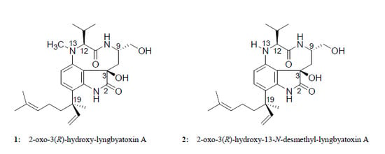
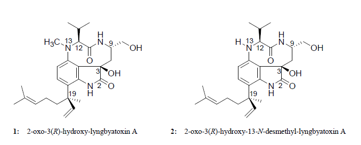
Two New Lyngbyatoxin Derivatives from the Cyanobacterium, Moorea producens

1
Department of Ocean Sciences, Tokyo University of Marine Science and Technology, Tokyo 108-8477, Japan
2
Division of Food Science and Biotechnology, Graduate School of Agriculture, Kyoto University, Kyoto 606-8502, Japan
3
National Research Institute of Fisheries Science, Yokohama 236-8648, Japan
4
Richard L. Roudebush VA Medical Center, Indianapolis, IN 46202, USA
*
Author to whom correspondence should be addressed.
These authors contributed equally to this work.
Mar. Drugs 2014, 12(12), 5788-5800; https://doi.org/10.3390/md12125788
Submission received: 10 October 2014 / Revised: 11 November 2014 / Accepted: 19 November 2014 / Published: 1 December 2014

Abstract

:
The toxin-producing cyanobacterium, Moorea producens, is a known causative organism of food poisoning and seaweed dermatitis (also known as “swimmer’s itch”). Two new toxic compounds were isolated and structurally elucidated from an ethyl acetate extract of M. producens collected from Hawaii. Analyses of HR-ESI-MS and NMR spectroscopies, as well as optical rotations and CD spectra indicated two new lyngbyatoxin derivatives, 2-oxo-3(R)-hydroxy-lyngbyatoxin A (1) and 2-oxo-3(R)-hydroxy-13-N-desmethyl-lyngbyatoxin A (2). The cytotoxicity and lethal activities of 1 and 2 were approximately 10- to 150-times less potent than lyngbyatoxin A. Additionally, the binding activities of 1 and 2 possessed 10,000-times lower affinity for the protein kinase Cδ (PKCδ)-C1B peptide when compared to lyngbyatoxin A. These findings suggest that these new lyngbyatoxin derivatives may mediate their acute toxicities through a non-PKC activation pathway.

Graphical Abstract

1. Introduction

Cyanobacteria, often called “blue-green algae”, have a long evolutionary and biochemical history [1]. Some cyanobacteria have been found to cause hepatic, neurologic and dermal system damage due to the “cyanotoxins” they produce [2]. One of the most toxic genera of filamentous marine cyanobacteria, Moorea producens (formerly classified as Lyngbya majuscula) [3], is a rich source of diverse compounds that possess a variety of biological activities [4,5,6]. Aplysiatoxin, debromoaplysiatoxin and lyngbyatoxin A are produced by M. producens and have been reported to be the causative agents of seaweed dermatitis (also known as “swimmer’s itch”) [7,8,9]. The first reported case of seaweed dermatitis was from Hawaii in 1958. On the island of Oahu, Hawaii, more than 125 people were reported to have dermatitis symptoms secondary to exposure to M. producens [10,11]. To date, numerous incidences of seaweed dermatitis (skin, eye and respiratory irritation) have been reported from the Pacific region [12]. Additionally, aplysiatoxin, debromoaplysiatoxin and lyngbyatoxin A are known compounds that can cause food poisoning. Symptoms of diarrhea, vomiting and skin ulcers from the consumption of meat from the marine turtle, Chelonia mydas, have been reported [13,14]. Lyngbyatoxin A was identified to be the cause of this intoxication [15]. In Hawaii, the ingestion of the red alga, Gracilaria coronopifolia, was reported to induce symptoms of food poisoning [16,17,18]. The characteristic symptoms of this intoxication were vomiting, diarrhea and a burning sensation of the mouth and throat [16]. Aplysiatoxin and its related compounds were identified as the causative agents in these toxic events [17,19,20,21]. The cyanobacteria-like organism, G. coronopifolia, was reported to be the true origin of these toxins [17]. Toxins from M. producens are known to possess potent tumor-promoting activity, which can induce tumor formation [22,23]. Lyngbyatoxin A is a known potent tumor promoter by its ability to strongly induce protein kinase C (PKC) activity [22,24].
Figure 1. Structures of 1 and 2.
Marinedrugs 12 05788 g001
1: 2-oxo-3(R)-hydroxy-lyngbyatoxin A, R: Me; 2: 2-oxo-3(R)-hydroxy-13-N-desmethyl-lyngbyatoxin A, R: H.
The isolation, structural elucidation and biological activities of two novel lyngbyatoxin derivatives, 2-oxo-3(R)-hydroxy-lyngbyatoxin A (1, Figure 1) and 2-oxo-3(R)-hydroxy-13-N-desmethyl-lyngbyatoxin A (2, Figure 1), from M. producens are presented.

2. Results and Discussion

2.1. Isolation of 1 and 2

After extraction of the freeze-dried sample, the crude extracts were evaporated to a dried residue. Liquid-liquid partition was then performed on the residue. The EtOAc fraction possessed the most potent activity determined by cytotoxic and crustacean lethal assays. This fraction was subjected to an open glass column for ODS chromatography and fractionated with various methanol solutions (50%, 70%, 90% and 100% methanol). The seventy percent methanol fraction was further purified using a reversed phase HPLC and recycling ODS HPLC system. Two pale yellow solid compounds, 1 (1.37 mg, 0.09%) and 2 (1.96 mg, 0.13%), were isolated, and the measurement of proton NMR spectra further certified the purity of the two newly-isolated compounds.

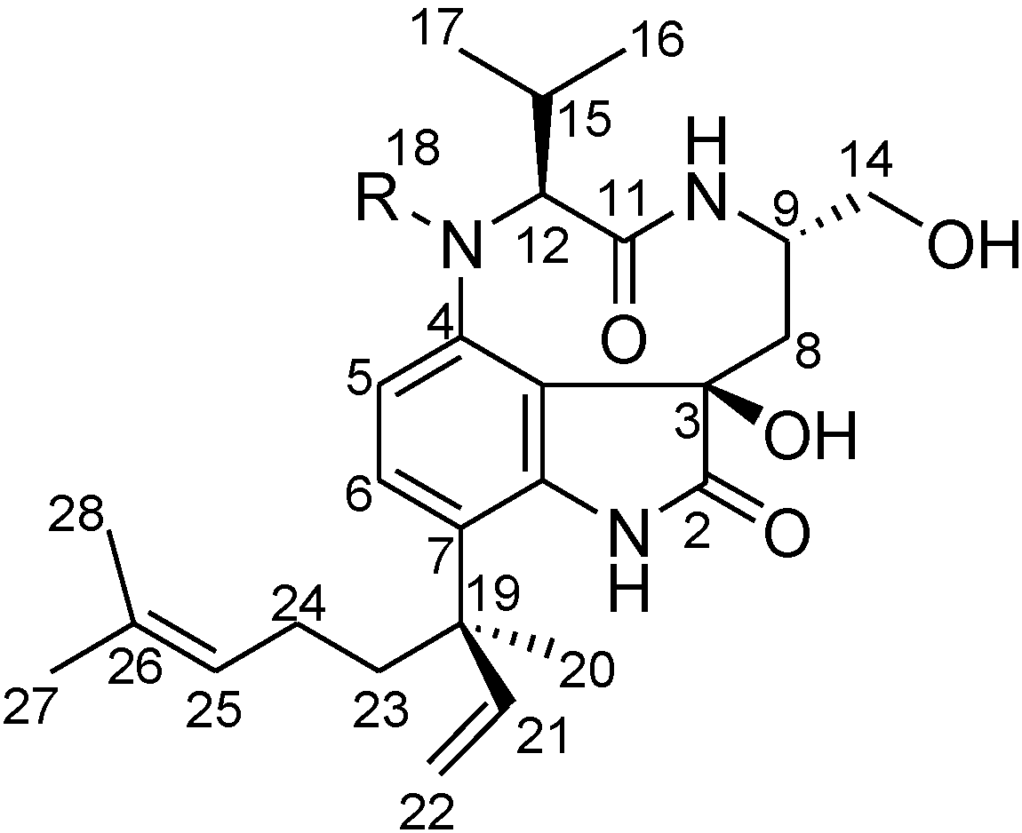
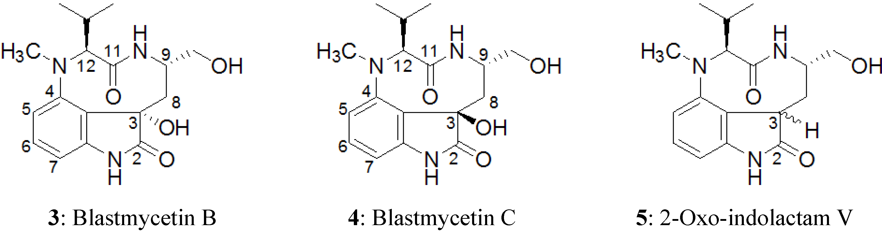
2.2. Elucidation of the Planar Structures of 1 and 2

Compound 1 possessed a molecular formula of C27H39N3O4 suggested by HR-ESI-MS according to the [M + H]+ ion peak at m/z 470.3051 (calcd. for C27H40N3O4, 470.3019) and the [M + Na]+ ion peak at m/z 492.2873 (calcd. for C27H39N3O4Na, 492.2838). A molecular formula of C26H37N3O4 was suggested for 2 based on the mass spectrum at m/z 456.2901 [M + H]+ (calcd. for C26H38N3O4, 456.2862), which had one less methyl or methylene moiety when compared to 1. The UV spectral data of 1 and 2 suggested the existence of a conjugated ring system (1, UV λmax (EtOH) nm (log ε), 239 (4.19), 284 (3.71); 2, UV λmax (EtOH) nm (log ε), 240 (4.54), 287 (3.73)).
The structure of 1 was predominantly determined by 1D and 2D NMR spectral analyses. The tabulated 13C and 1H NMR spectral data for 1 are shown in Table 1. The 1H NMR spectrum of 1 resembled lyngbyatoxin A. Comparing the 1H NMR spectrum of 1 in methanol-d4 (CD3OD) with that of lyngbyatoxin A [8], all of the proton signals in lyngbyatoxin A were observed, except for the signal corresponding to H-2. While lyngbyatoxin A shows an olefinic carbon at C-2 (δC 120.7) and C-3 (δC 114.1) from the 13C NMR spectral data of lyngbyatoxin A, the 13C NMR spectral data of 1 revealed a carbonyl carbon at C-2 (δC 182.1) and a quaternary carbon at C-3 (δC 76.0), indicating the existence of an oxindole moiety in 1. This was also supported by the UV spectra [25]. The correlations observed in the 1H-1H COSY and 1H–13C HMBC spectra of 1 are shown in Figure 2. From these results, 1 was inferred to be 2-oxo-3-hydroxy-lyngbyatoxin A (Figure 2, left). Furthermore, 2 lacked a C-18 N-methyl group from the data analysis of the 1D and 2D NMR spectra, indicating that 2 was 2-oxo-3-hydroxy-13-N-desmethyl-lyngbyatoxin A. The deduced planar structures were also supported by the close similarity of the 1H NMR spectral data of 2-oxo-teleocidin A-1 [26].
Table 1. 1H and 13C NMR chemical shifts observed for 1 and 2 in CD3OD.
No.12
13C1H (mult, J in Hz)13C1H (mult, J in Hz)
2182.1181.5
376.075.9
3a128.6122.5
4151.0145.6
5120.07.15, 1H (d, 8.6)116.96.69, 1H (d, 8.6)
6130.27.29, 1H (d, 8.6)129.87.15, 1H (d, 8.6)
7128.3123.7
7a139.7138.8
842.42.24, 1H (d, 15.1)42.92.15, 1H (m)
1.46, 1H (dd, 9.0, 15.1)1.50, 1H (dd, 9.4, 14.7)
951.15.63, 1H (m)51.64.49, 1H (m)
11175.1 176.4
1278.13.81, 1H (d, 5.5)74.33.78, 1H (d, 10.5)
1467.53.60, 1H (dd, 4.7, 10.9)67.13.54, 1H (dd, 5.0, 11.1)
3.54, 1H (dd, 5.6, 10.9)3.49, 1H (dd, 7.2, 11.1)
1530.02.43, 1H (m)32.62.15, 1H (m)
1622.71.22, 3H (d, 6.9)21.31.29, 3H (d, 6.5)
1719.41.20, 3H (d, 6.9)20.61.13, 3H (d, 6.5)
1842.32.67, 3H (s)
1944.644.4
2025.41.41, 3H (s)25.61.38, 3H (s)
21146.86.09, 1H (dd, 10.7, 17.6)147.26.08, 1H (dd, 10.7, 17.6)
22114.05.22, 1H (d, 10.7)113.65.19, 1H (d, 10.7)
5.06, 1H (d, 17.7)5.04, 1H (d, 17.7)
2340.21.86, 1H (m)40.51.83, 1H (m)
1.76, 1H (m)1.72, 1H (m)
2424.21.97, 1H (m)24.31.94, 1H (m)
1.76, 1H (m)1.74, 1H (m)
25125.35.10, 1H (t, 5.8)125.45.11, 1H (m)
26132.5132.4
2725.81.68, 3H (s)25.81.67, 3H (s)
2817.71.55, 3H (s)17.71.55, 3H (s)
s, singlet; d, doublet; t, triplet; m, multiplet.
Figure 2. The structure of 1 and its key 2D NMR correlations in CD3OD.
Marinedrugs 12 05788 g002

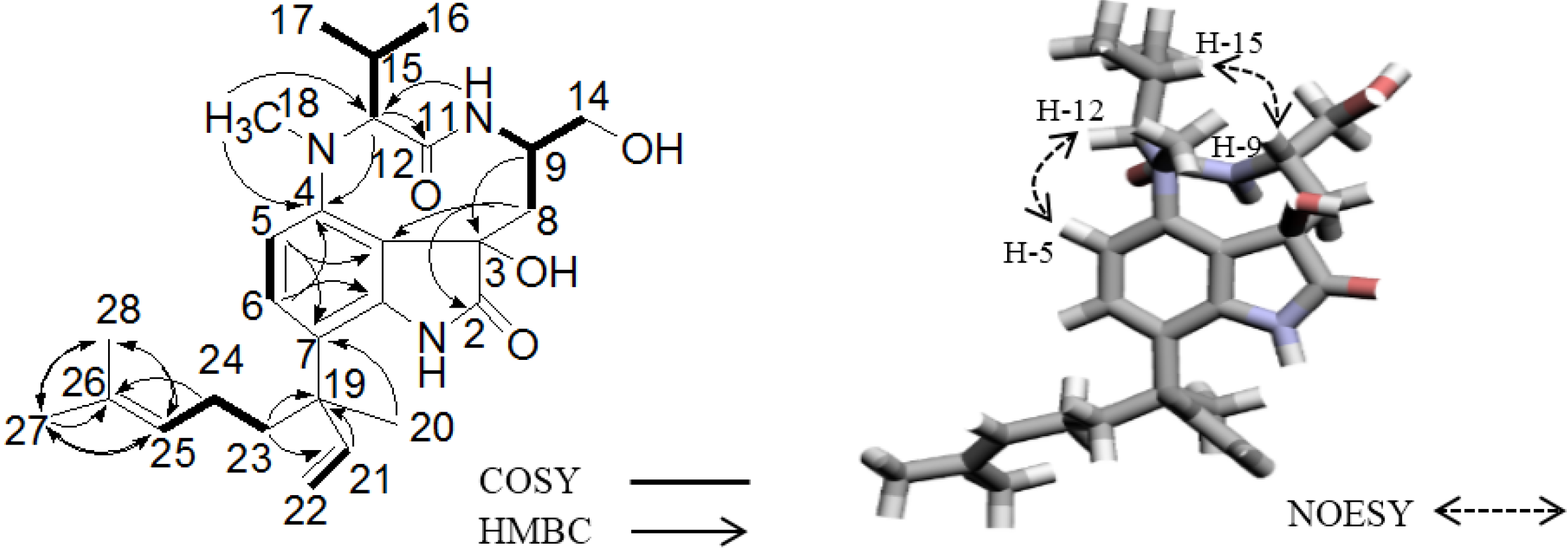
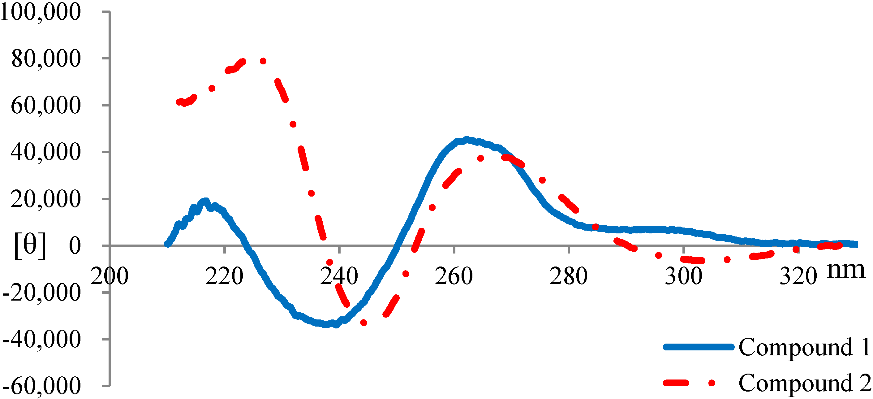
2.3. Elucidation of Absolute Stereochemistry of 1 and 2

The NOESY correlations (H-5/H-12 and H-9/H-15) of 1 indicated that the N-10/C-11 amide bond was cis by molecular model-building. One dominant conformer (cis amide conformer) was observed in the 1H NMR spectrum of 1, similar to lyngbyatoxin A. Therefore, the relative stereochemistry at C-9 and C-12 was suggested to be 9S*, 12S* (Figure 2, right). It was also deduced that 2 had 9S*, 12S* relative stereochemistry from the results of NOESY correlations. Blastmycetins B (3, Figure 3) and C (4, Figure 3) were previously isolated from the culture broth of the actinomycete, Streptoverticillium blastmyceticum [27]. The planar structures of 3 and 4 were a partial structure of 1. Furthermore, 3 and 4 had a 9S, 12S configuration. The only structural difference between 3 and 4 was the configuration at the C-3 hydroxyl group. Therefore, 3 and 4 could be the model compounds for the determination of the configuration on C-3 of 1 and 2. The reported proton chemical shifts were 2.05, 2.17 (H-8), 3.95 (H-9) and 5.96 (H-12) for 3 and 1.46, 2.22 (H-8), 5.61 (H-9) and 3.76 (H-12) for 4 [25]. The 1H chemical shifts obtained in this study were 1.46, 2.24 (H-8), 5.63 (H-9) and 3.81 (H-12) for 1 and 1.50, 2.15 (H-8), 4.49 (H-9) and 3.78 (H-12) for 2 (Table 1). The results indicated that both 1 and 2 had a 3R*, 9S*, 12S* relative stereochemistry. However, the downfield shift of H-9 (δH 4.49) of 2 was not so remarkable compared to the H-9 (δH 5.63) of 1. This might be caused by the lack of a C-18 N-methyl group in 2, leading to the conformational change of 2. To further analyze the configuration of 2, NMR spectra of 2 were measured in chloroform-d (see Supplementary Information). The 1H NMR spectra showed protons of two hydroxyl groups at C-3 and C-14 with chemical shifts of δH 4.36 and δH 3.37, respectively. Moreover, the NOESY spectra showed NOE correlations between H-9/H-13, H-14 and H-15, OH-3/H-13 and OH-14, H-8 (δ2.13, d)/H-14' and H-8' (δ1.59, dd)/H-10. These observations finally confirmed that 2 had the relative stereochemistry of 3R*, 9S*, 12S*.
Figure 3. Structures of blastmycetin B (3), blastmycetin C (4) and 2-oxo-indolactam V (5).
Marinedrugs 12 05788 g003
Furthermore, compound 3 showed levorotatory optical rotation, while 4 showed dextrorotatory optical rotation [25]. Both 1 and 2 showed dextrorotatory optical rotation (1, [ α ] D 24 +119.2° (c 0.18, CH3OH); 2, [ α ] D 24 +150.5° (c 0.30, CH3OH)), which was the same as that of 4. Therefore, the absolute configurations of both 1 and 2 were deduced to be 3R, 9S, 12S.
The inference in the absolute configurations on C-19 of the linalyl group in lyngbyatoxin A derivatives was elucidated by the analysis of the circular dichroism (CD) spectra at around 230 nm. The upward and downward curves around 230 nm indicated the absolute configurations to be 19-R and 19-S, respectively [28,29,30]. The CD spectra of 1 and 2 are shown in Figure 4. Both compounds showed upward curves around 230 nm, indicating that 1 and 2 had the same absolute configuration R at C-19. Therefore, it was deduced that both 1 and 2 had 3R, 9S, 12S, 19R absolute configurations. Consequently, 1 was 2-oxo-3(R)-hydroxy-lyngbyatoxin A and 2 was 2-oxo-3(R)-hydroxy-13-N-desmethyl-lyngbyatoxin A (Figure 1).
Figure 4. CD spectra of 1 and 2.
Marinedrugs 12 05788 g004
While compounds containing 2-oxo-indolactam V (5, Figure 3) have been isolated from actinomycetes [25,26], this is the first report describing 2-oxo derivatives of lyngbyatoxin A from the cyanobacterium, M. producens.
The LC-MS/MS analyses of the 70% methanol fraction were carried out to detect the existence of isomers of 1 and 2. The existence of another compound, which had the same molecular weight and MS/MS pattern with 1, was detected. Since 3 and 4 had been isolated from the same actinomycete species [27], this new unidentified compound might be 2-oxo-3(S)-hydroxy-lyngbyatoxin A. Unfortunately, we were unable to isolate this compound. While an isomer of 2 could not be detected by LC-MS/MS, 2-oxo-3(S)-hydroxy-13-N-desmethyl-lyngbyatoxin A may not have been present in the sample.

2.4. Biological Activity Tests

The effects of two lyngbyatoxin A derivatives, 1 and 2, on the inhibition of cell proliferation were determined using L1210 leukemia cells. The IC50 values determined for 1 and 2 were 98 and 321 μM, respectively. Recent reports indicated that the IC50 value of lyngbyatoxin A against L1210 cells was 8.1 μM [30]. The cytotoxic activities of 1 and 2 were approximately 10- to 40-times less potent than lyngbyatoxin A.
Crustacean lethal activity tests were performed using the shrimp, Palaemon paucidens. Injection of 1 and 2 caused paralysis of the tested shrimp in the first 30 min of exposure. The lethal dose 33% values (LD33) were used to estimate the lethal toxicity. While the LD33 values of 1 and 2 were 89 and 25 mg/kg, respectively, the LD33 value of lyngbyatoxin A was 0.6 mg/kg. The lethal activities of 1 and 2 were approximately 150- and 40-times less potent than lyngbyatoxin A, respectively.
Lyngbyatoxin A is a potent carcinogen by acting as a powerful activator of all conventional and novel PKC isozymes (PKCs) secondary to its direct binding of the C1 domain [22,23,31,32]. Since PKCδ is an important PKCs involved in carcinogenesis [33], the binding affinities of PKCδ were tested using a synthetic PKCδ-C1B peptide by a competitive binding assay with [3H]phorbol 12,13-dibutyrate (PDBu) [34]. The Ki value for the inhibition of [3H]PDBu-binding with lyngbyatoxin A is 0.11 nM [30]. In this study, it was shown that 1 and 2 had Ki values of 1400 and 940 nM, respectively.
The cytotoxicity and lethal activities of 1 and 2 were approximately 10- to 150-times less potent than lyngbyatoxin A. Additionally, the binding activities of 1 and 2 possessed 10,000-times lower affinity for the protein kinase Cδ (PKCδ)-C1B peptide when compared to lyngbyatoxin A. These results might be simply caused by the difference in bioavailability or localization of these new compounds and lyngbyatoxin A in the different assays. However, in the previous biological activity tests, 12-epi-lyngbyatoxin A and lyngbyatoxin A showed comparable acute toxicities, but 12-epi-lyngbyatoxin A had more than 100-times lower binding affinity for PKCδ compared to lyngbyatoxin A [30]. Therefore, we had proposed the hypothesis that the acute toxicities (cytotoxicity and lethality) of lyngbyatoxin-type compounds might be mediated through a non-PKC activation pathway [30]. In this study, acute toxicities (cytotoxicity and lethal activities) versus PKCδ binding activities of 1 and 2 showed big difference with those of lyngbyatoxin A. Thus, we considered that the results obtained in this study probably would also support the hypothesis we proposed and an alternate target instead of PKC isozymes might exist for the acute toxicities of these toxins.
Therefore, further studies are necessary to verify our proposed hypothesis, as well as to elucidate whether there exist other receptors in relation to the acute toxicities of lyngbyatoxin-type compounds.
M. producens producing lyngbyatoxin A, aplysiatoxin and their related compounds have been reported to be the causative agents of swimmer’s itch and food poisoning [8,14,15]. New Compounds 1 and 2 showed acute toxicity, albeit weak; thus, we might pay attention to these compounds also on health hazard cases involving M. producens.

3. Experimental Section

3.1. Instrumentation and General Methods

Optical rotations were measured on a JASCO P-2100 (JASCO Co., Tokyo, Japan). Circular dichroism (CD) spectra were measured on a JASCO J-715 (JASCO Co., Tokyo, Japan). UV spectra were measured on a HITACHI U-3000 (Hitachi High-Tech Fielding Co., Tokyo, Japan). NMR spectra were recorded on Bruker AVANCE III 800 MHz and 600 MHz spectrometers (Bruker Co., Bremen, Germany). HR-ESI-MS spectra data were recorded on a Bruker micrOTOF QII (Bruker Co., Bremen, Germany) mass spectrometer. LC-MS/MS analyses were performed using an Ultimate 3000 liquid chromatograph (Thermo Dionex, Sunnyvale, CA, USA), which was coupled to a Bruker micrOTOF QII (Bruker Co., Bremen, Germany) mass spectrometer. HPLC analyses were performed using a SHIMADZU HPLC system (SHIMADZU Co., Kyoto, Japan) equipped with a SPD-M10A diode array detector (SHIMADZU Co., Kyoto, Japan).

3.2. Sample Collection

The marine cyanobacterium, M. producens, was collected at Kahala Beach (Oahu, Hawaii) in 1998. The samples were freeze-dried immediately after the collection and stored in a freezer at −30 °C.

3.3. Extraction and Purification

A crude extract from the freeze-dried sample (823 g, dry weight) was prepared with organic solvents in sequence: ethanol, methanol and acetone. The three extracts were combined and evaporated. The residue (128 g, dry weight) was dissolved in 80% methanol and partitioned between hexane. After the 80% methanol layer was evaporated, the dried residue was re-suspended in distilled water and extracted with ethyl acetate (EtOAc). The EtOAc fraction (4.2 g, dry weight) was then separated by a 20 × 300 mm open glass column filled with ODS resin (Pegasil Prep ODS-7515-12A, Senshu Co., Tokyo, Japan) incorporating a stepwise increase in the methanol concentration (50%, 70%, 90% and 100% methanol). The various methanol solutions yielded the following: 50% methanol fraction (870 mg), 70% methanol fraction (1600 mg), 90% methanol fraction (1220 mg) and 100% methanol fraction (200 mg). From the 90% methanol fraction, we have previously isolated lyngbyatoxin A and 12-epi-lyngbyatoxin A [30]. In this study, reversed-phase high pressure liquid chromatography (HPLC) was utilized to further purify the 70% methanol fraction (1540 mg). The 70% methanol fraction was divided into 24 portions, and the HPLC was performed on each portion on a 20 × 250 mm column (SHISEIDO CAPCELL PAK C18, Shiseido Co., Tokyo, Japan) using a gradient elution system with the following conditions: a linear gradient from 75% to 100% methanol during the first 90 min, a flow rate of 3 mL/min and UV-Vis detection at 210, 230 and 270 nm. A recycle chromatography HPLC (system unit: Senshu Scientific SSC-1310, column: COSMOSIL C18-AR-II 10 × 250 mm, solvent: 73% MeOH for 1 and 72% MeOH for 2, flow rate: 2 mL/min, detection: 210 nm) was performed for the isolation of 1 and 2.

3.4. HR-ESI-MS and LC-MS/MS Analyses

The Bruker micrOTOF QII mass spectrometer was operated in both positive and negative ion modes. To detect the isomers of two newly-isolated compounds for LC-MS/MS analyses, a 2.0 × 250 mm column (Mightysil RP-18 GP, Kanto Chemical Co., Tokyo, Japan) was utilized. Eluent A was 0.1% formic acid in water and B was methanol-water (95:5), containing 0.1% formic acid. The mobile phase (25% A and 75% B) was isocratic with a flow rate of 0.2 mL/min.

3.5. Nuclear Magnetic Resonance Experiments

NMR assignment was achieved from 1D and 2D spectroscopies (1H, 13C, COSY, HSQC, HMBC and NOESY). Chemical shifts were reported in δ units (ppm) relative to CD3OD (δH at 4.87 ppm and δC at 49.0 ppm) and CDCl3H at 7.26 ppm and δC at 77.2 ppm) solvents at 300 K.

3.6. Circular Dichroism (CD) Spectroscopy

The CD spectral data were recorded in methanol with the concentration of 213 μmol/L for 1 and 220 μmol/L for 2, using a 2-mm path-length quartz cell. The measurements were performed at room temperature (25 °C). The spectra were measured from 380 nm to 195 nm, and a minimum of six scans was performed for each compound.

3.7. Cytotoxicity Assay

L1210 mouse leukemia cells were used for the cytotoxicity assay of 1 and 2, as previously described [35]. Five microliters of each sample or methanol alone were placed into individual wells of a 96-well microtiter plate. The solvent was removed by air drying from the samples at room temperature on a clean bench. Five microliters of sterilized water were then applied to each well. After the samples were dissolved, 100 μL of the L1210 mouse leukemia cell solution (3 × 10⁴ cells/mL) were plated into individual wells of the 96-well microtiter plate. The assay plate was placed in an incubator for 24 h at 37 °C with 5% CO2. After heating the 2,3-bis-(2-methoxy-4-nitro-5-sulfophenyl)-2H-tetrazolium-5-carboxanilide inner sodium salt (XTT, Sigma, St. Louis, MO, USA) working solution to 37 °C, the solution was gently swirled until a clear solution was obtained. Fifty microliters of the XTT working solution were added into each well and then incubated for 6 hours at 37 °C with 5% CO2. The XTT working solution was prepared as a mixture of 5.5 mL of XTT in RPMI-1640 medium (1 mg/mL) and 170 μL of phenazine methosulfate (Sigma, St. Louis, MO, USA) in RPMI-1640 medium (38 μg/mL). The cells without sample treatment were used as a negative control. The inhibition of cell proliferation was determined by measuring the absorbance of each well via a microplate reader (Model 550, Bio-Rad, Hercules, CA, USA) at a wavelength of 450 nm.

3.8. Crustacean Lethality Test

The crustacean lethality of lyngbyatoxin A, 1 and 2 were tested using the shrimp, Palaemon paucidens, as previously described [30]. The average weight of the shrimp was 0.5 g. A lethal dose 33% value (LD33) was used to estimate the lethality of the compounds.

3.9. Binding Assay of PKC Ligands Using the PKCδ-C1B Peptide

Using previously-described methods, the inhibition of [3H]phorbol 12,13-dibutyrate (PDBu) binding to the protein kinase Cδ (PKCδ)-C1B peptide was evaluated [36,37]. The standard assay mixture contained: 13.8 nM PKCδ-C1B peptide, 20 nM [3H]PDBu (18.7 Ci/mmol, Perkin-Elmer Life Sciences, Waltham, MA), 50 μg/mL 1,2-dioleoyl-sn-glycero-3-phospho-l-serine (Sigma, St. Louis, MO, USA), 3 mg/mL bovine γ-globulin (Sigma, St. Louis, MO, USA) and various concentrations of inhibitors in a 50 mM Tris-maleate buffer (pH 7.4 at 4 °C). Total binding was measured in the absence of an inhibitor (only ethanol), and non-specific binding was evaluated in the absence of the PKCδ-C1B peptide. The concentration of the properly-folded PKCδ-C1B peptide was estimated to be about 3 nM on the basis of the Bmax values from Scatchard analyses previously reported [36,37]. Binding affinity was evaluated as IC50 by the sample concentration required to cause 50% inhibition of the binding of [3H]PDBu to the PKCδ-C1B peptide [38]. The inhibition constant, Ki, was calculated by the method of Sharkey and Blumberg [39].

4. Conclusions

In this study, two new toxic compounds were isolated and structurally elucidated from the Hawaiian cyanobacterium, Moorea producens. From the analyses of HR-ESI-MS and NMR spectroscopies, as well as optical rotations and CD spectra, these new compounds were indicated to be the new lyngbyatoxin derivatives, 2-oxo-3(R)-hydroxy-lyngbyatoxin A (1) and 2-oxo-3(R)-hydroxy-13-N-desmethyl-lyngbyatoxin A (2). The cytotoxicity and lethal activities of 1 and 2 were approximately 10- to 150-times less potent than lyngbyatoxin A. However, the PKCδ binding activities of 1 and 2 possessed 10,000-times lower affinity when compared to lyngbyatoxin A. These findings supported our hypothesis that acute toxicities, such as cytotoxicity, and lethality of these new lyngbyatoxin derivatives might be mediated through a non-PKC activation pathway.

Supplementary Files

Supplementary File 1

Acknowledgments

This work was supported partly by Japan Society for the Promotion of Science (JSPS) Grant-in-Aid for Scientific Research Grant Number 24510294 (to Hiroshi Nagai).

Author Contributions

Hiroshi Nagai conceived of and designed the research. Hiroshi Nagai and Kazuhiro Irie supervised the research. Hiroshi Nagai and Bryan Sakamoto contributed to the sample collection. Weina Jiang and Satoshi Tan purified the compounds. Weina Jiang, Ryuichi Watanabe, Toshiyuki Suzuki, Hajime Uchida and Michiya Kamio performed the spectral measurement. Weina Jiang and Satoshi Tan carried out the structural identification and toxicity assay. Yusuke Hanaki performed the binding assay. Weina Jiang, Kazuhiro Irie and Hiroshi Nagai analyzed the data and wrote the paper. Bryan Sakamoto reviewed and edited the manuscript.

Conflicts of Interest

The authors declare no conflict of interest.

References

  1. Hoffman, L. Marine cyanobacteria in tropical regions: Diversity and ecology. Eur. J. Phycol. 1999, 34, 371–379. [Google Scholar] [CrossRef]
  2. Carmichael, W.W. Cyanobacteria secondary metabolites-the cyanotoxins. J. Appl. Microbiol. 1992, 72, 445–459. [Google Scholar]
  3. Engene, N.; Rottacker, E.C.; Kaštovský, J.; Byrum, T.; Choi, H.; Ellisman, M.H.; Komárek, J.; Gerwick, W.H. Moorea producens gen. nov., sp. nov. and Moorea bouillonii comb. nov., tropical marine cyanobacteria rich in bioactive secondary metabolites. Int. J. Syst. Evol. Microbiol. 2012, 62, 1171–1178. [Google Scholar] [CrossRef] [PubMed]
  4. Nunnery, J.K.; Mevers, E.; Gerwick, W.H. Biologically active secondary metabolites from marine cyanobacteria. Curr. Opin. Biotechnol. 2010, 21, 787–793. [Google Scholar] [CrossRef] [PubMed]
  5. Singh, R.K.; Tiwari, S.P.; Rai, A.K.; Mohapatra, T.M. Cyanobacteria: An emerging source for drug discovery. J. Antibiot. 2011, 64, 401–412. [Google Scholar] [CrossRef] [PubMed]
  6. Tan, L.T. Marine cyanobacteria: A prolific source of bioactive natural products as drug leads. In Marine Microbiology: Bioactive Compounds and Biotechnological Applications, 1st ed.; Kim, S.K., Ed.; Wiley-VCH: Weinheim, Germany, 2013; Volume 5, pp. 59–81. [Google Scholar]
  7. Mynderse, J.S.; Moore, R.E.; Kashiwagi, M.; Norton, T.R. Antileukemia activity in the osillatoriaceae: Isolation of debromoaplysiatoxin from Lyngbya. Science 1977, 196, 538–540. [Google Scholar] [PubMed]
  8. Cardellina, J.H.; Marner, F.J.; Moore, R.E. Seaweed dermatitis: Structure of lyngbyatoxin A. Science 1979, 204, 193–195. [Google Scholar] [CrossRef]
  9. Moore, R.E.; Blackman, A.J.; Cheuk, C.E.; Mynderse, J.S.; Matsumoto, G.K.; Clardy, J.; Woodard, R.W.; Craig, J.C. Absolute stereochemistries of the aplysiatoxins and oscillatoxin A. J. Org. Chem. 1984, 49, 2484–2489. [Google Scholar] [CrossRef]
  10. Banner, A.H. A dermatitis-producing alga in Hawaii. Preliminary report. Hawaii Med. J. 1959, 19, 35–36. [Google Scholar] [PubMed]
  11. Grauer, F.H. Dermatitis escharotica caused by a marine alga. Hawaii Med. J. 1959, 19, 32–34. [Google Scholar] [PubMed]
  12. Osborne, N.J.; Webb, P.M.; Shaw, G.R. The toxins of Lyngbya majuscule and their human and ecological health effects. Environ. Int. 2001, 27, 381–392. [Google Scholar] [CrossRef]
  13. Champetier, R.G.; Ranaivoson, G.; Ravaonindrina, N.; Rakotonjanabelo, A.L.; Rasolofonirina, N.; Roux, J.F.; Yasumoto, T. Un problème de santé publique réémergent à madagascar: Les intoxications collectives par consommation d’animaux marins. Aspects épidémiologiques, cliniques et toxicologiques des épisodes notifiés de janvier 1993 à janvier 1998. Arch. Inst. Pasteur Madag. 1998, 64, 71–76. [Google Scholar]
  14. Ito, E.; Satake, M.; Yasumoto, T. Pathological effects of lyngbyatoxin A upon mice. Toxicon 2002, 40, 551–556. [Google Scholar] [CrossRef] [PubMed]
  15. Yasumoto, T. Fish poisoning due to toxins of microalgal origins in the Pacific. Toxicon 1998, 36, 1515–1518. [Google Scholar] [CrossRef] [PubMed]
  16. Hanne, M.; Matsubayashi, H.; Vogt, R.; Wakida, C.; Hau, S.; Nagai, H.; Hokama, Y.; Solorzano, L. Outbreak of gastrointestinal illness associated with consumption of seaweed-Hawaii, 1994. Morb. Mortal. Wkly. Rep. 1995, 44, 724–727. [Google Scholar]
  17. Nagai, H.; Yasumoto, T.; Hokama, Y. Aplysiatoxin and debromoaplysiatoxin as the causative agents of a red alga Gracilaria coronopifolia poisoning in Hawaii. Toxicon 1996, 34, 753–761. [Google Scholar] [PubMed]
  18. Marshall, K.L.E.; Vogt, R.L. Illness associated with eating seaweed, Hawaii, 1994. West. J. Med. 1998, 169, 293–295. [Google Scholar] [PubMed]
  19. Nagai, H.; Yasumoto, T.; Hokama, Y. Manauealides, some of the causative agents of a red alga Gracilaria coronopifolia poisoning in Hawaii. J. Nat. Prod. 1997, 60, 925–928. [Google Scholar] [CrossRef] [PubMed]
  20. Nagai, H.; Kan, Y.; Fujita, T.; Sakamoto, B.; Hokama, Y. Manuauealide C and anhydrodebromoaplysiatoxin, toxic constituents of the Hawaiian red alga, Gracilaria coronopifolia. Biosci. Biotech. Biochem. 1998, 62, 1011–1013. [Google Scholar] [CrossRef]
  21. Ito, E.; Nagai, H. Morphological observations of diarrhea in mice caused by aplysiatoxin, the causative agent of the red alga Gracilaria coronopifolia poisoning in Hawaii. Toxicon 1998, 36, 1913–1920. [Google Scholar] [CrossRef] [PubMed]
  22. Fujiki, H.; Mori, M.; Nakayasu, M.; Terada, M.; Sugimura, T.; Moore, R.E. Indole alkaloids: Dihydroteleocidin B, teleocidin, and lyngbyatoxin A as members of a new class of tumor promoters. Proc. Natl. Acad. Sci. USA 1981, 78, 3872–3876. [Google Scholar] [CrossRef] [PubMed]
  23. Fujiki, H.; Tanaka, Y.; Miyake, R.; Kikkawa, U.; Nishizuka, Y.; Sugimura, T. Activation of calcium-activated, phospholipid-dependent protein kinase (protein kinase C) by new classes of tumor promoters: Teleocidin and debromoaplysiatoxin. Biochem. Biophys. Res. Commun. 1984, 120, 339–343. [Google Scholar] [CrossRef] [PubMed]
  24. Fujiki, H.; Sugimura, T. New classes of tumor promoters: Teleocidin, aplysiatoxin, and palytoxin. Adv. Cancer Res. 1987, 49, 223–264. [Google Scholar] [PubMed]
  25. Hagiwara, N.; Irie, K.; Funaki, A.; Hayashi, H.; Arai, M.; Koshimizu, K. Structure and tumor-promoting activity of new teleocidin-related metabolites (blastmycetins) from Streptoverticillium blastmyceticum. Agric. Biol. Chem. 1988, 52, 641–648. [Google Scholar] [CrossRef]
  26. Izumikawa, M.; Khan, S.T.; Komaki, H.; Takagi, M.; Shin-ya, K. JBIR-31, a new teleocidin analog, produced by salt-requiring Streptomyces sp. NBRC 105896 isolated from a marine sponge. J. Antibiot. 2010, 63, 33–36. [Google Scholar] [CrossRef] [PubMed]
  27. Irie, K.; Hagiwara, N.; Kurome, T.; Hayashi, H.; Arai, M.; Koshimizu, K. New teleochidin-related mtabolites from Streptoverticillium blastmyceticum producing tumor-promoting indole alkaloids. Agric. Biol. Chem. 1987, 51, 285–287. [Google Scholar] [CrossRef]
  28. Aimi, N.; Odaka, H.; Sakai, S.I.; Fujiki, H.; Suganuma, M.; Moore, R.E.; Patterson, G.M.L. Lyngbyatoxins B and C, two new irritants from Lyngbya majuscula. J. Nat. Prod. 1990, 53, 1593–1596. [Google Scholar] [CrossRef] [PubMed]
  29. Muratake, H.; Okabe, K.; Natsume, M. Synthesis of teleocidins A, B and their congeners. Part 2. Synthesis of lyngbyatoxin A (teleocidin A-1), teleocidin A-2, pendolmycin, and (R,E)-and (S,E)-7-(3,7,11-trimethyl-1,6,10-dodecatrien-3-yl)-(−)-indolactams V. Tetrahedron 1991, 47, 8545–8558. [Google Scholar]
  30. Jiang, W.; Zhou, W.; Uchida, H.; Kikumori, M.; Irie, K.; Watanabe, R.; Suzuki, T.; Sakamoto, B.; Kamio, M.; Nagai, H. A new lyngbyatoxin from the Hawaiian cyanobacterium Moorea producens. Mar. Drugs 2014, 12, 2748–2759. [Google Scholar] [CrossRef] [PubMed]
  31. Moore, R.E.; Patterson, G.M.; Entzeroth, M.; Morimoto, H.; Suganuma, M.; Hakii, H.; Fujiki, H.; Sugimura, T. Binding studies of [3H] lyngbyatoxin A and [3H] debromoaplysiatoxin to the phorbol ester receptor in a mouse epidermal particulate fraction. Carcinogenesis 1986, 7, 641–644. [Google Scholar] [PubMed]
  32. Ono, Y.; Fujii, T.; Igarashi, K.; Kuno, T.; Tanaka, C.; Kikkawa, U.; Nishizuka, Y. Phorbol ester binding to protein kinase C requires a cysteine-rich zinc-finger-like sequence. Proc. Natl. Acad. Sci. USA 1989, 86, 4868–4871. [Google Scholar] [CrossRef] [PubMed]
  33. Szállási, Z.; Denning, M.F.; Smith, C.B.; Dlugosz, A.A.; Yuspa, S.H.; Pettit, G.R.; Blumberg, P.M. Bryostatin 1 protects protein kinase C-delta from down-regulation in mouse keratinocytes in parallel with its inhibition of phorbol ester-induced differentiation. Mol. Pharmacol. 1994, 46, 840–850. [Google Scholar] [PubMed]
  34. Irie, K.; Nakahara, A.; Nakagawa, Y.; Ohigashi, H.; Shindo, M.; Fukuda, H.; Konishi, H.; Kikkawa, U.; Kashiwagi, K.; Saito, N. Establishment of a binding assay for protein kinase C isozymes using synthetic C1 peptides and development of new medicinal leads with protein kinase C isozyme and C1 domain selectivity. Pharmacol. Ther. 2002, 93, 271–281. [Google Scholar] [CrossRef] [PubMed]
  35. Kawabata, T.; Lindsay, D.J.; Kitamura, M.; Konishi, S.; Nishikawa, J.; Nishida, S.; Kamio, M.; Nagai, H. Evaluation of the bioactivities of water-soluble extracts from twelve deep-sea jellyfish species. Fish Sci. 2013, 79, 487–494. [Google Scholar] [CrossRef]
  36. Irie, K.; Oie, K.; Nakahara, A.; Yanai, Y.; Ohigashi, H.; Wender, P.A.; Fukuda, H.; Konishi, H.; Kikkawa, U. Molecular basis for protein kinase C isozyme-selective binding: The synthesis, folding, and phorbol ester binding of the cysteine-rich domains of all protein kinase C isozymes. J. Am. Chem. Soc. 1998, 120, 9159–9167. [Google Scholar] [CrossRef]
  37. Shindo, M.; Irie, K.; Nakahara, A.; Ohigashi, H.; Konishi, H.; Kikkawa, U.; Fukuda, H.; Wender, P.A. Toward the identification of selective modulators of protein kinase C (PKC) isozymes: Establishment of a binding assay for PKC isozymes using synthetic C1 peptide receptors and identification of the critical residues involved in the phorbolester binding. Bioorg. Med. Chem. 2001, 9, 2073–2081. [Google Scholar] [CrossRef] [PubMed]
  38. Irie, K.; Isaka, T.; Iwata, Y.; Yanai, Y.; Nakamura, Y.; Koizumi, F.; Ohigashi, H.; Wender, P.A.; Satomi, Y.; Nishino, H. Synthesis and biological activities of new conformationally restricted analogues of (−)-indolactam-V: Elucidation of the biologically active conformation of the tumor-promoting teleocidins. J. Am. Chem. Soc. 1996, 118, 10733–10743. [Google Scholar] [CrossRef]
  39. Sharkey, N.A.; Blumberg, P.M. Highly lipophilic phorbolesters as inhibitors of specific [3H]phorbol 12,13-dibutyrate binding. Cancer Res. 1985, 45, 19–24. [Google Scholar] [PubMed]

