Article

Sinulolides A–H, New Cyclopentenone and Butenolide Derivatives from Soft Coral Sinularia sp.

1
Key Laboratory of Tropical Marine Bio-resources and Ecology, Guangdong Key Laboratory of Marine Materia Medica, Research Center for Marine Microbes, South China Sea Institute of Oceanology, Chinese Academy of Sciences, Guangzhou 510301, China
2
Key Laboratory of Plant Resources Conservation and Sustainable Utilization, South China Botanical Garden, Chinese Academy of Sciences, Guangzhou 510650, China
3
Zhongshan Ophthalmic Center, Sun Yat-Sen University, Guangzhou 510060, China
4
Jilin Provincial Academy of Chinese Medicine Sciences, Changchun 130021, China
*
Author to whom correspondence should be addressed.
Mar. Drugs 2014, 12(10), 5316-5327; https://doi.org/10.3390/md12105316
Submission received: 6 August 2014 / Revised: 7 October 2014 / Accepted: 8 October 2014 / Published: 23 October 2014

Abstract

:
Eight new compounds, sinulolides A–H (18), along with two known compounds, α-methoxy-2,3-dimethyl-butenolide (9) and sinularone D (10), were isolated from the soft coral Sinularia sp. The structures of these compounds were elucidated on the basis of extensive spectroscopic analysis. The absolute configurations were determined on the basis of electronic circular dichroism (ECD) data analysis. Compounds 5 and 10 exhibited moderate effects for the inhibition of NF-κB activation.

1. Introduction

Genus Sinularia is a soft coral belonging to the phylum, Cnidaria, class Alcyonaria and family Alcyoniidae. It constitutes a dominant portion of the biomass in the tropical reef environment [1]. Many bioactive metabolites, including sesquiterpenes [2,3,4], diterpenes [5,6,7,8,9] and polyhydroxylated steroids [10,11,12], have been studied, and the isolated components display a range of biological activities, such as antimicrobial, anti-inflammatory, glucose transport in rat adipocytes and cytotoxic activities [13,14,15,16,17]. During the course of our investigation on the bioactive chemical constituents from the soft coral, eight new compounds, sinulolides A–H (18), along with two known compounds, α-methoxy-2,3-dimethyl-butenolide (9) and sinularone D (10) (Figure 1), were isolated from Sinularia sp., collected off the Dongluo Island, Sanya, in July, 2009, at a depth of 10 m. The bioactivities of these compounds were determined through bioactivity tests using high-throughput screening (HTS). We describe herein the isolation, structure elucidation and bioactivities of these compounds.
Figure 1. Structures of metabolites 110.
Marinedrugs 12 05316 g001

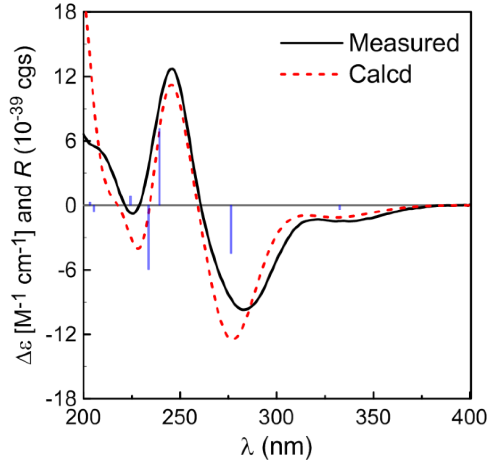
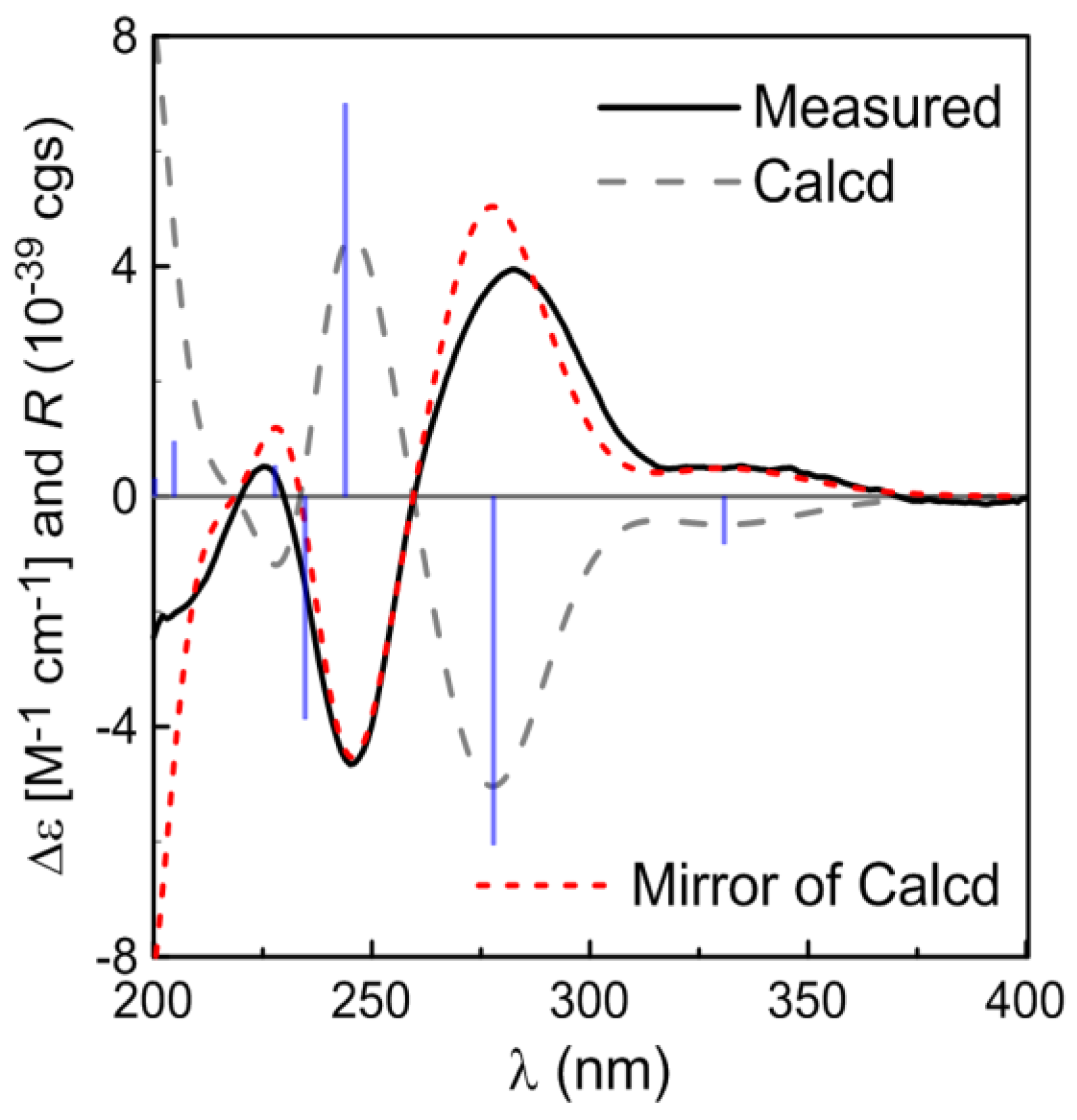
2. Results and Discussion
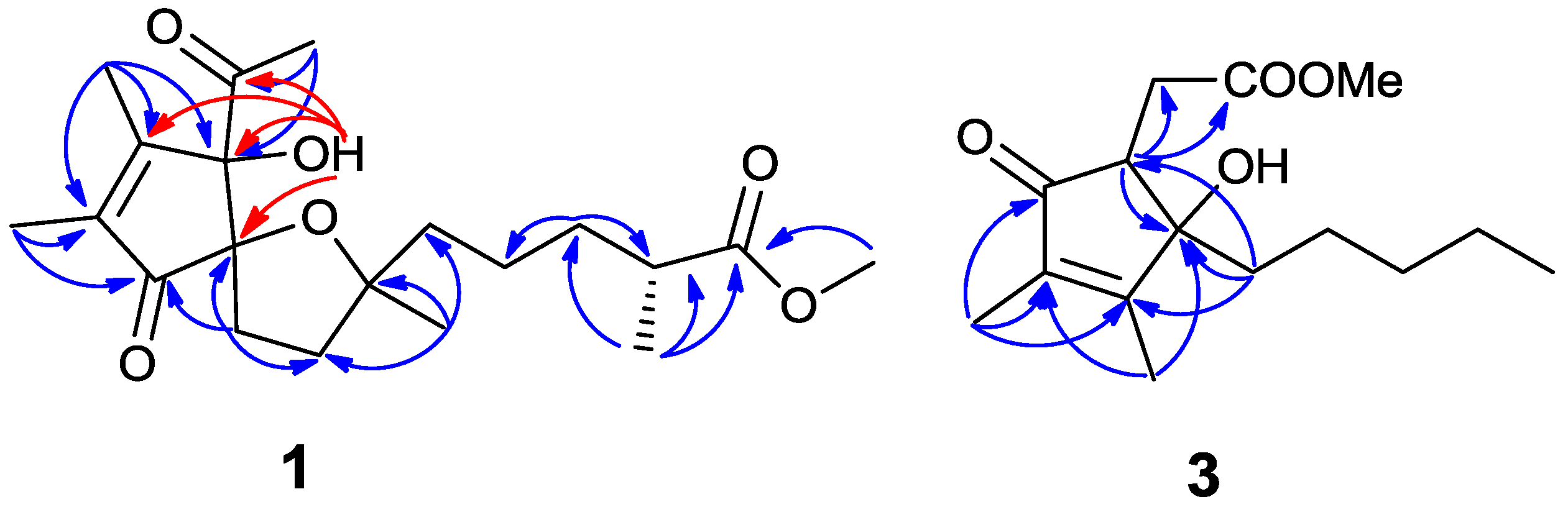
Compound 1 was isolated as a colorless oil. Its molecular formula was assigned as C20H30O6 based on the HRESIMS at m/z 367.2117 [M + H]+, accounting for six degrees of unsaturation. The 1H NMR spectrum indicated the presence of one methoxyl singlet (δH 3.66, s, H-16), four methyl singlets (δH 2.01, H-1, 1.83, H-19, 1.82, H-20, 1.04, H-18), one methyl doublet (δH 1.15, t, J = 7.0 Hz, H-17) and a low-field exchangeable hydroxyl proton (δH 4.72, s, H-3), in addition to 11 aliphatic protons (Table 1). The 13C-NMR and HMQC spectra of 1 showed the presence of six methyls, five methylenes, one methine and eight quaternary carbons, including two olefinic carbons (δC 163.1, 139.5), three oxygen-bearing quaternary carbons (δC 89.1, 92.3, 86.7), two ketones (δC 207.0, 204.7) and one carbonyl (δC 177.4). The above functionalities account for four of the six degrees of unsaturation in the molecule, revealing a bicyclic structure for 1. The 1H and 13C NMR spectra of 1 were similar to those of α-tocospiro A [18,19], with the difference of the side-chain, which was confirmed by the HMBC experiment. The HMBC interactions from H3-16 to C-15, H-13 to C-12, C-14, C-15 and C-17, H-14 to C-12, C-13, C-15 and C-17 and H3-17 to C-13, C-14 and C-15 led to the connectivity of the subunits to form a linear chain (Figure 2). Subsequently, the linear side chain was determined to be linked to the nucleus at C-10 on the basis of the HMBC interactions from H3-18 to C-9, C-10 and C-11. For the spiro moiety, the relative stereochemistry was shown to be the same as that of α-tocospiro A on the basis of similar specific rotation and the NOESY spectrum. Circular dichroism (CD) data also support the absolute configuration of 1 to be identical to that of α-tocospiro C [20]. The absolute configuration was further determined by electronic circular dichroism (ECD) (Figure 3). The calculated ECD showed diagnostic cotton effects around 226 (positive), 246 (negative) and 287 (positive) nm, consistent with the experimental ECD. Thus, the absolute configuration was established as 3S, 7R and 10S, whereas the configurations at C-14 remained to be determined.
Table 1. 1H and 13CNMR spectroscopic data for compounds 1 and 2 (500/125 MHz, in CDCl3, δ in ppm, J in Hz).
Position12
13C1H13C1H
125.02.01 s24.82.01 s
2207.0 207.0
389.14.72 s89.44.68 s
4163.1 163.2
5139.5 139.5
6204.7 205.0
792.3 92.7
836.51.87 m36.81.88 m
1.76 m 1.76 m
933.02.41 m33.32.38 m
1.76 m 1.76 m
1086.7 86.9
1141.01.62 m41.41.62 m
1222.51.41 m22.51.41 m
1.36 m 1.36 m
1334.41.67 m34.21.64 m
1.41 m 1.40 m
1439.52.48 m39.32.44 m
15177.4 177.2
1651.53.66 s51.53.67 s
1717.21.15 d (7.0)17.01.14 d (7.0)
1825.71.04 s25.41.29 s
1912.01.83 s11.81.83 s
208.91.82 s8.71.81 s
Figure 2. Key HMBC correlations of compounds 1 and 3.
Marinedrugs 12 05316 g002
Figure 3. Calculated and experimental electronic circular dichroism (ECD) spectra of compound 1.
Marinedrugs 12 05316 g003
The NMR spectroscopic data of 1 and 2 are very similar, except for the downfield shift of CH3-18 (1.04→1.29) (Table 1), implying 2 to be a C-10 epimer of 1, as is evident from α-tocospiro B [18,19]. However, the optical rotation and the CD spectrum of 2 are the opposite sign to the data of 1. The measured CD curve of 2 was very similar to the calculated ECD for 3R, 7S, 10S-isomer, opposite of the data for 1 (Figure 4), indicating 2 to be in agreement with 3R, 7S and 10S.
Figure 4. Calculated and experimental ECD spectra of compound 2.
Marinedrugs 12 05316 g004
Compound 3 was isolated as a colorless oil. The ion peak was observed in ESI-MS at m/z 291 [M + Na]+, 559 [2M + Na]+. The 1H NMR spectrum indicated the presence of one methoxyl singlet (δH 3.75, s, H-15), two methyl singlets (δH 2.00, H-13, 1.72, H-14) and one terminal methyl triplet (δH 0.83, t, J = 7.0 Hz, H-12) (Table 2). The 13C NMR and HMQC spectra of 3 showed the presence of four methyls, five methylenes one methine, as well as five quaternary carbons, including two olefinic carbons (δC 168.7, 135.5), one oxygen-bearing quaternary carbon (δC 80.4), one ketone (δC 202.8) and one carbonyl (δC 175.2). The 1H and 13C NMR spectra of 3 were almost the same as those of sinularone B [21]. The distinction was attributed to the presence of a methyl ester to replace an ethyl ester of the known analogue, as is evident from the molecular weight of 3, to be 14 amu less than that of the latter, as well as the presence of a methoxyl group in its NMR spectra (Figure 2).
Compound 4 was isolated as a colorless oil. The ion peak was observed in ESI-MS at m/z 253 [M − H]. The 1H and 13C NMR spectra of 4 were almost the same as those of 3, except for the absence of a methoxyl group at C-15 (δC 52.4) in 4 (Table 2).
Compound 5 was isolated as a colorless oil. The ion peak was observed in ESI-MS at m/z 299 [M + H]+, 321 [M + Na]+. Comparison with 5 showed almost the same NMR spectroscopic data as 3, except for the presence of a methoxyl carbon at δC 54.7 and a oxygenated methine at δC 86.9, while a methylene signal appeared at δC 37.0 in 3 (Table 2). This implied that a methoxyl group is located at C-8 in 5 instead of the methylene group in 3. The assumption was confirmed by the correlations of CH3-15 to C-8 and H-8 to C-9 and C-3 in the HMBC experiment.
Compound 6 was isolated as a colorless oil. The ion peak was observed in ESI-MS at m/z 283 [M − H]. Close comparison of the 13C NMR spectrum of Compound 6 to that of 5 showed a general similarity, except for the absence of a methoxyl carbon at δC 54.7 at the C-16 position in 5 (Table 2). The measured CD curve of 36 was very similar to the calculated ECD for 4R, 5R-isomer and opposite of the data for sinularone B [21], indicating 36 to be in agreement with 4R and 5R.
Compound 7 was isolated as a colorless oil. The ion peak was observed in ESI-MS at m/z: 279 [M + Na]+, 535 [2M + Na]+. The 1H NMR spectrum suggested the presence of two methyl singlets (δH 1.85, s, H-10, 11) and two methoxyl singlets (δH 3.66, s, H-12, 3.08, s, H-13). The 13C NMR spectrum indicated the presence of four methyls, four methylenes and five quaternary carbons, including two carbonyl groups, two olefinic carbons and one oxygen-bearing quaternary carbon. Both the 1H and 13C NMR spectra of 7 showed a close similarity to 2,3-dimethyl butenolide [22,23], except for the absence of eight methylenes in the methoxycarbonyl side chain and the presence of an additional methoxy group. Comparison of the 1H and 13C NMR spectrum of 7 and 8 revealed that there were two fewer methylenes in the side chain in 8. The negative specific rotation and the opposite Cotton effect in comparison with those of sinularone H indicated that C-4 had an R configuration [21,22].
By comparing the 1H, 13C-NMR and MS data with the literature values, the known Compounds 9 and 10 were identified as ã-methoxy-2,3-dimethyl-butenolide [24] and sinularone D [21], respectively.
Table 2. 1H and 13CNMR spectroscopic data for compounds 36 (500/125 MHz, in CDCl3, δ in ppm, J in Hz).
Position3456
13C1H13C1H13C1H13C1H
1175.2 174.3 175.5 174.4
229.83.15 dd (4.5, 15.5)31.72.98 m29.63.05 dd (3.0, 18.0)28.63.05 dd (4.5, 8.0)
2.39 dd (7.0, 11.5) 2.62 d (15.5) 2.69 dd (6.5, 11.5 ) 2.61 dd (4.5, 14.5)
355.02.85 dd (3.5, 8.5)46.62.93 m60.92.98 dd (3.0, 11.5)60.32.94 dd (7.0,12.5)
4202.8 204.1 203.1 204.4
5135.5 139.0 136.9 140.2
6168.7 167.1 165.9 165.7
780.4 92.3 83.7 80.8
837.01.76 td (19.0, 5.5)34.51.97 td (14.0, 4.0)86.93.29 dd (4.5, 8.0)93.03.61 dd (2.5, 8.5)
1.52 td (13.0, 3.5) 1.81 m
925.10.75 m23.30.88 m31.31.64 m30.61.68 m
0.63 m 1.10 m 1.22 m 1.30 m
1031.91.19 m32.51.32 m32.01.24 m32.21.34 m
1122.41.19 m22.41.25 m22.61.39 m22.81.34 m
1.07 m 1.51 m
1213.90.83 t (7.0)13.90.89 t (7.0)13.90.83 t (7.0)14.00.92 t (7.0 )
137.81.72 s8.21.75 s8.01.72 s8.31.75 s
1411.52.00 s12.22.06 s11.81.99 s13.02.06 s
1552.43.75 s 52.13.74 s44.03.38 s
16 54.73.42 s
Using HTS, all compounds were tested toward Forkhead box O 3α (Foxo3α), 3-hydroxy-3-methylglutaryl CoA reductase gene fluorescent protein (HMGCR-GFP), nuclear factor kappa B (NF-κB) luciferase, peroxisome proliferator-activated receptor-g co-activator 1α (PGC-1α), protein-tyrosine phosphatase 1B (PTP1B), mitochondrial membrane permeabilization (MMP) and adenosine monophosphate-activated protein kinase (AMPK) activity. Compounds 38 and 10 were evaluated for inhibition of NF-κB activation, and the inhibitory rates are listed in Table 3. At a concentration of 10 μg/mL, sinulolide E and sinularone D exhibited moderate effects with inhibitory rates of 38.12% and 43.00%, respectively. However, all compounds were inactive against other biological targets.
Table 3. Inhibitory rates of NF-κB activation of Compounds 38 and 10.
ConcentrationIR (%)
34567810
10 μg/mL27.8528.7538.1228.2427.0825.2843.00

3. Experimental Section

3.1. General Experimental Procedures

The NMR spectra were recorded on a Bruker AC 500NMR spectrometer (Bruker BioSpin, Fällanden, Switzerland) with tetramethylsilane (TMS) as an internal standard. ESI-MS data were measured on a Bruker amaZon SL spectrometer (Bruker, Fällanden, Switzerland). HR-ESI-MS data were measured on a Bruker micro TOF-QII mass spectrometer (Bruker, Fällanden, Switzerland). CD spectra were measured with a Chirascan circular dichroism spectrometer (Applied Photophysics Ltd., Leatherhead, UK). Optical rotation values were measured with an Anton Paar MCP500 polarimeter (Anton Paar, Graz, Austria). YMC gel (ODS-A, 12 nm, S-50 µm, YMC, Kyoto, Japan) was used for column chromatography. The SiO2 GF254 used for TLC was supplied by the Qingdao Marine Chemical Factory, Qingdao, China. Sephadex LH-20 gel (GE Healthcare, Uppsala, Sweden) was used. HPLC was carried out on a Hitachi L-2400 (Hitachi, Tokyo, Japan) with a YMC ODS column. Spots were detected on TLC under UV light or by heating after spraying with 5% H2SO4 in EtOH (v/v).

3.2. Animal Material

The soft coral Sinularia sp. was collected from Dongluo Island, Hainan province of China, in July, 2009 (7–10-m depth) and identified by Professor Hui Huang, South China Sea Institute of Oceanology, Chinese Academy of Sciences. A voucher specimen (No. 0907010) was deposited in the CAS Key Laboratory of Tropical Marine Bio-resources and Ecology, South China Sea Institute of Oceanology, Chinese Academy of Sciences, Guangzhou, China.

3.3. Extraction and Isolation

The fresh soft coral (wet, 6 kg) was extracted three times with 95% EtOH (20 L). The extract was concentrated under reduced pressure and partitioned between H2O (4 L) and CHCl3 (4 L); the CHCl3 layer (120 g) was further partitioned between 85% EtOH (4 L) and petroleum ether (PE; 4 L) to yield 85% EtOH (34 g) and PE (75.6 g) fractions. The 85% EtOH fraction was separated by silica gel column using CHCl3/MeOH to yield 11 portions (Fr. s1–s11). Fr. s3 was purified by silica gel column to yield 12 portions, and Portion 10 was further purified with semi-preparative HPLC, eluting with MeOH/H2O = 65:35 at a flow rate of 2 mL/min, to afford 1 (4.5 mg) and 2 (2.4 mg). Fr. s5 was purified by Sephadex LH-20 using CHCl3/MeOH = 1:1 to yield 3 portions, and Portion 3 was further purified with semi-preparative HPLC, eluting with MeOH/H2O = 57:43 at a flow rate of 2 mL/min, to afford 7 (3.0 mg), 8 (4.1 mg) and 9 (10.0 mg). Fr. s6 was further purified with semi-preparative HPLC, eluting with MeOH/H2O = 60:40 at a flow rate of 2 mL/min, to afford 3 (2.4 mg), 4 (2.8 mg) and 10 (6.7 mg). Fr. s7 was purified by Sephadex LH-20 to yield three portions, and Portion 3 was further purified with semi-preparative HPLC, eluting with MeOH/H2O = 60:40 at a flow rate of 2 mL/min, to afford 5 (3.5 mg) and 6 (4.2 mg).
Sinulolide A (1): colorless oil; [α] D 25 = −172.5 (c = 0.35, MeOH); CD (MeOH; c 0.2): Δε287 −9.72, Δε246 +12.72, Δε226 −0.76; 1H and 13C NMR data: see Table 1; HRESIMS m/z 367.2117 [M + H]+ (calcd. for C20H31O6, 367.2115), 389.1942 [M + Na]+ (calcd. for C20H30O6Na, 389.1935).
Sinulolide B (2): colorless oil; [α] D 25 = +103.2 (c = 0.07, MeOH); CD (MeOH; c 0.2): Δε287 +3.94, Δε246 −4.66, Δε226 +0.51; 1H and 13C NMR data: see Table 1; ESI-MS m/z 367 [M + H]+, 389 [M + Na]+, 755 [2M + Na]+.
Sinulolide C (3): colorless oil; [α] D 25 = −3.2 (c = 0.01, MeOH); CD (MeOH; c 0.2): Δε210 −0.23; 1H and 13C NMR data: see Table 2; ESI-MS m/z 291 [M + Na]+, 559 [2M + Na]+.
Sinulolide D (4): colorless oil; [α] D 25 = −2.5 (c = 0.01, MeOH); CD (MeOH; c 0.2): Δε207 –0.48, Δε238 −0.46; 1H and 13C NMR data: see Table 2; ESI-MS m/z 253 [M – H].
Sinulolide E (5): colorless oil; [α] D 25 = −3.8 (c = 0.01, MeOH); CD (MeOH; c 0.2): Δε218 –0.52; 1H and 13C NMR data: see Table 2; ESI-MS m/z 299 [M + H]+, 321 [M + Na]+.
Sinulolide F (6): colorless oil; [α] D 25 = −6.6 (c = 0.01, MeOH); CD (MeOH; c 0.2): Δε206 −0.48; 1H and 13C NMR data: see Table 2; ESI-MS m/z 283 [M − H].
Sinulolide G (7): colorless oil; [α] D 25 = −4.4 (c = 0.05, MeOH); CD (MeOH; c 0.2): Δε229 −0.4; 1H NMR (500 MHz, CDCl3) δ: 3.66 (3H, s), 3.08 (3H, s), 2.29 (2H, m), 1.99 (1H, m), 1.85 (6H, s), 1.70 (1H, m), 1.61 (2H, m), 1.23 (2H, m); 13C NMR (125 MHz, CDCl3) δ: 173.4 (C, C-9), 171.9 (C, C-1), 155.6 (C, C-3), 127.4 (C, C-2), 109.5 (C, C-4), 51.6 (CH3, C-10), 50.1 (CH3, C-11), 35.3 (CH2, C-5), 33.8 (CH2, C-8), 24.8 (CH2, C-6), 22.3 (CH2, C-7), 10.7 (CH3, C-13), 8.5 (CH3, C-12); ESI-MS m/z: 279 [M + Na]+, 535 [2M + Na]+.
Sinulolide H (8): colorless oil; [α] D 25 = −3.2 (c = 0.03, MeOH); CD (MeOH; c 0.2): Δε229 −0.4; 1H NMR (500 MHz, CDCl3) δ: 3.66 (3H, s), 3.07 (3H, s), 2.52 (1H, m), 2.39 (1H, m), 2.30 (1H, m), 1.96 (1H, m), 1.88 (3H, s), 1.85 (3H, s); 13C NMR (125 MHz, CDCl3) δ: 173.2 (C, C-7), 171.2 (C, C-1), 155.7 (C, C-3), 127.7 (C, C-2), 108.9 (C, C-4), 51.8 (CH3, C-8), 50.2 (CH3, C-9), 31.2 (CH2, C-5), 27.8 (CH2, C-6), 10.7 (CH3, C-11), 8.5 (CH3, C-10); ESI-MS at m/z: 251 [M + Na]+, 479 [2M + Na]+.

3.4. Assays for Bioactivities

Bioactivity assays were performed by the National Center for Drug Screening, the State Key Laboratory of Drug Research, Shanghai Institute of Materia Medica, Chinese Academy of Sciences, using HTS [25]. Previously reported procedures were followed for assaying the bioactivity against Foxo3α [26], HMGCR-GFP [27], NF-κB luciferase [28], PGC-1α [29], PTP1B [30], MMP [31] and AMPK [32].

3.5. Computational Calculation

The computational ECD, specific rotation and 13C NMR calculations were performed by the B3LYP functional and a generic basis set, employing the 6-311+G (d,p) basis set [21,33]. This generic basis set has been shown to be effective, both efficient and reliable, in predicting structural and reactivity properties for homogeneous systems. Molecular Merck force field (MMFF) and density functional theory/time dependent density functional theory (DFT/TDDFT) calculations were performed with Spartan’14 software package (Wavefunction Inc., Irvine, CA, USA) and the Gaussian 09 program package, respectively, using default grids and convergence criteria.

4. Conclusions

Our study revealed the chemical constituents of soft coral Sinularia sp., which is rich in the South China Sea. Ten compounds were isolated and purified, including seven cyclopentenone derivatives and three butenolide derivatives. Using HTS, their bioactivities toward several targets, such as Foxo3α, HMGCR-GFP, NF-κB-luciferase, PGC-1a, PTP1B, MMP and AMPK, were evaluated. Compounds 5 and 10 exhibited moderate effects for the inhibition of NF-κB activation.

Supplementary Files

Supplementary File 1

Acknowledgments

This study was supported by grants from the National Key Basic Research Program of China (973)’s Project (2011CB915503), the National High Technology Research and Development Program (863 Program, 2012AA092104), National Natural Science Foundation of China (21302198, 31270402, 21172230, 30973679, 41176148, and 21002110), the Strategic Priority Research Program of the Chinese Academy of Sciences (XDA11030403), and Guangdong Marine Economic Development and Innovation of Regional Demonstration Project (GD2012-D01-001), and the Knowledge Innovation Program of Chinese Academy of Science (SQ201117 and SQ201019).

Author Contributions

Bin Yang performed whole experiments and wrote the paper. Xiaoyi Wei contributed to the absolute configuration assignments, especially to the computational ECD. Jingxia Huang contributed to the analysis of the data of biological activity and also contributed to the manuscript preparation. Xiuping Lin, and Juan Liu performed the screening assays. Shengrong Liao, Junfeng Wang, Xuefeng Zhou, and Lishu Wang supported the Yang’s experiments, and shared the work of the structural investigations. Yonghong Liu conceived and designed the experiments, and contributed to the manuscript preparation.

Conflicts of Interest

The authors declare no conflict of interest.

References

  1. Lakshmi, V.; Kumar, R. Metabolites from Sinularia species. Nat. Prod. Res. 2009, 23, 801–850. [Google Scholar] [CrossRef] [PubMed]
  2. Chao, C.H.; Hsieh, C.H.; Chen, S.P.; Lu, C.K.; Dai, C.F.; Sheu, J.H. Sinularianins A and B, novel sesquiterpenoids from the Formosan soft coral Sinularia sp. Tetrahedron Lett. 2006, 47, 5889–5891. [Google Scholar] [CrossRef]
  3. Huang, C.Y.; Su, J.H.; Liu, Y.C.; Wen, Z.H.; Hsu, C.H.; Chiang, M.Y.; Sheu, J.H. Oppositane-type sesquiterpenoids from the Formosan soft coral Sinularia leptoclados. Bull. Chem. Soc. Jpn. 2010, 83, 678–682. [Google Scholar] [CrossRef]
  4. Yang, B.; Liao, S.R.; Lin, X.P.; Wang, J.F.; Liu, J.; Zhou, X.F.; Yang, X.W.; Liu, Y.H. New sinularianin sesquiterpenes from soft coral Sinularia sp. Mar. Drugs 2013, 11, 4741–4750. [Google Scholar] [CrossRef] [PubMed]
  5. Ahmed, A.F.; Wen, Z.H.; Su, J.H.; Hsieh, Y.T.; Wu, Y.C.; Hu, W.P.; Sheu, J.H. Oxygenated cembranoids from a Formosan soft coral Sinulatia gibberosa. J. Nat. Prod. 2008, 71, 179–185. [Google Scholar] [CrossRef] [PubMed]
  6. Chen, B.W.; Chao, C.H.; Su, J.H.; Huang, C.Y.; Dai, C.F.; Wen, Z.H.; Sheu, J.H. A novel symmetric sulfur-containing biscembranoid from the Formosan soft coral Sinularia flexibilis. Tetrahedron Lett. 2010, 51, 5764–5766. [Google Scholar] [CrossRef]
  7. Haidy, N.K.; Marc, S. Terpenoids of sinularia: Chemistry and biomedical applications. Pharm. Biol. 2005, 43, 253–269. [Google Scholar] [CrossRef]
  8. Yang, B.; Zhou, X.F.; Lin, X.P.; Liu, J.; Peng, Y.; Yang, X.W.; Liu, Y.H. Cembrane diterpenes chemistry and biological properties. Curr. Org. Chem. 2012, 16, 1512–1539. [Google Scholar] [CrossRef]
  9. Yang, B.; Zhou, X.F.; Huang, H.; Yang, X.W.; Liu, J.; Lin, X.P.; Li, X.B.; Peng, Y.; Liu, Y.H. New cembrane diterpenoids from a Hainan soft coral Sinularia sp. Mar. Drugs 2012, 10, 2023–2032. [Google Scholar] [CrossRef] [PubMed]
  10. Jin, P.F.; Deng, Z.W.; Pei, Y.H.; Fu, H.Z.; Li, J.; Van Ofwegen, L.; Proksch, P.; Lin, W.H. Polyhydroxylated steroids from the soft coral Sinularia dissecta. Steroids 2005, 70, 487–493. [Google Scholar] [CrossRef]
  11. Chen, B.W.; Su, J.H.; Dai, C.F.; Wu, Y.C.; Sheu, J.H. Polyoxygenated steroids from a Formosan soft coral Sinularia facile. Bull. Chem. Soc. Jpn. 2008, 81, 1304–1307. [Google Scholar] [CrossRef]
  12. Ahmed, A.F.; Hsieh, Y.T.; Wen, Z.H.; Wu, Y.C.; Sheu, J.H. Polyoxygenated sterols from the formosan soft coral Sinularia gibberosa. J. Nat. Prod. 2006, 69, 1275–1279. [Google Scholar] [CrossRef] [PubMed]
  13. Su, J.H.; Lo, C.L.; Lu, Y.; Wen, Z.H.; Huang, C.Y.; Dai, C.F.; Sheu, J.H. Anti-Inflammatory polyoxygenated steroids from the soft coral Sinularia sp. B. Chem. Soc. Jap. 2008, 81, 1616–1620. [Google Scholar] [CrossRef]
  14. Lu, Y.; Huang, C.Y.; Lin, Y.F.; Wen, Z.H.; Su, J.H.; Kuo, Y.H.; Chiang, M.Y.; Sheu, J.H. Anti-inflammatory cembranoids from the soft corals Sinularia querciformis and Sinularia granosa. J. Nat. Prod. 2008, 71, 1754–1759. [Google Scholar] [CrossRef] [PubMed]
  15. Duh, C.Y.; Hou, R.S. Cytotoxic cembranoids from the soft corals Sinularia gibberosa and Sarcophyton trocheliophorum. J. Nat. Prod. 1996, 59, 595–598. [Google Scholar] [CrossRef]
  16. Li, G.Q.; Zhang, Y.L.; Deng, Z.W.; van Ofwegen, L.P.; Proksch, P.; Lin, W.H. Cytotoxic cembranoid diterpenes from a soft coral Sinularia gibberosa. J. Nat. Prod. 2005, 68, 649–652. [Google Scholar] [CrossRef] [PubMed]
  17. Shoji, N.; Umeyama, A.; Takei, M.; Arihara, S. Potent inhibitors of histamine release polyhydroxylated sterols from the Okinawan soft coral Sinularia abrupta. J. Pharm. Sci. 1994, 83, 761–762. [Google Scholar] [CrossRef] [PubMed]
  18. Chiang, Y.M.; Kuo, Y.H. Two novel alpha-tocopheroids from the aerial roots of Ficus microcarpa. Tetrahedron Lett. 2003, 44, 5125–5128. [Google Scholar] [CrossRef]
  19. Matsuo, M.; Matsumoto, S.; Iitaka, Y. Oxygenations of vitamin-E (alpha-tocopherol) and its model-compound 2,2,5,7,8-pentamethylchroman-6-ol in the presence of the superoxide radical solubilized in aprotic-solvents-unique epoxidations and recyclizatons. J. Org. Chem. 1987, 52, 3514–3520. [Google Scholar] [CrossRef]
  20. Yuan, Z.Z.; Duan, H.M.; Xu, Y.Y.; Wang, A.L.; Gan, L.S.; Li, J.J.; Liu, M.T.; Shang, X.Y. α-Tocospiro C, a novel cytotoxic α-tocopheroid from Cirsium setosum. Phytochem. Lett. 2014, 8, 116–120. [Google Scholar] [CrossRef]
  21. Shi, H.Y.; Yu, S.J.; Liu, D.; van Ofwegen, L.; Proksch, P.; Lin, W.H. Sinularones A-I, new cyclopentenone and butenolide derivatives from a marine soft coral Sinularia sp. and their antifouling activity. Mar. Drugs 2012, 10, 1331–1344. [Google Scholar] [CrossRef] [PubMed]
  22. Lee, J.; Wang, W.; Hong, J.; Lee, C.O.; Shin, S.; Im, K.S.; Jung, J.H. A new 2,3-dimethyl butenolide from the brittle star Ophiomastix mixta. Chem. Pharm. Bull. 2007, 55, 459–461. [Google Scholar] [CrossRef] [PubMed]
  23. Nomura, Y.; Kusumi, T.; Ishitsuka, M.; Kakisawa, H. 2,3-dimethyl-4-methoxybuenolides from red algae, Coeloseira pacifica and Ahnfeltia paradoxa. Chem. Lett. 1980, 955–956. [Google Scholar]
  24. Koshino, H.; Yoshihara, T.; Sakamura, S.; Shimanuki, T.; Sato, T.; Tajimi, A. Novel C-11 expoxy fatty-acid from stromata of Epichloe typhina on Phleum pratense. Agric. Biol. Chem. 1989, 53, 2527–2528. [Google Scholar] [CrossRef]
  25. Zhu, Y.H.; Zhang, Z.Y.; Zhang, M.; Mais, D.E.; Wang, M.W. High throughput screening for bioactive components from traditional Chinese medicine. Comb. Chem. High Throughput. Screen. 2010, 13, 837–848. [Google Scholar] [CrossRef] [PubMed]
  26. Zanella, F.; Rosado, A.; Garcia, B.; Carnero, A.; Link, W. Chemical genetic analysis of FOXO nuclear-cytoplasmic shuttling by using image-based cell screening. Chembiochem 2008, 9, 2229–2237. [Google Scholar] [CrossRef] [PubMed]
  27. Schulz, M.M.P.; Reisen, F.; Zgraggen, S.; Fischer, S.; Yuen, D.; Kang, G.J.; Chen, L.; Schneider, G.; Detmar, M. Phenotype-based high-content chemical library screening identifies statins as inhibitors of in vivo lymphangiogenesis. Proc. Natl. Acad. Sci. USA 2012, 109, E2665–E2674. [Google Scholar] [CrossRef]
  28. Kang, M.I.; Henrich, C.J.; Bokesch, H.R.; Gustafson, K.R.; McMahon, J.B.; Baker, A.R.; Young, M.R.; Colburn, N.H. A selective small-molecule nuclear factor-kappa B inhibitor from a high-throughput cell-based assay for “activator protein-1 hits”. Mol. Cancer Ther. 2009, 8, 571–581. [Google Scholar] [CrossRef] [PubMed]
  29. Arany, Z.; Wagner, B.K.; Ma, Y.; Chinsomboon, J.; Laznik, D.; Spiegelman, B.M. Gene expression-based screening identifies microtubule inhibitors as inducers of PGC-1 alpha and oxidative phosphorylation. Proc. Natl. Acad. Sci. USA 2008, 105, 4721–4726. [Google Scholar] [CrossRef] [PubMed]
  30. Shi, L.; Yu, H.P.; Zhou, Y.Y.; Du, J.Q.; Shen, Q.; Li, J.Y.; Li, J. Discovery of a novel competitive inhibitor of PTP1B by high-throughput screening. Acta Pharmacol. Sin. 2008, 29, 278–284. [Google Scholar] [CrossRef]
  31. Farrelly, E.; Amaral, M.C.; Marshall, L.; Huang, S.G. A high-throughput assay for mitochondrial membrane potential in permeabilized yeast cells. Anal. Biochem. 2001, 293, 269–276. [Google Scholar] [CrossRef] [PubMed]
  32. Anderson, S.N.; Cool, B.L.; Kifle, L.; Chiou, W.; Egan, D.A.; Barrett, L.W.; Richardson, P.L.; Frevert, E.U.; Warrior, U.; Kofron, J.L.; et al. Microarrayed compound screening (mu ARCS) to identify activators and inhibitors of AMP-activated protein kinase. J. Biomol. Screen. 2004, 9, 112–121. [Google Scholar] [CrossRef] [PubMed]
  33. Bruhn, T.; Schaumloffel, A.; Hemberger, Y.; Bringmann, G. SpecDis: Quantifying the comparison of calculated and experimental clectronic circular dichroism dpectra. Chirality 2013, 25, 243–249. [Google Scholar] [CrossRef] [PubMed]

