Article

New Spirotetronate Antibiotics, Lobophorins H and I, from a South China Sea-Derived Streptomyces sp. 12A35

1
Department of Natural Products Chemistry, Shenyang Pharmaceutical University, Shenyang 110016, China
2
Institute of Applied Ecology, Chinese Academy of Sciences, Shenyang 110016, China
*
Authors to whom correspondence should be addressed.
Mar. Drugs 2013, 11(10), 3891-3901; https://doi.org/10.3390/md11103891
Submission received: 4 July 2013 / Revised: 12 August 2013 / Accepted: 22 August 2013 / Published: 15 October 2013

Abstract

:
Strain 12A35 was isolated from a deep-sea sediment collected from the South China Sea and showed promising antibacterial activities. It was identified as Streptomyces sp. by the 16S rDNA sequence analysis. Bioassay-guided fractionation using HP20 adsorption, flash chromatography over silica gel and octadecylsilyl (ODS) and semi-preparative HPLC, led to the isolation and purification of five metabolites from the fermentation culture of 12A35. Two new spirotetronate antibiotics, lobophorins H (1) and I (2), along with three known analogues, O-β-kijanosyl-(1→17)-kijanolide (3), lobophorins B (4) and F (5) were characterized by 1D, 2D-NMR and MS data. These compounds exhibited significant inhibitory activities against Bacillus subtilis. Compounds 1 and 5 exhibited moderate activities against Staphylococcus aureus. In particular, the new compound lobophorin H (1) showed similar antibacterial activities against B. subtilis CMCC63501 to ampicillin.

1. Introduction

Infectious disease is one of the most deadly diseases threatening public health. Meanwhile, drug-resistant bacteria are steadily rising, increasing the difficulty of treatment. Thus, an efficient way to address this issue is to develop new antibiotics both to help to overcome the antibiotic resistance and to treat new pathogenic diseases. Nowadays, natural products from marine-derived microorganisms have been an important source of novel lead structures for drug discovery. Marine actinomycetes are one of the most efficient groups of secondary metabolite producers. Many intriguing compounds with potent and various bioactivities have been found from marine microbes for lead compounds [1]. For instance, marinisporolides A and B were polyene macrolides isolated from the culture of an actinomycete from a new genus Marinispora. Marinisporolide A showed modest activity against Candida albicans [2]. The deep-sea-derived Streptomyces sp. SCSIO 03032 was capable of producing new bisindole alkaloids, spiroindimicins A–D, which exhibited moderate cytotoxicities against several cancer cell lines [3].
During our continuous screening for new antibiotics, the organic extract of the fermentation broth of a deep-sea-derived actinobacterial strain 12A35 showed potent antibacterial activities. Five spirotetronate antibiotics were isolated from an actinomycete 12A35 as a member of the genus Streptomyces by 16S rDNA analysis. Two compounds were determined to be new lobophorin analogues, designated as lobophorins H (1) and I (2), and the other three were characterized as known O-β-kijanosyl-(1→17)-kijanolide (3), lobophorins B (4) and F (5). Notably, the new compound lobophorin H (1) showed similar antibacterial activities against Bacillus subtilis to ampicillin as a positive control drug.

2. Results and Discussion

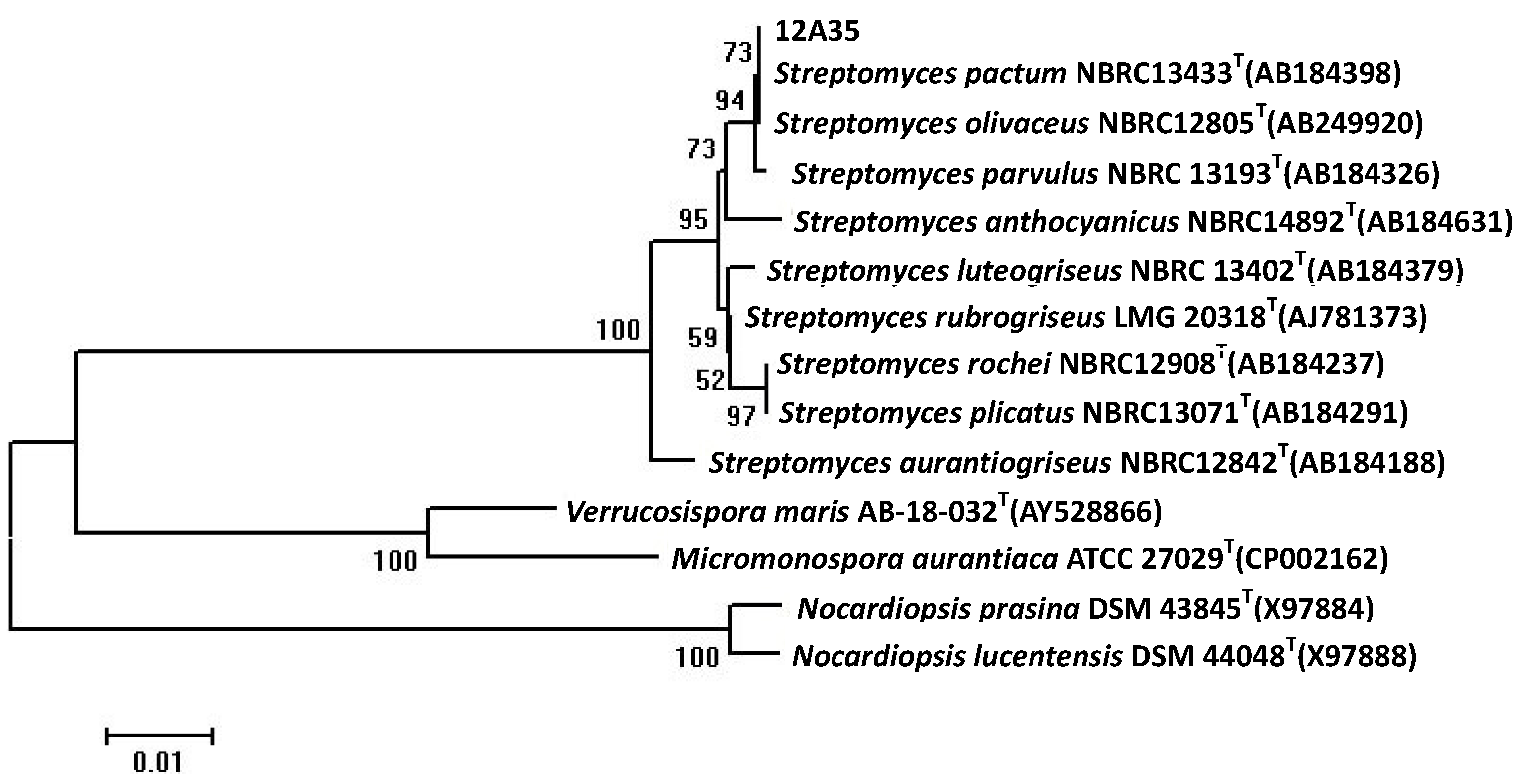
2.1. Taxonomy of the Strain 12A35

The 16S rDNA of producing strain 12A35 was polymerase chain reaction (PCR) amplified and sequenced. The strain 12A35 16S ribosomal RNA gene was submitted in the GenBank Database with the accession number KF313921. Sequence analysis showed that 16S rRNA gene sequence of 12A35 to be most similar to those of Streptomyces pactum NBRC 13433T, Streptomyces olivaceus NBRC 12805T, and Streptomyces parvulus NBRC 13193T, with sequence identities of 100%, 100%, and 99.33%, respectively. The phylogenetic tree generated by a neighbor-joining method based on 16S rRNA gene sequence clearly revealed the evolutionary relationship of the strain 12A35 with a group of Streptomyces species (Figure 1). So, this strain was designated as Streptomyces sp. 12A35.
Figure 1. Phylogenetic tree of 16S rDNA sequences of 12A35 strain by the neighbor-joining method.
Marinedrugs 11 03891 g001

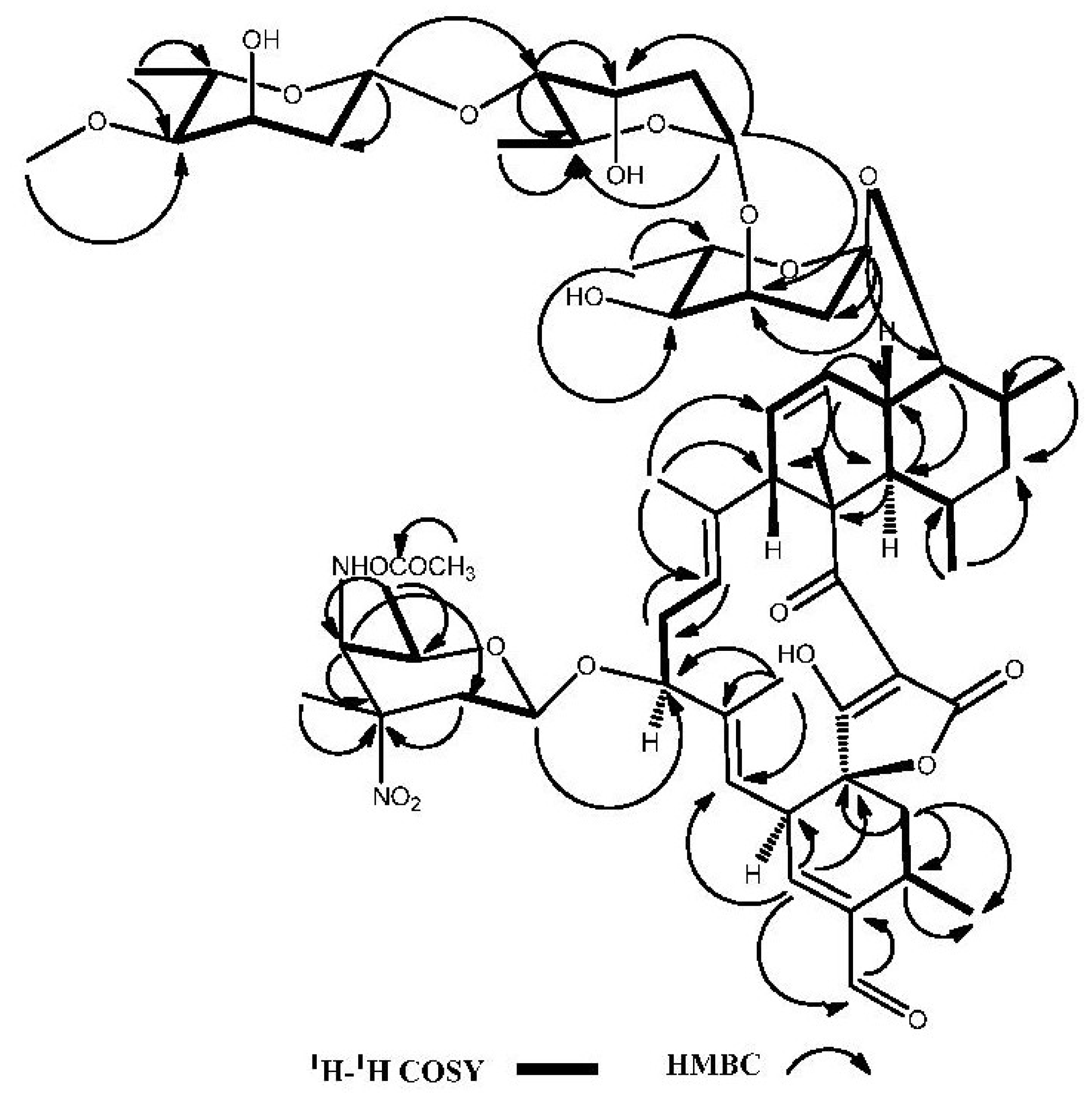
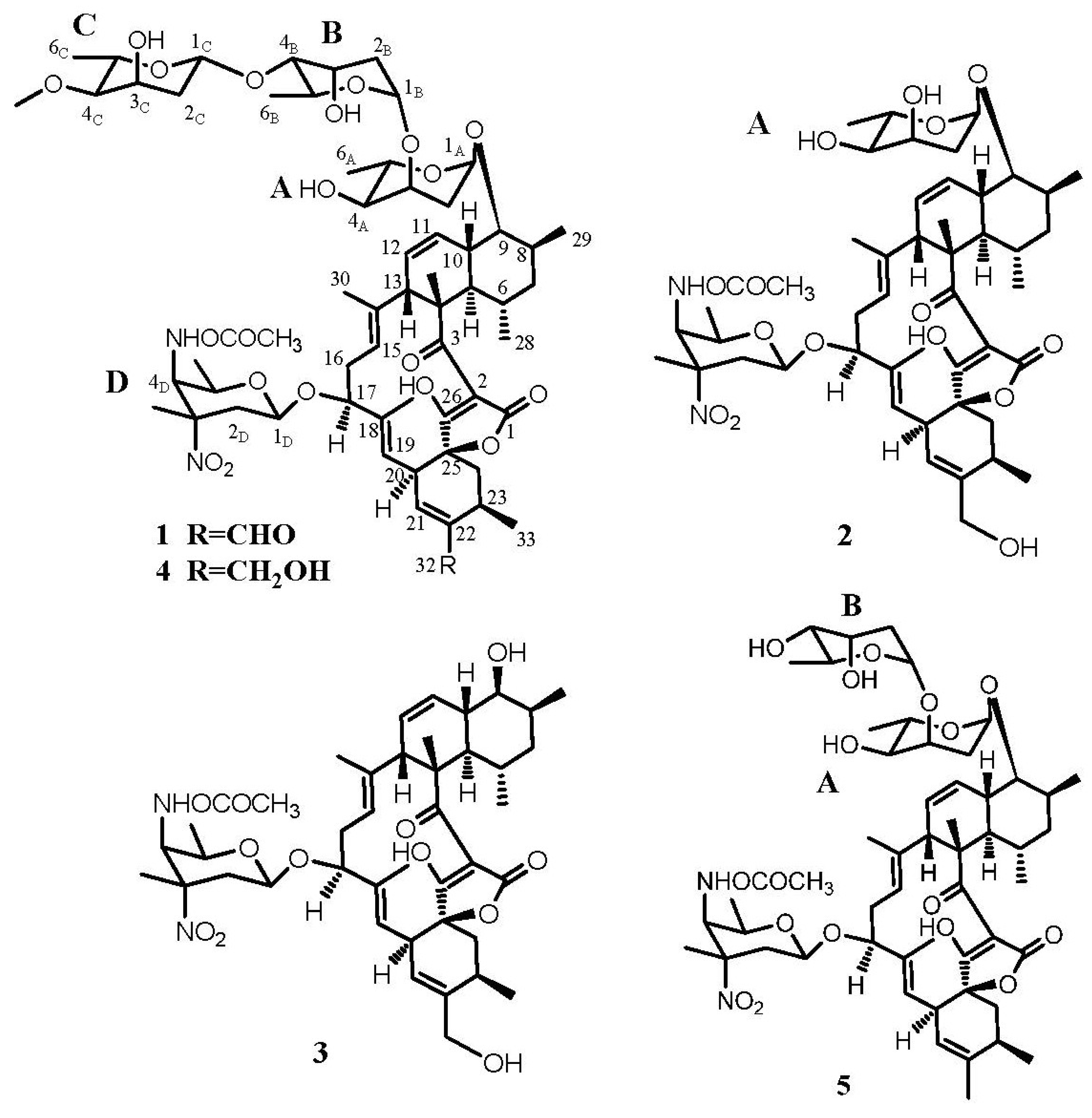
2.2. Structure Determination

Compound 1 was obtained as a white powder. Its high-resolution electrospray ionization mass spectrometry (HRESIMS) exhibited a [M − H] ion at m/z 1183.5807, corresponding to a molecular formula of C61H88N2O21, with 19 degrees of unsaturation. The 1H NMR (600 MHz, CDCl3) spectrum (Table 1) exhibited four methyl singlets at δ 1.35, 1.45, 1.61, 1.59 and seven methyl doublets at δ 0.64, 1.09, 1.16, 1.20, 1.25, 1.26, 1.36, two methoxy groups at δ 3.41 and 3.72, five olefinic protons at δ 5.17, 5.28, 5.38, 5.73, 6.56 and one aldehyde at δ 9.51. The 13C NMR (150 MHz, CDCl3) (Table 1) and HSQC spectra revealed 61 carbon signals, including four carbonyls (including an aldehyde), ten olefinic carbons, four sugar anomeric carbons, eleven methyls. The 1H and 13C NMR spectra readily indicated the presence of four 2,6-dideoxysugar units through easily identifiable signals for anomeric and 6-methyl protons and carbons. The four monosaccharide moieties were identified as two digitoxose, one 4-O-methyl-digitoxose and one kijanose by the interpretation of 1H-1H COSY, HMQC and HMBC spectra. The remaining 33 signals were suggestive of a spiroteronate skeleton [4,5]. Further, compound 1 showed similar 1H and 13C NMR spectra to lobophorin B (4) except for the hydroxymethyl signals at C-22 [4]. The oxygenated methylene (δH 4.22 (2H, m, H-32) and δC 64.9 (C-32)) in lobophorin B was replaced by an aldehyde group at δH 9.51 (1H, s, H-32) and δC 193.3 (C-32) in 1, which resulted downfield shift of H-21 proton signal from δ 5.50 to δ 6.55. The HMBC correlations (Figure 2) from proton signals at δH 9.51 (1H, s, H-32) to δC 144.6 (C-22) and 25.2 (C-23), as well as from δH 6.55 (1H, s, H-21) to δC 193.3 (C-32), confirmed the aldehyde group located at C-22 in compound 1, designated as lobophorin H (Figure 3). Furthermore, the similar coupling constant values of 1 to lobophorin B indicated the same relative stereochemistry [5]. The relative configuration of anomeric carbon of the sugars was easily determined to be α-configuration for sugars A and B and β-configuration for sugars C and D from the coupling constants for anomeric protons. The NOESY correlations between H-5/H-9, H-8/H-9, H-10/H-6, H-10/H-29, H-13/H-15 and H-16/H-30 supported the above relative stereochemistry.
Table 1. 1H and 13C NMR data of compounds 14 in CDCl3.
Position1234Lobophorin F [6]
δCδH ( J in Hz)δCδH ( J in Hz)δCδCδC
1166.71 167.24 167.12167.23167.3
2101.68 101.85 101.89101.76101. 9
3206.47 206.07 206.43206.33206.3
450.93 50.90 51.0250.9251.0
543.112.00 (m)43.201.99 (m)42.7943.1043.4
631.281.61 (m)31.291.61 (m)31.1931.3031.2
741.661.58 (m), 1.50 (m)41.731.59 (m), 1.51 (m)41.7241.6941.9
834.372.22 (m)34.981.89 (m)34.7134.3934.5
984.083.44 (m)85.573.55 (dd, 10.9, 5.4)76.0884.1686.7
1038.392.08 (m)38.372.15 (m)39.2238.4238.2
11125.975.73 (d, 10.6)124.985.71 (d, 10.9)125.51125.84125.9
12126.295.38 (m)127.185.38 (d, 9.7)126.51126.45126.6
1353.243.46 (m)52.983.48 (m)53.1753.1553.1
14135.92 135.45 135.83135.73135.7
15123.225.17 (d, 9.2)123.595.16 (d, 9.3)123.32123.34123.5
1630.022.34 (m), 1.71 (m)30.942.34 (m), 2.25 (m)31.0830.0331.2
1778.694.22 (m)78.334.19 (m)78.4778.3678.9
18139.46 137.09 137.03137.08136.7
19116.725.28 (d, 10.5)119.175.11 (d, 10.9)119.23119.19119.9
2041.243.80 (d, 11.1)40.173.85 (d, 10.9)40.1840.1740.4
21148.126.56 (s)121.525.49 (s)121.43121.52120.6
22144.59 141.24 141.35141.28137.8
2325.363.01 (m)27.872.66 (m)27.9327.9531.9
2434.802.36 (m), 1.89 (m)35.312.37 (m), 1.82 (m)35.3335.3535.5
2582.94 83.32 83.2483.3083.3
26200.97 201.37 201.49201.63201.7
27 (4-CH3)15.131.62 (s, 3H)14.981.59 (s, 3H)15.0315.0515.1
28 (6-CH3)22.230.64 (d, 4.9)22.150.63 (d, 5.8)22.2822.2322.2
29 (8-CH3)14.111.09 (d, 6.4)14.391.09 (d, 7.0)12.9914.1214.6
30 (14-CH3)13.731.35 (s, 3H)13.701.31 (s, 3H)13.7013.7013.8
31 (18-CH3)15.181.45 (s, 3H)15.031.45 (s, 3H)15.1115.0915.1
32 (22-C)193.329.51 (s)64.854.20 (s, 2H)64.9364.8921.8
33 (23-CH3)19.971.36 (d, 6.4, 3H)20.141.30 (m, 3H)20.1820.1820.2
A198.004.78 (d, 4.7)99.584.90 (d, 2.7) 97.9899.1
A231.022.30 (m), 2.35 (m)34.052.25 (m, 2H) 30.9533.4
A366.614.00 (m)67.263.98 (m) 66.6774.1
A471.803.26 (dd, 8.7, 3.5)72.603.19 (d, 8.9) 71.8172.4
A564.944.00 (m)65.233.82 (m) 64.9565.0
A617.721.26 (d, 6.0, 3H)17.691.30 (m, 3H) 17.7117.7
B190.975.13 (m) 90.9996.5
B234.082.12 (m), 1.91 (m) 34.0735.3
B365.484.22 (m) 65.5366.9
B482.103.23 (dd, 7.7, 2.3) 82.172.5
B562.123.98 (m) 62.1365.6
B617.891.20 (d, 6.0, 3H) 17.8717.7
C198.284.91 (dd, 9.3, 2.0) 98.30
C236.652.16 (m), 1.67 (m) 36.64
C363.964.25 (d, 2.6) 63.96
C482.102.84 (dd, 9.2, 2.6) 82.05
C568.373.76 (q, 5.8) 68.37
C618.251.25 (d, 6.0, 3H) 18.25
C4-OCH357.373.41 (s, 3H) 57.37
D197.394.46 (dd, 9.2, 1.5)96.984.42 (dd, 9.8, 1.9)97.0396.9697.4
D235.722.77 (m), 1.63 (m)35.652.75 (m), 1.59 (m)35.7135.7135.8
D391.09 90.98 91.0191.1290.9
D453.724.39 (d, 10.4)53.664.35 (d, 10.1)53.6653.6753.9
D569.123.48 (m)69.073.47 (d, 7.4)69.0769.0568.9
D616.971.16 (d, 6.0, 3H)16.941.16 (d, 6.4, 3H)16.9716.9717.0
D3-CH325.221.59 (s, 3H)25.251.56 (s, 3H)25.2825.2725.3
D4 C=O157.34 157.44 157.35157.38157.4
D4-OCH352.743.72 (s, 3H)52.723.72 (s, 3H)52.6952.7352.7
Figure 2. Selected key 1H-1H COSY and HMBC correlations of lobophorin H (1).
Marinedrugs 11 03891 g002
Compound 2 was obtained as a white amorphous powder. The HRESIMS data (m/z 911.4544 [M − H] and m/z 913.4522 [M + H]+) of 2 established the molecular formula to be C48H68N2O15. The 1H NMR (600 MHz, CDCl3) spectrum (Table 1) showed four methyl singlets at δ 1.31, 1.45, 1.56, 1.59 and five methyl doublets at δ 0.63, 1.09, 1.16, 1.30, 1.30, a methoxy groups at δ 3.72, one oxygenated methylene at δ 4.20 and five olefinic protons at δ 5.11, 5.16, 5.38, 5.49, 5.71. The 13C NMR (150 MHz, CDCl3) (Table 1) and HSQC spectra revealed 48 carbon signals, including three carbonyls, ten olefinic carbons, two sugar anomeric carbons, nine methyls. According to the 1H and 13C NMR spectral data, compound 2 was determined as an analogue of compound 1. Comparing with the 1H and 13C NMR data of lobophorin B (4), the signals of sugar B (digitoxose) and sugar C (4-O-methyl-digitoxose) in lobophorin B were absent in compound 2 (Table 1). The HMBC correlations between δH 4.79 (H-1A), 2.25 (H-2A) and δC 67.26 assigned the carbon signal at δ 67.26 to C-3A. In comparison with 13C-NMR data of lobophorin F, 6.8 ppm upfield deglycosidation shift of in C-3A was consistent with the loss of the sugar B and C moieties in 2. The 1H- and 13C-NMR signals of 2 were assigned by HSQC, HMBC and 1H-1H COSY spectral analyses (Table 1). Based on these MS and NMR data, the structure of 2 was determined to be lobophorin I (Figure 3). Furthermore, the relative stereochemistry could be determined by comparison of NMR data with 1.
Figure 3. Chemical structures of compounds 15.
Marinedrugs 11 03891 g003
Compound 3 was isolated as a white powder. Its molecular formula was determined as C42H58N2O12 by HRESIMS (m/z 781.3917 [M − H]). By comparing 1H NMR (600 MHz, CDCl3) and 13C NMR (150 MHz, CDCl3) data (Table 1) with those previously reported, 3 was identified as O-β-kijanosyl-(1→17)-kijanolide (Figure 3) which was reported as a methanolysis product of kijanimicin [7]. Compound 3 was found for the first time as a natural product.
Compound 4, white powder, afforded HRESIMS data (m/z 1185.5970 [M − H] and 1209.5901 [M + Na]+) consistent with the molecular formula C61H90N2O21. According to 1H NMR (600 MHz, CDCl3) and 13C NMR (150 MHz, CDCl3) (Table 1), 4 was identified as lobophorin B (Figure 3), which was previously found from the fermentation broths of a marine bacterium showed potent antiinflammatory activities in the PMA (Phorbol-Myristate-Acetate)-induced mouse ear edema model [5].
Compound 5 was obtained as a white powder, whose HRESIMS gave the molecular formula C54H78N2O17 base on quasi-molecular ion peaks at m/z 1049.5055 [M + Na]+ and 1025.5203 [M − H]. The 1H NMR (600 MHz, CDCl3) data of 5 was identical to those published data of lobophorin F (Figure 3), which was previously isolated from a Streptomyces sp. SCSIO 01127 and showed antibacterial activities and cytotoxic activities [6].
The spirotetronate antibiotics are a class of natural products that exhibit broad biological activities, including antibacterial, antitumor, antiviral, antimalaria effects and cholesterol biosynthesis inhibition [5,8,9]. They feature an unusual macrolide that contains a characteristic tetronic acid (spiro-linked to a cyclohexene ring) conjugated with a trans-decalin system either by a carboxylic ester or a carbonyl group [10]. In order to continue to search for new drugs, these compounds are continually found including kijanimicin [11], tetrocarcins [12], chlorothricin [10], decatromicins [13], saccharocarcins [14], antlermicins [15], versipelostatins [16], arisostatins [17], quartromicins [9] and chrolactomycin [18]. One representative of this group is lobophorins, among which lobophorins A and B were produced from an alga-associated acintobacterium [5], lobophorins C and D were produced by a marine spongerelated Streptomyces [19], and lobophorins E, F and G were isolated from marine-derived Streptomyces [6,20]. The discovery of more spirotetronate antibiotics will throw more light on the structure-activity relationships and potential applications of these compounds.

2.3. Antimicrobial Activity of Compounds 1–5

Regarding the results shown in Table 2, the tested lobophorins did not exhibit inhibitory activity on Gram-negative bacteria (E. coli) and fungi (C. albicans, F. moniliforme). Only compounds 1 and 5 exhibited moderate activities against Staphylococcus aureus ATCC29213 with minimum inhibitory concentrations (MIC) values of 50 and 6.25 μg·mL−1, respectively. All the tested compounds exhibited inhibitory activities against Bacillus subtilis CMCC63501. Compounds 1 and 4 showed strong activities against Bacillus subtilis CMCC63501 with MIC values of 3.13 and 1.57 μg·mL1, respectively, while compounds 2, 3 and 5 possessed moderate activities against Bacillus subtilis CMCC63501 with MIC values of 6.25, 50, 50 μg·mL1, respectively. With the increase of the amount of monosaccharide units, the inhibitory activity increased indicating that monosaccharide might play an important role for the antimicrobial activity of lobophorins. These findings support the proposal that the change of the length of saccharide chains alters the biological activity of the natural product, in which the sugars contribute to specific interactions with the biological target [21]. It is noticeable that lobophorin H (1) showed similar antibacterial activities against Bacillus subtilis CMCC63501 to ampicillin. Given the significant inhibitory activities against Gram-positive bacteria, lobophorins F (5) and H (1) may potentially find applications in anti-infective drug development.
Table 2. Minimum inhibitory concentrations for compounds 15 (μg·mL−1).
CompoundsS. aureus ATCC29213B. subtilis CMCC63501E. coil ATCC25922C. albicans ATCC10231F. moniliforme S16
1501.57>100>200>200
2>10050>100>200>200
310050>100>200>200
41003.13>100>200>200
56.256.25>100>200>200
ampicillin3.131.5725NANA
nystatinNANANA6.2525
NA: Not assayed.

3. Experimental Section

3.1. General Experimental Procedures

The chromatographic silica gel (500–600 mesh) was purchased from Qingdao Ocean Chemical Factory (Qingdao, China) and Diaion HP 20 macroporous resin was purchased from Mitsubishi Chemical Co., Ltd., Tokyo, Japan. RP-HPLC analysis and semi-preparation were conducted using a U3000 HPLC system (Dionex, Sunnyvale, CA, USA) and performed with a C18 YMC-Pack ODS-A column (5 μm, φ 10 × 250 mm). HRESIMS was recorded on a Bruker QTOF-ESI mass spectrometer. One-dimensional and two-dimensional NMR spectroscopy (1H-NMR, 13C-NMR, HSQC, HMBC, 1H-1H COSY) were conducted with a Bruker AV600 spectrometer (Rheinstetten, Germany). Deuterated NMR solvents were purchased from Cambridge Isotopes (Andover, MA, USA). Specimens of the strains for antimicrobial activity were deposited at the Group of Microbial Biotechnology, Institute of Applied Ecology, Chinese Academy of Sciences.

3.2. Microorganisms and Fermentation Conditions

The sediment was collected from the South China Sea at the depth of 2134 m (17°59.928′N, 111°36.160′E). To effective isolation of culturable marine actinomycetes by making the culture conditions similar to true marine environment, the strain 12A35 was isolated after incubation at 28 °C for 1 week on modified Gauze’s synthetic medium NO. 1 with artificial sea water instead of NaCl and distilled water (soluble starch 20.0 g; KNO3 1.0 g; MgSO4·7H2O 0.5 g; K2HPO4 0.5 g; FeSO4·7H2O 10.0 mg; agar 15.0 g; artificial sea water 1.0 L; adjust pH 7.0). Genomic DNA isolation, PCR amplification of 16S rDNA, and sequencing were performed with conventional methods. Sequence analysis of 16S rDNA were performed using BLASTN. Phylogenetic tree were constructed using Neighbor-Joining method of MEGA (version 5.0). Tree topologies were evaluated by bootstrap analysis with 1000 replicates.
The strain 12A35 was maintained on modified Gauze’s synthetic medium NO. 1 at 28 °C, and the agar was cut into pieces (1 × 1 cm) and inoculated into 5 × 100 mL of seed medium (potato sucrose broth containing sucrose 20 g, infusion from 200 g potatoes, artificial sea water 1 L at pH 6.0 before sterilization) in 500-mL Erlenmeyer flasks, then cultivated at 28 °C for 2 days with shaking at 180 rpm. Seed cultures were transferred into 24 × 650 mL production medium in 3.0 L Erlenmeyer flasks, with an inoculation volume of 3%–4% (v/v), and incubated under the same conditions for 7 days.

3.3. Bioactivity-Guided Isolation and Purification

Fermentation broth (16 L) of 12A35 was centrifuged at 4000× g for 30 min. The supernatant was subjected to HP20 macroporous adsorption resin by two-step gradient elution with EtOH/H2O solutions of 10% and 100%. The EtOH fraction was evaporated to dryness under vacuum with a rotary evaporator. The mycelia were extracted three times with 3 L acetone and the acetone solution was evaporated to dryness. Two above residues were combined and extracted three times with methanol as the crude extract for further isolation. The crude extract (30.04 g) were subjected to vacuum flash chromatography over silica gel (500–600 mesh) and gradiently eluted with dichloromethane and methanol at ratios of (v/v) 100:0, 98:2, 95:5, 90:10, 80:20, 70:30, 60:40, 50:50 and 0:100 to give nine fractions (f1–f9). F4 (yield: 1.77 g) obtained by elution with dichloromethane/methanol (90:10, v/v) showed antibacterial activity. F4 was subjected to vacuum flash chromatography over ODS and eluted with CH3OH/H2O (1:9–10:0), to give ten sub-fractions (rf1–rf10). Sub-fractions rf8 (458 mg) and rf9 (698 mg) with antibacterial activities were combined, and further separated by a semi-preparative HPLC system (Dionex U3000, Sunnyvale, CA, USA) using a C18 YMC-Pack ODS-A column (5 μm, φ 10 × 250 mm) eluted with 90% methanol containing 0.05% trifluoroacetic acid (TFA) at a flow rate of 2.5 mL/min with UV detection at 220 nm. Compounds 1 (18.1 mg), 2 (12.4 mg), 3 (6.3 mg), 4 (67.3 mg), and 5 (11.6 mg) were obtained at retention time of 15.2 min, 9.6 min, 8.9 min, 11.5 min, 16.8 min, respectively.

3.4. Antimicrobial Activity of Compounds 1–5

Antimicrobial activity of compounds 15 were performed as described methods previously [22], and the minimum inhibitory concentrations (MIC) of compounds (15) were determined against five microbial strains, including Staphylococcus aureus ATCC 29213, Bacillus subtilis CMCC63501, Escherichia coil ATCC25922, Candida albicans ATCC10231 and Fusarium moniliforme S16. Ampicillin for bacteria and nystatin for fungi were used as positive controls with medium as a negative control.

4. Conclusions

Two new spirotetronate antibiotics lobophorins H (1) and I (2), along with three known analogues, O-β-kijanosyl-(1→17)-kijanolide (3), lobophorins B (4) and F (5) were isolated and characterized from a deep-sea-derived Streptomyces sp. 12A35. These compounds exhibited significant inhibitory activities against Bacillus subtilis. Compounds 1 and 5 exhibited moderate activities against Staphylococcus aureus. Notablely, the compound 1 showed similar antibacterial activities against Bacillus subtilis to ampicillin as a positive control drug. Our results threw some light on the structure-activity relationships and potential applications of these compounds.

Acknowledgments

This work was supported by grants from the National Science Foundation for Young Scientists of China (41006088), National High Technology Research and Development Program of China (863 Program) (No. 2012AA092104, 2011AA09070404), the National Science & Technology Pillar Program (No. 2011BAE06B04). We are grateful to Wen Li, Ying Peng and Yi Sha of the Shenyang Pharmaceutical University for recording NMR and MS spectra.

Conflicts of Interest

The authors declare no conflict of interest.

References

  1. Blunt, J.W.; Copp, B.R.; Keyzers, R.A.; Munro, M.H.; Prinsep, M.R. Marine natural products. Nat. Prod. Rep. 2013, 30, 237–323. [Google Scholar] [CrossRef]
  2. Kwon, H.C.; Kauffman, C.A.; Jensen, P.R.; Fenical, W. Marinisporolides, polyene-polyol macrolides from a marine actinomycete of the new genus Marinispora. J. Org. Chem. 2008, 74, 675–684. [Google Scholar]
  3. Zhang, W.; Liu, Z.; Li, S.; Yang, T.; Zhang, Q.; Ma, L.; Tian, X.; Zhang, H.; Huang, C.; Zhang, S. Spiroindimicins A–D: New bisindole alkaloids from a deep-sea-derived actinomycete. Org. Lett. 2012, 14, 3364–3367. [Google Scholar] [CrossRef]
  4. Mallams, A.K.; Puar, M.S.; Rossman, R.R.; McPhail, A.T.; Macfarlane, R.D.; Stephens, R.L. Kijanimicin. Part 3. Structure and absolute stereochemistry of kijanimicin. J. Chem. Soc. Perkin Trans. 1983, 1, 1497–1534. [Google Scholar]
  5. Jiang, Z.-D.; Jensen, P.R.; Fenical, W. Lobophorins A and B, new antiinflammatory macrolides produced by a tropical marine bacterium. Bioorg. Med. Chem. Lett. 1999, 9, 2003–2006. [Google Scholar] [CrossRef]
  6. Niu, S.; Li, S.; Chen, Y.; Tian, X.; Zhang, H.; Zhang, G.; Zhang, W.; Yang, X.; Zhang, S.; Ju, J.; et al. Lobophorins E and F, new spirotetronate antibiotics from a South China Sea-derived Streptomyces sp. SCSIO 01127. J. Antibiot. 2011, 64, 711–716. [Google Scholar] [CrossRef]
  7. Mallams, A.K.; Puar, M.S.; Rossman, R.R.; McPhail, A.T.; Macfarlane, R.D. Kijanimicin. 2. Structure and absolute stereochemistry of kijanimicin. J. Am. Chem. Soc. 1981, 103, 3940–3943. [Google Scholar] [CrossRef]
  8. Tomita, F.; Tamaoki, T.; Shirahata, K.; Kasai, M.; Morimoto, M.; Ohkubo, S.; Mineura, K.; Ishii, S. Novel antitumor antibiotics, tetrocarcins. J. Antibiot. 1980, 33, 668–670. [Google Scholar] [CrossRef]
  9. Kusumi, T.; Ichikawa, A.; Kakisawa, H.; Tsunakawa, M.; Konishi, M.; Oki, T. The structures of quartromicins A1, A2, and A3: Novel macrocyclic antiviral antibiotics possessing four tetronic acid moieties. J. Am. Chem. Soc. 1991, 113, 8947–8948. [Google Scholar] [CrossRef]
  10. Jia, X.Y.; Tian, Z.H.; Shao, L.; Qu, X.D.; Zhao, Q.F.; Tang, J.; Tang, G.L.; Liu, W. Genetic characterization of the chlorothricin gene cluster as a model for spirotetronate antibiotic biosynthesis. Chem. Biol. 2006, 13, 575–585. [Google Scholar] [CrossRef]
  11. Waitz, J.A.; Horan, A.; Kalyanpur, M.; Lee, B.K.; Loebenberg, D.; Marquez, J.A.; Miller, G.; Patel, M.G. Kijanimicin (Sch 25663), a novel antibiotic produced by Actinomadura kijaniata SCC 1256. Fermentation, isolation, characterization and biological properties. J. Antibiot. 1981, 34, 1101–1106. [Google Scholar] [CrossRef]
  12. Tamaoki, T.; Kasai, M.; Shirahata, K.; Tomita, F. Tetrocarcins E1, E2, F and F-1, new antibiotics. Fermentation, isolation and characterization. J. Antibiot. 1982, 35, 979–984. [Google Scholar] [CrossRef]
  13. Momose, I.; Hirosawa, S.; Nakamura, H.; Naganawa, H.; Iinuma, H.; Ikeda, D.; Takeuchi, T. Decatromicins A and B, new antibiotics produced by Actinomadura sp. MK73-NF4. II. Structure determination. J. Antibiot. 1999, 32, 787–796. [Google Scholar]
  14. Horan, A.C.; Shearer, M.C.; Hedge, V.; Beyazova, M.L.; Brodsky, B.C.; King, A.; Berrie, R.; Cardaci, K.; Nimeck, M. A family of novel macrocyclic lactones, the saccharocarcins produced by Saccharothrix aerocolonigenes subsp. antibiotica. I. Taxonomy, fermentation, isolation and biological properties. J. Antibiot. 1997, 50, 126–134. [Google Scholar] [CrossRef]
  15. Kobinata, K.; Uramoto, M.; Mizuno, T.; Isono, K. A new antibiotic, antlermicin A. J. Antibiot. 1980, 33, 244–246. [Google Scholar] [CrossRef]
  16. Park, H.R.; Furihata, K.; Hayakawa, Y.; Shin-ya, K. Versipelostatin, a novel GRP78/Bip molecular chaperone down-regulator of microbial origin. Tetrahedron Lett. 2002, 43, 6941–6945. [Google Scholar] [CrossRef]
  17. Furumai, T.; Takagi, K.; Igarashi, Y.; Saito, N.; Oki, T. Arisostatins A and B, new members of tetrocarcin class of antibiotics from Micromonospora sp. TP-A0316-I. Taxonomy, fermentation, isolation and biological properties. J. Antibiot. 2000, 53, 227–232. [Google Scholar] [CrossRef]
  18. Nakai, R.; Kakita, S.; Asai, A.; Chiba, S.; Akinaga, S.; Mizukami, T.; Yamashita, Y. Chrolactomycin, a novel antitumor antibiotic produced by Streptomyces sp. J. Antibiot. 2001, 54, 836–839. [Google Scholar] [CrossRef]
  19. Wei, R.B.; Xi, T.; Li, J.; Wang, P.; Li, F.C.; Lin, Y.C.; Qin, S. Lobophorin C and D, new kijanimicin derivatives from a marine sponge-associated actinomycetal srain AZS17. Mar. Drugs 2011, 9, 359–368. [Google Scholar] [CrossRef]
  20. Chen, C.; Wang, J.; Guo, H.; Hou, W.; Yang, N.; Ren, B.; Liu, M.; Dai, H.; Liu, X.; Song, F.; et al. Three antimycobacterial metabolites identified from a marine-derived Streptomyces sp. MS100061. Appl. Microbiol. Biotechnol. 2013, 97, 3885–3892. [Google Scholar]
  21. Griffith, B.R.; Langenhan, J.M.; Thorson, J.S. “Sweetening” natural products via glycorandomization. Curr. Opin. Biotechnol. 2005, 16, 622–630. [Google Scholar] [CrossRef]
  22. Engelhardt, K.; Degnes, K.F.; Kemmler, M.; Bredholt, H.; Fjaervik, E.; Klinkenberg, G.; Sletta, H.; Ellingsen, T.E.; Zotchev, S.B. Production of a new thiopeptide antibiotic, TP-1161, by a marine Nocardiopsis species. Appl. Environ. Microbiol. 2010, 76, 4969–4976. [Google Scholar] [CrossRef] [Green Version]

Supplementary Files

  • Supplementary File 1:

    Supplementary Information (PDF, 1890 KB)

  • Share and Cite

    MDPI and ACS Style

    Pan, H.-Q.; Zhang, S.-Y.; Wang, N.; Li, Z.-L.; Hua, H.-M.; Hu, J.-C.; Wang, S.-J. New Spirotetronate Antibiotics, Lobophorins H and I, from a South China Sea-Derived Streptomyces sp. 12A35. Mar. Drugs 2013, 11, 3891-3901. https://doi.org/10.3390/md11103891

    AMA Style

    Pan H-Q, Zhang S-Y, Wang N, Li Z-L, Hua H-M, Hu J-C, Wang S-J. New Spirotetronate Antibiotics, Lobophorins H and I, from a South China Sea-Derived Streptomyces sp. 12A35. Marine Drugs. 2013; 11(10):3891-3901. https://doi.org/10.3390/md11103891

    Chicago/Turabian Style

    Pan, Hua-Qi, Song-Ya Zhang, Nan Wang, Zhan-Lin Li, Hui-Ming Hua, Jiang-Chun Hu, and Shu-Jin Wang. 2013. "New Spirotetronate Antibiotics, Lobophorins H and I, from a South China Sea-Derived Streptomyces sp. 12A35" Marine Drugs 11, no. 10: 3891-3901. https://doi.org/10.3390/md11103891

    Article Metrics

    Back to TopTop