Article

Sarcocrassocolides M–O, Bioactive Cembranoids from the Dongsha Atoll Soft Coral Sarcophyton crassocaule

1
Department of Marine Biotechnology and Resources, National Sun Yat-sen University, Kaohsiung 804, Taiwan
2
National Museum of Marine Biology & Aquarium, Pingtung 944, Taiwan
3
Graduate Institute of Marine Biotechnology, National Dong Hwa University, Pingtung 944, Taiwan
4
Institute of Oceanography, National Taiwan University, Taipei 112, Taiwan
5
National Research Institute of Chinese Medicine, Taipei 112, Taiwan
6
Asia-Pacific Ocean Research Center, National Sun Yat-sen University, Kaohsiung 804, Taiwan
*
Author to whom correspondence should be addressed.
Mar. Drugs 2012, 10(3), 617-626; https://doi.org/10.3390/md10030617
Submission received: 23 November 2011 / Revised: 29 February 2012 / Accepted: 29 February 2012 / Published: 8 March 2012

Abstract

:
Three new cembranoids, sarcocrassocolides M–O (13), have been isolated from the soft coral Sarcophyton crassocaule. The structures of the metabolites were determined by extensive spectroscopic analysis. Compounds 13 were shown to exhibit moderate cytotoxicity toward a limited panel of cancer cell lines and display significant in vitro anti-inflammatory activity in LPS-stimulated RAW264.7 macrophage cells by inhibiting the expression of the iNOS protein.

1. Introduction

Cembrane-type compounds have been found to be the important diterpenoidal constituents in marine coelenterates [1,2,3,4,5,6,7,8]. In the investigation of the bioactive metabolites from soft corals of Taiwanese waters, many bioactive cembranoids have been isolated from octocorals (Alcyonaceae) belonging to the genera Sinularia [9,10,11,12], Lobophytum [13,14], Sarcophyton [15,16,17,18,19,20] and Pachyclavularia [21]. Some of these metabolites have been shown to exhibit cytotoxic activity against the growth of various cancer cell lines [9,11,14,15,16,17,18,19,20,21], and/or anti-inflammatory activity [10,14]. Our recent study of the chemical constituents of the Dongsha Atoll soft coral Sarcophyton crassocaule [22,23] has yielded cembranoids sarcocrassocolides A–L, of which some were found to exhibit significant cytotoxic and anti-inflammatory activities. Our continuing chemical investigation on the same collection of this organism, with the aim of discovering other biologically active natural products, again led to the isolation of three new cembranoids, sarcrocrassocolides M–O (13) (Chart 1). The structures of 13 were established by extensive spectroscopic analysis, including careful examination of 2D NMR (1H–1H COSY, HMQC, HMBC and NOESY) correlations. The cytotoxicity of compounds 13 against human breast carcinoma (MCF-7), human colon carcinoma (WiDr), human laryngeal carcinoma (HEp-2) and human medulloblastoma (Daoy) cell lines was studied, and the ability of 13 to inhibit the up-regulation of pro-inflammatory iNOS (inducible nitric oxide synthase) and COX-2 (cyclooxygenase-2) proteins in LPS (lipopolysaccharide)-stimulated RAW264.7 macrophage cells was also examined. It was found that compounds 13 were cytotoxic towards the above cancer cells; 2 being the most cytotoxic. Compounds 13 were found to significantly inhibit the expression of iNOS protein.
Chart 1. Structures of metabolites 13.
Marinedrugs 10 00617 g001

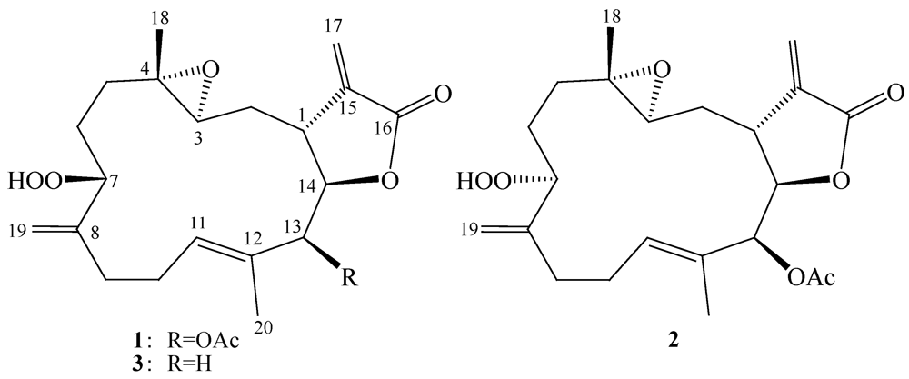
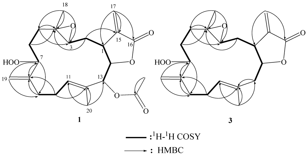
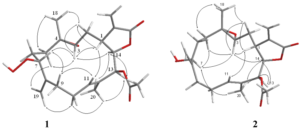
2. Results and Discussion

The HRESIMS (m/z 429.1892 [M + Na]+) of sarcrocrassocolide M (1) established the molecular formula C22H30O7, appropriate for eight degrees of unsaturation, and the IR spectrum revealed the presence of lactonic carbonyl (1757 cm−1) group. The 13C NMR and DEPT (Table 1) spectroscopic data showed signals of three methyls (including one acetate methyl), five sp3 methylenes, two sp2 methylenes, five sp3 methines (including four oxymethines), one sp2 methines, one sp3 and five sp2 quaternary carbons (including two ester carbonyls). The NMR signals (Table 1) observed at δC 169.1 (C), 139.3 (C), 121.6 (CH2), 81.4 (CH), and 37.4 (CH), and δH 6.28, 5.63 (each, 1H, d, J = 2.0 Hz), 4.61 (1H, t, J = 2.5 Hz), and 3.07 (1H, dt, J = 11.5, 2.5 Hz) showed the presence of an α-methylene-γ-lactonic group by comparing with similar NMR data of known cembranoids with the same five-membered lactone ring [22,23]. Signals resonating at δC 60.1 (C), 60.0 (CH) and δH 2.53 (1H, dd, J = 7.0, 4.0 Hz) revealed the presence of a trisubstituted epoxide. One trisubstituted and one 1,1-disubstituted double bond were also identified from NMR signals appearing at δC 129.4 (C), 128.1 (CH), and δH 5.46 (1H, dd, J = 7.0, 5.5 Hz), and at δC 113.5 (CH2), 146.6 (C), δH 5.16 and 5.12 (1H, s, each), respectively. In the 1H–1H COSY spectrum, it was possible to identify three different structural units, which were assembled with the assistance of an HMBC experiment. Key HMBC correlations of H3-18 to C-3, C-4 and C-5; H2-19 to C-7, C-8 and C-9; H3-20 to C-11, C-12 and C-13 and H2-17 to C-1, C-15 and C-16 permitted the establishment of the carbon skeleton (Figure 1). Furthermore, the acetoxy group positioned at C-13 was confirmed from the HMBC correlations of the methyl protons of an acetate (δH 2.02) to the ester carbonyl carbon at δC 169.2 and the oxymethine signal at 77.4 (C-13, CH). The 13C NMR signals at δC 87.1 (CH) and HRESIMS showed the presence of a hydroperoxy group at a methine carbon C-7 [9]. On the basis of the above analysis, the planar structure of 1 was established unambiguously. The relative structure of 1 was elucidated by the analysis of NOE correlations, as shown in Figure 2. The NOE interaction of H-1 (δ 3.07) with H-2β (δ 1.71), H-3 (δ 2.57) and H-11 (δ 5.46) revealed the β-orientation of H-1 and H-3. H-3 did not exhibit NOE correlation with H3-18 (δ 1.32, s) instead it correlated with one proton of H2-5, reflecting the trans stereochemistry of 3,4-epoxide. The proton H-7 (δ 4.30) showed NOE interactions with H-3, and both H-7 and H-11 had NOE correlations with one proton of H2-9 (δ 2.14). Thus, H-7 was placed on the β-face. The E geometry of the trisubstituted double bond at C-11 and C-12 was assigned from the NOE correlation of H3-20 (δ 1.76) with one proton of H2-10 (δ 2.40), but not with the olefinic proton H-11, in addition to the upper field chemical shift of C-20 (δ 14.8). H-14 (δ 4.61) exhibited NOE correlations with both H-13 (δ 5.40) and H3-20, but not with H-1, indicating the α-orientation of both H-13 and H-14. These results, together with other detailed NOE correlations of 1 (Figure 2), unambiguously established the structure of sarcocrassocolide M, as shown in formula 1. Therefore, the relative structure of compound 1 was determined.
Figure 1. 1H–1H COSY and HMBC correlations for 1 and 3.
Marinedrugs 10 00617 g002
Table 1. NMR spectropic data for sarcrocrassocolides M–O (13).
PositionSarcrocrassocolide M (1)Sarcrocrassocolide N (2)Sarcrocrassocolide O (3)
δC, mult.aδH (J in Hz) bδC, mult.aδH (J in Hz)bδC, mult.cδH (J in Hz) d
137.4, CH3.07, dt (11.5, 2.5)37.5, CH3.07, brd (11.5)41.2, CH2.78, ddd (11.2, 4.8, 2.8)
234.8, CH21.87, ddd (14.5, 11.5, 4.0)35.1, CH21.86, ddd (15.5, 11.5, 6.0)33.2, CH21.97, m
1.71, m 1.71, ddd (15.5, 6.0, 2.0) 1.64, m
360.0, CH2.53, dd (7.0, 4.0)59.9, CH2.59, t (6.0)59.8, CH2.63, dd (8.8, 4.6)
460.1, C 60.9, C 60.8, C
532.8, CH21.98, ddd (14.0, 7.0, 3.5)32.9, CH21.96, m32.6, CH21.96, m
1.46, dt (14.0, 7.0) 1.42, m 1.39, m
626.2, CH21.72, m28.6, CH21.62, m26.4, CH21.78, m
1.58, m 1.43, m 1.52, m
787.1, CH4.30, t (5.5)86.1, CH4.38, t (5.0)86.6, CH4.38, t (5.2)
8146.6, C 147.9, C 146.3, C
932.5, CH22.43, m31.9, CH22.47, m32.5, CH22.29, m
2.14, ddd (14.5, 8.0, 4.0) 2.01, m 2.21, m
1026.1, CH22.40, m25.5, CH22.33, m26.0, CH22.37, m
2.29, m 2.23, m
11128.1, CH5.46, dd (7,0, 5.5)127.4, CH5.41, t (7.5)128.8, CH5.26, t (6.8)
12129.4, C 129.4, C 130.5, C
1377.4, CH5.40, s77.4, CH5.39, s44.9, CH22.58, brd (14.4)
2.32, dd (14.4, 8.0)
1481.4, CH4.61, t (2.5)81.7, CH4.62, s82.1, CH4.47, dt (8.0, 4.0)
15139.3, C 139.3, C 138.8, C
16169.1, C 169.1, C 169.4, C
17121.6, CH26.28, d (2.0)122.0, CH26.30, d (2.0)122.8, CH26.32, d (2.4)
5.63, d (2.0) 5.64, d (2.0) 5.62, d (2.4)
1817.5, CH31.32, s17.7, CH31.29, s17.0, CH31.33, s
19113.5, CH25.16, s111.9, CH25.09, s113.8, CH25.17, s
5.12, s 5.08, s 5.12, s
2014.8, CH31.76, s15.1, CH31.78, s17.3, CH31.70, s
OAc20.7, CH32.02, s20.7, CH32.03, s
169.2, C 169.2, C
a Spectra recorded at 125 MHz in CDCl3; b Spectra recorded at 500 MHz in CDCl3; c Spectra recorded at 100 MHz in CDCl3; d Spectra recorded at 400 MHz in CDCl3.
Compound 2 possessed the same molecular formula (C22H30O7) as that of 1, as revealed from HRESIMS. Furthermore, it was found that the NMR spectroscopic data of 2 (Table 1) were similar to those of 1. Analysis of the 2D NMR (1H–1H COSY, HMQC, and HMBC) correlations revealed that compound 2 possesses the same planar structure as that of 1. From the NOESY spectrum, it was found that H-7 (δ 4.38) showed a weak NOE interaction with H3-20 (1.78), but not with H-11 (δ 5.41), revealing the α-orientation of H-7. Further analysis of other NOE interactions revealed that 2 possessed the same relative configurations at C-1, C-3, C-4, C-13 and C-14, as those of 1 (Figure 2). Therefore, 2 was found to be the C-7 epimer of 1.
Figure 2. Key NOESY correlations for 1 and 2.
Marinedrugs 10 00617 g003
Compound 3 was shown by HRESIMS to possess the molecular formula C20H28O5 (m/z 371.1835 [M + Na]+). The IR spectrum of 3 also revealed the presence of lactonic carbonyl (1752 cm−1) group. Comparison of the 1H and 13C NMR data (Table 1) of compounds 1 and 3 showed that the structure of 3 should be very close to that of 1, with the exception of signals assigned to C-13, where an acetoxymethine (δH 5.40, 1H, s; δC 77.4) in 1 was replaced by a methylene (δH 2.58, 1H, brd, J = 14.4 Hz, δH 2.32, 1H, dd, J = 14.4, 8.0 Hz; δC 44.9) in 3. The planar structure of 3 was elucidated by analyzing the 1H–1H COSY and HMBC correlations (Figure 1). The relative stereochemistry of 3 was confirmed from the key NOESY correlations (Figure 3), and the structure of sarcocrassocolide O, as shown in formula 3, was established unambiguously. Thus, 3 is the 13-deacetoxy derivative of 1.
Figure 3. Key NOESY correlations for 3.
Marinedrugs 10 00617 g004
Similar to sarcocrassocolides F–L [22], 13 should be the oxidized products of the related 3α,4α-epoxycembranolides with 7,8-olefinic group, although we have not yet discovered the similar oxidation from cembranolides possessing 7,8-double bond and 3β,4β-epoxide, such as sarcocrassolide (4), sinularolide E (5), and 13-acetoxysarcocrassolide (6) (Chart 2), which were isolated by our previous study [24,25].
Chart 2. Structures of compounds 46.
Marinedrugs 10 00617 g005
The cytotoxicity of compounds 13 against the proliferation of a limited panel of cancer cell lines, including Daoy, HEp-2, MCF-7 and WiDr carcinoma cell lines was evaluated. The results (Table 2) showed that all compounds 13 were found to exhibit cytotoxicity against all or part of the above carcinoma cell lines. In this assay, the in vitro anti-inflammatory effects of compounds 13 were also tested. The inhibition of LPS-induced up-regulation of pro-inflammatory proteins, iNOS and COX-2 in RAW264.7 macrophage cells was measured by immunoblot analysis (Figure 4). At a concentration of 10 μM, compounds 13 were found to significantly reduce the levels of iNOS protein to 4.2 ± 1.6%, 52.9 ± 12.8%, and 22.7 ± 2.8%, respectively, relative to the control cells stimulated with LPS only. At the same concentration metabolites 2 and 3 did not show activity in inhibiting the expression of the pro-inflammatory COX-2 expression with LPS treatment, but compound 1 could reduce the expression of COX-2 to 62.8 ± 22.4%. Thus, compounds 13 might be useful anti-inflammatory agents, while 1 is a promising anti-inflammatory lead compound. Compound 1 could inhibit the expression of both iNOS and COX-2 which might be arisen from the presence of β-hydroperoxy group at C-7 by comparison with compound 2.
Figure 4. Effect of compounds 13 on inducible nitric oxide synthetase (iNOS) and cyclooxygenase-2 (COX-2) proteins expression of RAW264.7 macrophage cells by immunoblot analysis.(A) Immunoblots of iNOS and β-actin; (B) Immunoblots of COX-2 and β-actin. The values are mean ± SEM (n = 6). Relative intensity of the lipopolysaccharide (LPS) alone stimulated group was taken as 100%. * Significantly different from LPS alone stimulated group (*P < 0.05). a stimulated with LPS; b stimulated with LPS in the presence of 13 (10 μM).
Marinedrugs 10 00617 g006
Table 2. Cytotoxicity (ED50μM) of compounds 13.
CompoundDaoyHEp-2MCF-7WiDr
16.6 ± 0.810.4 ±1.110.6 ± 0.5>40
25.2 ± 0.612.3 ± 1.610.1 ± 2.330.1 ± 2.8
35.0 ± 0.712.4 ± 2.16.4 ± 0.5>40
Mitomycin-C0.44 ± 0.060.30 ± 0.060.30 ± 0.120.47 ± 0.12

3. Experimental Section

3.1. General Experimental Procedures

Optical rotations were measured on a JASCO P-1020 polarimeter. Ultraviolet spectra were recorded on a JASCO V-650 spectrophotometer. IR spectra were recorded on a JASCO FT/IR-4100 infrared spectrophotometer. NMR spectra were recorded on a Varian 400MR FT-NMR (or Varian Unity INOVA500 FT-NMR) instrument at 400 MHz (or 500 MHz) for 1H and 100 MHz (or 125 MHz) for 13C in CDCl3. LRMS and HRMS were obtained by ESI on a Bruker APEX II mass spectrometer. Silica gel (Merck, 230–400 mesh) was used for column chromatography. Precoated silica gel plates (Merck, Kieselgel 60 F-254, 0.2 mm) were used for analytical TLC. High-performance liquid chromatography was performed on a Hitachi L-7100 HPLC apparatus with a Merck Hibar Si-60 column (250 × 21 mm, 7 μm) and on a Hitachi L-2455 HPLC apparatus with a Supelco C18 column (250 × 21.2 mm, 5 μm).

3.2. Animal Material

S. crassocaule (specimen No. 20070402) was collected by hand using scuba off the coast of Dongsha, Taiwan, in April 2007, at a depth of 5–10 m, and stored in a freezer until extraction. A voucher sample was deposited at the Department of Marine Biotechnology and Resources, National Sun Yat-sen University.

3.3. Extraction and Separation

The frozen bodies of S. crassocaule (0.5 kg, wet wt.) were minced and exhaustively extracted with EtOAc (1 L × 5). The EtOAc extract (7.3 g) was chromatographed over silica gel by column chromatography and eluted with EtOAc in n-hexane (0–100%, stepwise) then with acetone in EtOAc (50–100%, stepwise) to yield 28 fractions. Fraction 10, eluting with n-hexane–EtOAc (6:1), was further purified over silica gel using n-hexane-EtOAc (7:1) to afford six subfractions (A1–A6). Subfraction A5 was separated by normal-phase HPLC using CH2Cl2-Acetone (40:1) to afford 3 (2.1 mg). Fraction 14, eluting with n-hexane-EtOAc (3:1), was further purified over silica gel using n-hexane-acetone (5:1) to afford eight subfractions (B1–B8). Subfraction B8 was separated by C18 column chromatography and further purified by normal-phase HPLC using CH2Cl2-Acetone (25:1) to afford 1 (4.6 mg). Fraction 17, eluting with n-hexane-EtOAc (1:1), was further purified over silica gel using n-hexane-acetone (3:1) to afford seven subfractions (C1–C7). Subfraction C7 was separated by reversed-phase HPLC using MeOH-H2O (7:5) to afford 2 (2.2 mg).
Sarcocrassocolide M (1): colorless oil; [α]25D −61 (c 0.4, CHCl3); UV (MeOH) λmax 207 (log ε = 3.6); IR (neat) vmax 3421, 2961, 2926, 2855, 1757, 1647, 1371 and 1228 cm–1; 13C and 1H NMR data, see Table 1; ESIMS m/z 429 [M + Na]+ ;HRESIMS m/z 429.1892 [M + Na]+ (calcd for C22H30O7Na, 429.1889).
Sarcocrassocolide N (2): colorless oil; [α]25D −153 (c 0.2, CHCl3); UV (MeOH) λmax 205 (log ε = 3.5); IR (neat) vmax 3419, 2927, 1757, 1659, 1434, 1372, 1272 and 1227 cm–1; 13C and 1H NMR data, see Table 1; ESIMS m/z 429 [M + Na]+ ; HRESIMS m/z 429.1886 [M + Na]+ (calcd for C22H30O7Na, 429.1889).
Sarcocrassocolide O (3): colorless oil; [α]25D −140 (c 0.2, CHCl3); UV (MeOH) λmax 209 (log ε = 3.7); IR (neat) vmax 3456, 2970, 2927, 2855, 1752, 1659, 1434, 1381, and 1271 cm–1; 13C and 1H NMR data, see Table 1; ESIMS m/z 371 [M + Na]+ ; HRESIMS m/z 371.1835 [M + Na]+ (calcd for C20H28O5Na, 371.1834).

3.4. Cytotoxicity Testing

Cell lines were purchased from the American Type Culture Collection (ATCC). Cytotoxicity assays of compounds 13 were performed using the MTT [3-(4,5-dimethylthiazol-2-yl)-2,5-diphenyltetrazolium bromide] colorimetric method [26].

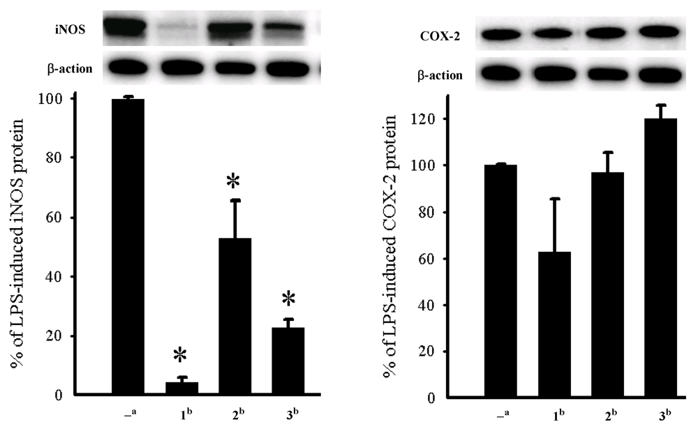
3.5. In Vitro Anti-Inflammatory Assay

Macrophage (RAW264.7) cells were purchased from ATCC. In vitro anti-inflammatory activities of compounds 13 were measured by examining the inhibition of lipopolysaccharide (LPS) induced upregulation of iNOS (inducible nitric oxide synthetase) and COX-2 (cyclooxygenase-2) proteins in macrophages cells using western blotting analysis [27].

4. Conclusions

Our investigation demonstrated that the soft coral, S. crassocaule, could be a good source of bioactive substances. The isolated compounds 13, in particular 1, are potentially anti-inflammatory and may become lead compounds in the future drug development. Also, it is noteworthy to mention that cembranoids 13 possessing an α-methylene-γ-lactonic group with a rarely found 1,1-disubstituted double bond at C-19/C-8 and containing a hydroperoxy group at C-7, were discovered for the first time from corals of this species.

Acknowledgements

This work was supported by grants from the Ministry of Education (00C030205) and National Science Council of Taiwan (NSC 98-2113-M-110-002-MY3 and 100-2320-B-110-001-MY2) awarded to J.-H.S.

References

  1. Bishara, A.; Rudi, A.; Benayahu, Y.; Kashman, Y. Three biscembranoids and their monomeric counterpart cembranoid, a biogenetic diels-alder precursor, from the soft coral Sarcophyton elegans. J. Nat. Prod. 2007, 70, 1951–1954. [Google Scholar] [CrossRef]
  2. Bensemhoun, J.; Rudi, A.; Bombarda, I.; Gaydou, E.M.; Kashman, Y.; Aknin, M. Flexusines A and B and epimukulol from the soft coral Sarcophyton flexuosum. J. Nat. Prod. 2008, 71, 1262–1264. [Google Scholar] [CrossRef]
  3. Marrero, J.; Benítez, J.; Rodríguez, A.D.; Zhao, H.; Raptis, R.G. Bipinnatins K-Q, Minor cembrane-type diterpenes from the west Indian gorgonian Pseudopterogorgia kallos: Isolation, structure assignment, and evaluation of biological activities. J. Nat. Prod. 2008, 71, 381–389. [Google Scholar] [CrossRef]
  4. Shi, Y.-P.; Rodríguez, A.D.; Barnes, C.L.; Sánchez, J.A.; Raptis, R.G.; Baran, P. New terpenoid constituents from Eunicea pinta. J. Nat. Prod. 2002, 65, 1232–1241. [Google Scholar] [CrossRef]
  5. Rashid, M.A.; Gustafson, K.R.; Boyd, M.R. HIV-inhibitory cembrane derivatives from a Philippines collection of the soft coral Lobophytum species. J. Nat. Prod. 2000, 63, 531–533. [Google Scholar] [CrossRef]
  6. König, G.M.; Wright, A.D. New cembranoidditerpenes from the soft coral Sarcophyton ehrenbergi. J. Nat. Prod. 1998, 61, 494–496. [Google Scholar] [CrossRef]
  7. Iwashima, M.; Matsumoto, Y.; Takahashi, H.; Iguchi, K. New marine cembrane-type diterpenoids from the Okinawan soft coral Clavularia koellikeri. J. Nat. Prod. 2000, 63, 1647–1652. [Google Scholar] [CrossRef]
  8. Iguchi, K.; Fukaya, T.; Takahashi, H.; Watanabe, K. Stolonilactone, a novel terpenoid-related compound, isolated from the Okinawan soft coral Clavularia koellikeri. J. Org. Chem. 2004, 69, 4351–4355. [Google Scholar] [CrossRef]
  9. Su, J.-H.; Ahmed, A.F.; Sung, P.-J.; Chao, C.-H.; Kuo, Y.-H.; Sheu, J.-H. ManaarenolidesA-I, new diterpenoids from the soft coral Sinularia manaarensis. J. Nat. Prod. 2006, 69, 1134–1139. [Google Scholar] [CrossRef]
  10. Lu, Y.; Huang, C.-Y.; Lin, Y.-F.; Wen, Z.-H.; Su, J.-H.; Kuo, Y.-H.; Chiang, M. Y.; Sheu, J.-H. Anti-inflammatory cembranoids from the soft corals Sinularia querciformis and Sinularia granosa. J. Nat. Prod. 2008, 71, 1754–1759. [Google Scholar] [CrossRef]
  11. Ahmed, A.F.; Tai, S.-H.; Wen, Z.-H.; Su, J.-H.; Wu, Y.-C.; Hu, W.-P.; Sheu, J.-H. A C-3 methylatedisocembranoid and 10-oxocembranoids from a Formosan soft coral Sinularia grandilobata. J. Nat. Prod. 2008, 71, 946–951. [Google Scholar] [CrossRef]
  12. Chen, B.-W.; Chao, C.-H.; Su, J.-H.; Huang, C.-Y.; Dai, C.-F.; Wen, Z.-H.; Sheu, J.-H. A novel symmetric sulfur-containing biscembranoid from the Formosan soft coral Sinularia flexibilis. Tetrahedron Lett. 2010, 44, 5764–5766. [Google Scholar]
  13. Cheng, S.-Y.; Wen, Z.-H.; Wang, S.-K.; Chiou, S.-F.; Hsu, C.-H.; Dai, C.-F.; Chiang, M.Y.; Duh, C.-Y. Unprecedented hemiketalcembranolides with anti-inflammatory activity from the soft coral Lobophytum durum. J. Nat. Prod. 2009, 72, 152–155. [Google Scholar] [CrossRef]
  14. Chao, C.-H.; Wen, Z.-H.; Wu, Y.-C.; Yeh, H.-C.; Sheu, J.-H. Cytotoxic and anti-inflammatory cembranoids from the soft coral Lobophytum crassum. J. Nat. Prod. 2008, 71, 1819–1824. [Google Scholar] [CrossRef]
  15. Huang, H.-C.; Ahmed, A.F.; Su, J.-H.; Wu, Y.-C.; Chiang, M.Y.; Sheu, J.-H. Crassocolides A–F, new cembranoids with a trans-fused lactone from the soft coral Sarcophyton crassocaule. J. Nat. Prod. 2006, 69, 1554–1559. [Google Scholar] [CrossRef]
  16. Huang, H.-C.; Chao, C.-H.; Kuo, Y.-H.; Sheu, J.-H. Crassocolides G-M, cembranoids from a Formosan soft coral Sarcophyton crassocaule. Chem. Biodivers. 2009, 6, 1232–1242. [Google Scholar] [CrossRef]
  17. Cheng, Y.-B.; Shen, Y.-C.; Kuo, Y.-H.; Khalil, A.T. Cembranediterpenoids from the Taiwanese soft coral Sarcophyton stolidotum. J. Nat. Prod. 2008, 71, 1141–1145. [Google Scholar] [CrossRef]
  18. Cheng, S.-Y.; Wang, S.-K.; Chiou, S.-F.; Hsu, C.-H.; Dai, C.-F.; Chiang, M.Y.; Duh, C.-Y. Cembranoids from the octocoral Sarcophyton ehrenbergi. J. Nat. Prod. 2010, 73, 197–203. [Google Scholar] [CrossRef]
  19. Su, J.-H.; Lu, Y.; Lin, W.-Y.; Wang, W.-H.; Sung, P.-J.; Sheu, J.-H. A cembranoid, trocheliophorol, from the cultured soft coral Sarcophyton trocheliophorum. Chem. Lett. 2010, 39, 172–173. [Google Scholar] [CrossRef]
  20. Wang, G.-H.; Huang, H.-C.; Su, J.-H.; Huang, C.-Y.; Hsu, C.-H.; Kuo, Y.-H.; Sheu, J.-H. Crassocolides N–P, three cembranoids from the Formosan soft coral Sarcophyton crassocaule Bioorg. Med. Chem. Lett. 2011, 21, 7201–7204. [Google Scholar]
  21. Sheu, J.-H.; Wang, G.-H.; Duh, C.-Y.; Soong, K. Pachyclavulariolides M-R, six novel diterpenoids from a Taiwanese soft coral Pachyclavulariaviolacea. J. Nat. Prod. 2003, 66, 662–666. [Google Scholar] [CrossRef]
  22. Lin, W.-Y.; Lu, Y.; Su, J.-H.; Wen, Z.-H.; Dai, C.-F.; Kuo, Y.-H.; Sheu, J.-H. Bioactive cembranoids from the Dongsha Atoll soft coral Sarcophyton crassocaule. Mar. Drugs 2011, 9, 994–1006. [Google Scholar] [CrossRef]
  23. Lin, W.-Y.; Su, J.-H.; Lu, Y.; Wen, Z.-H.; Dai, C.-F.; Kuo, Y.-H.; Sheu, J.-H. Cytotoxic and anti-inflammatory cembranoids from the Dongsha Atoll soft coral Sarcophyton crassocaule. Bioorg. Med. Chem. 2010, 18, 1936–1941. [Google Scholar]
  24. Duh, C.-Y.; Wang, S.-K.; Chung, S.-G.; Chou, G.-C.; Dai, C.-F. Cytotoxiccembrenolides and steroids from the Formosan soft coral Sarcophyton crassocaule. J. Nat. Prod. 2000, 63, 1634–1637. [Google Scholar] [CrossRef]
  25. Li, G.; Zhang, Y.; Deng, Z.; Ofwegen, L. V.; Proksch, P.; Lin, W. Cytotoxic cembranoid diterpenes from a soft coral Sinularia gibberosa. J. Nat. Prod. 2005, 68, 649–652. [Google Scholar] [CrossRef]
  26. Scudiero, D.A.; Shoemaker, R.H.; Paull, K.D.; Monks, A.; Tierney, S.; Nofziger, T.H.; Currens, M.J.; Seniff, D.; Boyd, M.R. Evaluation of a soluble tetrazolium/formazan assay for cell growth and drug sensitivity in culture using human and other tumor cell lines. Cancer Res. 1988, 48, 4827–4833. [Google Scholar]
  27. Jean, Y.-H.; Chen, W.-F.; Duh, C.-Y.; Huang, S.-Y.; Hsu, C.-H.; Lin, C.-S.; Sung, C.-S.; Chen, I.-M.; Wen, Z.-H. Inducible nitric oxide synthase and cyclooxygenase-2 participate in anti-inflammatory and analgesic effects of the natural marine compound lemnalol from Formosan soft coral Lemnalia cervicorni. Eur. J. Pharmacol. 2008, 578, 323–331. [Google Scholar] [CrossRef]
  • Sample Availability: Not available.

