Aptamers as Diagnostic Tools in Cancer
by
 ,
,
Dario Ruiz Ciancio
1 ,
,
Mauricio R. Vargas
1,
William H. Thiel
2,
Martin A. Bruno
1,
Paloma H. Giangrande
2 and
María Belén Mestre
1,* 1
Biomedical Science Institute (ICBM), Catholic of Cuyo University, San Juan, CP 5400, Argentina
2
Department of Internal Medicine, University of Iowa, Iowa City, IA 52246, USA
*
Author to whom correspondence should be addressed.
Pharmaceuticals 2018, 11(3), 86; https://doi.org/10.3390/ph11030086
Submission received: 24 July 2018
/
Revised: 30 August 2018
/
Accepted: 2 September 2018
/
Published: 11 September 2018
(This article belongs to the Special Issue Aptamer-Based Diagnostics and Therapeutics)
Abstract
:Cancer is the second leading cause of death worldwide. Researchers have been working hard on investigating not only improved therapeutics but also on early detection methods, both critical to increasing treatment efficacy, and developing methods for disease prevention. The use of nucleic acids, or aptamers, has emerged as more specific and accurate cancer diagnostic and therapeutic tools. Aptamers are single-stranded DNA or RNA molecules that recognize specific targets based on unique three-dimensional conformations. Despite the fact aptamer development has been mainly restricted to laboratory settings, the unique attributes of these molecules suggest their high potential for clinical advances in cancer detection. Aptamers can be selected for a wide range of targets, and also linked with an extensive variety of diagnostic agents, via physical or chemical conjugation, to improve previously-established detection methods or to be used as novel biosensors for cancer diagnosis. Consequently, herein we review the principal considerations and recent updates in cancer detection and imaging through aptamer-based molecules.
1. Introduction
Cancer is a major public health problem and the second leading cause of death worldwide. The number of new cases of cancer is expected to grow rapidly as the population increases and continues to make lifestyle choices that increase cancer risk [1,2]. According to Hussain and Nguyen, focusing on early detection and accurate staging of cancer is as important as the development of new anticancer drugs and treatments intended to cure cancer [3].
The accumulation of genetic alterations in cells together with abnormal cell division and elevated resistance to apoptosis are known factors in cancer development. These changes allow mutant cells to acquire the ability to overexpress certain proteins not expressed by normal cells [4]. These cancer-specific markers are attractive targets for cancer diagnosis and treatment [5]. Recently, greater emphasis has been placed on investigating not only improved therapeutics but also early detection and diagnosis methods. In many cases, cancer metastasizes throughout the body before it is diagnosed, resulting in decreased probability of successful cure and/or survival. Therefore, early and accurate cancer diagnosis is critical to increasing treatment efficacy, decreasing secondary diseases post-therapy, and ultimately developing methods for disease prevention [6,7]. Based on these observations, the necessity of more sensitive, low-cost, and/or non-invasive methods for cancer diagnosis is still a challenge for research laboratories.
Aptamers have recently emerged as potential molecular probes in the development of more specific and accurate cancer diagnostics and therapeutics due to their unique characteristics. Aptamers are single-stranded nucleic acid molecules (ssDNA or RNA) that recognize specific targets based on unique three-dimensional conformations [8]. A wide range of diagnostic agents can be linked to aptamers via physical or chemical conjugation in order to further customize their function [9]. Specifically, over the past decade, aptamers that target proteins overexpressed in tumors have become ideal tools for the specific recognition of these cancer-specific markers [10]. Consequently, the adaptation of aptamer-based technologies to actual cancer diagnosis methods could provide a great opportunity for advances in cancer detection.
Thus, we present a state-of-the-art aptamer review with a specific focus on their application to cancer. We summarize the latest advances in aptamer use for cancer diagnosis, highlighting their potential future role as promising molecules in cancer detection and imaging.
2. Aptamers
Aptamers are single-stranded nucleic acid molecules (ssDNA or RNA) that recognize specific targets based on unique three-dimensional conformations [8]. Aptamers are developed in vitro through a process called systematic evolution of ligands by exponential enrichment (SELEX) (Figure 1) [11]. SELEX technology was established by Tuerk et al. and Ellington et al. in 1990 [12,13]. Typically, a selection cycle starts with the incubation of the protein target with a combinatorial DNA or RNA library, comprised of approximately 1012–1014 unique oligonucleotide sequences 20–80 bases in length. Protein-bound aptamers are eluted and amplified to create the library for the next round of selection. An enriched pool of potential binders generated from PCR amplification is used in the subsequent selection rounds. To increase the selection stringency, a negative selection step can be introduced. After multiple selection rounds, potential target-specific aptamers are obtained [14]. Additionally, a negative selection step can be introduced to eliminate sequences that bind to non-desirable targets, thereby increasing the specificity of the selection [15,16].
The resulting highly enriched pool of aptamers can be analyzed using next-generation sequencing for further characterization of individual aptamers [17]. In fact, as better sequencing technologies and bioinformatic software are developed, aptamer researchers have been able to identify functional oligonucleotides from earlier rounds by evaluating population dynamics during selections [17,18,19]. Proteins are by far the most common target used in SELEX. However, the difficulty of generating high-purity recombinant human proteins, with native conformation as SELEX-targets, has made it difficult to produce more reliable and robust aptamers. In addition, SELEX is not applicable for unknown proteins, insoluble proteins, or proteins that only function in a multiprotein complex [20,21,22]. Consequently, scientists have been working hard on different variants of aptamer selection methodology. The advances and improvements made in traditional SELEX have allowed the development of new methods that permit the selection of aptamers against not only proteins [23] but also against a wide range of targets, such as live cells [24,25,26,27], viruses [28], bacteria [29], and tissues [30]. Cell-based SELEX methodology (cell-SELEX) stands out among other new aptamer-selection methods: the oligonucleotides selected with this technique are able to bind not just cell-surface molecules in their native state, but also unknown membrane receptors [31,32]. Therefore, cell-SELEX offers a new opportunity for working with aptamers that recognize the native state of proteins.
Over the past two decades, cell-SELEX technology has been used for the selection of specific aptamers with diagnostic and therapeutic purposes over a wide variety of cell surface targets, particularly for tumor cells. Multiple groups have reported the discovery of new important biomarkers in different types of cancer and other diseases through the use of cell-SELEX [18,33,34,35].
The general protocol of cell-SELEX includes a positive selection step of incubation, elution, and amplification of binding aptamers, and a negative selection step is also necessary to remove sequences which bind to normal cells. In other words, the cell-SELEX method first starts with the incubation of the nucleic acid library with normal or control cells that express either normal surface proteins or do not express the target protein. This step is called the negative selection step. In the positive selection step, unbound aptamers are incubated with the target cancer cells. Candidate aptamers are recovered from the positive cell lines and amplified to become the library for the next round. This cycle of selection is repeated for as many rounds as necessary [36]. Changing the conditions of each selection round is crucial to increasing stringency and yielding high-affinity aptamers that bind to the target cell population [21,37]. The stringency can be increased after each cycle by increasing the washing steps and/or increasing the number of non-target competitors. Reducing the number of target cells will achieve the same goal, as it will force the aptamers to compete with each other for the target, increasing target affinity and the overall efficiency of the process. Finally, the enriched pools are sequenced, and representative aptamers are chosen for subsequent characterization. With the incorporation of flow cytometry and high-throughput sequencing to cell-SELEX technology, a shorter selection round and high-quality aptamer identification have been achieved [38,39].
Despite its enormous potential, cell-SELEX presents technical limitations such as cell conditions and the complexity of some cancer cell lines. It is important for the aptamer selection to avoid nonspecific uptake. Cell death leads to altered protein expression. Therefore, the elimination of these cells will favor the identification of specific aptamers. Second, it is important to choose the proper cell line because cell surface proteins are very complex in some cancers. Because of the complexity and heterogenicity of some cancer cells, it is critical to perform additional selection rounds against non-target cells in order to improve aptamer specificity. However, this selection process can be quite complex and may have a negative impact in terms of time and resources spent by the researcher [40].
Currently, new selection methods based on cell-SELEX have emerged to improve the success rate of aptamer selection, such as target expressed on cell surface-SELEX (TECS-SELEX) [41], fluorescence-activated cell sorting-SELEX (FACS-SELEX) [42], 3D SELEX [43], automated SELEX [44], click-SELEX [45], cell-internalization SELEX [46], and in vivo SELEX [24], among others.
Several factors need to be taken into consideration before the selection process begins. One of these aspects is the design of the library for SELEX procedures. Researchers must decide whether they are working with DNA or RNA aptamers—a decision that depends on the purpose of the selection. DNA and RNA aptamers are functionally similar, but each has its own unique benefits. DNA aptamers are inherently more stable, cheaper, and easy to produce. RNA aptamers typically have more diverse three-dimensional conformations due to the 2′-OH group and stronger RNA-RNA interactions, which probably increase binding affinity and specificity. However, RNA aptamers need more steps between selection rounds than DNA aptamers, making the method potentially more cost- and labor-intensive. Whereas PCR is used to recover the DNA aptamer-bound sequences, RNA aptamers must first be reverse-transcribed into cDNA to enable subsequent PCR amplification and RNA in vitro transcription for the next selection round [15,47].
There are also several challenges in terms of in vivo application that remain unresolved. Aptamers are vulnerable to nuclease degradation and have short circulation times. They can be chemically modified to enhance their nuclease resistance and in vivo stability. Replacing the 2′-OH position with fluoro (F), amino (NH2), or O-methyl (OCH3) groups can increase the half-lives by reducing aptamer degradation. Capping the 3′end with an inverted dT residue can provide resistance to serum nucleases as well. The latest results of Kratschmer and Levy indicate that fully modified oligonucleotides (100% 2′-O-Methyl or 2′-O-Methyl A, C, and U in combination with 2′-fluoro G) had the longest half-lives. They also demonstrated little degradation in human serum even after prolonged incubation [48,49,50]. In practice, these modifications can be accomplished by two different methods: in-SELEX and post-SELEX. In the in-SELEX strategy for aptamer selection, a DNA or RNA library containing the modified nucleotides is produced by using a mutant T7 RNA polymerase [51,52]. However, according to Rozemblum et al., backbone modifications can reduce the ability of polymerase to amplify the DNA in the PCR amplification step [53]. Conversely, the post-SELEX strategies introduce modifications to aptamers after the selection, during the solid-phase chemical synthesis of individual aptamers [54]. To solve the problem of renal filtration, aptamers are generally linked to polyethylene glycol (PEG), cholesterol, or proteins. Using a multivalent aptamer design can also help to achieve the same goal [55,56,57]. Regarding toxicity, while it is very limited in humans, chemical modifications should be used carefully in order to avoid undesired effects. In the Phase III study of the Regado Biosciences aptamer-based anticoagulation system (REG1 system) [58], serious allergic responses to PEG were observed [59]. Thus, evaluation and optimization of aptamer-modifications are still a challenge.
Several authors have reviewed the importance of aptamer development and research by emphasizing their advantages over antibodies [60,61,62]. However, the clinical success of aptamers is far behind antibodies, with only one FDA-approved aptamer available to-date [63]. The use of antibodies in cancer diagnosis and therapy has dominated the marketplace, while aptamer development has remained mainly confined to laboratory settings. Nevertheless, aptamers are still notable molecules with great potential for clinical application. Instead of comparing them with antibodies, scientists should rather concentrate on the unique attributes of aptamers and take advantage of them. Aptamers present excellent stability to variations in pH, temperature, and ionic conditions of the environment [64]. They have low immunogenicity and toxicity, and they are easily synthesized and modified in large quantities without batch-to-batch variation [64,65]. Furthermore, the size of the aptamer enables deeper tumor penetration [66]. They can be selected over a wide range of targets: ions, organic and inorganic molecules, nucleic acids, proteins, peptides, toxins, viral particles, whole cells, entire organs, and live animals [64]. More importantly, aptamers could be used as a delivery tool to release diagnostic or therapeutic cargoes into the intracellular space of target cells. In this way, aptamers could be linked with drugs, non-coding RNAs, proteins, and nanoparticles, making them promising tools for specific tumor diagnosis and therapy [67].
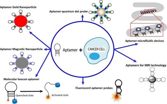
3. Applications of Aptamers in Cancer Imaging and Diagnostics
For the past decade, researchers have been working in the development of new methods for cancer imaging and diagnosis that can identify the presence of cancer biomarkers at early stages, before the appearance of any symptoms. Being able to detect cancer early on would allow for earlier treatment responses and increased probability of curing the disease. Nowadays, physicians use magnetic resonance imaging (MRI), computed tomography, and other imaging techniques combined with flow cytometry, immunohistochemistry, and cancer biomarker assays in serum to reach a diagnosis in cancer patients [68]. However, these methods face various technical issues, especially due to the often low concentrations of cancer markers mixed with other proteins and cells that make their identification in bodily fluids quite difficult. These methods can also be time- and labor-intensive [69,70,71]. Consequently, aptamers appear as a promising tool for cancer imaging and detection in biological samples because they are able to recognize almost any oncological biomarker, cancer metabolite, or cancer cell with high affinity and specificity at low target concentrations. Researchers may also benefit from the adaptability and easy chemical manipulation of aptamers. Thus, aptamers could be linked with a wide range of existing cancer detection and imaging platforms to improve sensitivity and selectivity [6,72]. They could also be used to detect and measure the expression or activity of target molecules that influence tumor behavior or change in response to therapy [73].
3.1. Aptamer-Nanoparticle System
One detection system used for cancer diagnosis is aptamers linked to nanoparticles (Apt-NP). The use of this chimera is a good alternative to achieve the specific extraction and detection of cancer cells in complex fluids such as blood. Due to the low molecular weight of the aptamer, this method allows cell-specific aptamers to be immobilized on the surface of nanoparticles to create an aptamer–gold nanoparticle (Apt-AuNP) system (Figure 2) or aptamer-magnetic nanoparticle (Apt-MNP) cluster (Figure 3), among others. These Apt-NP chimeras are expected to improve cancer diagnosis with higher sensitivity and selectivity compared to cancer-cell-detection strategies that use aptamers or nanoparticles alone. Interestingly, nanomaterials protect aptamers from being digested by nucleases [29,74].
Liu et al. developed an aptamer-conjugated gold nanoparticle strip biosensor (ANSB) for the detection of Ramos cells, even in human blood [75]. In short, Liu et al. used a pair of previously identified aptamers through cell-SELEX for Burkitt’s lymphoma [33], a thiolated aptamer TD05 (Thiol-TD05), and a biotinylated aptamer TE02 (Biotin-TE02). In this work, both aptamers Thiol-TD05 and Biotin-TE02 were immobilized on the nanoparticle surface and on the test zone of ANSB, respectively.
A sample containing Ramos cells and Thiol-TD05-NP migrate along the strip. The Thiol-TD05-AuNP-Ramos cell complex is captured on the test zone through the immobilized aptamer, and its accumulation produces a quantifiable red band. As a result of ANSB, approximately 4000 Ramos cells could be detected with the researcher’s eye and 800 cells with a portable strip reader. In similar studies, Medley et al. and Borghei et al. developed a colorimetric assay based on the color produced by the conjugated aptamer-nanoparticle [76,77]. In Medley’s study, a thiol-aptamer for T- acute lymphoblastic leukemia cells was linked to gold-nanoparticles. When the Thiol-Apt-AuNP recognized the target cells, a change in color was observed. Thus, by measuring the color change, researchers were able to differentiate between target and non-target cells. Borghei et al. used the ability of the target cells to capture the aptamer. A specific aptamer for nucleolin, a protein overexpressed in metastatic cells, was used. When the sample contained the cancer cells, the aptamer bound nucleolin receptors. The solution containing the single-stranded DNA (ssDNA) joined to gold nanoparticles, revealing a red color because of the aptamers’ removal. However, when aptamers, but no cancer cells, were present in the sample, the solution was blue because aptamers interact with ssDNA-AuNP probes. This procedure had a detection limit of 10 MCF-7 cells. In contrast, Hu et al. developed an approach in which the aptamer and the nanoparticle were separated but interacted with each other via biotin-streptavidin interaction [78]. In this study, HepG2 cells were incubated with biotin-conjugated TLS11a aptamer (Bio-TLS11a). Then, the cancer cells-Bio-TLS11a complexes with streptavidin-labeled fluorescent silica nanoparticles (SA-FSNPs) were detected due to strong and specific biotin-streptavidin association. This approach allowed the sensitive and specific detection of hepatoma cancer cells in vitro. Moreover, an aptamer-modified fluorescent silica nanoparticle (FSNPs) system was used by Tan et al. for the detection of leukemia cells. This group used Sgc8 aptamer labeled with amine linked to carboxyl-modified FSNPs. This system could detect leukemia cells with high sensitivity and specificity [79]. Finally, the same Sgc8 aptamer, as well as 41t and TE17 aptamers, were linked to a sophisticated nanorobot and tested in response to target proteins. When aptamers recognized their targets, active previously-loaded cargoes such as gold nanoparticles, were delivered to the target cell. This study demonstrated the promising future of aptamers for the specific delivery of nanoparticles loaded into DNA-based nanorobots for cancer cell detection [80].
A similar approach to an Apt-AuNP system was pursued using aptamers conjugated to magnetic nanoparticles (Apt-MNPs). Tan’s group developed an effective method for the detection of a minimum of 10 cancer cells in 250 μL of sample. The method consists of measuring the change of spin-spin relaxation time (ΔT2) of the surrounding media water protons. It is well-known that MNPs alone enhance the magnetic resonance signal of protons from surrounding water molecules. In this way, when aptamer-magnetic nanoparticle clusters recognize the cancer cells, they generate a magnetic field that brings a consequent decrease of the T2. Tan et al. were able to recognize human T-leukemic cells (CCRF-CEM) and colorectal cancer cell lines (DLD-1 and HCT 116) using specific aptamers for each cell line in different complex biological media, including fetal bovine serum, human plasma, and whole blood. This proof-of-concept study demonstrated the ability of the Apt-MNPs clusters to be a sensitive and specific method for cancer cells detection and diagnosis [81].
A different approach was developed combining the biological recognition property of Apt-AuNP with a physical transduction technique (i.e., electrochemiluminescence, ECL), to achieve the detection of target cancer cells [82,83]. Zhang et al. could detect cancer cells using an Apt-magnetic complex and a reporter DNA-AuNP combined with a sensitive ECL approach. In this study, a DNA-1 was hybridized in a CdS nanocrystal (NC) film. HL-60-specific aptamers were linked to magnetic beads (Apt-MBs) and hybridized with a DNA-2 labeled with AuNPs. In the presence of HL-60 cells, the Apt-MBs conjugated with the cell surface and the DNA-2-AuNPswere released. After magnetic separation, the DNA-2-AuNPs hybridized with the captured DNA-1 on the CdS NC film, increasing the ECL signal. Between 20 to 1.0 × 10⁶ cells/mL of HL-60 cells could be detected using this approach. In another study, Zhang et al. demonstrated the electrochemical detection of CCRF-CEM cells using Apt-MNPs conjugated with ferrous ferric oxide (Fe3O4). A competitive binding assay was performed in which T-acute leukemia cells competed efficiently with an AuNP-conjugated cDNA for binding specifically to the aptamer of the Fe3O4-Apt-MNP complex. The released AuNPs were treated via silver deposition in the presence of target cells. The AuNP-catalyzed silver deposition enhancement showed high sensitivity with a detection limit of 10 cells [84]. Another highly sensitive and selective method for the early detection of leukemia cells was developed by Khoshfetrat and Mehrgardi [85]. In this study, the T-cell leukemia sgc8c aptamer, a truncated version of sgc8 aptamer, was linked to gold nanoparticles-coated magnetic Fe3O4 nanoparticles (Apt-GMNPs). Ethidium bromide (EB) was intercalated into the stem of the aptamer hairpin. The presence of cancer cells caused the disruption of the hairpin structure of the aptamer, forcing the release of EB and a consequent decrease in electrochemical signal. Under optimal conditions, this technology could detect a range of leukemia cancer cells from 10 to 1 × 10⁶ cell/mL. However, most of the ECL approaches achieve cell detection by capturing cells on the electrode surface. A novel sandwich sensing system through an aptamer-cell-aptamer architecture has been developed by several authors [86,87,88,89]. In their studies, one of the aptamers was combined with AuNPs and immobilized on the electrode surface. Cancer cell lines could bind to this sandwich sensing system, and they could be measured by cyclic voltammetry (CV) and electrochemical impedance spectroscopy (EIS) using ferricyanide (Fe(CN)63−/4−) as a redox probe. More recently, Crulhas et al. described a system to detect vascular endothelial growth factor (VEGF) and prostate-specific antigen (PSA) released by prostate cancer cells based on specific thiolated aptamers bound to a gold-covered electrode surface [90]. This electrochemical aptamer-based biosensor could detect 0.08 ng/mL of PSA and 0.15 ng/mL of VEGF, released in vitro by three different prostate cancer cell lines (RWPE-1, LNCaP, and PC3). The precise detection of these biomarkers make this aptasensor a potentially important non-invasive tool for prostate cancer diagnosis. It is clear that the use of aptamer-based electrochemical biosensors is gaining popularity due to its simplicity, rapid response, low-cost fabrication, and high sensitivity [91].
3.2. Aptamer-Microfluidic Devices
The use of aptamers for cancer diagnosis has also spread to other platforms. In 2009, Phillips and colleagues revealed the combination of aptamers with microfluidic devices to be an excellent platform for cancer diagnosis, since this device is cheap, simple, and able to detect multiple cancer cells [92]. Briefly, aptamers are first immobilized on the surface of a microfluidic channel. Then, a mixture of cells is passed along this device, allowing specific immobilized aptamers to capture target cells (Figure 4). The percentage of captured cells can be measured by optical microscopy. Xu et al. proved that captured cells could be sorted and cultured for further studies using this platform [93]. To achieve this goal, three different aptamers for leukemia cells (Sgc8, TD05, and Sgd5) were immobilized on the surface of the microfluidic device and the leukemia cells were isolated with high purity (50% to 100% purity for the different leukemia cell lines). In a similar study, a DNA aptamer against protein tyrosine kinase 7 (PTK7), a protein overexpressed in multiple human cancers, was reportedly immobilized on a microfluidic surface [94]. An aptamer-3D network was synthesized in this study using rolling circle amplification (RCA) to increase the capture and isolation of the target cells. RCA is a DNA amplification technique in which short DNA is elongated with the assistance of a circular DNA template. Consequently, RCA is employed to extend the aptamer sequence, creating a 3D network with multiple aptamer domains on the microfluidic surface. CCRF-CEM cells (T-human acute lymphoblastic leukemia cells) were captured with higher efficiency than monovalent aptamers and antibodies immobilized with the same device. Captured cells could be easily released for further analysis using restriction enzymes to cleave the aptamers. Interestingly, other authors have also incorporated the specific cell aptamer recognition with the RCA technology in their experiments to augment the specificity and sensitivity for the detection of cancer cells [95,96]. Recently, Chen et al. investigated how cells and aptamers interact under different flow conditions in microfluidic devices [97]. They used T- acute leukemia cells (CCRF-CEM) and their specific aptamer, Sgc8, to analyze the effects of flow rates and device shapes. The results of CCRF-CEM capture efficiency in this study will help researchers to choose the best experimental conditions and device shapes. Another elegant study conducted by Sheng et al. used DNA nanostructures combined with microfluidics for circulating tumor cell (CTCs) isolation [98]. In this work, DNA nanospheres were constructed by immobilizing up to 95 aptamers to gold nanoparticles (AuNP), and then these DNA nanospheres were immobilized onto the channel of microfluidic devices. Efficient isolation of CTCs from cell mixture and whole blood was achieved using this platform. This study showed that the combination of two different technologies, nanotechnology and microfluidics, has enormous potential in the cancer field.
3.3. Aptamer-Quantum Dots Probes
Quantum dots (QDs) appear to be promising tools for cancer imaging and diagnosis because they can be easily conjugated with nucleic acids that specifically target biomolecules. QDs with distinct emission wavelengths can be ligated to different aptamers for multiple cancer detection (Figure 5). Kang et al. presented a system that simultaneously targets three cancer molecular markers: tenascin-C, nucleolin, and mucin (MUC-1). To achieve this goal, three aptamers (TTA1, AS1411. and MUC-1) were conjugated to QDs with distinct emission wavelengths of 605, 655, and 705 nm, respectively. Healthy and cancer cell lines were incubated with QD-aptamer conjugates. Results showed that these QDs-aptamer complexes could not only produce a visible fluorescence signal in the presence of target cells, but could also differentiate between different types of cancer cells [99]. Similarly, Lian et al. used the AS1411 aptamer conjugated with QDs to recognize breast cancer cells through confocal microscopy, being suitable for in vitro diagnostic biosensing [100]. In 2015, two different aptamer-conjugated QDs (TTA-1 and AS1411 aptamer) were used by Lee et al. to simultaneously demonstrate the presence of the cancer biomarkers tenascin-C and nucleolin in different cancer cell lines [101]. In similar studies, two different researchers created polymeric structures by conjugating acrylamide, aptamers, and QDs [102,103]. These polymeric QD-aptamer systems successfully exhibited fluorescence in the presence of cancer cells. Furthermore, they proved to be suitable systems for long-term fluorescent cellular imaging. Recently, Tang’s group developed a new system for use in fluorescence-guided surgery for glioma, using A32 aptamer labeled with QDs. A32 aptamers specifically recognized epidermal growth factor receptor variant III, highly dispersed on the surface of glioma cells. The system generated strong fluorescence in the mouse model of glioma, and was nontoxic both in vitro and in vivo. Most importantly, this system could be applied for preoperative diagnosis and postoperative examination of glioma [104]. Generally, aptamers are first labeled to QDs. Then, the aptamer-QDs recognize the cancer cells. However, Wu et al. developed a recognition-before-labeling strategy, where the aptamer first recognizes the target cells and then fluorescent QDs bind to the aptamers-cell conjugate via simple streptavidin-biotin interaction. This new method for in vitro diagnostic assays of cancer cells has some advantages over other strategies because it avoids the impact on aptamer configuration when conjugated to QDs [105].
3.4. Molecular Beacon Linked to Aptamers
Molecular beacon aptamers have been used to monitor different kinds of targets, including cancer cells. A molecular beacon (MB) is a dual-labeled single stranded DNA with a fluorophore at the 5’ end and a quencher at the 3’ end. The molecule has a stem-loop structure where the fluorophore and quencher do not fluoresce when in close proximity to each other. However, if the probes hybridize to a target sequence, separating the fluorophore and quencher, they will fluoresce (Figure 6). Shi et al. developed an activated fluorescence aptamer probe using molecular beacon technology [106]. While fluorescence is quenched in free Sgc8 aptamers, fluorescence is activated after targeting CCRF-CEM cancer cells due to an aptamer-conformational alteration. Activated fluorescence signals were observed in vitro and in vivo in the CCRF-CEM tumor sites. Another proof-of-concept study conducted by Zeng et al. [107] featured an aptamer-reporter conjugated with a pair of fluorochrome-quencher molecules that selectively target circulating tumor cells (CTCs). In the absence of CTCs, the probe is optically silent. However, when it recognizes and is taken up by tumor cells, the aptamer-reporter is rapidly degraded, resulting in the separation of the fluorochrome and quencher, with the subsequent activation of the fluorochrome. This method was used to detect CTCs with no background noise in whole blood and bone marrow aspirates. Using the same principle, aptamers were used by different researchers as internalizing carriers for the specific delivery of MB probes targeting mRNA and miRNAs. So far, the AS1411 aptamer was used labeled with a MB probe specific for miRNAs. Both Li et al. and Kim et al. used the great cancer selectivity of AS1411 to achieve cell-specific delivery of the MB probe that allows intracellular miRNA imaging of the miRNA target [108,109]. Interestingly, aptamer-MB for miRNA imaging could be applied to other cancers by changing the target miRNA sequence of the MB. Another study conducted by Qui and colleagues investigated intracellular mRNA analysis in live cells using aptamer-based molecular beacon probes. The internalizing aptamer AS1411 with an extended cDNA sequence was linked to a single-stranded MB specific for the mRNA. As expected, the MB was delivered into the cytoplasm of cancer cells, allowing detection of the mRNA after fluorescence was activated [110].
Two years ago, Zhang et al. identified abnormal DNA methyltransferase activity which is closely associated with cancer, through the detection of DNA adenine methylation methyltransferase (Dam MTase) activity [111]. Both a streptavidin-specific aptamer (SA-apt) labeled with a single fluorophore and an allosteric molecular beacon (aMB) were used in this study. The SA-apt could not bind with SA beads because a stable hairpin structure was formed in the absence of a target. Nevertheless, the presence of Dam MTase methylated the aMB, making it available for the restriction nuclease Dpnl to cut the methylated probe and release the fluorophore-labeled aptamer. The free SA-apt bound to SA beads, making the beads highly fluorescent. DNA methyltransferase activity can be quantified by a microscope or by flow cytometry. Zhao’s group similarly took advantage of conformational changes after binding to develop an “activatable” aptamer-based fluorescence probe (AAFP) to detect cancer cells and frozen cancer tissue [112]. In this study, the TLS11a aptamer, specific for HepG2 cells, was linked to two short-complementary DNA sequences, with a 5’-fluorophore and 3’-quencher, respectively. In the absence of a target, the AAFP formed a hairpin structure capable of auto-quenching. In the presence of cancer cells, the fluorophore and quencher separated, making the AAFP emit a strong fluorescence signal. Under optimal incubation conditions, AAFP was able to detect cancer cells at concentrations as low as ~100 cells/mL. These results indicate the AAFP could be a promising tool for the specific detection of cancer cells with low signal-to-background ratio. Another elegant study designed by Hwang et al. detected the exogenous EpCAM (epithelial cell adhesion molecule) or muc1 (mucin1) expression correlated to cancer metastasis [113]. In this study, a quantum dot-based aptamer beacon was used for CTC diagnosis, conjugated with a 5’-quantum dot and a 3’-black hole quencher. In the absence of target EpCAM/muc1 of CTCs, the aptamer remained in the quenched state because of its conformational state. When the target molecule was present, a fluorescence signal was emitted due to the activation of the EpCAM/muc1 aptamer. A change in its conformation, when targeting EpCAM/muc1, allowed separation of the QD and the quencher and the subsequent fluorescence signal emission.
3.5. Fluorescent Aptamer Probes
Fluorescent aptamer probes appear to be one of the most widely used imaging tool in aptamer research due to their low cost and high sensitivity (Figure 7). One of the first authors to apply fluorescent aptamer probes to cancer research was Zhang et al., who used a fluorescent-labeled RNA aptamer specific for CD30—a protein overexpressed in lymphoma cell lines [114]. Specific binding of the aptamer to lymphoma cells was confirmed using flow cytometry and fluorescence microscopy. While the anti-CD30 antibody is currently the gold standard for CD30 detection, this study suggests that CD30 aptamer could be used in combination with CD30 antibody for the improved detection and diagnosis of lymphoma. A study by Shi et al. used a novel aptamer-based fluorescence imaging approach to selectively detect Ramos tumor in mice using Cy5-labeled TD05 aptamer (Cy5-TD05), specific to Ramos cells (B-cell lymphoma cell line). Cy5-TD05 was injected intravenously into Ramos tumor-bearing nude mice. The fluorescent probe could effectively recognize Ramos tumors and determine their spatial and temporal distribution, up to 5–6 h after binding the targets. This study was the first to use aptamers obtained through cell-SELEX for in vivo fluorescence imaging [115]. Later, the same group developed an aptamer-based fluorescence probe for human lung cancer imaging using Cy5-labeled S6 aptamer obtained using whole cell-SELEX [116]. These aptamers specifically targeted A549 lung carcinoma cells in both buffer and serum. After Cy5-S6 was intravenously injected into nude mice, the aptamer recognized, with high specificity, A549 lung carcinomas over Tca8113 tongue carcinomas (off-target), presenting a clear imaging result for in vivo fluorescence molecular imaging of carcinomas. In the same study, two aptamers for liver carcinoma cells recognition, LS2 and ZY8, were used to confirm the efficacy of the whole-cell SELEX method in generating molecular imaging probes that target different cancer types and even subtypes in complex systems. Another good example of fluorescent-labeled aptamers is the J3 aptamer for metastatic cancer. Yuan and colleagues identified a new DNA aptamer named J3, specific for the metastatic colorectal carcinoma LoVo cells through cell-SELEX [117]. The Cy5-labeled J3 aptamer was able to recognize colorectal carcinoma metastasis with a detection rate of 73.9%, whereas a small percentage of non-metastatic colorectal carcinoma cells were recognized using the J3-Cy5 aptamer. These results illustrate the exciting potential of J3-Cy5 for clinical diagnosis of cancer metastasis. Another potential target for fluorescent-labeled aptamers, human matrix metalloprotease-9 (hMMP-9), recently emerged due to its overexpression in malignant tumor cells, especially in cutaneous malignant melanoma. Kryza et al. evaluated the chemically modified RNA aptamer F3B as an imaging agent for malignant tumor diagnosis [118]. In this study, both fluorescent- and isotope-labelled aptamers were used to evaluate, both ex vivo and in vivo, the target efficiency of F3B in melanoma diagnosis. Optical fluorescence imaging and isotope tumor uptake confirmed the specific binding to hMMp-9 protein in A375 melanoma-bearing mice. The specificity of F3B was also confirmed ex vivo in human melanoma samples. The results of this study, together with the previous studies, indicate that fluorescent-labeled aptamers have excellent potential to improve upon current tumor imaging methods.
3.6. Aptamers in MRI Technology
Magnetic resonance imaging (MRI) is an imaging technique that uses the behavior of protons in a magnetic field to construct 3D images of biological systems. In order to improve the sensitivity of this technique, small exogenous probes based on gadolinium (Gd(III)) or manganese (Mn(II)) complexes could be used as contrast agents due to their measurable influence on magnetic relaxation time (T1 and T2) [119]. Over the last decade, researchers have explored the use of aptamers with MRI. These smart vectors could be linked to contrast agents for target-specific molecular and cellular imaging (Figure 8). Li et al. were able to specifically identify MCF-7 cells by coupling MRI and fluorescence imaging in vitro. Fluorescence reporters made with AS1411 aptamer silver nanoclusters (aptamer-Ag NCs) were conjugated with PEG-Gd2O3 nanoparticles (NPs) and used as MRI contrast agents. The formation of PEG-Gd2O3/aptamer-Ag NCs nanoprobes demonstrated their application as multimodal molecular imaging probes by enhancing the fluorescence emissions of each molecule in vitro [120]. Newer contrast agents such as superparamagnetic iron oxide nanoparticles (SPIONs) appear to be more promising than traditional gadolinium-based MRI contrast agents due to their lower toxicity and detection limits. In 2008, Wang et al. linked the A10 RNA aptamer with SPION for prostate cancer cell imaging in vitro [121]. They showed a dramatic decrease in the longitudinal and transverse relaxation times (T1 and T2) when interacting with prostate membrane antigen (PSMA)-positive cells, while only a small change in T1 and T2 was observed in control cells. Additionally, the chemotherapeutic agent doxorubicin (DOX) was intercalated with the aptamer-SPION complex for PSMA cell therapy. The SPION-Apt was found useful for the detection and treatment of prostate cancer cells in vitro. Three years later, the same RNA aptamer-SPION-DOX construct was evaluated both in vitro and in vivo for MRI detection and therapy [122]. This complex was found effective for the detection of prostate cancer cells using MRI. It was also able to deliver DOX to the tumor site and monitoring the response of tumors in a mouse model. Another elegant study used the same contrast agent, ultrasmall superparamagnetic iron oxide nanoparticles (USPION), linked to the tumor vascular endothelial growth factor 165 aptamer (VEGF165-aptamer), for in vitro and in vivo MRI imaging [123]. In this study, the binding activity of the probe was assessed in vitro, then in vivo with liver cancer cells that express VEGF165 in a mouse model. The results indicated that the imaging effect could be seen within 3 h after the administration of the probe, but disappeared after 6 h, making their application in vivo quite promising. In another study, Fe3O4 nanoparticles with a fluorescent silicon dioxide (SiO2) shell (MFS) were conjugated to TLS11a aptamers for liver cancer-cell-specific targeting and imaging [124]. The use of HepG2 cells demonstrated the specific uptake of the aptamer-conjugated nanoprobe by both fluorescence and MRI. These results were confirmed in tumor-bearing mice in vivo with MRI images of the liver at various time points. The nanoprobe exhibited low toxicity and good biocompatibility, making it a good candidate for further studies in the biomedical imaging field. A similar study conducted by Keshtkar et al. used a construct for the detection of nucleolin-expressing breast cancer cells [125]. The MRI results showed a statistically significant difference in the signal intensity of the aptamer-conjugated Fe3O4-Au nanoparticle when interacted with different cancer cell lines. The authors concluded that the designed nanoprobe could both specifically bind breast cancer cells (4T1 cells) and be used as an MRI contrast agent.
4. Aptamers in Clinical Trials
Despite the vast number of aptamers mentioned in this review (Table 1), only a few have reached clinical trials for cancer diagnosis. The Sgc8 aptamer is currently being evaluated as a specific imaging agent in healthy volunteers and colorectal cancer patients [126]. The study began in 2017 and is expected to conclude in 2019. Additionally, Landman’s group conducted an observational clinical trial for bladder cancer detection [127]. This investigation aims to develop novel molecular sensors specific for urinary biomarkers of bladder cancer. Furthermore, in 2017, Aptamer Sciences (Gyeonggi-do, Republic of Korea) started commercializing a panel to detect non-small cell lung cancer using an aptamer-based protein biomarker technology [128]. Jung et al. conducted the study, and a total of 200 clinical samples were assessed to develop and validate the test, showing a 75% sensitivity and a 91% specificity with benign nodules controls.
Regarding clinical trials for oncology treatment, several aptamers have undergone clinical evaluation for the treatment of different diseases. However, only one aptamer-based drug was approved by the Food and Drug Administration (FDA). In 2004, the anti-vascular endothelial growth factor (VEGF) aptamer, pegaptanib (Macugen®) received FDA approval for age-related macular degeneration (AMD) therapy [63]. This RNA aptamer was soon relegated by more effective antibodies for AMD treatment, such us bevacizumab (Avastin®) [129], ranibizumab (Lucentis®) [130], and aflibercept (Eylea®) [131].
Two aptamers for cancer therapy have failed to pass clinical trials: AS1411 [132] and NOX-A12 [133]. The first aptamer to undergo a clinical trial for cancer therapy was AS1411, a nucleolin-specific DNA aptamer. Its evaluation in Phase II clinical trials showed a minimal response to treatment in both acute myeloid leukemia and renal carcinoma patients [134,135]. The spiegelmer NOX-A12 aptamer, an L-form RNA aptamer specific against C-X-C chemokine ligand 12 (CXCL12), has also been implicated in two different clinical trials. In a Phase II clinical trial for the treatment of chronic lymphocytic leukemia (CLL), NOX-A12 was evaluated in combination with bendamustine and rituximab [136]. Additionally, in a Phase II study with multiple myeloma patients, NOX-A12 combined with bortezomib and dexamethasone was evaluated [137]. Both studies were terminated with unknown results. Recently, an open-label Phase I/II study to evaluate NOX-A12 has started [138]. This study will assess the treatment efficacy of NOX-A12 combined with pembrolizumab in colorectal and pancreatic cancer patients.
5. Future Perspectives
The number of available applications of in vitro-selected aptamers has increased significantly over the past 25 years, indicating an exciting future for aptamers as early, personalized cancer diagnostics [139]. Aptamers have demonstrated enormous potential due to their ability to improve previously-established detection methods and their use as novel biosensors for cancer cell imaging and detection [87,107,108]. The simplicity with which aptamers can be modified and conjugated with various molecules (e.g., fluorescent agents, nanoparticles, quantum dots, etc.) will permit their application in various areas of cancer research [140,141]. One such recent application using modified oligonucleotides has made aptamers even more specific and useful [142]. SomaLogic (Boulder, CO, USA) has been generating SOMAmers (slow off-rate modified aptamers) through the incorporation of modified ribose sugars to the aptamer backbone in order to enhance functionality and streamline post-selection optimization [143,144]. The future of aptamer-based cancer diagnosis and biomarker discovery seems to be close to SomaLogic’s development [145]. The creation of new SOMAmer array platforms for targeting more than 1300 different proteins, such as SOMAscan or SOMApanel, suggest that this technology could have a substantial impact in the diagnostic field in the coming years.
Despite its promising future, the use of aptamers as cancer diagnostics is still mostly restricted to early stages. For several reasons, most currently-available aptamers have not gone beyond the laboratory setting. Cancer cells have complex membrane proteins and vary in composition across both tumor type and subtype, making the accurate identification of tumor cells complex. Further investigation into SELEX techniques are needed to identify high-quality aptamers with improved stability, simplified synthesis, and optimal target specificity. The use of next-generation sequencing (NGS) and bioinformatics coupled with the SELEX method appears to be the critical step needed to address these issues and push aptamers into the clinical setting [19]. In addition to the issues created by the cancer cells themselves, limitations inherent to the aptamers, such as issues with pharmacokinetics, toxicity, and cross-reactivity, remain mostly unresolved and must be addressed before clinical application [146]. Aptamers must be tested under physiologic conditions to validate pharmacokinetic and off-target reactions, and to test for potential changes in toxicity after each modification made to the aptamer [147]. Finally, several authors agree that more standardization and information about aptamers needs to be published, so that more studies of in vitro-selected oligonucleotides can be independently repeated by others [139,148]. The scientific community is being encouraged to publish—at minimum—the sequence information, characterization (Kd values), specificity (cross-reactivity data), secondary structure predictions and the biological, chemical, or thermal stability (buffer conditions, temperature) of their aptamers. This would allow others to reproduce their results—a mechanism critical to the improvement of aptamer-based assays.
6. Conclusions
It is important to not only continue with further research and the optimization of existing aptamers, but also to focus on the development of new aptamers with new molecular targets. It is clear that the full potential of aptamer technology has not been reached. Addressing the challenges presented above is crucial to their successful future in personalized medicine. Therefore, the aptamer-based technologies discussed in this review provide an excellent alternative to traditional cancer diagnostic methods, and have the potential to soon radically change how we diagnose and treat cancer.
Author Contributions
D.R.C., M.A.B., and M.B.M. wrote the manuscript and designed the figures. M.R.V., W.H.T., and P.H.G. provided critical revision of the article for important intellectual content. All authors gave final approval of the version to be published.
Funding
This work was supported by the National Council for Scientific and Technological Research of Argentina (CONICET) and the National Cancer Institute of Argentina.
Acknowledgments
We acknowledge Michelle Tamplin for careful reading and editing of this manuscript.
Conflicts of Interest
The authors declare no conflict of interest.
References
- Siegel, R.; Miller, K.; Jemal, A. Cancer statistics, 2017. CA-Cancer J. Clin. 2017, 67, 7–30. [Google Scholar] [CrossRef] [PubMed]
- Torre, L.A.; Siegel, R.L.; Ward, E.M.; Jemal, A. Global Cancer Incidence and Mortality Rates and Trends—An Update. Cancer Epidemiol. Biomark. Prev. 2015, 25, 1–12. [Google Scholar] [CrossRef] [PubMed]
- Hussain, T.; Quyen, N. Molecular Imaging for Cancer Diagnosis and Surgery. Adv. Drug Deliv. Rev. 2014, 66, 90–100. [Google Scholar] [CrossRef] [PubMed]
- Hanahan, D.; Weinberg, R.A. Hallmarks of cancer: The next generation. Cell 2011, 144, 646–674. [Google Scholar] [CrossRef] [PubMed]
- Tan, H.; Bao, J.; Zhou, X. Genome-wide mutational spectra analysis reveals significant cancer-specific heterogeneity. Sci. Rep. 2015, 5, 1–14. [Google Scholar] [CrossRef] [PubMed]
- Ma, H.; Liu, J.; Ali, M.M.; Mahmood, M.A.I.; Labanieh, L.; Lu, M.; Iqbal, S.M.; Zhang, Q.; Zhao, W.; Wan, Y. Nucleic acid aptamers in cancer research, diagnosis and therapy. Chem. Soc. Rev. 2015, 44, 1240–1256. [Google Scholar] [CrossRef] [PubMed]
- Dickey, D.D.; Giangrande, P.H. Oligonucleotide Aptamers: A Next-Generation Technology for the Capture and Detection of Circulating Tumor Cells. Methods 2016, 97, 94–103. [Google Scholar] [CrossRef] [PubMed]
- Berg, K.; Lange, T.; Mittelberger, F.; Schumacher, U.; Hahn, U. Selection and Characterization of an α6β4 Integrin blocking DNA Aptamer. Mol. Ther.-Nucl. Acids 2016, 5, e294. [Google Scholar] [CrossRef] [PubMed]
- Kim, M.W.; Jeong, H.Y.; Kang, S.J.; Choi, M.J.; You, Y.M.; Im, C.S.; Lee, T.S.; Song, I.H.; Lee, C.G.; Rhee, K.J.; et al. Cancer-targeted Nucleic Acid Delivery and Quantum Dot Imaging Using EGF Receptor Aptamer-conjugated Lipid Nanoparticles. Sci. Rep. 2017, 7, 9474. [Google Scholar] [CrossRef] [PubMed]
- Chiang, S.C.; Han, C.L.; Yu, K.H.; Chen, Y.J.; Wu, K.P. Prioritization of cancer marker candidates based on the immunohistochemistry staining images deposited in the Human Protein Atlas. PLoS ONE 2013, 8, e81079. [Google Scholar] [CrossRef] [PubMed]
- Urak, K.T.; Shore, S.; Rockey, W.M.; Chen, S.; Mccaffrey, A.P.; Giangrande, P.H.; City, I.; States, U.; Diego, S.; States, U.; et al. In vitro RNA SELEX for the generation of chemically-optimized therapeutic RNA drugs. Methods 2016, 103, 167–174. [Google Scholar] [CrossRef] [PubMed] [Green Version]
- Szostak, J.; Ellington, A. In vitro selection of RNA molecules that bind specific ligands. Nature 1990, 346, 818–822. [Google Scholar]
- Tuerk, C.; Gold, L. Systematic Evolution of Ligands by Exponential Enrichment: RNA Ligands to Bacteriophage T4 DNA Polymerase. Science 1990, 249, 505–510. [Google Scholar] [CrossRef] [PubMed]
- Bock, L.C.; Griffin, L.C.; Latham, J.; Vermaas, E.H.; Toole, J.J. Selection of single-stranded DNA molecules that bind and inhibit human thrombin. Nature 1992, 355, 564–566. [Google Scholar] [CrossRef] [PubMed]
- Thiel, K.W.; Giangrande, P.H. Therapeutic Applications of DNA and RNA Aptamers. Oligonucleotides 2009, 19, 209–222. [Google Scholar] [CrossRef] [PubMed]
- Xiang, Q.; Tan, G.; Jiang, X.; Wu, K.; Tan, W.; Tan, Y. Suppression of FOXM1 Transcriptional Activities via a Single-Stranded DNA Aptamer Generated by SELEX. Sci. Rep. 2017, 7, 45377. [Google Scholar] [CrossRef] [PubMed] [Green Version]
- Thiel, W.H.; Giangrande, P.H. Analyzing HT-SELEX data with the Galaxy Project tools—A web based bioinformatics platform for biomedical research. Methods 2014, 97, 3–10. [Google Scholar] [CrossRef] [PubMed]
- Thiel, W.H.; Bair, T.; Peek, A.S.; Liu, X.; Dassie, J.; Stockdale, K.R.; Behlke, M.A.; Miller, F.J.; Giangrande, P.H. Rapid Identification of Cell-Specific, Internalizing RNA Aptamers with Bioinformatics Analyses of a Cell-Based Aptamer Selection. PLoS ONE 2012, 7, e43836. [Google Scholar] [CrossRef] [PubMed]
- Thiel, W.H. Galaxy Workflows for Web-based Bioinformatics Analysis of Aptamer High-throughput Sequencing Data. Mol. Ther.-Nucl. Acids 2016, 5, e345. [Google Scholar]
- Avci-adali, M. Selection and Application of Aptamers and Intramers. Med. Biol. 2016, 917, 241–258. [Google Scholar]
- Cerchia, L.; de Franciscis, V. Targeting cancer cells with nucleic acid aptamers. Trends Biotechnol. 2010, 28, 517–525. [Google Scholar] [CrossRef] [PubMed]
- Pestourie, C.; Cerchia, L.; Gombert, K.; Aissouni, Y.; Boulay, J.; De Franciscis, V.; Libri, D.; Tavitian, B.; Ducongé, F. Comparison of different strategies to select aptamers against a transmembrane protein target. Oligonucleotides 2006, 16, 323–335. [Google Scholar] [CrossRef] [PubMed]
- Tasset, D.M.; Kubik, M.F.; Steiner, W. Oligonucleotide inhibitors of human thrombin that bind distinct epitopes. J. Mol. Biol. 1997, 272, 688–698. [Google Scholar] [CrossRef] [PubMed]
- Mi, J.; Liu, Y.; Rabbani, Z.N.; Yang, Z.; Urban, J.H.; Sullenger, A.; Clary, B.M. In vivo selection of tumor-targeting RNA motifs. Nat. Chem. Biol. 2010, 6, 22–24. [Google Scholar] [CrossRef] [PubMed]
- Shangguan, D.; Li, Y.; Tang, Z.; Cao, Z.C.; Chen, H.W.; Mallikaratchy, P.; Sefah, K.; Yang, C.J.; Tan, W. Aptamers evolved from live cells as effective molecular probes for cancer study. Proc. Natl. Acad. Sci. USA 2006, 103, 11838–11843. [Google Scholar] [CrossRef] [PubMed] [Green Version]
- Cao, H.Y.; Yuan, A.H.; Shi, X.S.; Chen, W.; Miao, Y. Evolution of a gastric carcinoma cell-specific DNA aptamer by live cell-SELEX. Oncol. Rep. 2014, 32, 2054–2060. [Google Scholar] [CrossRef] [PubMed]
- Rong, Y.; Chen, H.; Zhou, X.F.; Yin, C.Q.; Wang, B.C.; Peng, C.W.; Liu, S.P.; Wang, F.B. Identification of an aptamer through whole cell-SELEX for targeting high metastatic liver cancers. Oncotarget 2016, 7, 8282–8294. [Google Scholar] [CrossRef] [PubMed] [Green Version]
- Lange, M.J.; Nguyen, P.D.M.; Callaway, M.K.; Johnson, M.C.; Burke, D.H. RNA-protein interactions govern antiviral specificity and encapsidation of broad spectrum anti-HIV reverse transcriptase aptamers. Nucleic Acids Res. 2017, 45, 6087–6097. [Google Scholar] [CrossRef] [PubMed]
- Gedi, V.; Kim, Y.P. Detection and characterization of cancer cells and pathogenic bacteria using aptamer-based nano-conjugates. Sensors 2014, 14, 18302–18327. [Google Scholar] [CrossRef] [PubMed]
- Zamay, G.S.; Ivanchenko, T.I.; Zamay, T.N.; Grigorieva, V.L.; Glazyrin, Y.E.; Kolovskaya, O.S.; Garanzha, I.V.; Barinov, A.A.; Krat, A.V.; Mironov, G.G.; et al. DNA Aptamers for the Characterization of Histological Structure of Lung Adenocarcinoma. Mol. Ther.-Nucl. Acids 2017, 6, 150–162. [Google Scholar] [CrossRef] [PubMed]
- Ohuchi, S. Cell-SELEX Technology. Biores. Open Access 2012, 1, 265–272. [Google Scholar] [CrossRef] [PubMed]
- Sefah, K.; Shangguan, D.; Xiong, X.; O’Donoghue, M.B.; Tan, W. Development of DNA aptamers using Cell-SELEX. Nat. Protoc. 2010, 5, 1169–1185. [Google Scholar] [CrossRef] [PubMed]
- Tang, Z.; Shangguan, D.; Wang, K.; Shi, H.; Sefah, K.; Mallikratchy, P.; Chen, H.W.; Li, Y.; Tan, W. Selection of Aptamers for Molecular Recognition and Characterization of Cancer Cells. Anal. Chem. 2007, 79, 4900–4907. [Google Scholar] [CrossRef] [PubMed]
- Chen, H.; Yuan, C.H.; Yang, Y.F.; Yin, C.Q.; Guan, Q.; Wang, F.B.; Tu, J.C. Subtractive Cell-SELEX Selection of DNA Aptamers Binding Specifically and Selectively to Hepatocellular Carcinoma Cells with High Metastatic Potential. BioMed Res. Int. 2016, 2016, 5735869. [Google Scholar] [CrossRef] [PubMed]
- Haghighi, M.; Khanahmad, H.; Palizban, A. Selection and characterization of single-stranded DNA aptamers binding human B-cell surface protein CD20 by cell-SELEX. Molecules 2018, 23, 715. [Google Scholar] [CrossRef] [PubMed]
- Zhao, L.; Tan, W.; Fang, X. Introduction to Aptamer and Cell-SELEX. In Aptamers Selected by Cell-SELEX for Theranostics; Fang, X., Tan, W., Eds.; Springer-Verlag: Heidelberg/Berlin, Germany, 2016; pp. 1–11. [Google Scholar]
- Pereira, R.L.; Nascimento, I.C.; Santos, A.P.; Ogusuku, I.E.Y.; Lameu, C.; Mayer, G.; Ulrich, H. Aptamers: Novelty tools for cancer biology. Oncotarget 2018, 9, 26934–26953. [Google Scholar] [CrossRef] [PubMed]
- Quang, N.N.; Perret, G.; Ducongé, F. Applications of High-Throughput Sequencing for In Vitro Selection and Characterization of Aptamers. Pharmaceutical 2016, 9, 76. [Google Scholar] [Green Version]
- Scoville, D.J.; Uhm, T.K.B.; Shallcross, J.A.; Whelan, R.J. Selection of DNA Aptamers for Ovarian Cancer Biomarker CA125 Using One-Pot SELEX and High-Throughput Sequencing. J. Nucleic Acids 2017, 2017, 9879135. [Google Scholar] [CrossRef] [PubMed]
- Yan, A.C.; Levy, M. Aptamer-Mediated Delivery and Cell-Targeting Aptamers: Room for Improvement. Nucleic Acid Ther. 2018, 28, 194–199. [Google Scholar] [CrossRef] [PubMed]
- Ohuchi, S.P.; Ohtsu, T.; Nakamura, Y. Selection of RNA aptamers against recombinant transforming growth factor-β type III receptor displayed on cell surface. Biochimie 2006, 88, 897–904. [Google Scholar] [CrossRef] [PubMed]
- Mayer, G.; Ahmed, M.S.L.; Dolf, A.; Endl, E.; Knolle, P.A.; Famulok, M. Fluorescence-activated cell sorting for aptamer SELEX with cell mixtures. Nat. Protoc. 2010, 5, 1993–2004. [Google Scholar] [CrossRef] [PubMed]
- Souza, A.G.; Marangoni, K.; Fujimura, P.T.; Alves, P.T.; Silva, M.J.; Bastos, V.A.F.; Goulart, L.R.; Goulart, V.A. 3D Cell-SELEX: Development of RNA aptamers as molecular probes for PC-3 tumor cell line. Exp. Cell Res. 2016, 341, 147–156. [Google Scholar] [CrossRef] [PubMed]
- Cox, J.C.; Ellington, A.D. Automated selection of anti-protein aptamers. Bioorg. Med. Chem. 2001, 9, 2525–2531. [Google Scholar] [CrossRef]
- Pfeiffer, F.; Tolle, F.; Rosenthal, M.; Brändle, G.M.; Ewers, J.; Mayer, G. Identification and characterization of nucleobase-modified aptamers by click-SELEX. Nat. Protoc. 2018, 13, 1153–1180. [Google Scholar] [CrossRef] [PubMed]
- Thiel, W.H.; Thiel, K.W.; Flenker, K.S.; Bair, T.; Dupuy, A.J.; McNamara, J.O., II; Miller, F.J.; Giangrande, P.H. Cell-Internalization SELEX: Method for Identifying Cell-Internalizing RNA Aptamers for Delivering siRNAs to Target Cells. Methods Mol. Biol. 2015, 1218, 187–199. [Google Scholar] [PubMed]
- Ulrich, H.; Trujillo, C.A.; Nery, A.A.; Alves, J.M.; Majumder, P.; Resende, R.R.; Martins, A.H. DNA and RNA aptamers: From tools for basic research towards therapeutic applications. Comb. Chem. High Throughout Screen. 2006, 9, 619–632. [Google Scholar] [CrossRef]
- Healy, J.M.; Lewis, S.D.; Kurz, M.; Boomer, R.M.; Thompson, K.M.; Wilson, C.; McCauley, T.G. Pharmacokinetics and biodistribution of novel aptamer compositions. Pharm. Res. 2004, 21, 2234–2246. [Google Scholar] [CrossRef] [PubMed]
- Keefe, A.D.; Cload, S.T. SELEX with modified nucleotides. Curr. Opin. Chem. Biol. 2008, 12, 448–456. [Google Scholar] [CrossRef] [PubMed]
- Kratschmer, C.; Levy, M. Effect of Chemical Modifications on Aptamer Stability in Serum. Nucleic Acid Ther. 2017, 27, 335–344. [Google Scholar] [CrossRef] [PubMed]
- Padilla, R.; Sousa, R. Efficient synthesis of nucleic acids heavily modified with non-canonical ribose 2’-groups using a mutant T7 RNA polymerase (RNAP). Nucleic Acids Res. 1999, 27, 1561–1563. [Google Scholar] [CrossRef] [PubMed]
- Hirao, I.; Kimoto, M.; Mitsui, T.; Fujiwara, T.; Kawai, R.; Sato, A.; Harada, Y.; Yokoyama, S. An unnatural hydrophobic base pair system: Site-specific incorporation of nucleotide analogs into DNA and RNA. Nat. Methods 2006, 3, 729–735. [Google Scholar] [CrossRef] [PubMed]
- Rozenblum, G.T.; Lopez, V.G.; Vitullo, A.D. Aptamers: Current Challenges and Future Prospects. Expert Opin. Drug Discov. 2016, 11, 127–135. [Google Scholar] [CrossRef] [PubMed]
- Li, K.; Deng, J.; Jin, H.; Yang, X.; Fan, X.; Li, L.; Zhao, Y.; Guan, Z.; Wu, Y.; Zhang, L.; et al. Chemical modification improves the stability of the DNA aptamer GBI-10 and its affinity towards tenascin-C. Org. Biomol. Chem. 2017, 15, 1174–1182. [Google Scholar] [CrossRef] [PubMed]
- Mas, S.; Gassò, P.; Álvarez, S.; Parellada, E.; Bernardo, M.; Lafuente, A. Intuitive pharmacogenetics: Spontaneous risperidone dosage is related to CYP2D6, CYP3A5 and ABCB1 genotypes. Pharmacogenomics J. 2012, 12, 255–259. [Google Scholar] [CrossRef] [PubMed]
- Haruta, K.; Otaki, N.; Nagamine, M.; Kayo, T.; Sasaki, A.; Hiramoto, S.; Takahashi, M.; Hota, K.; Sato, H.; Yamazaki, H. A Novel PEGylation Method for Improving the Pharmacokinetic Properties of Anti-Interleukin-17A RNA Aptamers. Nucleic Acids Ther. 2016, 27, 36–44. [Google Scholar] [CrossRef] [PubMed]
- Boomer, R.M.; Lewis, S.D.; Healy, J.M.; Kurz, M.; Wilson, C.; McCauley, T.G. Conjugation to Polyethylene Glycol Polymer Promotes Aptamer Biodistribution to Healthy and Inflamed Tissues. Oligonucleotides 2005, 15, 183–195. [Google Scholar] [CrossRef] [PubMed]
- Lincoff, A.M.; Mehran, R.; Povsic, T.J.; Zelenkofske, S.L.; Huang, Z.; Armstrong, P.W.; Steg, P.G.; Bode, C.; Cohen, M.G.; Buller, C.; et al. Effect of the REG1 anticoagulation system versus bivalirudin on outcomes after percutaneous coronary intervention (REGULATE-PCI): A randomised clinical trial. Lancet 2016, 387, 349–356. [Google Scholar] [CrossRef]
- Ganson, N.J.; Povsic, T.J.; Sullenger, B.A.; Alexander, J.H.; Zelenkofske, S.L.; Sailstad, J.M.; Rusconi, C.P.; Hershfield, M.S. Pre-existing anti-polyethylene glycol antibody linked to first-exposure allergic reactions to pegnivacogin, a PEGylated RNA aptamer. J. Allergy Clin. Immun. 2016, 137, 1610–1613. [Google Scholar] [CrossRef] [PubMed]
- Keefe, A.D.; Pai, S.; Ellington, A. Aptamers as therapeutics. Nat. Rev. Drug Discov. 2010, 9, 537–550. [Google Scholar] [CrossRef] [PubMed]
- Zhou, J.; Rossi, J. Aptamers as targeted therapeutics: Current potential and challenges. Nat. Rev. Drug Discov. 2016, 16, 181–202. [Google Scholar] [CrossRef] [PubMed]
- Thiviyanathan, V.; Gorenstein, D.G. Aptamers and the next generation of diagnostic reagents. Proteomics Clin. Appl. 2012, 6, 563–573. [Google Scholar] [CrossRef] [PubMed] [Green Version]
- Ng, E.W.M.; Shima, D.T.; Calias, P.; Cunningham, E.T.; Guyer, D.R.; Adamis, A.P. Pegaptanib, a targeted anti-VEGF aptamer for ocular vascular disease. Nat. Rev. Drug Discov. 2006, 5, 123–132. [Google Scholar] [CrossRef] [PubMed]
- Mayer, G. The chemical biology of aptamers. Angew. Chem. Int. Ed. 2009, 48, 2672–2689. [Google Scholar] [CrossRef] [PubMed]
- Dassie, J.P.; Liu, X.; Thomas, G.S.; Whitaker, R.M.; Kristina, W.; Stockdale, K.R.; Meyerholz, D.K.; Mccaffrey, A.P.; Mcnamara, J.O.; Giangrande, P.H. Systemic administration of optimized aptamer-siRNA chimeras promotes regression of PSMA-expressing tumors. Nat. Biotechnol. 2009, 27, 839–849. [Google Scholar] [CrossRef] [PubMed]
- Xiang, D.; Zheng, C.; Zhou, S.F.; Qiao, S.; Tran, P.H.L.; Pu, C.; Li, Y.; Kong, L.; Kouzani, A.Z.; Lin, J.; et al. Superior performance of aptamer in tumor penetration over antibody: Implication of aptamer-based theranostics in solid tumors. Theranostics 2015, 5, 1083–1097. [Google Scholar] [CrossRef] [PubMed]
- Orava, E.W.; Cicmil, N.; Gariépy, J. Delivering cargoes into cancer cells using DNA aptamers targeting internalized surface portals. Biochim. Biophys. Acta-Biomembr. 2010, 1798, 2190–2200. [Google Scholar] [CrossRef] [PubMed]
- Nakamura, R.; Grody, W. General Considerations in the Use and Application of Laboratory Tests for the Evaluation of Cancer. In Cancer Diagnostic: Current and Future Trends; Nakamura, R., Grody, W., Wu, J., Nagle, R., Eds.; Humana Press Inc.: Totowa, NJ, USA, 2004; pp. 3–14. [Google Scholar]
- Fass, L. Imaging and cancer: A review. Mol. Oncol. 2008, 2, 115–152. [Google Scholar] [CrossRef] [PubMed] [Green Version]
- Nimse, S.B.; Sonawane, M.D.; Song, K.S.; Kim, T. Biomarker detection technologies and future directions. Analyst 2015, 141, 740–755. [Google Scholar] [CrossRef] [PubMed]
- Badalà, F.; Nouri-mahdavi, K.; Raoof, D.A. Aptamer in Bioanalytical Applications. Anal. Chem. 2011, 83, 4440–4452. [Google Scholar] [Green Version]
- Xiang, D.; Shigdar, S.; Qiao, G.; Wang, T.; Kouzani, A.Z.; Zhou, S. Nucleic Acid Aptamer-Guided Cancer Therapeutics and Diagnostics: The Next Generation of Cancer Medicine. Theranostics 2015, 5, 23–42. [Google Scholar] [CrossRef] [PubMed]
- Xing, H.; Hwang, K.; Li, J.; Torabi, S.F.; Lu, Y. DNA Aptamer Technology for Personalized Medicine. Curr. Opin. Chem. Eng. 2014, 4, 79–87. [Google Scholar] [CrossRef] [PubMed]
- Medley, C.D.; Bamrungsap, S.; Tan, W.; Smith, J.E. Aptamer-Conjugated Nanoparticles for Cancer Cell Detection. Anal. Chem. 2012, 83, 727–734. [Google Scholar] [CrossRef] [PubMed]
- Liu, G.; Mao, X.; Phillips, J.A.; Xu, H.; Tan, W.; Zeng, L. Aptamer-nanoparticle strip biosensor for sensitive detection of cancer cells. Anal. Chem. 2009, 81, 10013–10018. [Google Scholar] [CrossRef] [PubMed]
- Medley, C.D.; Smith, J.E.; Tang, Z.; Wu, Y.; Bamrungsap, S.; Tan, W. Gold nanoparticle-based colorimetric assay for the direct detection of cancerous cells. Anal. Chem. 2008, 80, 1067–1072. [Google Scholar] [CrossRef] [PubMed]
- Borghei, Y.S.; Hosseini, M.; Dadmehr, M.; Hosseinkhani, S.; Ganjali, M.R.; Sheikhnejad, R. Visual detection of cancer cells by colorimetric aptasensor based on aggregation of gold nanoparticles induced by DNA hybridization. Anal. Chim. Acta 2016, 904, 92–97. [Google Scholar] [CrossRef] [PubMed]
- Hu, Z.; Tan, J.; Lai, Z.; Zheng, R.; Zhong, J.; Wang, Y.; Li, X.; Yang, N.; Li, J.; Yang, W.; et al. Aptamer Combined with Fluorescent Silica Nanoparticles for Detection of Hepatoma Cells. Nanoscale Res. Lett. 2017, 12, 96. [Google Scholar] [CrossRef] [PubMed]
- Tan, J.; Yang, N.; Hu, Z.; Su, J.; Zhong, J.; Yang, Y.; Yu, Y.; Zhu, J.; Xue, D.; Huang, Y.; et al. Aptamer-Functionalized Fluorescent Silica Nanoparticles for Highly Sensitive Detection of Leukemia Cells. Nanoscale Res. Lett. 2016, 11, 298. [Google Scholar] [CrossRef] [PubMed]
- Douglas, S.M.; Bachelet, I.; Church, G.M. A Logic-Gated Nanorobot for Targeted Transport of Molecular Payloads. Science 2012, 335, 831–834. [Google Scholar] [CrossRef] [PubMed]
- Bamrungsap, S.; Chen, T.; Shukoor, M.I.; Chen, Z.; Sefah, K.; Chen, Y.; Tan, W. Pattern Recognition of Cancer Cells Using Aptamer-Conjugated Magnetic Nanoparticles. ACS Nano 2012, 6, 3974–3981. [Google Scholar] [CrossRef] [PubMed] [Green Version]
- Ding, C.; Wei, S.; Liu, H. Electrochemiluminescent Determination of Cancer Cells Based on Aptamers, Nanoparticles, and Magnetic Beads. Chem. Eur. J. 2012, 18, 7263–7268. [Google Scholar] [CrossRef] [PubMed]
- Zhang, H.; Xia, X.; Xu, J.; Chen, H. Sensitive cancer cell detection based on Au nanoparticles enhanced electrochemiluminescence of CdS nanocrystal film supplemented by magnetic separation. Electrochem. Commun. 2012, 25, 112–115. [Google Scholar] [CrossRef]
- Zhang, K.; Tan, T.; Fub, J.J.; Zhengb, T.; Zhub, J.J. A novel aptamer-based competition strategy for ultrasensitive electrochemical detection of leukemia cells. Analyst 2013, 138, 6323–6330. [Google Scholar] [CrossRef] [PubMed]
- Khoshfetrat, S.M.; Mehrgardi, M.A. Amplified detection of leukemia cancer cells using an aptamer-conjugated gold-coated magnetic nanoparticles on a nitrogen-doped graphene modified electrode. Bioelectrochemistry 2016, 114, 24–32. [Google Scholar] [CrossRef] [PubMed]
- Hashkavayi, A.B.; Raoof, J.B.; Ojani, R.; Kavoosian, S. Ultrasensitive electrochemical aptasensor based on sandwich architecture for selective label-free detection of colorectal cancer (CT26) cells. Biosens. Bioelectron. 2016, 92, 630–637. [Google Scholar] [CrossRef] [PubMed]
- Wang, K.; He, M.Q.; Zhai, F.H.; He, R.H.; Yu, Y.L. A novel electrochemical biosensor based on polyadenine modified aptamer for label-free and ultrasensitive detection of human breast cancer cells. Talanta 2017, 166, 87–92. [Google Scholar] [CrossRef] [PubMed]
- Kashefi-Kheyrabadi, L.; Mehrgardi, M.A.; Wiechec, E.; Turner, A.P.F.; Tiwari, A. Ultrasensitive Detection of Human Liver Hepatocellular Carcinoma Cells Using a Label-Free Aptasensor. Anal. Chem. 2014, 86, 4956–4960. [Google Scholar] [CrossRef] [PubMed] [Green Version]
- Heydari-bafrooei, E.; Shamszadeh, N.S. Electrochemical bioassay development for ultrasensitive aptasensing of prostate specific antigen. Biosens. Bioelectron. 2017, 15, 284–292. [Google Scholar] [CrossRef] [PubMed]
- Crulhas, B.P.; Karpik, A.E.; Delella, F.K.; Castro, G.R.; Pedrosa, V.A. Electrochemical aptamer-based biosensor developed to monitor PSA and VEGF released by prostate cancer cells. Anal. Bioanal. Chem. 2017, 409, 6771–6780. [Google Scholar] [CrossRef] [PubMed]
- Mohammad, S.; Mohammad, N.; Ramezani, M. A novel electrochemical aptasensor based on Y-shape structure of dual-aptamer-complementary strand conjugate for ultrasensitive detection of myoglobin. Biosens. Bioelectron. 2016, 80, 532–537. [Google Scholar]
- Phillips, J.A.; Xu, Y.; Xia, Z.; Fan, Z.H.; Tan, W. Enrichment of cancer cells using aptamers immobilized on a microfluidic channel. Anal. Chem. 2009, 81, 1033–1039. [Google Scholar] [CrossRef] [PubMed]
- Xu, Y.; Phillips, J.A.; Yan, J.; Li, Q.; Fan, Z.H.; Tan, W. Aptamer-based microfluidic device for enrichment, sorting, and detection of multiple cancer cells. Anal. Chem. 2009, 81, 7436–7442. [Google Scholar] [CrossRef] [PubMed]
- Zhao, W.; Cui, C.H.; Bose, S.; Guo, D.; Shen, C.; Wong, W.P. Bioinspired multivalent DNA network for capture and release of cells. Proc. Natl. Acad. Sci. USA 2012, 109, 19626–19631. [Google Scholar] [CrossRef] [PubMed] [Green Version]
- Bi, S.; Ji, B.; Zhang, Z.; Zhang, S. A chemiluminescence imaging array for the detection of cancer cells by dual-aptamer recognition and bio-bar-code nanoprobe-based rolling circle amplification. Chem. Commun. 2013, 49, 3452–3454. [Google Scholar] [CrossRef] [PubMed]
- Zhu, Y.; Wang, H.; Wang, L.; Zhu, J.; Jiang, W. Cascade Signal Amplification Based on Copper Nanoparticle-Reported Rolling Circle Amplification for Ultrasensitive Electrochemical Detection of the Prostate Cancer Biomarker. ACS Appl. Mater. Interfaces 2016, 8, 2573–2581. [Google Scholar] [CrossRef] [PubMed]
- Chen, K.; Georgiev, T.Z.; Sheng, W.; Zheng, X.; Varillas, J.I.; Zhang, J.; Hugh Fan, Z. Tumor cell capture patterns around aptamer-immobilized microposts in microfluidic devices. Biomicrofluidics 2017, 11, 54110. [Google Scholar] [CrossRef] [PubMed]
- Sheng, W.; Chen, T.; Tan, W.; Fan, Z.H. Multivalent DNA Nanospheres for Enhanced Capture of Cancer Cells in Microfluidic Devices. ACS Nano 2013, 7, 7067–7076. [Google Scholar] [CrossRef] [PubMed] [Green Version]
- Kang, W.J.; Chae, J.R.; Cho, Y.L.; Lee, J.D.; Kim, S. Multiplex imaging of single tumor cells using quantum-dot-conjugated aptamers. Small 2009, 5, 2519–2522. [Google Scholar] [CrossRef] [PubMed]
- Lian, S.H.; Zhang, P.F.; Gong, P.; Hu, D.H.; Shi, B.H.; Zeng, C.C.; Cai, L.T. A Universal Quantum Dots-Aptamer Probe for Efficient Cancer Detection and Targeted Imaging. J. Nanosci. Nanotechnol. 2012, 12, 7703–7708. [Google Scholar] [CrossRef] [PubMed]
- Lee, J.; Kang, H.J.; Jang, H.; Lee, Y.J.; Lee, Y.S.; Ali, B.A.; Al-Khedhairy, A.A.; Kim, S. Simultaneous imaging of two different cancer biomarkers using aptamer-conjugated quantum dots. Sensors 2015, 15, 8595–8604. [Google Scholar] [CrossRef] [PubMed]
- Jie, G.; Zhao, Y.; Qin, Y. A fluorescent polymeric quantum dot/aptamer superstructure and its application for imaging of cancer cells. Chem.-Asian J. 2014, 9, 1261–1264. [Google Scholar] [CrossRef] [PubMed]
- Li, Z.; He, X.; Luo, X.; Wang, L.; Ma, N. DNA-Programmed Quantum Dot Polymerization for Ultrasensitive Molecular Imaging of Cancer Cells. Anal. Chem. 2016, 88, 9355–9358. [Google Scholar] [CrossRef] [PubMed]
- Tang, J.; Huang, N.; Zhang, X.; Zhou, T.; Tan, Y.; Pi, J.; Pi, L.; Cheng, S.; Zheng, H.; Cheng, Y. Aptamer-conjugated PEGylated quantum dots targeting epidermal growth factor receptor variant III for fluorescence imaging of glioma. Int. J. Nanomed. 2017, 12, 3899–3911. [Google Scholar] [CrossRef] [PubMed]
- Wu, C.; Liu, J.; Zhang, P.; Li, J.; Ji, H.; Yang, X.; Wang, K. A recognition-before-labeling strategy for sensitive detection of lung cancer cells with a quantum dot–aptamer complex. Analyst 2015, 140, 6100–6107. [Google Scholar] [CrossRef] [PubMed]
- Shi, H.; He, X.; Wang, K.; Wu, X.; Ye, X.; Guo, Q.; Tan, W.; Qing, Z.; Yang, X.; Zhou, B. Activatable aptamer probe for contrast-enhanced in vivo cancer imaging based on cell membrane protein-triggered conformation alteration. Proc. Natl. Acad. Sci. USA 2011, 108, 3900–3905. [Google Scholar] [CrossRef] [PubMed] [Green Version]
- Zeng, Z.; Tung, C.H.; Zu, Y. A cancer cell-activatable aptamer-reporter system for one-step assay of circulating tumor cells. Mol. Ther. Nucleic Acids 2014, 3, e184. [Google Scholar] [CrossRef] [PubMed]
- Li, H.; Mu, Y.; Qian, S.; Lu, J.; Wan, Y.; Fu, G.; Liu, S. Synthesis of fluorescent dye-doped silica nanoparticles for target-cell-specific delivery and intracellular microRNA imaging. Analyst 2015, 140, 567–573. [Google Scholar] [CrossRef] [PubMed]
- Kim, J.K.; Choi, K.J.; Lee, M.; Jo, M.H.; Kim, S. Molecular imaging of a cancer-targeting theragnostics probe using a nucleolin aptamer- and microRNA-221 molecular beacon-conjugated nanoparticle. Biomaterials 2012, 33, 207–217. [Google Scholar] [CrossRef] [PubMed]
- Qiu, L.; Wu, C.; You, M.; Han, D.; Chen, T.; Zhu, G.; Jiang, J.; Yu, R.; Tan, W. A Targeted, Self-delivered and Photocontrolled Molecular Beacon for mRNA Detection in Living Cells Liping. J. Am. Chem. Soc. 2013, 135, 12925–12955. [Google Scholar] [CrossRef] [PubMed]
- Zhang, W.; Zu, X.; Song, Y.; Zhu, Z.; Yang, C.J. Detection of DNA methyltransferase activity using allosteric molecular beacons. Analyst 2016, 141, 579–584. [Google Scholar] [CrossRef] [PubMed]
- Lai, Z.; Tan, J.; Wan, R.; Tan, J.; Zhang, Z.; Hu, Z.; Li, J.; Yang, W.; Wang, Y.; Jiang, Y.; et al. An “activatable” aptamer-based fluorescence probe for the detection of HepG2 cells. Oncol. Rep. 2017, 37, 2688–2694. [Google Scholar] [CrossRef] [PubMed]
- Hwang, J.Y.; Kim, S.T.; Han, H.S.; Kim, K.; Han, J.S. Optical aptamer probes of fluorescent imaging to rapid monitoring of circulating tumor cell. Sensors 2016, 16, 1909. [Google Scholar] [CrossRef] [PubMed]
- Zhang, P.; Zhao, N.; Zeng, Z.; Feng, Y.; Tung, C.H.; Chang, C.C.; Zu, Y. Using an RNA aptamer probe for flow cytometry detection of CD30-expressing lymphoma cells. Lab. Investig. 2009, 89, 1423–1432. [Google Scholar] [CrossRef] [PubMed] [Green Version]
- Shi, H.; Tang, Z.; Kim, Y.; Nie, H.; Huang, Y.F.; He, X.; Deng, K.; Wang, K.; Tan, W. In vivo fluorescence imaging of tumors using molecular aptamers generated by cell-SELEX. Chem.-Asian J. 2010, 5, 2209–2213. [Google Scholar] [CrossRef] [PubMed]
- Shi, H.; Cui, W.; He, X.; Guo, Q.; Wang, K.; Ye, X.; Tang, J. Whole Cell-SELEX Aptamers for Highly Specific Fluorescence Molecular Imaging of Carcinomas In Vivo. PLoS ONE 2013, 8, e70476. [Google Scholar] [CrossRef] [PubMed]
- Yuan, B.; Jiang, X.; Chen, Y.; Guo, Q.; Wang, K.; Meng, X.; Huang, Z.; Wen, X. Metastatic cancer cell and tissue-specific fluorescence imaging using a new DNA aptamer developed by Cell-SELEX. Talanta 2017, 170, 56–62. [Google Scholar] [CrossRef] [PubMed]
- Kryza, D.; Debordeaux, F.; Azema, L.; Hassan, A.; Paurelle, O.; Schulz, J.; Savona-Baron, C.; Charignon, E.; Bonazza, P.; Taleb, J.; et al. Ex vivo and in vivo imaging and biodistribution of aptamers targeting the human Matrix MetalloProtease-9 in melanomas. PLoS ONE 2016, 11, e0149387. [Google Scholar] [CrossRef] [PubMed]
- Boros, E.; Gale, E.M.; Caravan, P. MR Imaging Probes: Design and Applications. Dalt Trans. 2015, 44, 4804–4818. [Google Scholar] [CrossRef] [PubMed]
- Li, J.; You, J.; Dai, Y.; Shi, M.; Han, C.; Xu, K. Gadolinium oxide nanoparticles and aptamer-functionalized silver nanoclusters-based multimodal molecular imaging nanoprobe for optical/magnetic resonance cancer cell imaging. Anal. Chem. 2014, 86, 11306–11311. [Google Scholar] [CrossRef] [PubMed]
- Wang, A.Z.; Bagalkot, V.; Vasilliou, C.C.; Gu, F.; Alexis, F.; Zhang, L.; Shaikh, M.; Yuet, K.; Cima, M.J.; Langer, R.; et al. Superparamagnetic Iron Oxide Nanoparticle-Aptamer Bioconjugates for Combined Prostate Cancer Imaging and Therapy. ChemMedChem 2008, 3, 1311–1315. [Google Scholar] [CrossRef] [PubMed] [Green Version]
- Yu, M.K.; Kim, D.; Lee, I.H.; So, J.S.; Jeong, Y.Y.; Jon, S. Image-Guided Prostate Cancer Therapy Using Aptamer-Functionalized Thermally Cross-Linked Superparamagnetic Iron Oxide Nanoparticles. Small 2011, 7, 2241–2249. [Google Scholar] [CrossRef] [PubMed]
- You, X.G.; Tu, R.; Peng, M.L.; Bai, Y.J.; Tan, M.; Li, H.J.; Guan, J.; Wen, L.J. Molecular magnetic resonance probe targeting VEGF165: Preparation and in vitro and in vivo evaluation. Contrast Media Mol. Imaging 2014, 9, 349–354. [Google Scholar] [CrossRef] [PubMed]
- Wei, Z.; Wu, Y.; Zhao, Y.; Mi, L.; Wang, J.; Wang, J.; Zhao, J.; Wang, L.; Liu, A.; Li, Y.; et al. Multifunctional nanoprobe for cancer cell targeting and simultaneous fluorescence/magnetic resonance imaging. Anal. Chim. Acta 2016, 938, 156–164. [Google Scholar] [CrossRef] [PubMed]
- Keshtkar, M.; Shahbazi-Gahrouei, D.; Khoshfetrat, S.M.; Mehrgardi, M.A.; Aghaei, M. Aptamer-conjugated Magnetic Nanoparticles as Targeted Magnetic Resonance Imaging Contrast Agent for Breast Cancer. J. Med. Signals Sens. 2016, 6, 243–247. [Google Scholar] [PubMed]
- Clinicaltrials.gov US National Library of Medicine. Available online: https://clinicaltrials.gov/ct2/show/NCT03385148 (accessed on 28 December 2017).
- Clinicaltrials.gov US National Library of Medicine. Available online: https://clinicaltrials.gov/ct2/show/NCT02957370 (accessed on 7 November 2016).
- Jung, Y.J.; Katilius, E.; Ostroff, R.M.; Kim, Y.; Seok, M.; Lee, S.; Jang, S.; Kim, W.S.; Choi, C.M. Development of a Protein Biomarker Panel to Detect Non-Small-Cell Lung Cancer in Korea. Clin. Lung Cancer 2017, 18, e99–e107. [Google Scholar] [CrossRef] [PubMed]
- Ferrara, N.; Hillan, K.J.; Novotny, W. Bevacizumab (Avastin), a humanized anti-VEGF monoclonal antibody for cancer therapy. Biochem. Biophys. Res. Commun. 2005, 333, 328–335. [Google Scholar] [CrossRef] [PubMed]
- Lowe, J.; Araujo, J.; Yang, J.; Reich, M.; Oldendorp, A.; Shiu, V.; Quarmby, V.; Lowman, H.; Lien, S.; Gaudreault, J.; et al. Ranibizumab inhibits multiple forms of biologically active vascular endothelial growth factor in vitro and in vivo. Exp. Eye Res. 2007, 85, 425–430. [Google Scholar] [CrossRef] [PubMed]
- Semeraro, F.; Morescalchi, F.; Duse, S.; Parmeggiani, F.; Gambicorti, E.; Costagliola, C. Aflibercept in wet AMD: Specific role and optimal use. Drug Des. Dev. Ther. 2013, 7, 711–722. [Google Scholar] [CrossRef] [PubMed]
- Bates, P.J.; Kahlon, J.B.; Thomas, S.D.; Trent, J.O.; Miller, D.M. Antiproliferative activity of G-rich oligonucleotides correlates with protein binding. J. Biol. Chem. 1999, 274, 26369–26377. [Google Scholar] [CrossRef] [PubMed]
- Hoellenriegel, J.; Zboralski, D.; Maasch, C.; Rosin, N.Y.; Wierda, W.G.; Keating, J.; Kruschinski, A.; Burger, J.A.; Hoellenriegel, J.; Zboralski, D.; et al. The Spiegelmer NOX-A12, a novel CXCL12 inhibitor, interferes with chronic lymphocytic leukemia cell motility and causes chemosensitization. Blood 2014, 123, 1032–1039. [Google Scholar] [CrossRef] [PubMed]
- Clinicaltrials.gov US National Library of Medicine. Available online: https://clinicaltrials.gov/ct2/show/NCT00512083 (accessed on 7 August 2007).
- Clinicaltrials.gov US National Library of Medicine. Available online: https://clinicaltrials.gov/ct2/show/NCT00740441 (accessed on 25 August 2008).
- Clinicaltrials.gov US National Library of Medicine. Available online: https://clinicaltrials.gov/ct2/show/study/NCT01486797 (accessed on 7 December 2011).
- Clinicaltrials.gov US National Library of Medicine. Available online: https://clinicaltrials.gov/ct2/show/NCT01521533 (accessed on 30 January 2012).
- Clinicaltrials.gov US National Library of Medicine. Available online: https://clinicaltrials.gov/ct2/show/NCT03168139 (accessed on 30 May 2017).
- Dunn, M.R.; Jimenez, R.M.; Chaput, J.C. Analysis of aptamer discovery and technology. Nat. Rev. Chem. 2017, 1, 1–16. [Google Scholar] [CrossRef]
- Hwang, D.W.; Ko, H.Y.; Lee, J.H.; Kang, H.; Ryu, S.H.; Song, I.C.; Lee, D.S.; Kim, S. A Nucleolin-Targeted Multimodal Nanoparticle Imaging Probe for Tracking Cancer Cells Using an Aptamer. J. Nucl. Med. 2010, 51, 98–105. [Google Scholar] [CrossRef] [PubMed]
- Li, J.J.; Fang, X.; Tan, W. Molecular aptamer beacons for real-time protein recognition. Biochem. Biophys. Res. Commun. 2002, 292, 31–40. [Google Scholar] [CrossRef] [PubMed]
- Vaught, J.D.; Bock, C.; Carter, J.; Fitzwater, T.; Otis, M.; Schneider, D.; Rolando, J.; Waugh, S.; Wilcox, S.K.; Eaton, B.E. Expanding the chemistry of DNA for in vitro selection. J. Am. Chem. Soc. 2010, 132, 4141–4151. [Google Scholar] [CrossRef] [PubMed]
- Wu, D.; Katilius, E.; Olivas, E.; Dumont Milutinovic, M.; Walt, D.R. Incorporation of Slow Off-Rate Modified Aptamers Reagents in Single Molecule Array Assays for Cytokine Detection with Ultrahigh Sensitivity. Anal. Chem. 2016, 88, 8385–8389. [Google Scholar] [CrossRef] [PubMed]
- Park, N.J.; Wang, X.; Diaz, A.; Goos-Root, D.M.; Bock, C.; Vaught, J.D.; Sun, W.; Strom, C.M. Measurement of Cetuximab and Panitumumab-Unbound Serum EGFR Extracellular Domain Using an Assay Based on Slow Off-Rate Modified Aptamer (SOMAmer) Reagents. PLoS ONE 2013, 8, e71703. [Google Scholar] [CrossRef] [PubMed]
- Rohloff, J.C.; Gelinas, A.D.; Jarvis, T.C.; Ochsner, U.A.; Schneider, D.J.; Gold, L.; Janjic, N. Nucleic acid ligands with protein-like side chains: Modified aptamers and their use as diagnostic and therapeutic agents. Mol. Ther.-Nucl. Acids 2014, 3, e201. [Google Scholar] [CrossRef] [PubMed]
- Cerchia, L.; Esposito, C.L.; Camorani, S.; Rienzo, A.; Stasio, L.; Insabato, L.; Affuso, A.; De Franciscis, V. Targeting Axl with an high-affinity inhibitory aptamer. Mol. Ther. 2012, 20, 2291–2303. [Google Scholar] [CrossRef] [PubMed]
- Choi, D.Y.; Ortube, M.C.; McCannel, C.A.; Sarraf, D.; Hubschman, J.P.; McCannel, T.A.; Gorin, M.B. Sustained elevated intraocular pressures after intravitreal injection of bevacizumab, ranibizumab, and pegaptanib. Retina 2011, 31, 1028–1035. [Google Scholar] [CrossRef] [PubMed]
- Cho, E.J.; Lee, J.W.; Ellington, A.D. Applications of Aptamers as Sensors. Annu. Rev. Anal. Chem. 2009, 2, 241–264. [Google Scholar] [CrossRef] [PubMed]
Figure 1.
Systematic evolution of ligands by exponential enrichment (SELEX), for the generation of either single-stranded DNA or RNA oligonucleotides that bind to a target ligand. SELEX starts with the incubation of a nucleic acid library with the target protein. Unbound aptamers are washed off and protein-bound aptamers are eluted and amplified to create the library for the next round of selection. After iterative selection rounds, potential target-specific sequences are enriched and dominate the population of aptamers.

Figure 2.
Schematic illustration of aptamer-gold nanoparticle (Apt-AuNP) molecules for cancer cell detection. The aptamer (Apt) linked to gold nanoparticles (AuNP) bind specifically to cancer cells and start to behave as a larger gold structure because of the particle’s proximity. The short proximity to each other causes a shift in the absorption spectra of the particles. However, the Apt-AuNP complex cannot recognize normal cells, which results in no alteration of the absorption signal.

Figure 3.
Aptamer-magnetic nanoparticle (Apt-MNP) clusters bind to cancer cells and lead to the aggregation of these magnetic molecules. The accumulation of Apt-MNPs around tumor cells cause a decrease in the T2 of adjacent water protons. Whereas, in the presence of normal cells, Apt-MNPs are well dispersed, resulting in a high T2 of surrounding water protons. T2 = spin-spin relaxation time or transverse relaxation time is a measure of the rate of decay of transverse magnetization within the x-y plane, and is reported in milliseconds.
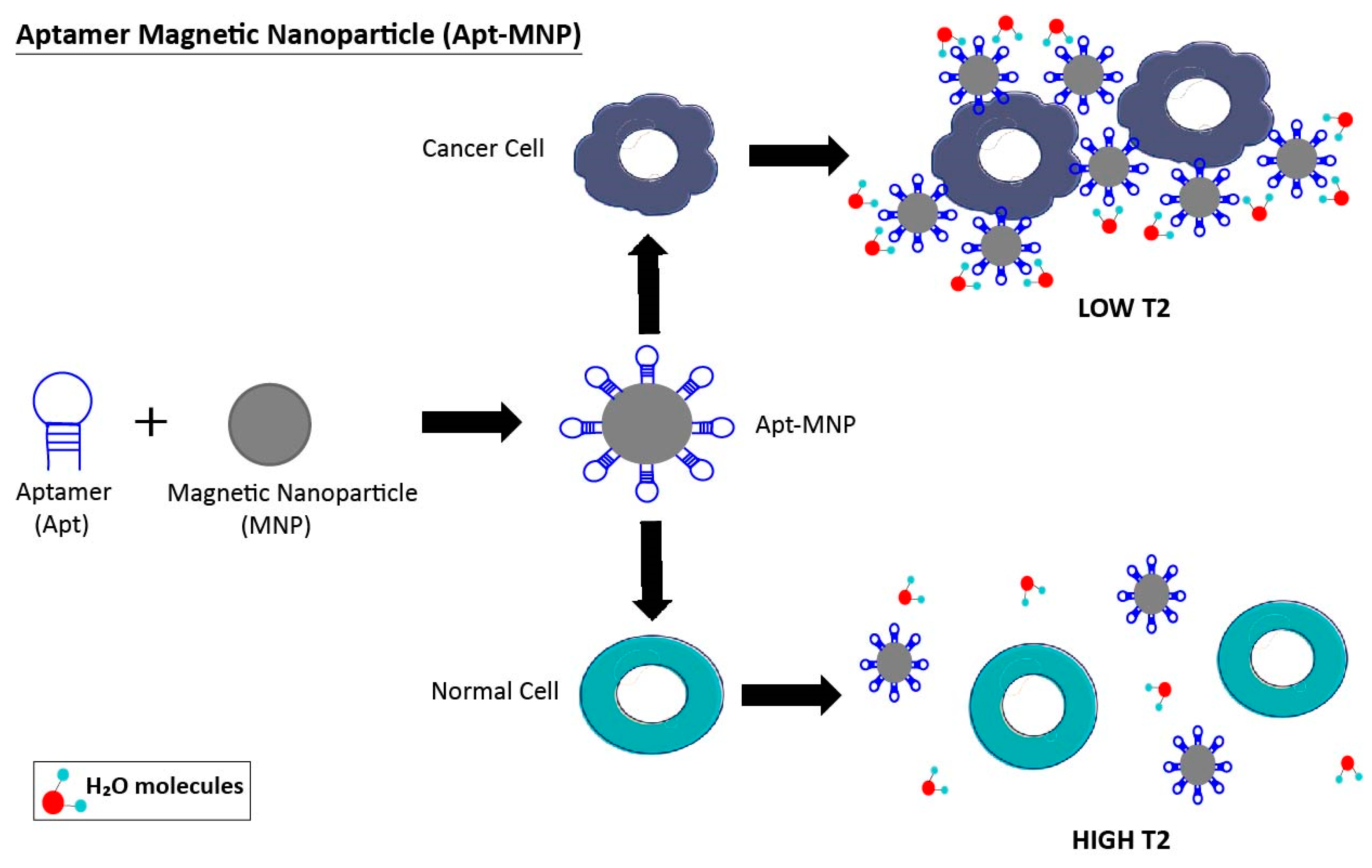
Figure 4.
Schematic illustration of a microfluidic device, showing a middle region with immobilized aptamers. After the injection of the sample, cancer cells are trapped by specific aptamer-cell interaction, whereas normal cells pass through the device. In this sense, cancer cells can be detected by optical microscope observation of the middle zone, and also eluted for future experiments. This device allows the isolation of cancer cells from a heterogeneous mixture of cells by selective cell-capture of immobilized aptamers.
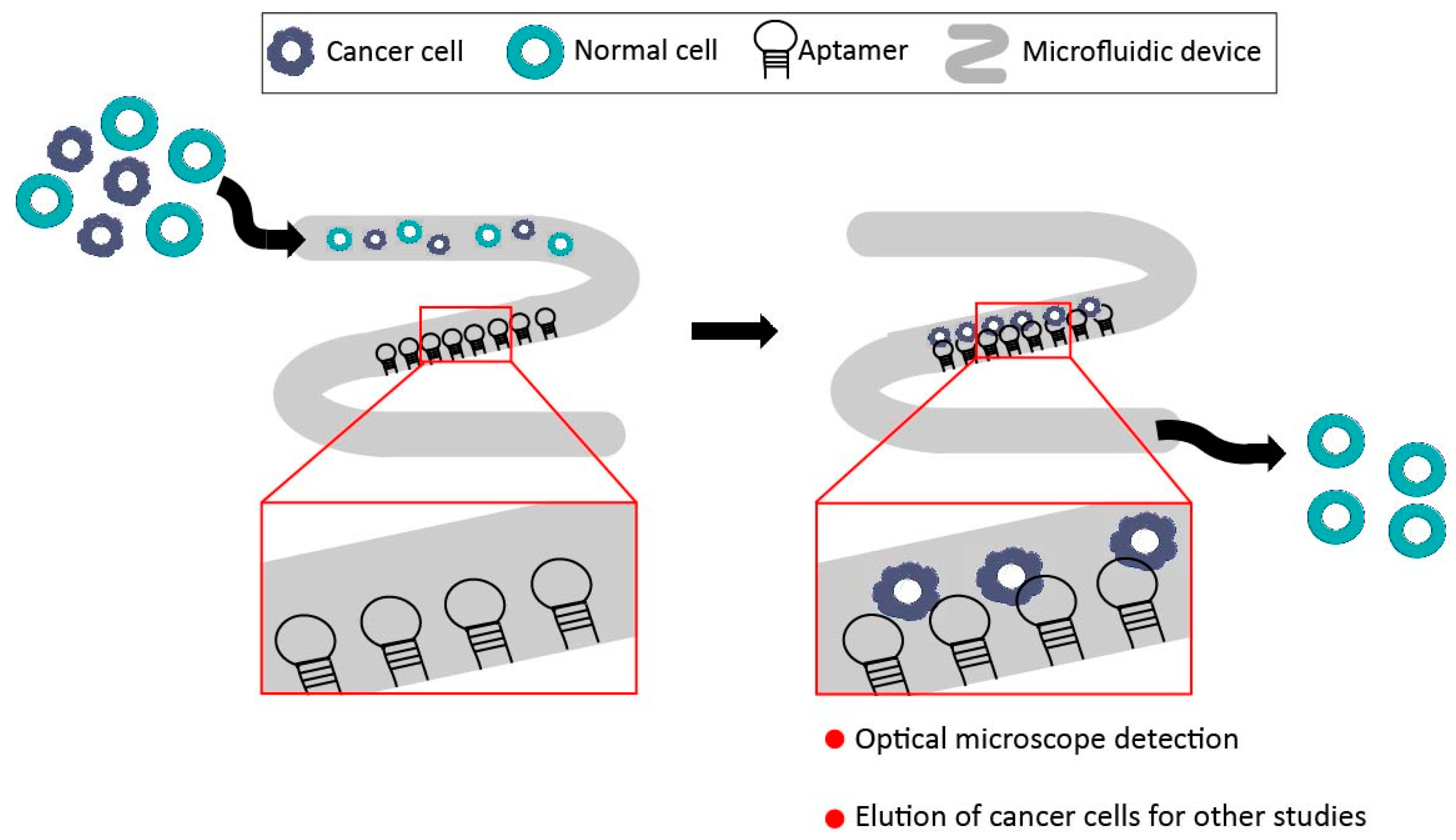
Figure 5.
Schematic representation of functionalized aptamer-quantum dot (Apt-QD) probe. A group of biotinylated aptamers (biotinylated-Apt) that target a cancer cell are conjugated with streptavidin-quantum dot (Streptavidin-QD) via biotin-streptavidin-specific interaction. When Apt-QDs recognize the cancer cell, the fluorescence emission spectra of QDs is observed.
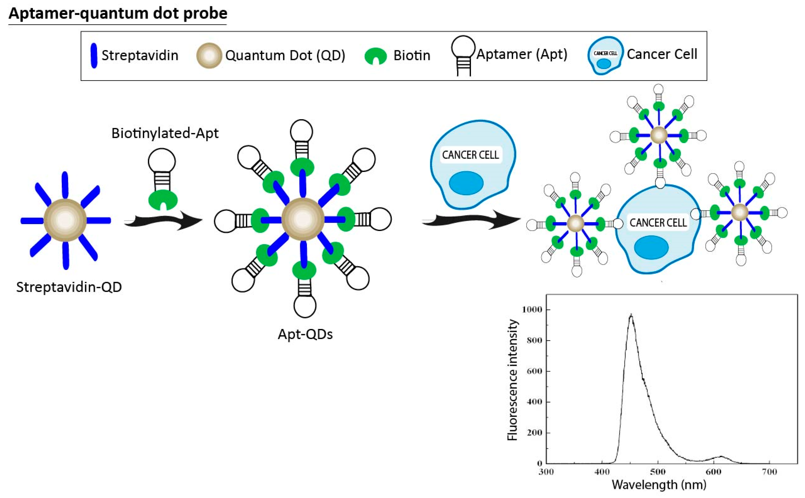
Figure 6.
Molecular beacon aptamers (MB-Apt) for cancer cell detection with fluorophore and quencher at the 5′- and 3′-ends of the aptamer, respectively. In the absence of a target, the hairpin structure of the MB-Apt holds the quencher molecule close to the fluorophore, resulting in no fluorescence emission. When the MB-Apt is bound to membrane receptors of the cancer cell, its conformation is altered, thus resulting in an activated fluorescence signal because of fluorophore-quencher physical separation.

Figure 7.
Illustration of fluorescent aptamer probes. The aptamer is labeled with fluorochrome at its 5′ end. This chimeric molecule could be used for fluorescence staining of cancer cell as the top image, as well as in vivo cancer cell imaging in tumor-bearing mice, as in the picture beneath.
Figure 7.
Illustration of fluorescent aptamer probes. The aptamer is labeled with fluorochrome at its 5′ end. This chimeric molecule could be used for fluorescence staining of cancer cell as the top image, as well as in vivo cancer cell imaging in tumor-bearing mice, as in the picture beneath.

Figure 8.
Schematic depiction of aptamer-gadolinium and aptamer-SPION nanoprobes for cancer cell detection with magnetic resonance imaging (MRI) technology. The aptamer can conjugate with gadolinium (Gd(III)) or magnetite (Fe3O4) nanoparticles via strong and specific interactions between the aptamer and the nanoparticle. In this sense, the chimeric molecule could be used as a contrast agent because it could change the magnetic relaxation time in a measurable way when targeting the cancer cell.


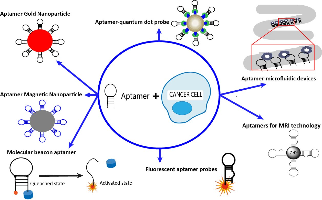
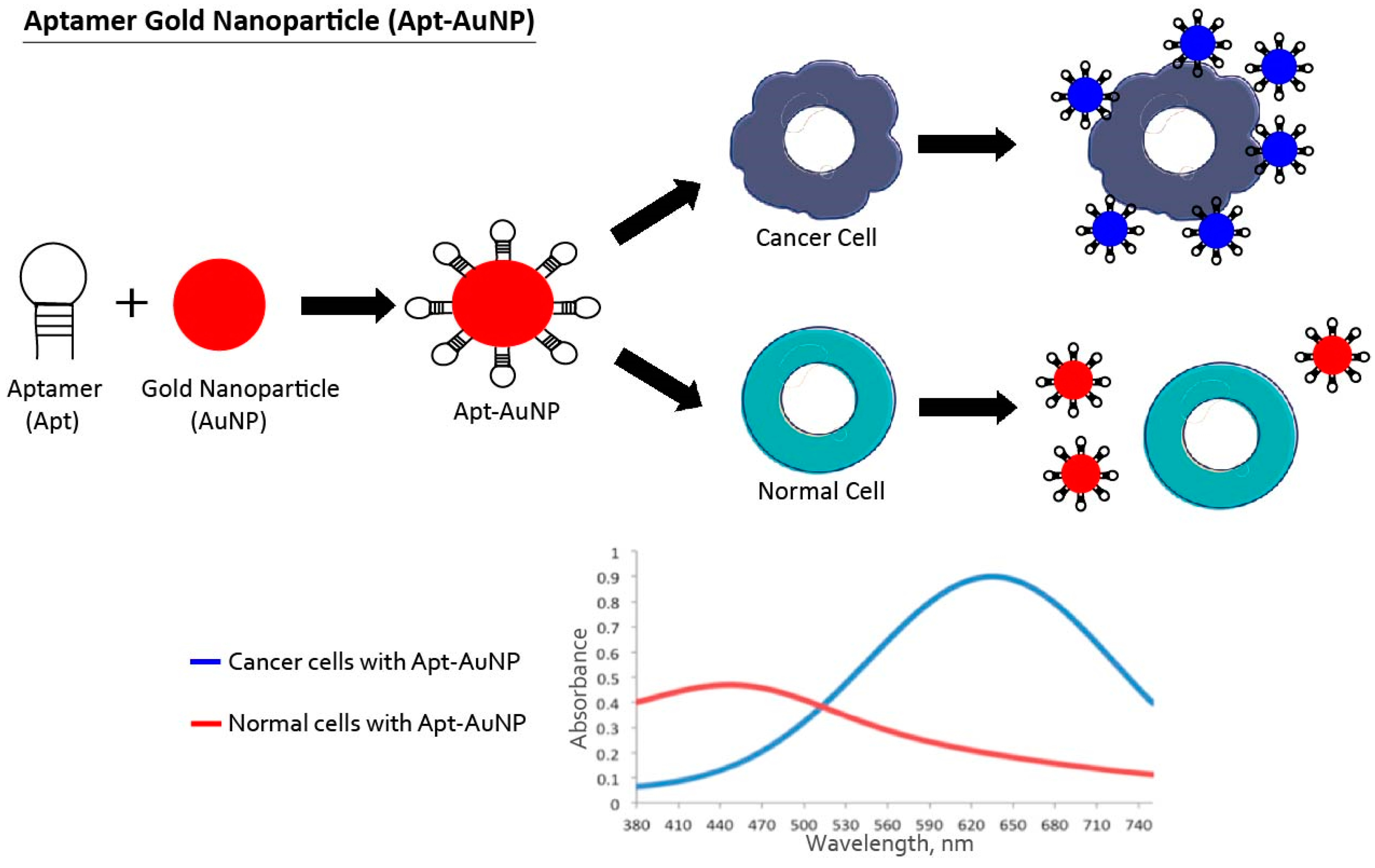
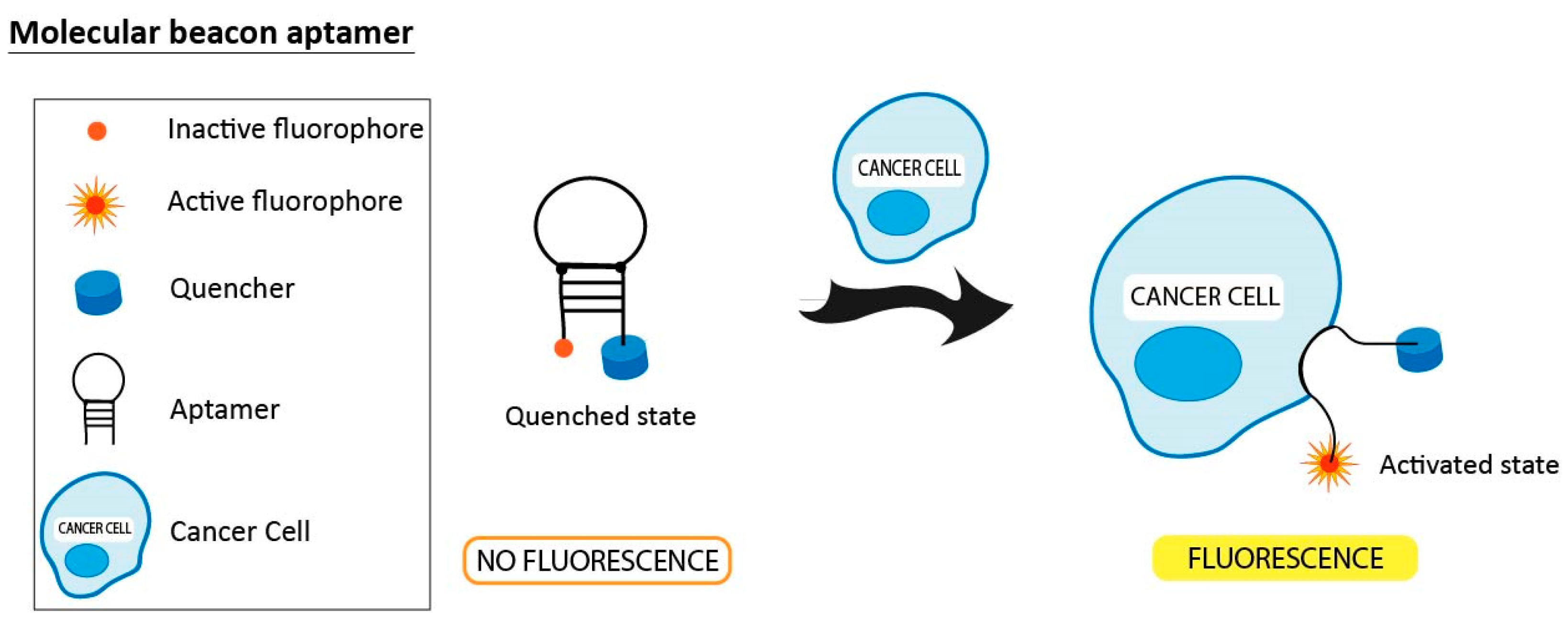
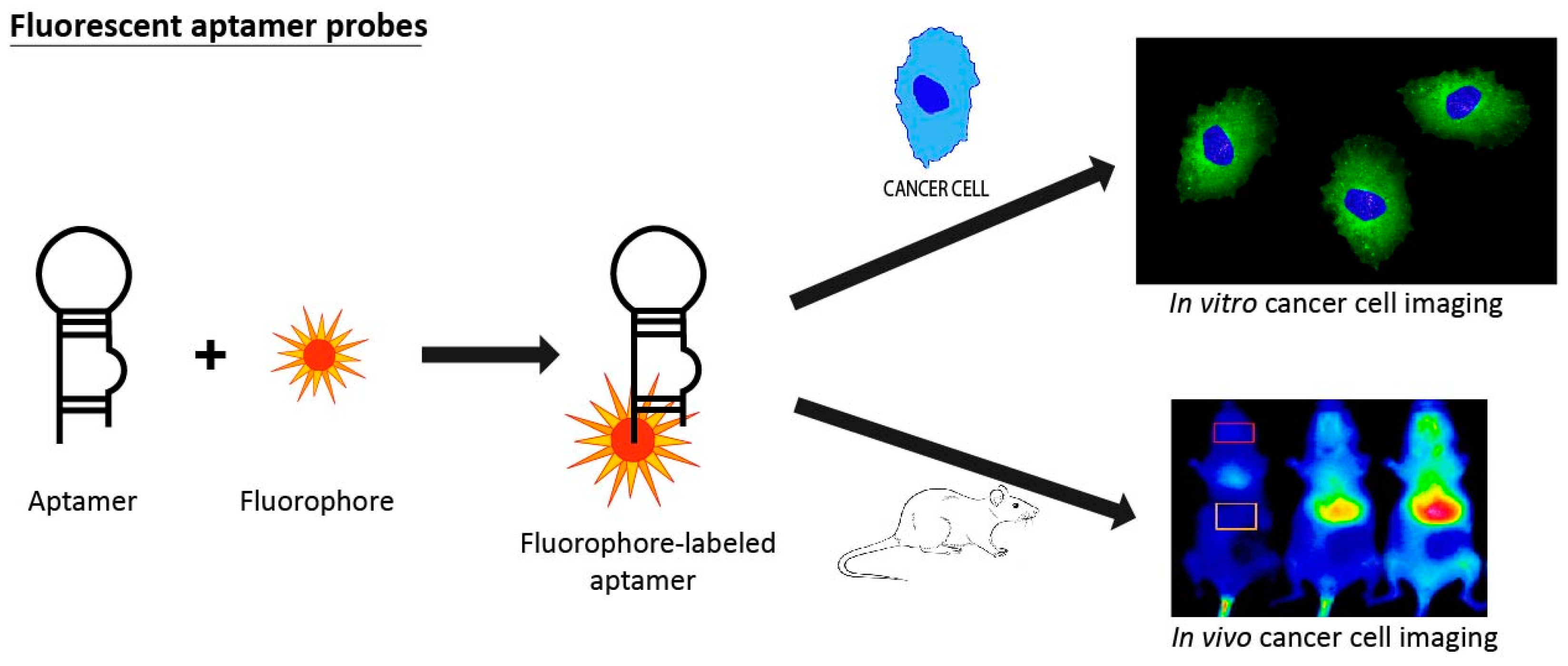
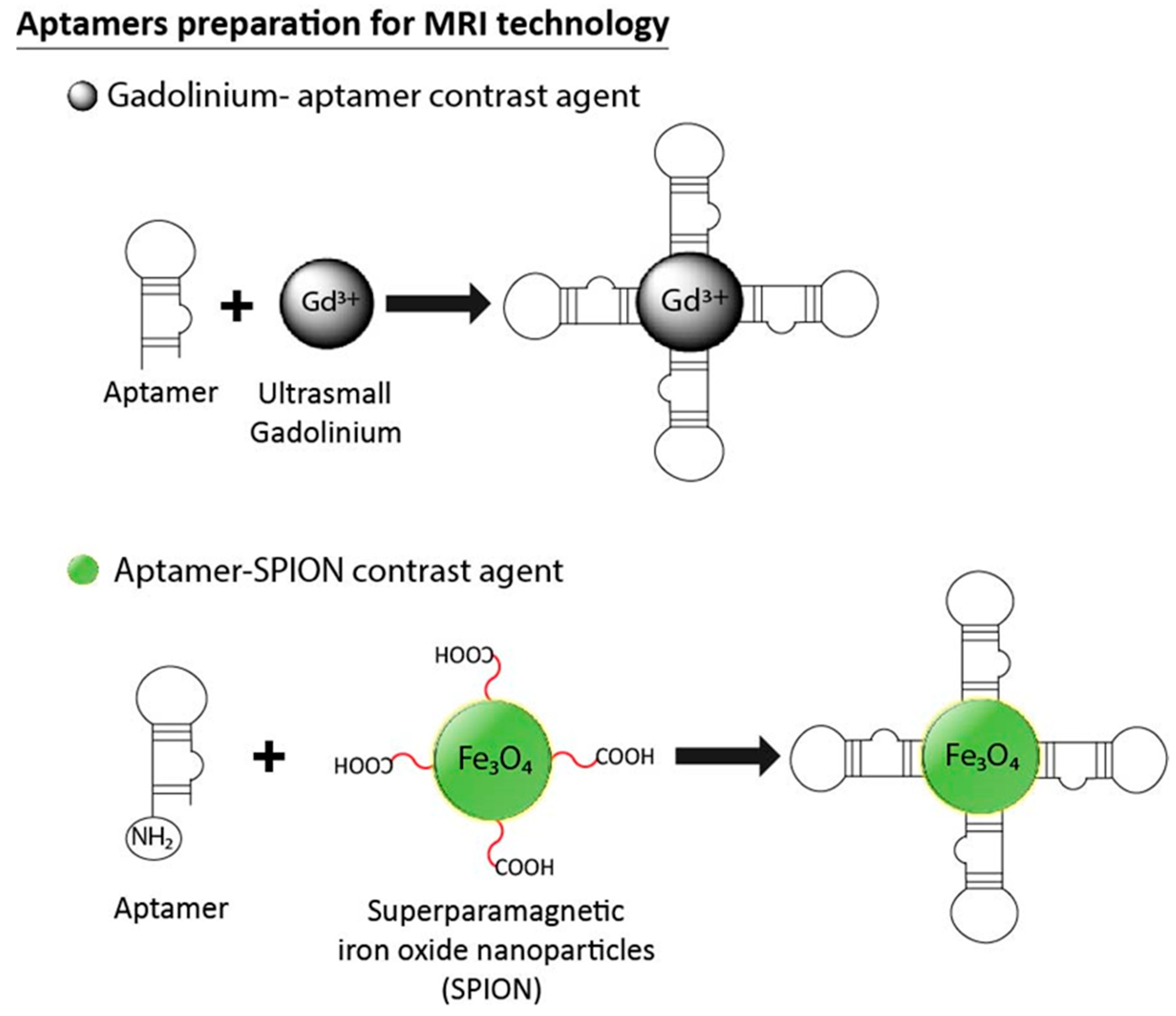
Table 1.
A list of reviewed aptamers.
| Aptamer Name | Nature | Ligand/Target | Cancer | Affinity (nM) 1 | Reference in the Review |
|---|---|---|---|---|---|
| TD05 | DNA | Immunoglobulin heavy constant mu (IGHM) | Burkitt Lymphoma | 74.7 | [33,75,81,115] |
| TE02 | DNA | Ramos cells | Burkitt Lymphoma | 0.76 | [33,75] |
| AS1411 | DNA | Nucleolin | Expressed in different cancers | 169 | [77,99,100,101,108,109,110,120,125] |
| TLS11a | DNA | HepG2 cells (also LH86 cells) | Hepatocellular carcinoma | 7 | [78,112,124] |
| Sgc8 | DNA | Tyrosine-protein kinase-like 7 (PTK7) | T-cell leukemia | 0.8 | [79,80,81,85,92,93,94,97,106] |
| 41t | DNA | Platelet-derived growth factor (PDGF) | --- | 1 | [80] |
| TE17 | DNA | CCRF-CEM cells | T-cell Acute Lymphoblastic Leukemia | 675 | [80] |
| KDED 2a-3 | DNA | DLD-1 cells | Colorectal cancer | 29.2 | [81] |
| KCHA10 | DNA | HCT 116 cells | Colorectal cancer | 21.3 | [81] |
| Sgd5 | DNA | Toledo cells | nonHodgkin’s B cell lymphoma | 70.8 | [93] |
| TTA1 | DNA | Tenacin C | Expressed in different cancers | 5 | [99,101] |
| MUC-1 | DNA | Mucin 1 | Adenocarcinoma | 27 | [99] |
| A32 | DNA | Epidermal growth factor receptor III (EGFRIII) | Glioma | 0.62 | [104] |
| S11e | DNA | A549 cells | Lung | 46.2 | [105] |
| --- | RNA | CD30 | Lymphoma | 0.11 | [114] |
| S6 | DNA | A549 cells | Lung | 28.2 | [116] |
| J3 | DNA | LoVo cells | Colorectal carcinoma | 138.2 | [117] |
| F3B | RNA | Human matrix metalloprotease-9 (hMMP-9) | Malignant melanoma | 20 | [118] |
| A10 | RNA | Prostate membrane antigen (PSMA) | Prostate | 11.9 | [121,122] |
| VEGF165 | RNA | Vascular endothelial growth factor 165 (VEGF-165) | Expressed in different cancers | 50 | [123] |
1 Affinity value using reported dissociation constant (Kd) of each aptamer in nanomoles (nM).
© 2018 by the authors. Licensee MDPI, Basel, Switzerland. This article is an open access article distributed under the terms and conditions of the Creative Commons Attribution (CC BY) license (http://creativecommons.org/licenses/by/4.0/).
Share and Cite
MDPI and ACS Style
Ruiz Ciancio, D.; Vargas, M.R.; Thiel, W.H.; Bruno, M.A.; Giangrande, P.H.; Mestre, M.B. Aptamers as Diagnostic Tools in Cancer. Pharmaceuticals 2018, 11, 86. https://doi.org/10.3390/ph11030086
AMA Style
Ruiz Ciancio D, Vargas MR, Thiel WH, Bruno MA, Giangrande PH, Mestre MB. Aptamers as Diagnostic Tools in Cancer. Pharmaceuticals. 2018; 11(3):86. https://doi.org/10.3390/ph11030086
Chicago/Turabian StyleRuiz Ciancio, Dario, Mauricio R. Vargas, William H. Thiel, Martin A. Bruno, Paloma H. Giangrande, and María Belén Mestre. 2018. "Aptamers as Diagnostic Tools in Cancer" Pharmaceuticals 11, no. 3: 86. https://doi.org/10.3390/ph11030086
Note that from the first issue of 2016, this journal uses article numbers instead of page numbers. See further details here.