Article

Biosensors and Bio-Bar Code Assays Based on Biofunctionalized Magnetic Microbeads

by
Nicole Jaffrezic-Renault
1,*,
Claude Martelet
2,
Yann Chevolot
3 and
Jean-Pierre Cloarec
3
1
LSA, Claude Bernard University Lyon 1, 69622 Villeurbanne Cedex, France
2
AMPERE, Ecole Centrale de Lyon, 69134 Ecully Cedex, France
3
INL, Ecole Centrale de Lyon, 69134 Ecully Cedex, France
*
Author to whom correspondence should be addressed.
Sensors 2007, 7(4), 589-614; https://doi.org/10.3390/s7040589
Submission received: 5 March 2007 / Accepted: 10 April 2007 / Published: 30 April 2007

Abstract

:
This review paper reports the applications of magnetic microbeads in biosensors and bio-bar code assays. Affinity biosensors are presented through different types of transducing systems: electrochemical, piezo electric or magnetic ones, applied to immunodetection and genodetection. Enzymatic biosensors are based on biofunctionalization through magnetic microbeads of a transducer, more often amperometric, potentiometric or conductimetric. The bio-bar code assays relie on a sandwich structure based on specific biological interaction of a magnetic microbead and a nanoparticle with a defined biological molecule. The magnetic particle allows the separation of the reacted target molecules from unreacted ones. The nanoparticles aim at the amplification and the detection of the target molecule. The bio-bar code assays allow the detection at very low concentration of biological molecules, similar to PCR sensitivity.

1. Introduction

Magnetic microbeads (MMB) used in biomedical applications present usually a core/shell structure. Such microbeads have an inorganic core e.g. iron oxide, surrounded by an outer layer of shell wall that consists of long-chain organic ligands or inorganic/organic polymers. The attachment of bioactive ligands to the surface of the outer shell is the key to bioapplication of magnetic microbeads. Table 1 summarizes the main immobilization procedures of bioactive species, on magnetic microbeads used in this chapter. A more exhaustive view can be found in [1].
This paper is devoted to biosensing applications of magnetic microbeads. It is divided into three parts covering three biosensing systems: affinity biosensors, enzymatic biosensors, bio-bar codes.

2. Use of magnetic microbeads for affinity biosensors

2.1. Immunomagnetic electrochemical sensors (ELIME)

Electrochemical immunosensors are designed through the immobilization of the specific antibody on the surface of the electrochemical transducer. The main problem affecting immunosensors is reproducible regeneration of the sensing surface. The need of renewal of the sensing surface arises from the affinity constants derived from the strong antigen-antibody interaction. This renewal is a difficult task since the drastic procedures required alter immunoreagent bound to the surface of the transducer. This drawback makes immunosensors difficult to be integrated into automatic systems. An alternative approach avoiding regeneration consists of using disposable antibody-coated magnetic microbeads and building up in situ immunosensing surface by localizing the immunomagnetic beads on the electrode area with the aid of a magnet. Moreover, the use of immunomagnetic beads is particularly evident in the detection of analytes contained in complex sample matrices (e.g. heterogeneous food mixtures) that may exhibit either poor mass transport to immunosensor or physical blockage of immunosensor surface by non-specific adsorption. The schematic representation of the enzyme-linked immunomagnetic electrochemical assay (ELIME) is presented in figure 1. Immunogenic analyte (bacteria, for example) is sandwiched between an antibody-coated magnetic microbead (immunomagnetic bead) and an antibody-enzyme conjugate. The immunomagnetic bead is trapped magnetically on the electrode surface, exposed to the enzymatic substrate, the electroactive product is detected electrochemically. This type of immunomagnetic electrochemical assay was applied for different analytes with different transducer/enzyme combinaisons, gathered in table 2. A good reproducibility of 2% relative standard deviation was observed [8].

2.2. Label-free immunomagnetic impedancemetric sensors [7]

The electrochemical impedance measurements of the electrical properties of an antibody layer immobilized on a gold electrode allows the direct monitoring of the variation of these properties when antigen-antibody interaction occurs. This technique allows label-free detection of the antigen concentration in biological samples. The problem of regeneration of the sensing surface has been solved, in this example, by using streptavidin magnetic microbeads for the immobilization of the antibody specific of a small pesticide molecule, the atrazine. The antibody, biotinyl-anti-atrazine Fab fragment K47, forms a quite stable layer onto the streptavidin-magnetic microbeads immobilized on gold electrode using a 300mT magnet, due to the high affinity of the biotin/ streptavidin interaction. After the antibody layer formation an antigen, atrazine was injected and interacted with the antibody (cf figure 2).
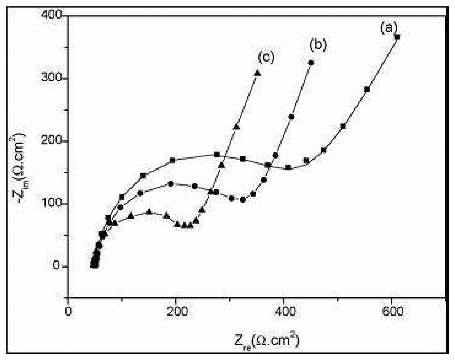
Complex impedance plots of the successive building-up of the sensing layers: magnetic microbeads/ Au electrode, biotinyl-Fab fragment K47 antibody/ magnetic microbeads/Au electrode and after a 600 ng/ml of atrazine injection in cell are shown in figure 3.
Using Randles' equivalent circuit model, an excellent fitting between the simulated and experimental spectra was obtained. The electron transfer resistance values were 304.6 Ω.cm2 for the magnetic monolayer, 204.5 Ω.cm2 for the antibody layer and 188.5 Ω.cm2 after injection of 600 ng/ml of atrazine. The decreases of electron transfer resistance could be attributed to a reorganization of the microbead layer as the specific capacitance decreases too. The specific capacitance, extracted from the computer fitting for the same steps was 17μF/cm2, 15μF/cm2 and 14.29μF/cm2, respectively. This decrease should be due to a thickness increase.
In order to obtain a calibration data set, the values of electron transfer resistance differences ΔRet versus the added atrazine concentrations were plotted, as shown in figure 4. The change of electron transfer resistance was calculated according to the equation:
Δ R m = R et ( Ab ) R et ( Ab Ag )
Where Ret(Ab) is the value of electron transfer resistance after antibody immobilization, Ret(Ab-Ag) is the value of the electron transfer resistance after antigen binding to the antibody.
As it can be seen in figure 4, the plot is linear for high concentrations of atrazine and then reaches saturation. A linear relationship between ΔRet values and the concentration of atrazine was established in the range from 50 to 500ng.ml-1. A detection limit of 10ng.ml-1 is reached.

2.3. Piezoimmunosensors

A piezoelectric immunoassay based on a quartz crystal microbalance (QCM), with a renewable surface, was also proposed using immunomagnetic beads localized on the surface of a quartz using a permanent magnet [13]. Human IgG as analyte was detected using goat-anti-human IgG antibody covalently bonded on aminomodified silica coated magnetic microbeads. A detection limit of 0.36μg.ml-1 and a dynamic range of 0.6-34.9μg.ml-1 were obtained using a 9MHz QCM.
Another piezoelectric immunosensor, based on a 5MHz quartz crystal resonator, was used for the detection of biological pathogens such as Salmonella typhinium [14, 15]. The Salmonella cells were captured by antibody-coated magnetic microbeads and then these complexes were moved magnetically to the sensing quartz and were captured by antibodies immobilized on the crystal surface. An impedance analyser measured the impedance behavior of the oscillating quartz crystal exposed to various concentrations of Salmonella typhinium (102 – 108 cell per ml). The response of the crystal was expressed in terms of equivalent circuit parameters. The motional inductance and the motional resistance increased as a function of the concentration of Salmonella. The viscous damping was the main contributor to the resistance and the inductance in a liquid environment. The load resistance was the most effective and sensitive circuit parameter. A magnetic force was a useful method to collect complexes of Salmonella-immunomagnetic microbeads on the crystal surface and enhance the response of the sensor. In this system, the detection limit, based on resistance monitoring was about 103 cell per ml.

2.4. Genomagnetic electrochemical assays

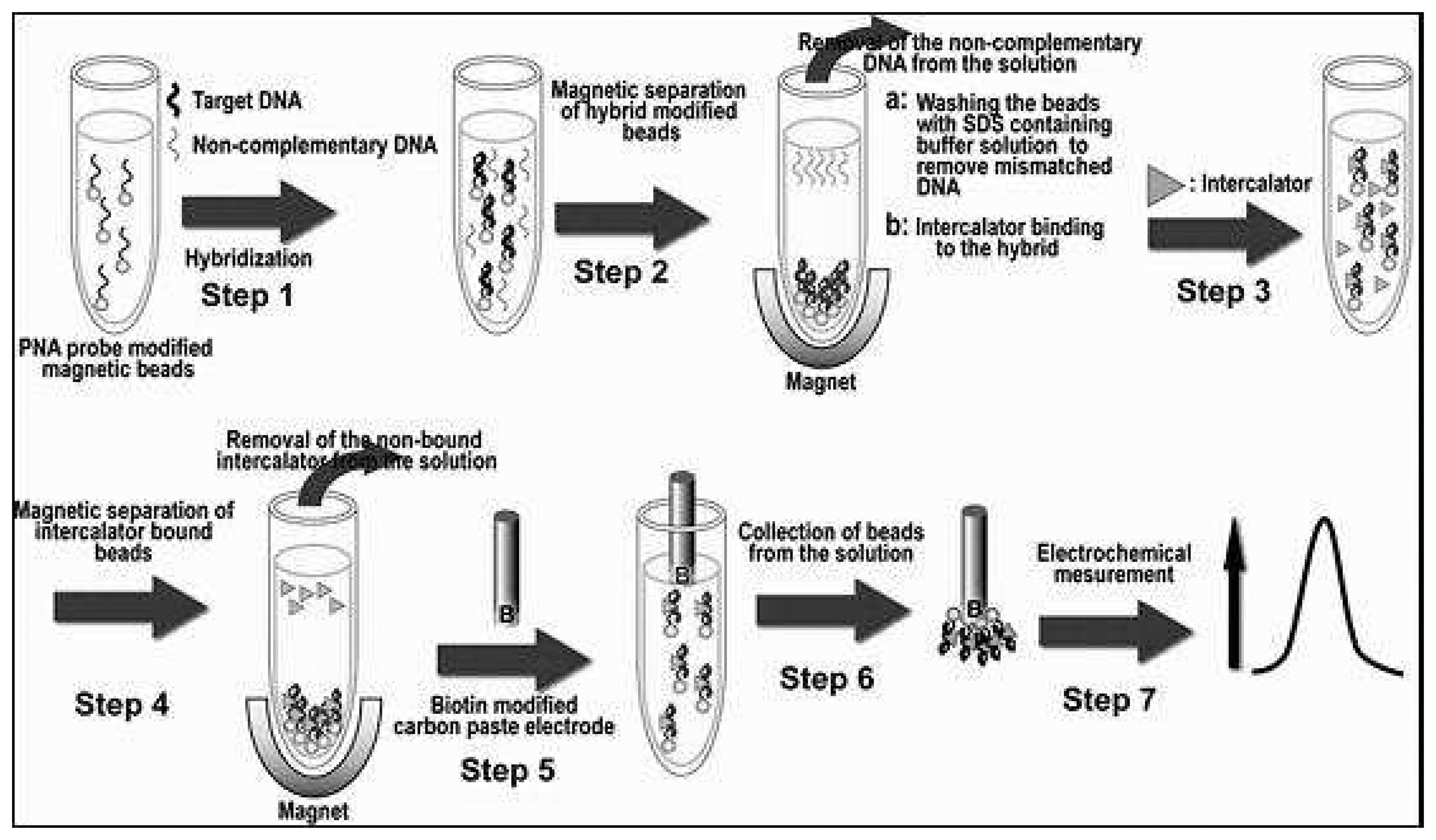
Currently developed DNA hybridization sensors are using single-stranded (ss) short (15-25 nucleotides) oligodeoxynucleotides (ODN, probe DNA) immobilized on an electrode. The ODN-modified electrode is immersed in target DNA solution. When the sequence of target DNA matches that of the probe (based complementary Watson-Crick pairing), a probe-target (hybrid) duplex DNA is formed at the electrode surface. The hybridization event (DNA duplex formation) is detected electrochemically in different ways. This system works quite well with synthetic ODNs when probe target DNAs are about the same lengths. In a real DNA sequence analysis with longer PCR products, viral or chromosomal DNAs, the target DNAs are substantially longer than the probe. With longer target DNAs, difficulties connected with non-specific target DNA adsorptions frequently arise, resulting in a loss of specificity and decreased sensitivity. Elimination of the non-specific DNA adsorption at the electrodes (such as carbon or gold ones) usually applied for the DNA hybridization is very difficult. A new method based on separation of DNA hybridizal step (on magnetic microbeads) from the electrochemical detection step has been proposed and successfully used [16, 17]. One procedure of the genomagnetic electrochemical bioassay (GEME), based on a PNA probe and on an electroactive intercalator, is schematized in Figure 5 [18].
The detection of target DNA can be done by label-free detection of guanine peak [16, 17, 19], by detection of chemically modified DNA prelabelled or postlabelled by redox probes [16], by detection of the product of the enzymatic reaction on DNA post-labelled by enzyme [20], or by detection of redox intercalators [18]. The features of the different genomagnetic electrochemical assays are presented in Table 3.

2.5. Magnetic detection

2.5.1. Magnetic Permeability measurements

Magnetic microbeads are now used as magnetic markers for bioassays. Measurements based on variation of magnetic permeability were performed using a transducer comprising a coil placed in a Maxwell bridge [21] (see Figure 6). The change in magnetic permeability of a compound is detected using inductance measurements. Furthermore, analyte detection has been achieved directly (the analyte being labelled ferromagnetically) or competitively (competition between ferromagnetic labelled and unlabelled analytes exists for binding sites on the transducer) using the aforementionned device. The use of magnetic markers has been shown to offer advantages such as, low interference, little or no background signal, no transducer fouling and no sample treatment. The ‘sandwich’ approach used in the magneto binding assay, where the target analyte is bound between silica carrier particles and the magnetic marker is presented in Figure 7.
Histone H1 conjugated magnetic microbeads were assessed for their ability to work as magnetic transducers for the detection and quantification of DNA [22]. For the quantification of calf thymus DNA, a linear relationship between the DNA concentration in the sample and the relative magnetic permeability of the pellet was found for DNA concentration up to 67μg.ml-1 in buffered solutions as well as in a lyzed cell culture. The detection limits were determined to be 12 and 31μg.ml-1, respectively. For the quantification of plasmid DNA in buffered solution a linear range was established for concentration up to 150μg.ml-1 and the detection limit was determined to be 52 μg.ml-1.
A rapid (6.5min) and simple one-step magnetic immunoassay has been developed for the analysis of human urinary albumin in near patient settings [23]. Polyclonal rabbit anti-human was used as a capture antibody and monoclonal mouse anti-human albumin as a detection antibody in a two-site immunomagnetic assay, requiring no additional washing procedures. The polyclonal anti-human albumin was conjugated to silica microparticles (cf Figure 7) and the monoclonal antibody to dextran coated magnetic microbeads (cf. Figure 7). Quantification of human albumin in undiluted urine was performed by adding 2μl urine to a measuring vial containing polyclonal Ab-silica microparticles, monoclonal Ab-magnetic microbeads and reaction buffer and then agitating the vial by hand for 20s. A linear response was obtained for 0-400μg.ml-1 albumin with a detection limit of 5μg.ml-1.

2.5.2. Frequency mixing

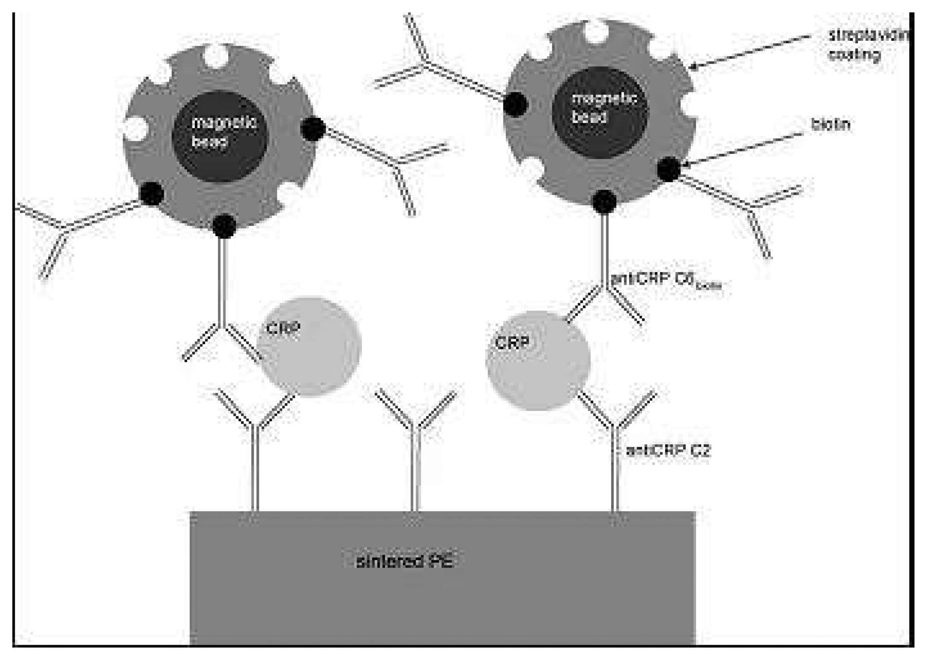
A new technique so-called frequency mixing was also used for detection of magnetic biomarkers. A two-frequency magnetic field excitation was used and the magnetic response at a third frequency was detected, which is a linear combination of the applied frequencies [24]. The so-called magnetic sandwich bioassay for the detection of c-reactive protein (CRP) is presented in figure 8. The detection principle is based on two different anti-CRP antibodies (monoclonal IgG) for CRP trapment (grafted on a polyethylene PE sintered filter) and labelling. The linear detection range of this immunosensor ranged from 25 ng.ml-1 to 2.5 μg.ml-1 and is therefore much more sensitive than typical hsCRP-sandwich-assays. Disavantages of this system is the sample size. Sample sizes are currently 0.5 ml, which is quite high for blood serum analysis.

2.5.3. Magnetic AFM [25]

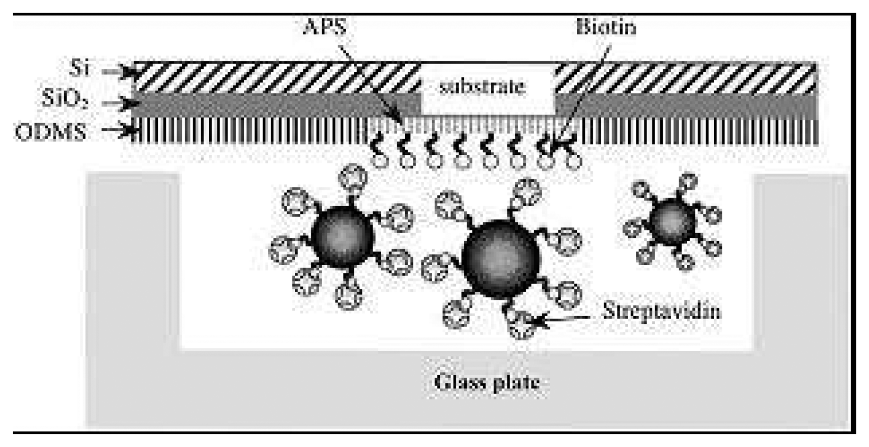
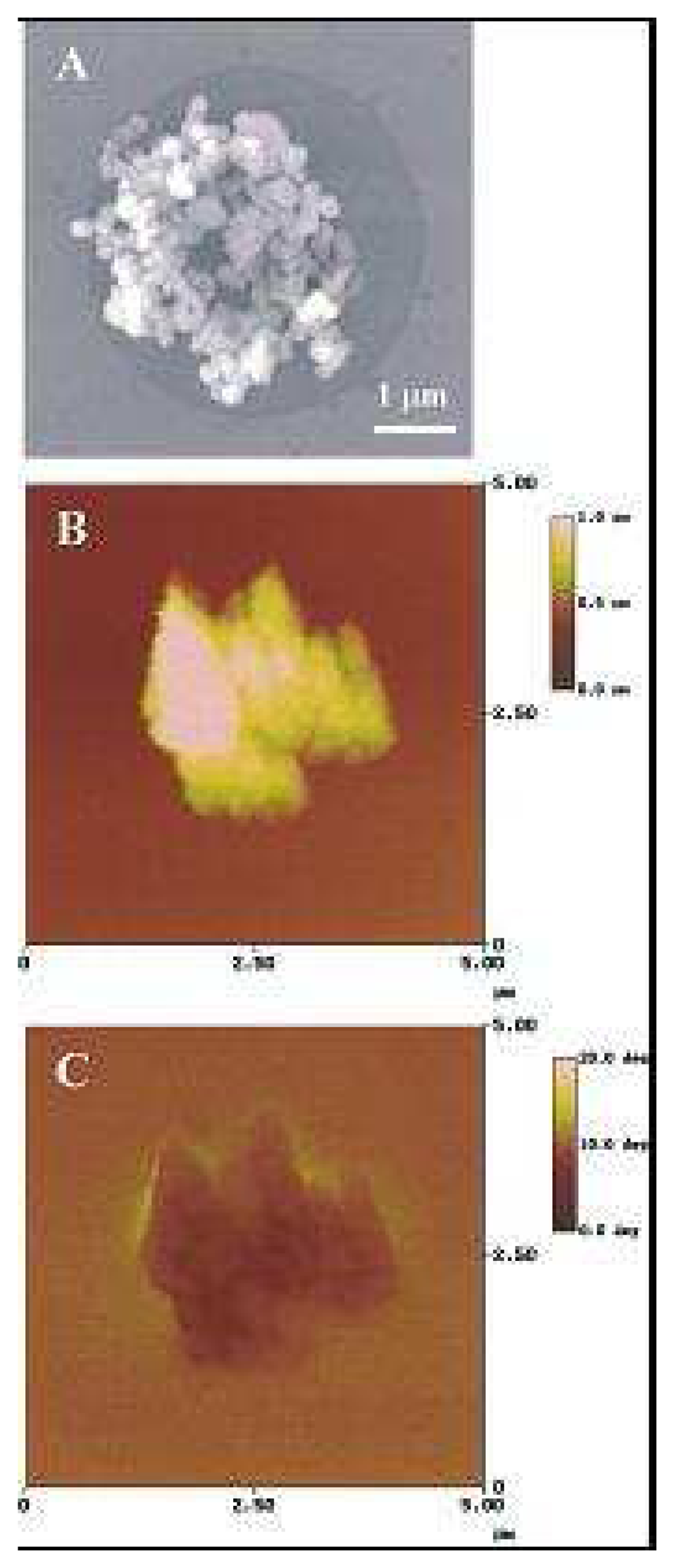
For developing a magnetic bioassay system, an investigation to determine the presence of a specific biomolecular interaction between biotin and streptavidin was done using paramagnetic nanoparticles and a silicon substrate with a self-assembled substrate (cf figure 9). The reaction of streptavidin-modified magnetic nanoparticles on the biotin-modified substrate was clearly observed under an optical microscope (cf. Figure 10). The magnetic signals from the particles were detected using a magnetic force microscope (MFM). The results of this study demonstrate that the combination of a monolayer-modified substrate with biomolecule-modified magnetic particles is useful for the detection of biomolecular interactions in medical and diagnostic analysis.

2.5.4. Magnetic Resonance Imaging (MRI)

An application of magnetic nanobeads is the development of in situ sensors. Based on biomolecular interaction, the self assembly of the superparamagnetic iron oxide core nanobeads can be realised, leading to the enhancement of the spin-spin relaxation times of the water molecular surrounding the self-assembly [26]. Indeed, the spins of surrounding water protons are more efficiently dephased. Therefore if one can design magnetic nanobeads that would self-assemble in the presence of a target and detect the T2 relation time of water protons, it would be possible to design a magnetic nanosensor for magnetic resonance imaging. A proof of concept was realised using DNA/DNA interaction, protein/protein interactions, protein/small molecules and enzyme reactions [26].
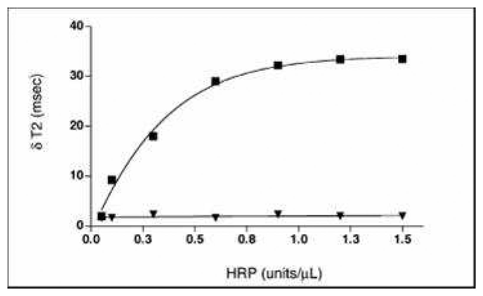
After immobilising serotonin on dextran caged iron oxide nanobeads (75nm), Perez et al [27] have shown that in the presence of Horse radish peroxydase or Myeloperoxidase and hydrogen peroxide, self assembly of the nanoparticles was observed leading to a shift in T2 relaxation that could be successfully imaged. (cf figures 11 and 12)
This group performed similar experiments for the detection of andeno virus and herpes virus (5 viral particles/10 μl) by immobilising antibody directed against these viruses at the surface of dextran caged iron nanoparticles [28] (cf figure 13). The antibodies were immobilised via the protein G attached to the dextran via N-succinimidyl-3-(2-pyridyldithio)propionate (SPDP).
Harris et al [29] have developed an MRI assay with nanoparticles (50 nm dextran caged iron core nanoparticles) for detecting Matrix Metalloproteinase-2 (MPP-2) activity. This protease activity when upregulated leads to invasive proliferation and metastasis of cancer cell. The strategy proposed by these authors relies on a peptide modified PEG that blocks the interaction between neutravidin and biotin. The peptide has an amino-acid sequence which is specific to MPP-2 protease activity. In the presence of MPP-2, PEG is removed from the particles by enzymatic cleavage. The particles can then assemble into nanoassemblies due to the interaction of biotin and neutravidin leading to a T2 shift (cf figure 14). The device could detect down to 9.2 Uml-1 of MPP-2. The authors have demonstrated that inhibition of biotin/neutravidin interaction increases with PEG molecular weight (10 kDa). Inversely, the rate of aggregation in the presence of MPP-2 increases with lower PEG molecular weight. In the presence of a MMP-2 inhibitor (Galardin), no aggregation occurs and then no variation in the T2 relaxation time is observed (cf figure 15).
In conclusion, self assembling of magnetic nanoparticles upon biological interaction is a promising tool for in vivo diagnostic as demonstrated. Indeed these authors have shown that this methodology can work in complex and turbid media. Its sensitivity is below the concentration encountered in tumoral cell for instance.

3. Enzymatic biosensors based onto magnetic microbeads

Most of proposed enzymatic biosensors are based on biofunctionnalization of a transducer more often amperometric, potentiometric or conductimetric. Compared to classical immobilization methods used for the development of enzymatic biosensors, the use of magnetic microbeads can bring a lot of advantages:
  • enzyme immobilization onto magnetic microbeads, having high specific area, allows a high loading of the sensitive matrix
  • such a matrix provides a good macroenvironment for retention of the bioactivity
  • the most decisive advantage is a good control of the localisation of the sensitive material through the use of magnets or of magnetised transducing parts allowing enzyme reactions to occur close to the detection device
  • the sensitive transducing part can be easily renewed
In parallel with biosensing applications, enzyme coated magnetic microbeads have been used to facilitate the enzyme handling. By this way, stock solutions containing such a material can be, when properly stored, used during for more than ten months without significant activity loss. Then this immobilization method has been for example validated for glucose oxidase, urease and alpha-amylase covalently immobilized onto polyacrolein beads [30].
Except the work of Miyabayashi et al. [31], where biomodified magnetic beads were used on a Clark electrode for glucose or Saccharomyces cerevisiae detection, one of the first applications of magnetic beads for biosensors was the combination of a covalent enzyme bonding onto magnetic beads with physical entrapment on the sensor surface. Latex beads, containing superparamagnetic material covalently modified with enzymes, have been patterned on a transducer by the means of screen printed thick films permanent magnets. Such a method, valuable for batch production independently of the nature of the substrate material, was evaluated for assessing glucose concentrations up to 20mM [32].
Enzymes immobilized on magnetic microparticles can be trapped by magnets and retained on an electrode surface at a specific location in flow analysis devices. But only few works using magnetic microparticles have been devoted to design real enzymatic biosensors. As an example of such devices, an enzyme-based electrochemical magnetobiosensor for environmental toxicity analysis can be quoted. The integration of the bioactive material as urease, cholinesterases classes, significantly increases the sensitivity and allows detection limits as low as 10-11M for heavy metal ions and 10-12M for organophosphates and carbamates [33]. In table 4, characteristics of some magnetic microbeads based enzymatic biosensors are gathered.
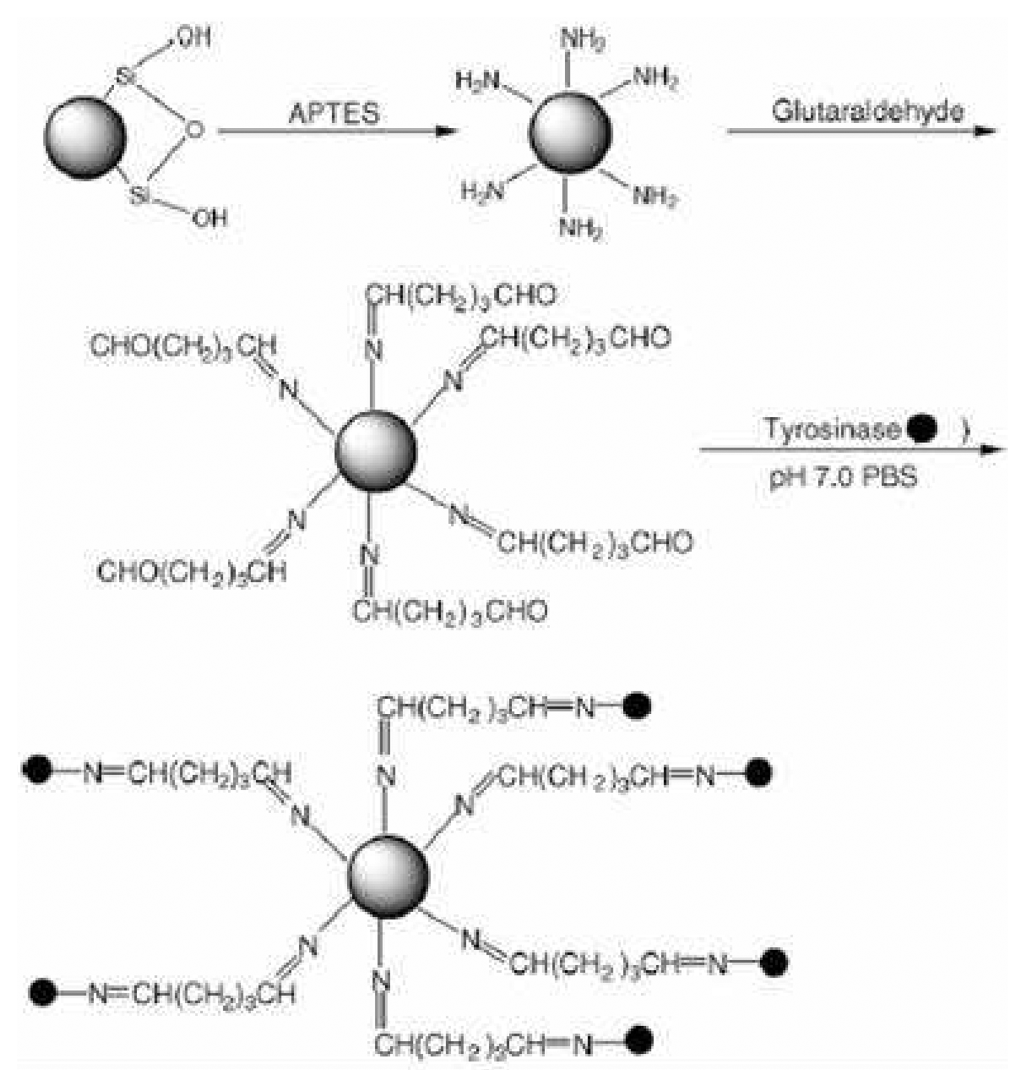
As an example a strategy of enzyme immobilization is given in figure 16 where a covalent bonding of the biospecie can be achieved allowing a quite good storage stability.
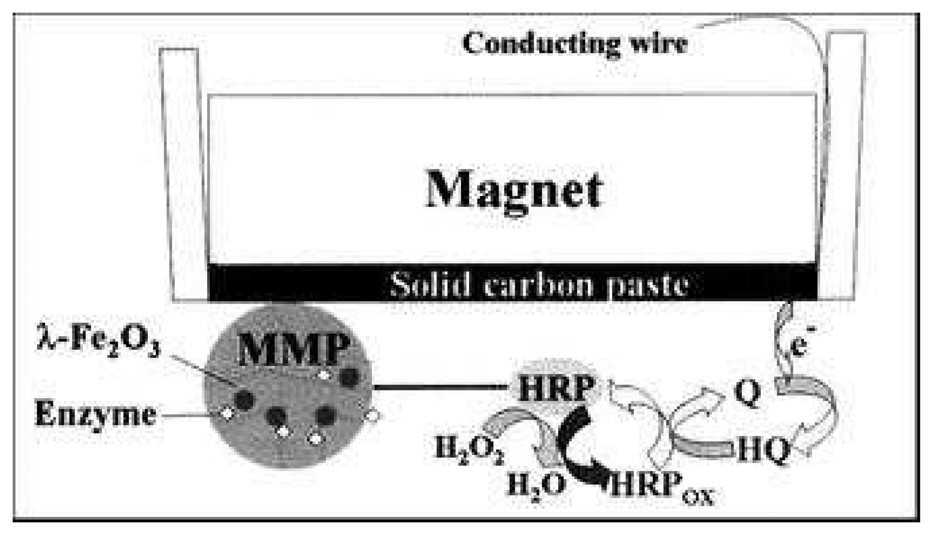
Recently, the potentialities of nanoporous silica microparticles containing superparamagnetic defect spinel-type iron oxide nanoparticles inside their pores have been underlined for biosensors applications. This approach allows the obtaining of a high sensitivity and selectivity even in complex media. Interest of such a support has been validated for the immobilization of horseradish peroxidase leading to hydroquinone and hydrogen peroxide biosensing at level as low as 4×10⁻⁷ M [36]. The design of a magnetised carbon paste electrode with trapped magnetic nanoporous silica microparticles (MMPs) is given on figure 17.
A similar strategy has been used for the immobilization of horseradish peroxidase with a high density of nanopores of MMPs having a diameter of 5 micrometers and applied for studying peroxidation of clozapine. Clozapine is a drug belonging to the dibenzoazepine class and often used to treat neurological disorders. The resulting amperometric biosensor allows the drug quantification in the micromolar range and presents a quite good stability, no significant signal loss being observed after one month of storage [37].
Enzymatic MMPs based devices present also specific advantages, as MMPs do not act as a barrier for the diffusion of the analyte to the electrode surface. Only a decrease of few percent of the voltamperometric signal is observed in presence of microparticles compared to the bare electrode. Furthermore MMPs constitute a valuable tool for inhibition studies, immobilized enzymes on such macroparticles being less sensitive, through a screening effect, to inhibitors than soluble enzymes. Thus, it has been recently shown that immobilized HRP, through MMP strategy, was protected from inactivation by inhibitors as thiols which can react with intermediary quinoneimine derivatives produced during the enzymatic reaction [38].
Magneto-switchable electrocatalysis and bioelectrocatalysis are accomplished by the surface modification of magnetic microbeads with redox-relay units. By the attraction of the modified magnetic microbeads to the electrode support, or their retraction from the electrode, by means of an external magnet, the electrochemical functions of the magnetic microbeads tethered relays can be switched between “ON” and “OFF” states, respectively. The magneto-switchable redox functionalities of the modified particles activate electrocatalytic transformations, such as a biocatalytic chemoluminescence cascade that leads to magneto-switchable light emission or the activation of bioelectrocatalytic processes [39, 40].

4. Bio-bar code

Polymerase chain reaction was introduced in 1985 and has revolutionized biology and molecular biology since then. Its sensitivity allows the detection of 5-10 copies of DNA. However some drawbacks such as its complexity, time consuming procedure and narrow target have motivated the findings of new technologies.
Furthermore PCR is related to DNA detection, and some authors have been looking for new procedures allowing the detection of proteins with similar sensitivity offered by PCR.
The bio–bar code assay appeared in the early 2000's as a promising analytical tool for high sensitivity detection of protein [6] and DNA [4].
In the following section, the principle of bio–bar-code for protein and DNA detection will be presented followed by examples of biological applications.
The bio–bar code assay relies on a sandwich structure based on specific biological interaction of a magnetic particles and a nanoparticle with a defined biological molecule in a medium. The magnetic particle allows the separation of reacted target molecules from unreacted ones. The nanoparticles aim at amplifying and detecting the target of interest.
Therefore, the bio–bar code approach relies on two components:
  • Magnetic Micro Beads (MMB) bearing the biological probe (DNA or Antibody, Figure 18) for target recognition.
  • Nanoparticles (NP) bearing a target binding molecule (DNA or polyclonal antibody, Figure 18), and the so-called bio–bar-code (an oligonucleotide, Figure 18).
To be clear the oligonucleotide sequence immobilized on the Nanoparticles (Figure 18) will be called bio–bar code complement, whereas the bio–bar-code (Figure 18) will refer to the oligonucleotide that hybridised with this immobilised sequence. The bio–bar-code sequence is the one that is captured on the DNA chip and further detected.
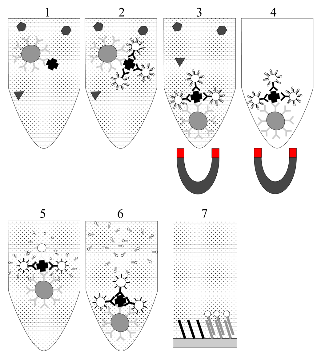
The steps involved in the bio–bar-code assay are presented in Figure 19.
First, the MMB are added to a solution of interest and allowed to interact with the biological target to be detected generally via DNA/DNA interaction or Antibody/Antigen interaction (Figure 19.1). Next, the NPs are added to the mixture and interact with the biological target (via DNA/DNA interaction or Antibody/Antigen interaction) to form a sandwich like structure (Figure 19.2). This structure is separated from the medium and from unreacted material thanks to the magnetic properties of the MMB (Figure 19.3). The beads can then be separated from the medium (Figure 19.4). The sandwich is redispersed in pure water. The bio–bar code DNA is then dehybridized (Figure 19.5), purified from the particles by centrifugation (Figure 19.6). It is then captured on a DNA chip and detected by fluorescence or silver enhancement (Figure 19.7). A promising approach based on using a Y junction dendrimer like DNA carrying two fluorophores (Alexa Fluor 480 and Bodipy 630/650). The coding is not detected via a DNA chip but by intensity encoding. In consequence, no nanoparticles are required [41]. So far this approach was carried out on polystyrene based microbeads (non magnetic) but would be an interesting alternative in the bio–bar-code assay.

4.1. Nanoparticles (NP)

The NP is aimed at detecting and signal amplifying of the molecule through the so called bio–bar code, after the interaction with the MMB/target complex. Two materials are preferentially employed namely gold nanoparticles [2, 4] and polystyrene [42].
Two different cases will be considered depending on the target molecule: 1) antigen detection and 2) nucleic acid detection. If the target molecule is an antigen, a polyclonal antibody (directed against the target molecule) is immobilised at the surface of the particles. In the case of polystyrene, amine bearing particles are used and the antibody is immobilised using a glutaraldehyde protocole through its N-termini. Next, the bio–bar code complement is immobilised at the surface of the particles. In the case of gold NP, it is immobilised through thiol/gold interactions. In the case of polystyrene, immobilisation is achieved through amine glutaraldehyde strategy (1530 ± 181 Abs). The particles in the later case are passivated with ethanol amine.
The sequence is typically 30 to 50 bases long, it can bear at the 3' end an A10 spacer and an amine or thiol terminated alkyl (usually C6). Sequence analysis of the complement bio–bar code leads to the following: The GC content is near 50 % with a melting temperature between 65°C and 75°C (calculated with the next neighbouring theory).
The bio–bar code is loaded by hybridization at room temperature in PBS 0.1 M. The average number of loaded bio–bar code is estimated to be a few hundreds (approx. 360) [5] on 30 nm gold particles and 3.5 105 on polystyrene particles (1 μm diameter). Under these hybridization conditions on polystyrene particles, the hybridisation yield is near 10 % as observed by UV spectroscopy and fluorescence [42].
If the target molecule is a nucleic acid, two different oligonucleotides are immobilised at the surface of the particle using a solution containing both molecules. One is an oligonucleotide complementary to the target and one corresponding to the bio–bar-code complement. The ratio between the two oligonucleotides leads to signal amplification.
In 2005, Taxton et al developed simplified protocole of the bio–bar-code NP. Instead of using double stranded DNA, the bio–bar-code consists in thiol immobilized oligonucleotides on gold NP. The DNA is released for on chip DNA capture by DTT [43].

4.2. Magnetic Microbeads (MMB)

The Magnetic Microbeads aim at interacting with the target molecule through biological recognition, namely DNA/DNA complementary sequence (hybridization) or antigene/antibody interaction. It gives to the sandwich structure magnetic properties allowing its separation from unreacted material and medium.
Typically, Magnetic Micro Beads are iron oxide based particles that may be encapsulated in a polymeric layer, the total particle having a diameter in the range of one to several μm. Mirkin group used amine bearing MMB obtained by silanisation of iron oxide [5] with an amine modified silane or purchased with a polyamine layer [2, 5]. Immobilisation of the monoclonal antibody is achieved by reaction of glutaraldehyde with the primary amine at the surface of the particle and with free amine of the antibody. In the case of oligonucleotide target complement, the particle is modified with surface maleimido groups using succinimidyl 4-(p-maleimidophenyl) butyrate (SMPB) [4] or sulfosuccinimidyl 4-N-maleimidomethyl cyclohexane-1-carboxylate (sulfo-SMCC) [5]. The oligonucleotide is then immobilised through thiol addition to the double bound of the maleimido group. The average number of DNA strand per particles is 3 105. In the case of antibody immobilisation, the average number of immobilised molecules is 3500 (small) per MMB as estimated by optical density at 280 nm. Capping is accomplished with BSA or sulfo-NHS acetate.

4.3. Detection

Three main detection methods are described in the literature namely: scanometric detection, fluorescent detection and rolling circle amplification (RCA).
Scanometric detection is performed as followed. After collection of the sandwich structure from the media, the DNA bar code is released from the NP by dehybridization of the bio–bar-code from its complement at high strigency (in DI water 55 °C) or by competition between thiolated oligonucleotide and DTT [43, 2]. The bio–bar-code is recovered from the supernatant after centrifugation and captured on DNA chip by hybridization. The DNA chip is usually supported on maleimide modified glass slides bearing 12 mer oligonucleotides. After hybridization of the bio–bar-code (30-50 mer) on the DNA chip, a labelled oligonucleotide gold nanoparticle (13 nm) further hybridizes with the free end of the bio–bar-code. The chip is then immersed in a modified photographic solution containing silver salt. Its reduction is being catalyzed by the gold nanoparticules. The bio–bar-code is then detected by light scattering at the surface of the chip due to the silver particles (around the gold nanoparticles).
Mirkin groups uses an alterative methods based on the detection of a fluorophore (Alexa type) instead of silver enhancement [42, 44].
Scanometric detection leads to detection as low as 500zM [5] in the case of DNA targeted molecules and 3 aM is the case of protein targeted molecules (PSA) [42]. With fluorescence, the detection limit is of a few hundreds of aM. In the later case, the detection limit was lowered by using micrometer scaled NP instead of nanoscaled. Indeed 3.105 bio-bar-code were loaded per particles, as compared to a few hundreds. However one would thing that smaller size NP would allow a higher density of NPs per MMB compared to microscale NP.
In fact, when using complex media or when multiplexing, the detection limit is increased by one to five orders of magnitude probably due to cross reactivity and non specific adsorption [4].
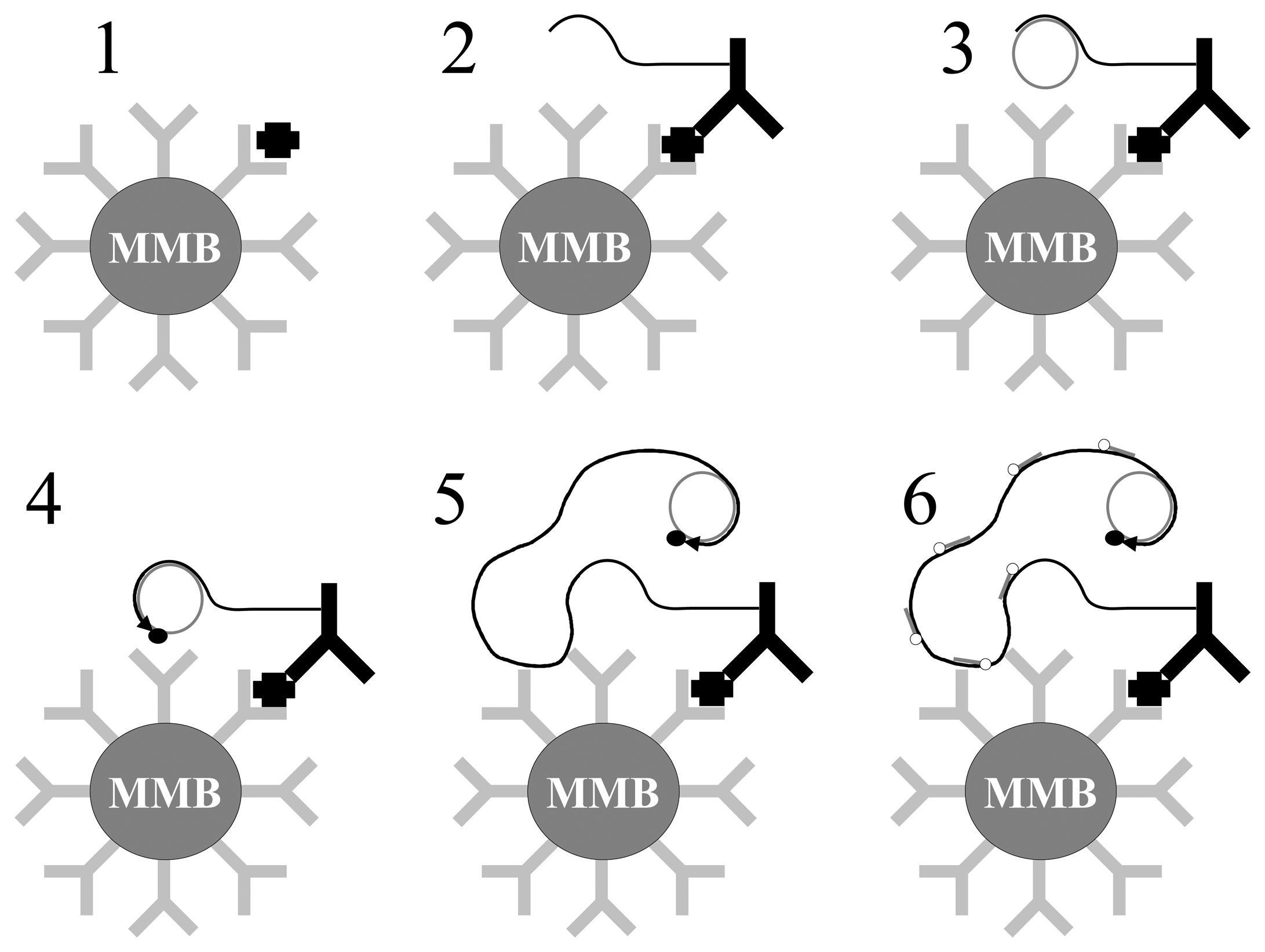
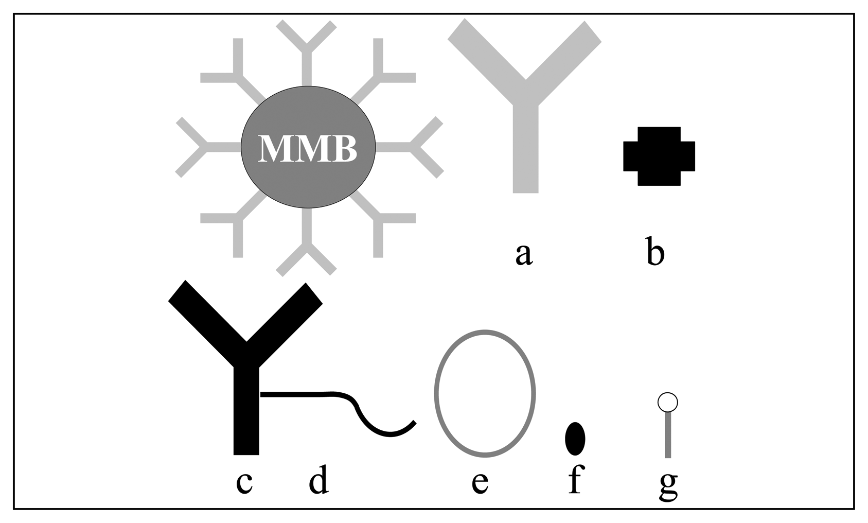
Other authors have developed rolling circle amplification (RCA) coupled to the use of magnetic bead [45]. This method allows an antibody / antigen interaction to be revealed and amplified using a DNA polymerase reaction. The method, which can be used in several different formats (ELISA, array), has been described using magnetic particles as solid support. The magnetic beads (Figure20) have been functionnalized with polyclonal anti-human IgE antibodies (Figure 20.a). The detection of target molecules (e.g.: human IgE, Figure 20.b) involved the use of monoclonal anti-human IgE antibodies (Figure 20.c) conjugated with an oligonucleotide (Figure 20.d). The rolling circle amplification procedure consists in extending this oligonucleotide using a complementary circular DNA (Figure 20.e), and a DNA polymerase reaction (Figure 20.f). After reaction the extended DNA is analysed using a labelled probe (Figure 20.g).
The steps of the rolling circle amplification are the following ones :
After binding of the target antigen on the bead (Figure 21.1), the interaction is revealed using the monoclonal antibody (Figure 21.2). The oligonucleotides d linked to the c are then hybridized with a partially complementary circular DNA (Figure 21.3). Extension of the oligonucleotide is then allowed by incubating a DNA polymerase in the mixture (Figure 21.4). The extension goes on using the circular DNA as a template several times, then leading to the “rolling circle amplification”: the final extended DNA includes several linear complementary copies of the initial circular DNA template (Figure 21.5). The analysis of this DNA is done using a labelled oligonucleotide probe that hybridizes on the extended DNA stand (Figure 21.6).
Such an assay has been compared to a more classical immunoassay using magnetic particle and an anti-IgE alkaline phosphatase conjugate. The RCA amplification gave approximately 75-fold more signal than the classical immunotest, for a quantity of 25 ng/ml of IgE to be detected. The limit of detection of the RCA assay was reported as 1 pg/ml.

4.4. Biological applications

Table 5 presents the summary of existing bio-bar code assays.
Most applications concern the diagnosis of cancer (PSA), pathogens (bacteria or viruses), the detection of Alzhiemer disease maker (ADDL).
In brief, Bio-bar-code allows the detection of biomolecules in the aM range when dealing with model solution. When detection is performed with a complex mixture for multiplex detection, the detection limit raises up to 500fM.
Furthermore Single Nucleotide Polymorphism selectivity was realized [43]. However, the assay was performed in with two separated solution with concentration of 500 aM. It did not allowed to test cross reactivity. The signal to noise ratio is not given. These limitations could be due to non-specific adsorption or cross-reactivity.

4.5. Conclusion

The Bio–bar code assays allow the detection at a very low concentration of biological molecules similar to PCR sensitivity. Further work will probably focus on optimising biological molecule immobilisation in order to reduce cross-reactivity and to lower non specific adsorption.

References

  1. Hermanson, G. T.; Mallia, A. K; Smith, P. K. Immobilised affinity ligand techniques.; Academic Press, 1992. [Google Scholar]Hermanson, G. T. Bioconjugate Techniques.; Academic Press, 1996. [Google Scholar]
  2. Georganopoulou, D. G.; Chang, L.; Nam, J-M.; Thaxton, C. S.; Mufson, E. J.; Klein, W. L.; Mirkin, C. A. Fluorescent and scanometric ultrasensitive detection technologies with the biobar code assay for alzheimer's disease diagnosis. PNAS 2005, 102, 2273–2276. [Google Scholar]
  3. Liu, Z.; Liu, Y.; Yang, H.; Yang, Y.; Shen, G.; Yu, R. A phenol biosensor based on immobilizing tyrosinase to modified core-shell magnetic nanoparticles supported at a carbon paste electrode. Analytica Chimica Acta 2005, 533(1), 3–9. [Google Scholar]
  4. Stoeva, S. I.; Lee, J-S.; Thaxton, S.; Mirkin, C. A. Multiplexed DNA detection with biobarcoded Nanoparticle probes. Angewandte Chemie 2006, 45, 3303–3306. [Google Scholar]
  5. Nam, J.; Stoeva, S. I.; Mirkin, C. A. Bio-Bar-code-Based DNA Detection with PCR like sensitivity. J. Am. Chem. Soc. 2004, 126, 5932–5933. [Google Scholar]
  6. Nam, J.; Taxton, C. S.; Mirkin, C. A. Nanoparticle-based Bio-barcodes for the ultrasensitive detection of proteins. Science. 2003, 301, 1884–1886. [Google Scholar]
  7. Helali, S.; Martelet, C.; Abdelghani, A.; Maaref, M.A.; Jaffrezic-Renault, N. A disposable immunomagnetic electrochemical sensor based on functionalised magnetic beads on gold surface for the detection of atrazine. Electrochimica Acta 2006, 51(24), 5182–5186. [Google Scholar]
  8. Santandreu, M.; Sole, S.; Fabregas, E.; Alegret, S. Development of electrochemical immunosensing systems with renewable surfaces. Biosensors Bioelectronics 1998, 13, 7–17. [Google Scholar]
  9. Gehring, A.G.; Brewster, J.D.; Irwin, P.L.; Tu, S.I.; Van Houten, L. J. 1-Naphtyl phosphate as an enzymatic substrate for enzyme-linked immunomagnetic electrochemistry. J. Electroanal. Chem. 1999, 469, 27–33. [Google Scholar]
  10. Sole, S.; Alegret, S.; Cespedes, F.; Fabregas, E. ; Flow injection immunoanalysis based on a magnetoimmunosensor system. Anal. Chem. 1998, 70, 1462–1467. [Google Scholar]
  11. Dequaire, M.; Degrand, C.; Limoges, B. An immunomagnetic electrochemical sensor based on a perfluorosulfonate-coated screen-printed electrode for the determination of 2,4-dichlorooxyacetic acid. Anal. Chem. 1999, 71, 2571–2577. [Google Scholar]
  12. Liu, Z.M.; Yang, H.F.; Li, Y.F.; Liu, Y.L.; Shen, G.L.; Yu, R.Q. Core-shell magnetic nanoparticles applied for immobilization of antibody on carbon paste electrode and amperometric immunosensing. Sensors and Actuators B 2006, 113, 956–962. [Google Scholar]
  13. Li, J.; He, X.; Wu, Z.; Wang, K.; Shen, G.; Yu, R. Piezoelectric based on magnetic nanoparticles with simple immobilization procedures. Analytica Chimica Acta 2003, 481, 191–198. [Google Scholar]
  14. Kim, G.H.; Rand, A.G.; Letcher, S.V. Impedance characterisation of a piezoelectric immunosensor. Part I: antibody coating and buffer solution. Biosensors Bioelectronics 2003, 18, 83–89. [Google Scholar]
  15. Kim, G.H.; Rand, A. G.; Letcher, S.V. Impedance characterization of a piezoelectric immunosensor. Part II: Salmonella typhinium detection using magnetic enhancement. Biosensors Bioelectronics 2003, 18, 91–99. [Google Scholar]
  16. Palacek, E.; Fojta, M; Jelen, F. New approaches in the developement of DNA sensors: hybridization and electrochemical detection of DNA and RNA at two different surfaces. Bioelectrochemistry 2002, 56, 85–90. [Google Scholar]
  17. Wang, J.; Kawde, A.N.; Erdem, A.; Salazar, M. Magnetic bead-based label-free elecrochemical detection of DNA hybridization. Analyst 2001, 126, 2020–2024. [Google Scholar]
  18. Kerman, K.; Matsubara, Y.; Morita, Y.; Takamura, Y. Peptide nucleic acid modified magnetic beads for intercalator based electrochemical detection of DNA hybridization. Science and technology of Advanced Materials 2004, 5, 351–357. [Google Scholar]
  19. Erdem, A.; Pividori, M.I.; Lermo, A.; Bonanni, A.; Del Valle, M.; Alegret, S. Genomagnetic assay based on label-free electrochemical detection using magneto-composite electrodes. Sensors and Actuators B 2006, 114, 591–598. [Google Scholar]
  20. Wang, J.; Xu, D.; Erdem, A.; Polsky, R.; Salazar, M. A. Genomagnetic electrochemical assays of DNA hybridization. Talanta 2002, 56, 931–938. [Google Scholar]
  21. Kriz, K.; Gehrke, J.; Kriz, D. Advancements toward magneto immunoassays. Biosensors Biolectronics 1998, 13, 817–823. [Google Scholar]
  22. Abrahamsson, D.; Kriz, K.; Lu, M.; Kriz, D. A preliminary sutdy on DNA detection based on relative magnetic permeability measurements and histone H1 conjugated superparamagnetic nanoparticles as magnetic tracers. Biosensors Bioelectronics 2004, 19, 1549–1557. [Google Scholar]
  23. Lu, M.; Ibraimi, F.; Kriz, D.; Kriz, K. A combination of magnetic permeability detection with nanometer-scaled puperparamagnetic tracer and its application for one-step detection of human urinary albumin in undiluted urine. Biosensors Bioelectronics 2006, 21, 2248–2254. [Google Scholar]
  24. Meyer, M.H.F.; Hartmann, M.; Krause, H.J; Blankenstein, G.; Mueller-Chorus, B.; Oster, J.; Miethe, P.; Keusgen, M. CRP determination based on a novel magnetic biosensor. Biosensors Bioelectronics 2007, 22(6), 973–979. [Google Scholar]
  25. Arakaki, A.; Hideshima, S.; Nakagawa, T; Niwa, D.; Tanaka, T.; Matsunaga, T. Detection of biomolecular interaction between biotin and streptavidin on a self-assembled monolayer using magnetic nanoparticles. Biotechnology & Bioengineering 2004, 88, 543–546. [Google Scholar]
  26. Perez, J. M.; Josephson, L.; O'Loughlin, T.; Högemann, D.; Weissleder, R. Magnetic relaxation switches capable of sensing molecular interactions. Nature Biotechnology 2002, 20, 816–820. [Google Scholar]
  27. Perez, J. M.; Simeone, F. J.; Tsourkas, A.; Josephson, L.; Weissleder, R. Peroxidase Substrate Nanosensors for MR imaging. Nanoletters 2004, 4, 119–122. [Google Scholar]
  28. Perez, J. M.; Simeone, F. J.; Saeki, Y.; Josephson, L.; Weissleder, R. Viral-Induced Self-Assembly of Magnetic Nanoparticles Allows the Detection of Viral Particles in Biological Media. JACS 2003, 125, 10192–10193. [Google Scholar]
  29. Harris, T. J.; von Maltzahn, G.; Derfus, A. M.; Ruoslahti, E.; Bhatia, S. N. Proteolytic actuation of Nanoparticles Self assembly. Angew.Chem. Int. Ed. 2006, 45, 3161–3165. [Google Scholar]
  30. Varlan, A.R.; Sansen, W.; Van Loey, A.; Hendrickx, M. Covalent enzyme immobilization on paramagnetic polyacrolein beads. Biosensors Bioelectronics 1996, 11(4), 443–448. [Google Scholar]
  31. Miyabayashi, A.; Mattiasson, B. An enzyme electrode based on electromagnetic entrapment of the biocatalyst bound to magnetic beads. Analytica Chimica Acta 1988, 213, 121–130. [Google Scholar]
  32. Varlan, A.R.; Suls, J.; Jacobs, P.; Sansen, W. A new technique of enzyme entrapment for planar biosensors. Biosensors Bioelectronics 1995, 10(8), XV-XIX. [Google Scholar]
  33. Solé, S.; Merkoci, A.; Alegret, S. New materials for electrochemical sensing III. Beads. TrAC Trends in Analytical Chemistry 2001, 20(2), 102–110. [Google Scholar]
  34. Liao, M-H.; Guo, J-C.; Chen, W.C. A disposable amperometric ethanol biosensor based on screen-printed carbon electrodes mediated with ferricyanide-magnetic nanoparticle mixture. Journal of Magnetism and Magnetic Materials 2006, 304(1), e421–e423. [Google Scholar]
  35. Lu, B-W.; Chen, W.C. A disposable glucose biosensor based on drop-coating of screen-printed carbon electrodes with magnetic nanoparticles. Journal of Magnetism and Magnetic Materials 2006, 304(1), e400–e402. [Google Scholar]
  36. Elyacoubi, A.; Zayed, S.; Blankert, B.; Kauffmann, J-M. Development of an Amperometric Enzymatic Biosensor Based on Gold Modified Magnetic Nanoporous Microparticles. Electroanalysis 2006, 18, 345–350. [Google Scholar]
  37. Yu, D.; Blankert, B.; Bodoki, E.; Bollo, S.; Viré, J-C; Sandulescu, R.; Nomura, A.; Kauffmann, J-M. Amperometric biosensor based on horseradish peroxidase-immobilised magnetic microparticles. Sensors and Actuators B 2006, 113(2), 749–754. [Google Scholar]
  38. Kauffmann, J-M.; Yu, D.; El Yacoubi, A.; Blankert, B. Magnetic nanoporous microparticles for biosensors and bioreactors. LabPlus international 2006, 20(3), 6–8. [Google Scholar]
  39. Willner, I.; Willner, B. Functional nanoparticles architectures for sensoric, optoelectronic and bioelectronic applications. Pure Appl. Chem. 2002, 74(9), 1773–1783. [Google Scholar]
  40. Willner, I; Katz, E. Magnetic control of electrocatalytic and bioelectrocatalytic process. Angew. Chem. Int. Ed 2003, 42, 4576–4588. [Google Scholar]
  41. Li, Y.; Hong Cu, Y.T.; Luo, D. Multiplexed detection of pathogen DNA with DNA-bases fluorescence nanobarcodes. Nature Biotechnology 2005, 23(7), 885–889. [Google Scholar]
  42. Oh, B-K.; Nam, J.M.; Lee, S. W; Mirkin, C. A. A fluorophore-base Bio-barcode Amplification Assay for Proteins. Small 2006, 2, 103–108. [Google Scholar]
  43. Thaxton, C. S.; Hill, H. D.; Georganopoulou, D. G.; Stoeva, S. I.; Mirkin, C. A. A Bio-Bar Code Assay Based upon Dithiothreitol-Induced Oligonucleotide Release. Anal. Chem 2005, 77, 8174–8178. [Google Scholar]
  44. Khan, S.; Klein, W.; Mirkin, C. A.; Chang, L.; Georganopoulou, D. Fluorescent and scanometric ultrasensitive detection technologies with the bio-bar code assay for alzheimer's disease diagnosis. Nanoscape 2005, 2(1), 7–15. [Google Scholar]
  45. Schweitzer, B.; Wiltshire, S.; Lambert, J.; O'Malley, S.; Kukanskis, K.; Zhu, Z.; Kingsmore, S. F.; Lizardi, P. M. Ward. D. C. Immunoassays with rolling circle DNA amplification: a versatile platform for ultrasensitive antigen detection. PNAS 2000, 97(18), 10113–10119. [Google Scholar]
  46. Lee, A.; Mirkin, C.; Georganopoulos, D. Real-time polymerase chain reaction completed detection of cardiac tropin I with the bio-bar code assay. Nanoscape 2005, 2(1), 17–25. [Google Scholar]
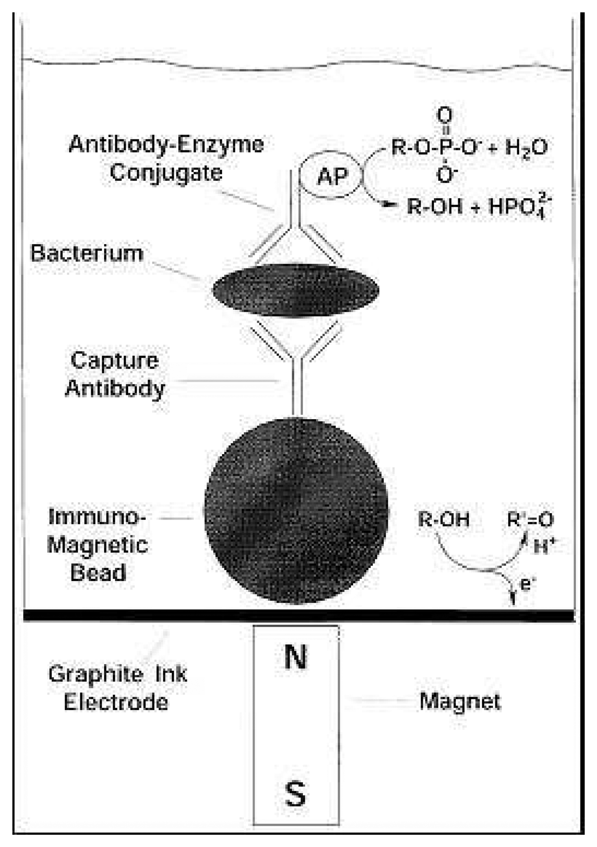
Figure 1. Schematic representation of the enzyme-linked immunomagnetic electrochemical assay (ELIME) (Reprinted from [9] with permission from Elsevier).
Sensors 07 00589f1
Figure 2. Different steps for building-up the immunomagnetic impedancemetric sensor (Reprinted from [7] with permission from Elsevier).
Sensors 07 00589f2
Figure 3. Nyquist diagram (Zre vs. Zim) for the non-faradaic impedance measurements corresponding to: (a) magnetic microbeads/Au-electrode; (b)anti-atrazine-Fab fragment K47 antibody/ magnetic microbeads/Au-electrode; (c) 600ng.ml-1 of atrazine/anti-atrazine-Fab fragment K47 antibody/magnetic microbeads/Au-electrode; Solid curve show the computer fitting of the data using Randles' equivalent circuit. Symbols show the experimental data (Reprinted from [7] with permission from Elsevier).
Sensors 07 00589f3
Figure 4. Calibration plot of the variation of electron transfer resistance ΔRet vs. concentration of atrazine (Reprinted from [7] with permission from Elsevier).
Sensors 07 00589f4
Figure 5. Procedure of the genomagnetic bioassay (Reprinted from [18] with permission from Elsevier).
Sensors 07 00589f5
Figure 6. The current magnetic permeability meter, a portable version of the device (Reprinted from [21] with permission from Elsevier).
Sensors 07 00589f6
Figure 7. The ‘sandwich’ approach used for in the magneto binding assay (Reprinted from [21] with permission from Elsevier).
Sensors 07 00589f7
Figure 8. Magnetic sandwich bioassay for the detection of c-reactive protein (CRP) (Reprinted from [24] with permission from Elsevier).
Sensors 07 00589f8
Figure 9. Schematic illustration of biotin-streptavidin reaction on a SAM-modified substrate. Biotin is attached to the aminopropyl silane (APS) patterned substrate. Streptavidin-modified magnetic microbeads are injected into a channel on a glass plate. The magnetic beads are bound by a specific interaction between biotin and streptavidin (Reprinted from [25] with permission from Wiley-VCH).
Sensors 07 00589f9
Figure 10. SEM (A), AFM (B) and MFM (C) images of magnetic microbeads immobilized on a patterned substrate by specific interaction between biotin and streptavidin. Dot size = 4μm (Reprinted from [25] with permission from Wiley-VCH).
Sensors 07 00589f10
Figure 11. Principle of peroxidase based assay. In the presence of peroxidase, free radicals are formed allowing self assembly (Reprinted from [27] with permission from ACS).
Sensors 07 00589f11
Figure 12. Effect of Horse radish peroxidase concentration on δT2 (Reprinted from [27] with permission from ACS).
Sensors 07 00589f12
Figure 13. δT2 as a function of virus particle concentration (Reprinted from [28] with permission from ACS).
Sensors 07 00589f13
Figure 14. Proteolytic activation. In the presence of MMP-2, PEG molecules are cleaved allowing self assembly of nanoparticles. (Reprinted from [29] with permission from Wiley-VCH).
Sensors 07 00589f14
Figure 15. In the presence of tumor derived cells (HT-1080), MMP-2 is secreted. (a) T2 mapping of Fe3O4 nanoparticles. In the presence pf an MMP-2 inhibitor (Galardin) no variation in the T2 relaxation time is observed. 5c) aggregation is observe when incubating the nanoparticles with HT-080, while it is inhibited in the presence of galardin (Reprinted from [29] with permission from Wiley VCH).
Sensors 07 00589f15
Figure 16. Preparation of magnetic bio-particles for a tyrosinase based biosensor (Reprinted from [3] with permission from Elsevier).
Sensors 07 00589f16
Figure 17. An enzymatic biosensor based on magnetic nanoporous silica microparticles (MMPs) where oxidation of hydroquinone (HQ) to quinone by the enzyme horseradish peroxidase (HRP) in presence of hydrogen peroxide and subsequent electroreduction of quinone (Q) is shown.
Sensors 07 00589f17
Figure 18. Components of the bio_bar-code assay: MMB Magnetic MicroBeads, NP Nanoparticle a Monoclonal antibody or oligonucleotide b Polyclonal antibody or oligonucleotide c bio–bar-code d Bio–bar-code complementary oligonucleotide e Target molecule (antigen or DNA).
Sensors 07 00589f18
Figure 19. Steps of the bio–bar-code assay
1
Interaction between MMB and target.
2
Recognition between target and particles in complex biological medium : sandwich MMB/target/NP
3
Magnetic separation of MMB
4
Removal of biological medium. Only sandwiches MMB/target/NP and MMB are kept in the tube
5
Redispersion of sandwiches in distilled water causes dehybridation of biobar-codes
6
Removal and analysis of bio–bar-codes using DNA microarray or other methods
Sensors 07 00589f19
Figure 20. Components of rolling circle amplification on magnetic particle MMB Magnetic microparticle; a Polyclonal anti-human IgE antibody; b Human IgE; c Monoclonal anti-human IgE antibody; d Oligonucleotide linked to the antibody c; e Circular DNA; f Polymerase; g Labeled oligonucleotide
Sensors 07 00589f20
Figure 21. Rolling circle amplification with magnetic particle 1 target antigen / primary antibody interaction; 2 secondary antibody recognition; 3 circular DNA hybridization with oligonucleotide; 4 starting polymerase reaction; 5 rolling circle extension using polymerase; 6 hybridization of labelled probes on extended oligonucleotide
Sensors 07 00589f21
Table 1. Immobilisation protocoles of biomolecules used to our knowledge on nanobeads.
Surface functionImmobilisation protocoleBiomoleculesAdvantageDisavantageReference
AmineActive Ester
AmineGlutaraldehydeAntibody (Ab) EnzymeEasy YieldNon reproducible Non Oriented[2], [3]
AmineMaleimido groups: SMPB sulfo-SMCCThiol derivatives[4], [5]
GoldThiolThio-DNASimpleLimited to thiol containing molecules[6]
Active esterAmine
Avidin, StreptavidinBiotinylated biomoleculesAntibodySimpleNeed biotinylated biomolecules[7]
Table 2. Features of enzyme-linked immunomagnetic electrochemical assays using different electrochemical transducers and different enzymatic labels.
AnalyteTransducerEnzymeDetection LimitDynamic rangeRefs
Rabbit IgGpH-ISFETurease8nM0 – 2.07μM[10]
Rabbit IgGGraphite composite electrodeHR Peroxidase9×10-6μg.l-10 – 0.26μM[8]
E. coli 0157:H7Graphite ink electrodeAlkaline Phosphatase4.7×103 cells.ml-10 – 105cells.ml-1[9]
2,4-D herbicideNafion-SPEAlkaline phosphatase0.01 μg.l-10.01 - 100μg.l-1[11]
Human IgGCarbon paste electrodeHR Peroxidase0.18μg.ml-10.51 – 30.17 μg.ml-1[12]
Table 3. Features of the different genomagnetic electrochemical assays.
AnalyteElectrochemical TransducerLabelDetection LimitDynamic rangeRefs
Breast-cancer BRCA1 genePotentiometric stripping measurements Graphite pencil electrodeNo100ng.ml-1 (ppb)100 ppb-20ppm[17]
Breast-cancer BRCA1 geneDifferential pulse voltammetryAlkaline phosphatase100ppb0.25-1.0ppm (20 min Hybridization)[20]
21 mer DNASquare wave voltammetryMeldola's blue intercalator2pM2 – 20pM[18]
DNA Sequence from SalmonellaDifferential pulse voltammetry Graphite composite electrodesNo9.68pM[19]
Table 4. Examples of magnetic microbeads based enzymatic biosensors.
AnalyteMagnetic beadsTransducerEnzymeSensitivityDynamic rangeRef.
PhenolMgFe2O4silica coated ϕ = 120nmC pasteTyrosinase54,2μA.mM-110-6 – .5 10-4 M[3]
EthanolPrecipitated Fe3O4 ϕ = 9.8nmScreen printed CYeast YADH/NAD+0,61μA.mM-11-9 mM[34]
GlucosePrecipitated Fe3O4 ϕ = 9.8nmScreen printed CGlucose oxydase1.74 mA mM-10-33 mM[35]
Table 5. Summary of biological target detected through a Bio–bar code assay.
#TargetRecognition eventDetectionLower detection limitAuthors
1PSA (Prostate Specific Antigen)Ab/AgScanometric30 aM[6]
2PSAAb/AgFluorescence300 aM[42]
3Cardiac troponin I (cTnI)Ab/AgRT-PCR500 fM[46]
4ADDL (Amyloid Beta Derived Diffusible Ligands)Ab/AgScanometric10 aM[2]
5Hepatitis B, Variolas virus, Ebola, HIVDNA/DNAScanometric500 fM[4]
6Anthrax lethal factorDNA/DNAScanometric500 zM[5]
7Mock RNARNA/DNAScanometric700 aM[43]