Article

Synthesis, Characterization and Metal Ion Detection of Novel Fluoroionophores Based on Heterocyclic Substituted Alanines

by
Susana P. G. Costa
1,*,
Elisabete Oliveira
2,
Carlos Lodeiro
2 and
M. Manuela M Raposo
1
1
Centro de Química, Universidade do Minho, Campus de Gualtar, 4710-057 Braga, Portugal
2
REQUIMTE, Faculdade de Ciências e Tecnologia, Universidade Nova de Lisboa, 2829-516 Monte de Caparica, Portugal
*
Author to whom correspondence should be addressed.
Sensors 2007, 7(10), 2096-2114; https://doi.org/10.3390/s7102096
Submission received: 19 September 2007 / Accepted: 27 September 2007 / Published: 3 October 2007
(This article belongs to the Special Issue Supramolecular Sensors)

Abstract

:
The synthesis of new fluorescent probes containing the thiophene and benzoxazole moieties combined with an alanine residue is described. The resulting highly fluorescent heterocyclic alanine derivatives respond via a quenching effect, with paramagnetic Cu(II) and Ni(II) metal ions and with diamagnetic Hg(II), as shown by the absorption and steady-state fluorescence spectroscopy studies. The formation of mononuclear or dinuclear metal complexes was postulated based on the presence of the free carboxylic acid as binding site and also with the interaction with the donor atoms in the chromophore. Interaction with other important biological metal ions such as Zn(II), Ca(II) and Na(I) was also explored.

1. Introduction

In the last few years various 2-substituted benzoxazole derivatives were studied extensively for their antitumor, antiviral, antimicrobial activities as non-nucleoside topoisomerase 1 poison, HIV-1 reverse transcriptase and/or DNA gyrase inhibitors [1]. Other applications include in vivo probes for positron emission tomography (PET) and single-photon emission computed tomography for early detection of amyloid plaque formation and for visualization of brain dopamine D3 receptors [2-4]. Derivatives containing the benzoxazole or the thiophene nucleus combined with amino acid moieties have also important biological applications such as potent inhibitors of human cystein proteases, as ligands of the NMDA receptor, in molecular recognition, as biomarkers or biosensors [5-8]. Owing to their excellent optical properties and high lipophilicity, it is possible for benzoxazole compounds to be used as sensing materials. Therefore, several authors described recently the application of benzoxazole derivatives as fluorescent and/or colorimetric sensors for metals, anionic species and pharmaceutical analysis [9-17].
The interesting biological activity and photophysical properties of thiophene and benzazole derivatives lead us to the synthesis of alanine derivatives containing these heterocyclic nuclei [18-24] The thienyl-benzoxazole amino acid derivatives can be incorporated into peptide chains and as such used as energy donors in conformational studies of peptides by means of fluorescence or be used as fluorescence markers. To our knowledge this is the first time that a fluorescent sensor containing the thiophene and benzoxazole fluorophores combined with the alanine residue selectively deprotected is described.
As a part of our ongoing research, in the present work we have combined the ability of amino acids for metal ion chelation with the strong fluorescence properties of the 2-substituted benzoxazole moiety. Thus, we were interested in studying the influence of both potential chelating units incorporated: i) the amino acid residue, bearing an amino and carboxylic acid terminal groups, partially or totally deprotected and ii) the aromatic chelating unit in the chromophore, with two possibilities for chelation: a NS or OS donor sets. The presence of two or more coordination centres in these compounds can lead to the design of interesting supramolecular structures for biological and environmental applications.

2. Results and Discussion
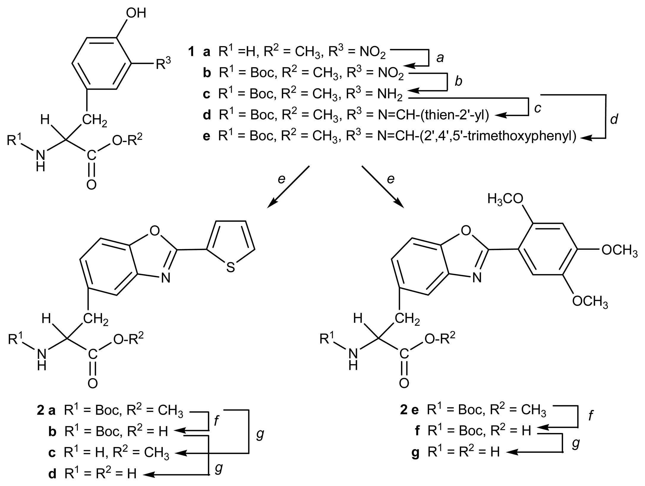
2.1. Synthesis

Starting from commercially available 3-nitro-l-tyrosine, reaction with thionyl chloride in methanol yielded the corresponding methyl ester 1a. Subsequent reaction with t-butyl pyrocarbonate resulted in the N-Boc protected derivative 1b. The nitro group in this compound was reduced to the amino group, by catalytic hydrogenation, yielding compound 1c [25], which was condensed with 2-formylthiophene, in order to obtain the imino derivative 1d. By reaction with lead tetraacetate (LTA) in DMSO [26], compound 1d was oxidised to the [2-(2′-thienyl)benzoxazol-5-yl] alanine derivative 2a.
Compound 2a was then selectively deprotected at its C- and N-terminus, yielding the corresponding N-Boc protected compound 2b and the methyl ester derivative 2c, as well as the fully deprotected alanine derivative 2d. By using similar procedures, compound 1c was reacted with 2,4,5-trimethoxybenzaldehyde, yielding imine 1e. Oxidation of this compound yielded [2-(2′,4′,5′-trimethoxyphenyl)benzoxazol-5-yl] alanine derivative 2e [27]. Selective removal of the protecting groups resulted in the corresponding N-Boc protected 2f and the fully deprotected 2g alanine derivatives (Scheme 1). Compounds 2e-g based on trimethoxyphenyl were prepared in order to confirm the involvement of the sulphur atom at the thiophene ring in the coordination process. All the compounds were obtained in good to excellent yields (Table 1) and were completely characterised by the usual techniques (MS, IR, UV-vis, 1H and 13C NMR spectroscopy).

2.2. Photophysical study

The absorption and emission spectra of benzoxazolyl-alanines 2a-d bearing a thiophene heterocycle and trimethoxy-phenyl alanine derivatives 2e-g in absolute ethanol solution at 298 K were performed. The absorption and emission bands of the 2-substituted benzoxazole derivatives were centered at 315-336 and 393-398 nm, respectively. The fluorescence quantum yields for compounds 2a-g were determined and the derivatives 2a-d with a thiophene moiety were found to be strongly emissive (0.66 < ΦF < 0.80) while compounds 2e-g displayed much lower quantum yields (0.26 < ΦF < 0.47) (Table 2). As can be seen by the results, the selective deprotection at the amino and carboxylic groups of the amino acid residue had only a minor influence on the fluorescence quantum yields. No solvatochromic effect in the absorption spectra was observed in absolute ethanol/water (1:1), absolute ethanol, acetonitrile, dioxane and cyclohexane for all compounds studied.
Compounds 2a-g are amino acid derivatives, which in solution are present in zwitterionic form, and during our studies their zwitterionic nature was taken into account. All of our studies were performed in absolute ethanol and/or in water-absolute ethanol mixtures and the measured pH value was always between 5-6, at which the zwitterionic form predominates. At this pH value, the amine group in alanine derivatives is protonated and the carboxylic group is deprotonated, being available for coordination with the metal ions studied, Cu(II), Hg(II) and Ni(II), in these conditions.

2.3. Spectrofluorimetric titrations and metal sensing effect

In order to evaluate the sensor ability of systems 2a-g in solution towards Zn(II), Hg(II), Cu(II), Ni(II), Ca(II) and Na(I) in low acidic or neutral conditions, UV-vis and fluorescence studies were performed.

2.3.1. Protonation effects

In the presence of increasing amount of HBF4, only compounds 2a and 2f, in acetonitrile or absolute ethanol were slightly affected. The maximum of absorption at 315 nm (2a) and 334 nm (2f), showed a 4 nm red shift upon acid addition. For compound 2f, a new band centered at 380 nm was observed when a larger amount of acid was added (30 to 190 equivalents). As previously reported for related quinoxalinyl derivatives [15], compounds 2b-d and 2e,g did not reveal any significant change in the absorption spectrum upon acid addition in absolute ethanol and absolute ethanol/water (1:1). With further addition of acid, a decrease in the intensity of the fluorescence emission was observed. This quenching may be a result of protonation on the benzoxazole nitrogen atom.

2.3.2. Deprotonation effects

Addition of bases such as potassium hydroxide, triethylamine and tetramethylammonium hydroxide solutions to ligands 2b, 2d, 2f and 2g in ethanolic solutions (with a free carboxylic acid group) were performed.
The maximum of absorption at 315 nm for 2a, and 334 nm for 2f, was slightly affected upon addition of 100 equivalents of tetrabutylammonium hydroxide solution. With further addition of base (more than 500 equivalents), a 50% quenching effect in fluorescence emission was observed. This effect can be attributed to a photoinduced electron transfer (PET) process from the NH2 deprotonated group to chromophore. At this point, the pH value was 10-11 and addition of Hg(II) ions to a basic solution of ligand 2d led to the precipitation of a solid (hydroxide complex), thus preventing complexation studies in basic conditions.

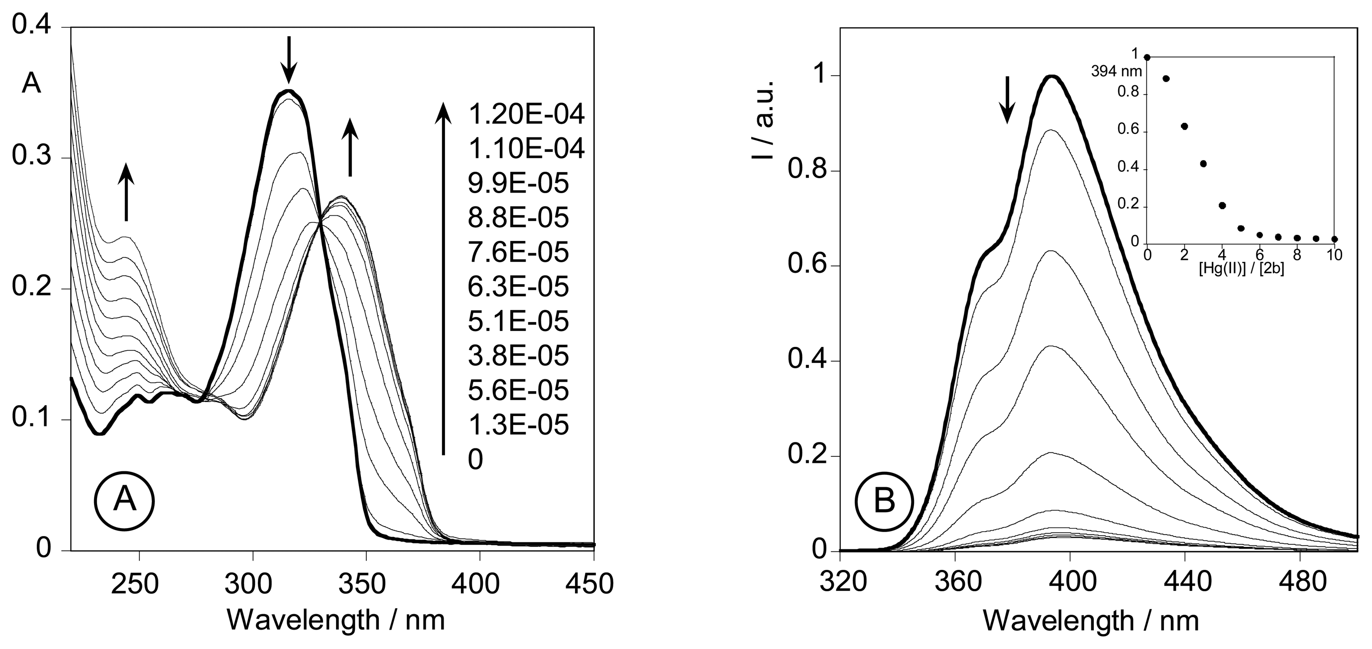
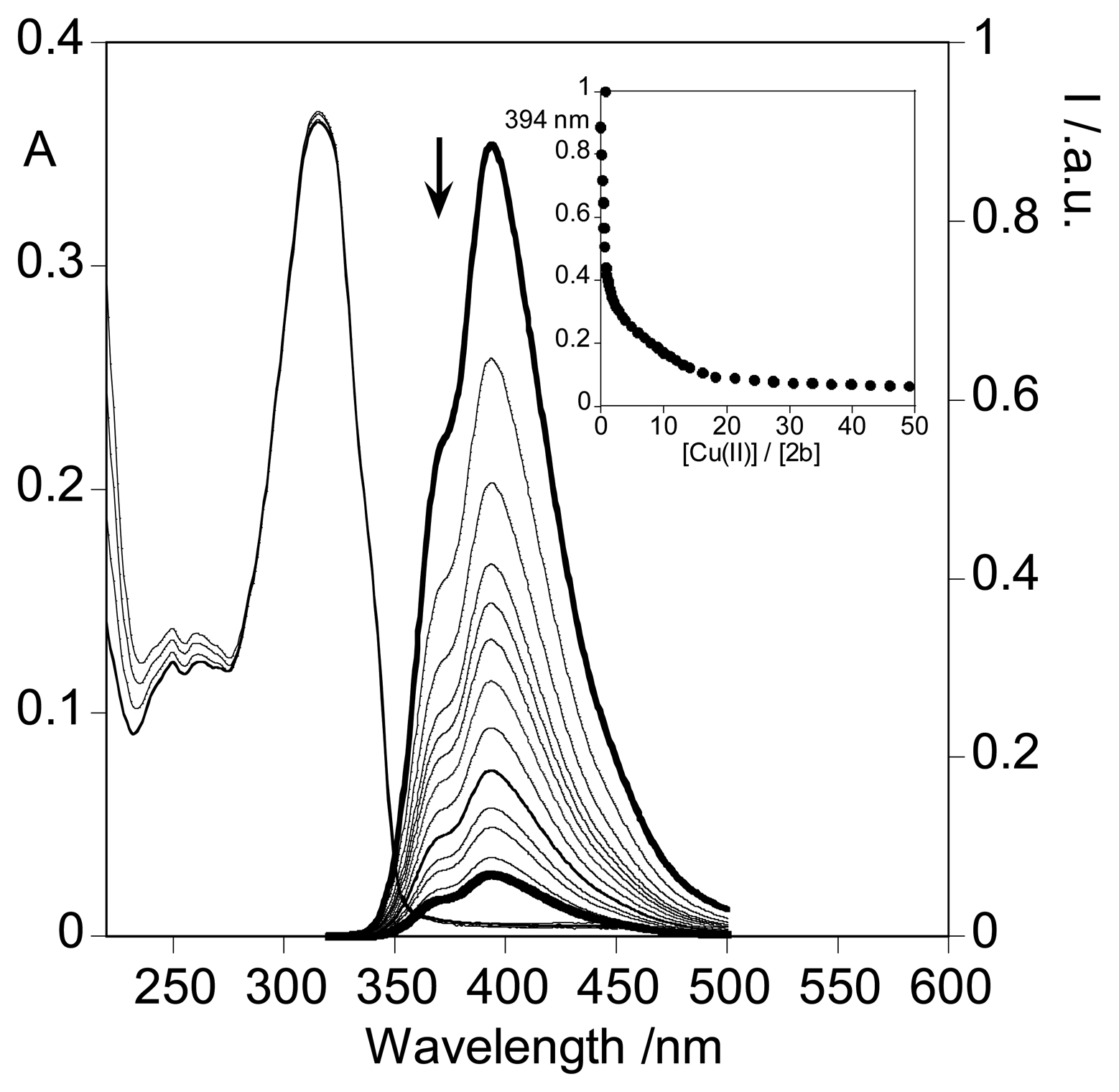
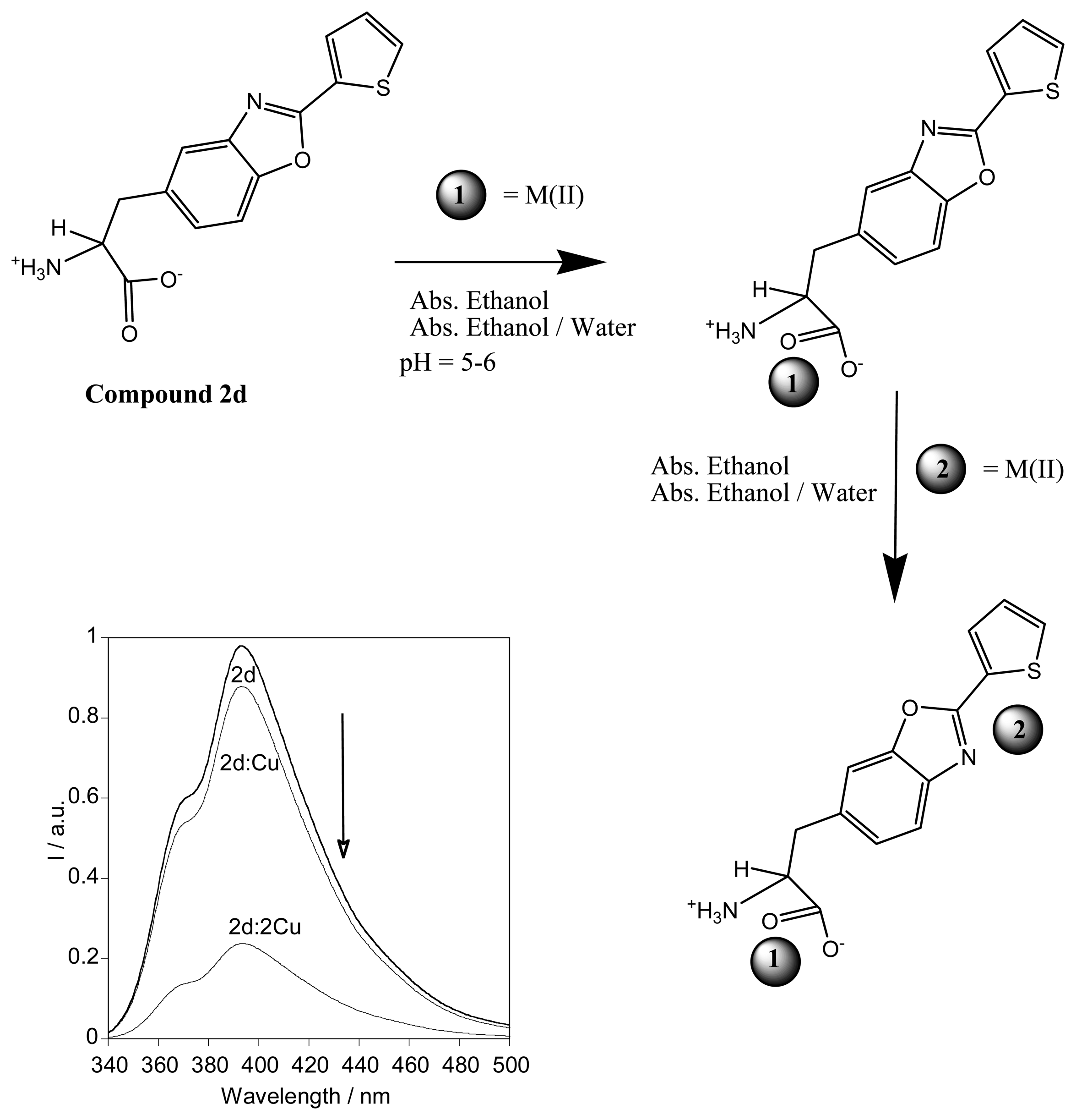
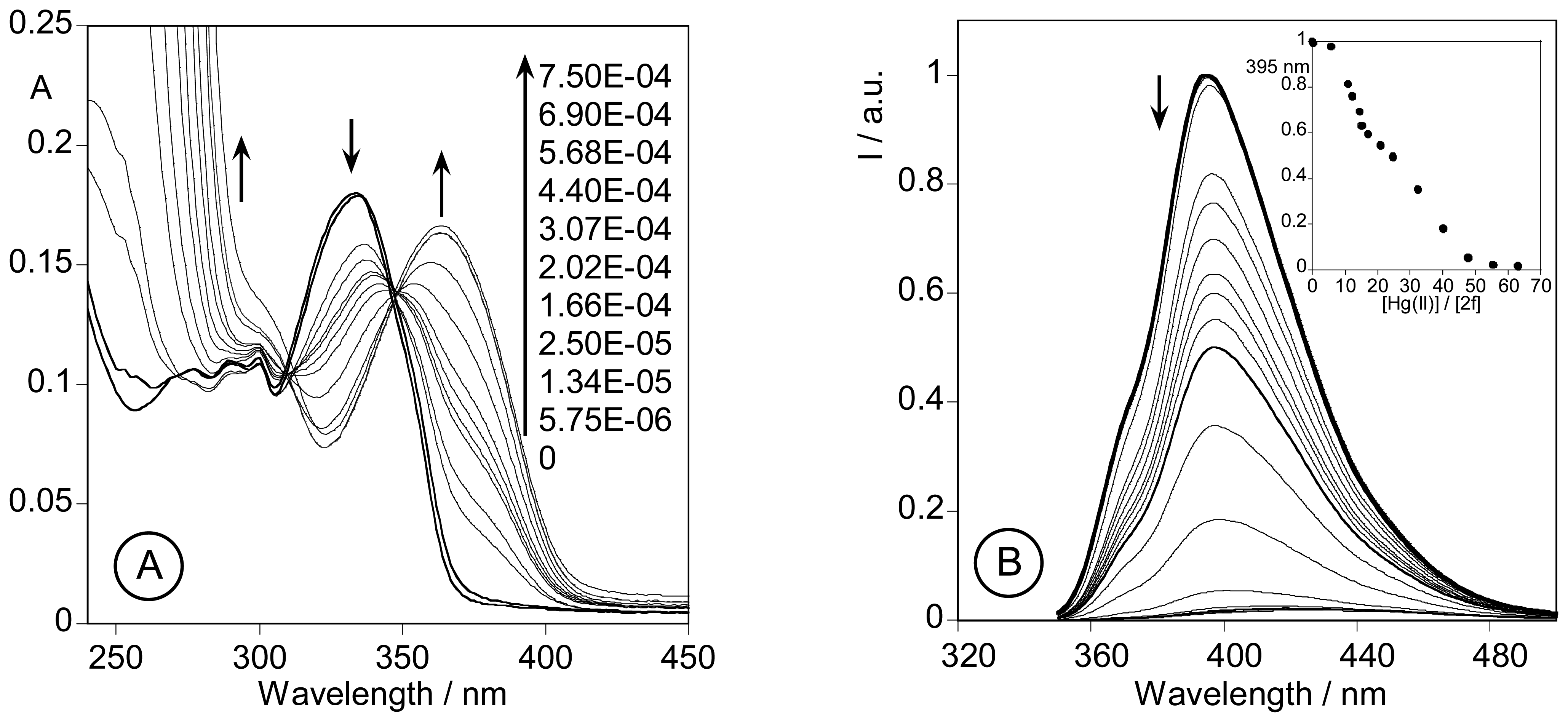
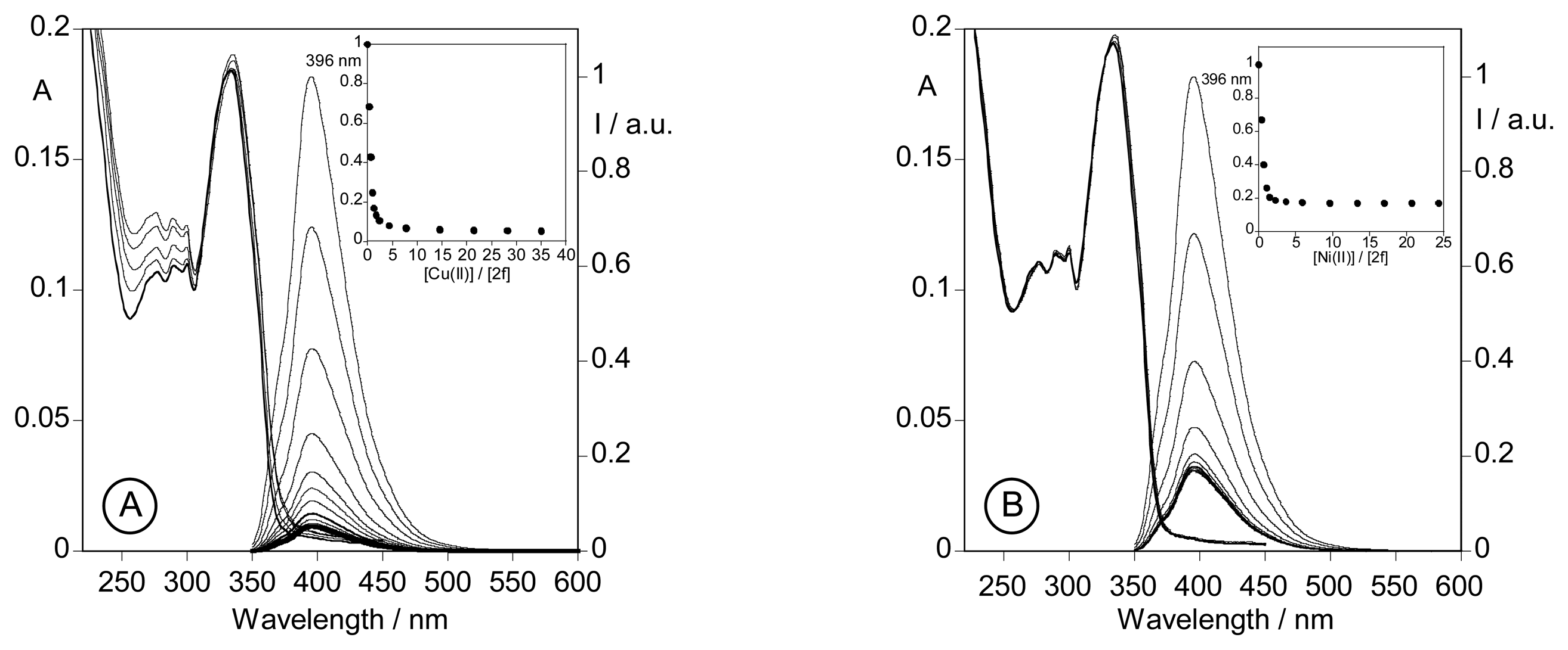
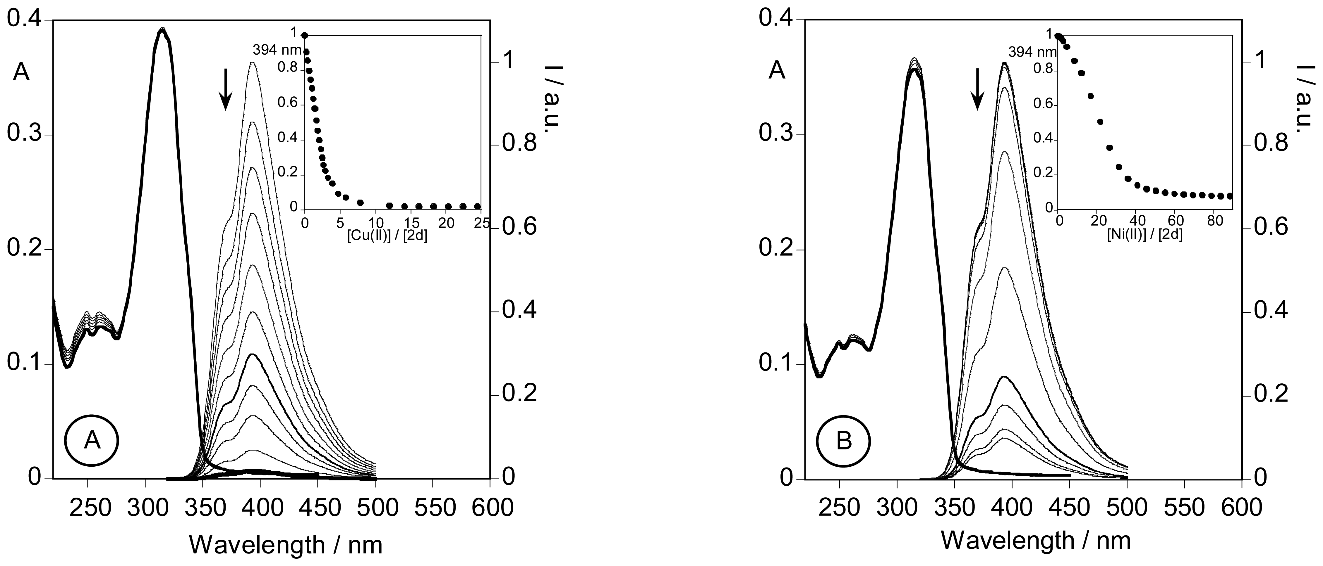
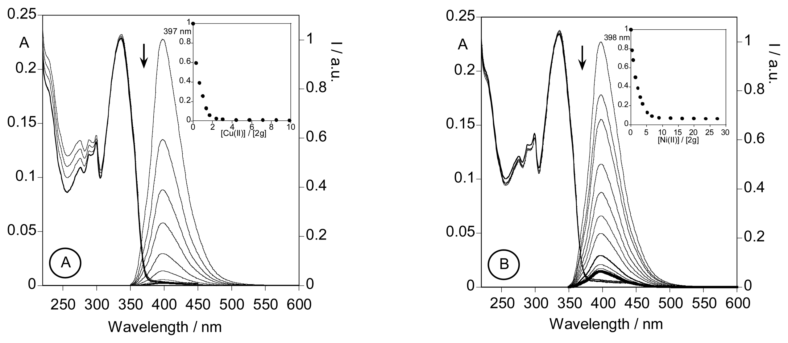
2.3.3. Metal sensing effects

Compounds 2a and 2e, protected at amino and carboxylic terminals, did not show any change in the ground (absorption) and excited (emission) states after the addition of Na(I), Ca(II), Zn(II), Cu(II) and Ni(II) cations. In the titration of 2a with a Hg(CF3SO2)2 ethanolic solution, the intensity of fluorescence was reduced in 30% after the addition of 30 equivalents of metal ion, while the spectrum of trimethoxy-phenyl alanine derivative 2e was not affected by Hg(II) addition. This result can be due to the involvement of the S atom in complexation.
Selective deprotection of the carboxylic terminal in 2a and 2e, gave compounds 2b and 2f. Both systems responded to the presence of Cu(II), Ni(II) and Hg(II), while addition of increasing amounts of Na(I) and Ca(II) did not change either the absorption and emission spectra. A rising CHEQ effect (Chelation Enhancement of the Quenching) [28-29] was observed in the cases of Cu(II), Ni(II) and Hg(II). This CHEQ effect is commonly observed in polyamine ligands containing aromatic fluorophores, and can be attributed to an energy transfer quenching of the π* emissive state through low-lying metal-centre unfilled d-orbitals for Cu(II), and to an intersystem crossing mechanism due to the heavy atom effect for Hg(II) [29]. While the quenching of fluorescence is predominantly connected with the nature of metal ion, the ligand fluorescence enhancement could be result from changes of the geometry between the steady and excited states, or be also induced by the ligand. A very small CHEF effect (Chelation enhancement of the Fluorescence Emission) was observed only when compound 2b was titrated with Zn(II) [30-31].
Although Cu(II) and Ni(II) affected only the excited state of the chromophore in both aforementioned ligands 2b and 2f, Hg(II) affected the ground and excited states when ligand 2b, bearing a thiophene unit, was used. This result suggests that the metal ion interacts with both chelating units present in the compound: the carboxylic acid group of the amino acid residue and the S atom of the thiophene heterocycle [32]. For ligand 2b, complexation constants using the SPECFIT/32 program [33] were obtained only for the interaction with Hg(II). The results suggested that two metal ions are coordinated to the ligand. In the case of ligand 2f all metal complexation studies showed values that agree with 2:1 metal to ligand molar ratio for Hg(II) and 1:1 metal to ligand ratio for Cu(II) and Ni(II) (Table 3).
In Figure 1 is represented the absorption (A) and emission (B) spectra of compound 2b in the presence of increasing amount of Hg(II). The increase of Hg(II) ion concentration in absolute ethanol solution caused the decrease in the absorbance in the range of 280-340 nm, and increase in absorption between 340-390 nm. A sharp isosbestic point at λ= 330 nm was detected (Fig. 1A). After the addition of small molar ratio equivalents of metal ions (0.25 to 1 equivalent) a small effect in absorption and emission was observed. By increasing the metal-ligand molar ratio, strong changes in absorption and emission were observed (inset Figure 1B).
The absorption and emission spectra of compound 2b with increasing amount of Cu(II) is represented in figure 2. It can be seen that while the emission was strongly affected by metal ion complexation, no major changes were seen in the absorption. These results did not allow us to calculate the stoichiometry of complexes with Cu(II) and Ni(II), formed in the ground state. Probably the formation of the 1:1 M:L metal complexes has relatively low values for the binding constants. Increasing the metal ion concentration forces the formation of dinuclear species. Due to the spectral response, complexation probably takes place firstly at the carboxylic acid group of the aminoacid residue, farther from the chromophore, and the second metal ion complexation occurs at the donor atoms present in the chromophore unit. As observed before, interaction of ligand 2b with Hg(II), Ni(II) and Cu(II) also caused a strong CHEQ effect, suggesting the interaction with these metal ions with the chromophore.
The complexation of ligand 2f with Hg(II), Cu(II) and Ni(II) in absolute ethanol can be seen in Figures 3 and 4, respectively. The amount of metal ion necessary to quench the fluorescence emission was higher than that used for the thiophenic ligand 2b. In the case of compound 2f, fifty equivalents were used, in contrast with the six equivalents of Hg(II) needed to quench ligand 2b. These results suggest lower complexation constants for compound 2f. In the cases of Ni(II) and Cu(II), 5 equivalents of metal ion were enough in all cases to reduce the intensity of emission in 80%.
Selective deprotection at the amino terminal of the amino acid gave compound 2c with a free NH2 group. Due to the zwitterionic form of compound 2c studied (pH = 5-6) in which the amine group is protonated, NH3+, a strong decrease in the coordination ability in absolute ethanol and absolute ethanol/water (1:1) was observed when compared with ligand 2b, with a free carboxylic acid group. After addition of Na(I), Ca(II), Zn(II), and Ni(II), only minor changes were observed in the absorption and emission spectra. In the case of Hg(II) and Cu(II) a decrease in the intensity of the fluorescence emission in 40% and 30%, respectively, was observed when a metal to ligand molar ratio of 30:1 was achieved.
Total deprotection of both amino and carboxylic acid terminals in alanines 2a and 2e gave compounds 2d and 2g in the form of free amino acid residues. In our conditions, the zwiterionic form was present in solution. Although higher solubility in water would be expected, ligands 2d and 2g were sparingly soluble in water probably due to the heteroaromatic moiety present at the ligands. Therefore, the complexation studies were carried out in acetonitrile, absolute ethanol and/or water-absolute ethanol mixtures, in order to compare the results for all ligands studied. Once again, no interaction was observed upon complexation with Na(I), Ca(II) and Zn(II) for both ligands, 2d and 2g, in the absorption and emission spectra.
In figure 5 is represented the Hg(II) titration with ligand 2d, showing that both, ground and excited states, are affected by the complexation and an isosbestic point at 325 nm is observed. In contrast, interaction of Hg(II) with compound 2g affected only the excited state, and in this case the intensity of the emission was reduced in 30%. This result is comparable with the results obtained for compounds 2a and 2b, suggesting the importance of the sulphur atom for the sensing of this metal ion, due to its role on the complexation. The values for the complexation constants were calculated and are presented in Table 3.
Cu(II) and Ni(II) complexation are represented in figure 6A and 6B for ligand 2d, and in figures 7A and 7B for ligand 2g. Stronger interaction with Cu(II) was observed in both ligands. Five and two Cu(II) equivalents were enough to totally quench the fluorescence emission of 2d and 2g, while 40 and 10 equivalents of Ni(II), respectively, were necessary to quench the fluorescence emission to 90%. A dinuclear complex could be postulated by the global log K calculated using the SPECFIT32 programme in the case of Ni(II), and a mononuclear species was postulated for Cu(II) (see table 3). A schematic drawing of the proposed coordination process is displayed in Scheme 2. Firstly, the metal ion binds through the carboxylic acid at the alanine residue with small change in the fluorescence emission, followed by another metal ion interaction at the coordinative site present in the chromophore, which strongly affects the emission spectrum.

2.4. Conclusions

The synthesis of new highly fluorescent probes containing the thiophene and benzoxazole moieties combined with an alanine residue was achieved by simple procedures in excellent yields.
The resulting heterocyclic alanine derivatives respond with a quenching effect to the presence of Cu(II), Ni(II) and Hg(II), as shown by the absorption and steady-state fluorescence spectroscopy studies. Selective deprotection of the carboxylic acid group increased the coordination ability, while that deprotection of the amino group reduced notably this effect. The sensor effect takes place by both coordinative sites, for systems 2b and 2d, involving first the terminal carboxylic acid group of the amino acid, and finally the donor atoms present in the chromophores. The synthetic, photophysical and metal ion sensing properties displayed by these compounds showed that they are promising candidates as fluorescent probes and also for sensory applications.

3. Experimental Section

3.1. Synthesis general

All melting points were measured on a Gallenkamp melting point apparatus and are uncorrected. TLC analyses were carried out on 0.25 mm thick precoated silica plates (Merck Fertigplatten Kieselgel 60F254) and spots were visualised under UV light. Chromatography on silica gel was carried out on Merck Kieselgel (230-240 mesh). IR spectra were determined on a Perkin Elmer FTIR-1600 using KBr discs. UV/visible spectra were run on a Shimadzu UV/2501PC or a Perkin Elmer lamda-35 spectrophotometers. 1H NMR spectra were recorded on a Varian 300 spectrometer in CDCl3 or DMSO-d6 at 300 MHz at 25 °C. All chemical shifts are given in ppm using δH Me4Si = 0 ppm as reference and J values are given in Hz. 13C NMR spectra were run in the same instrument at 75.4 MHz using the solvent peak as internal reference. Assignments were made by comparison of chemical shifts, peak multiplicities and J values and were supported by spin decoupling-double resonance and bidimensional heteronuclear HMBC and HMQC correlation techniques. Mass spectrometry analyses were performed at the “C.A.C.T.I. -Unidad de Espectrometria de Masas”, University of Vigo, Spain. Elemental analyses were carried out on a Leco CHNS 932 instrument. Fluorescence spectra were collected using a Perkin Elmer LS45.

3-Nitro-l-tyrosine methyl ester hydrochloride (1a)

Thionyl chloride (0.65 mL, 8.85 × 10⁻³ mol) was added drop wise with stirring to methanol (10 mL), cooled in an ice bath, followed by the addition of 3-nitrotyrosine (2.00 g, 8.85 × 10⁻³ mol). The solution was heated at 40 °C for 5 h. The solvent was evaporated under reduced pressure, yielding a yellowish green solid (2.44 g, quant.). M.p. > 300 °C [lit. [25] data not quoted]. 1H NMR (DMSO-d6): δ = 3.11 (d, J = 6.3 Hz, 2H, β CH2), 3.71 (s, 3H, CH3), 4.30 (t, J = 6.6 Hz, 1H, αH), 7.15 (d, J = 8.4 Hz, 1H, H-5), 7.38 (dd, J = 2.1 and 8.4 Hz, 1H, H-6), 7.78 (d, J = 2.1 Hz, 1H, H-2), 8.55 (br s, 3H, +NH3), 11.08 (br s, 1H, OH) ppm. 13C NMR (DMSO-d6): δ = 34.26 (β CH2), 52.75 (CH3), 52.94 (αC), 119.36 (C5), 125.46 (C1), 126.15 (C2), 136.30 (C3), 136.59 (C6), 151.51 (C4), 169.24 (C=O) ppm. IR (KBr): ν = 3388 (br), 3204, 2856, 1746, 1635, 1589, 1544, 1493, 1445, 1390, 1318, 1241, 1064, 990 cm−1.

N-tert-Butyloxycarbonyl-3-nitro-l-tyrosine methyl ester (1b)

Compound 1a (1.52 g, 5.5 × 10⁻³ mol) was added to 1,4-dioxane (11 mL), water (5.5 mL) and sodium hydroxide 1 m aqueous solution (5.5 mL), with stirring in an ice bath. t-Butyl pyrocarbonate (1.36 g, 1.1 eq, 6.05 × 10⁻³ mol) was added and the mixture stirred at room temperature for 2 days. The volume was reduced by half in a rotary evaporator under reduced pressure and ethyl acetate was added to cover the aqueous layer. The mixture was acidified by addition of KHSO4 1 m aqueous solution, until acidic pH with congo red paper, and extracted with ethyl acetate (3 × 10 mL), followed by washing of the organic layer with water (3 × 10 mL). The organic layer was dried with anhydrous magnesium sulphate and evaporated under reduced pressure, resulting a yellow solid (1.68 g, 90%). M.p. 97.3-98.4 °C [lit. [25] 90-91°C]. 1H NMR (CDCl3): δ = 1.43 (s, 9H, C(CH3)3), 2.95-3.04 (m, 1H, β CH2), 3.12-3.22 (m, 1H, β CH2), 3.76 (s, 3H, CH3), 4.54-4.60 (m, 1H, αH), 5.07 (d, J = 6.6 Hz, 1H, NH), 7.10 (d, J = 8.4 Hz, 1H, H-5), 7.37 (dd, J = 1.8 and 8.4 Hz, 1H, H-6), 7.87 (d, J = 1.8 Hz, 1H, H-2), 10.50 (s, 1H, OH) ppm. 13C NMR (CDCl3) δ = 28.21 (C(CH3)3), 37.23 (β CH2), 52.51 (CH3), 54.18 (αC), 80.27 (C(CH3)3), 120.09 (C5), 125.25 (C2), 128.62 (C1), 133.28 (C3), 138.65 (C6), 154.13 (C4 and C=O Boc), 171.69 (C=O ester) ppm. IR (KBr): ν = 3335, 3023, 3007, 2984, 2955, 2936, 1732, 1685, 1629, 1578, 1536, 1439, 1324, 1221, 1162, 1061, 994, 838 cm−1.

N-tert-Butyloxycarbonyl-3-amino-l-tyrosine methyl ester (1c)

Compound 1b (1.00 g, 2.94 × 10⁻³ mol), 1,4-cyclohexadiene (0.5 mL, 1.8 eq, 5.29 × 10⁻³ mol) and Pd/C (147 mg) were dissolved in methanol (15 mL) and refluxed for 24h. The solvent was evaporated and the residue submitted to column chromatography with silica gel (eluent: CHCl3/MeOH, 98:2). The fractions were combined and the product was obtained as a light brown solid (0.92 g, 98%). M.p. 52.0-52.9 °C [lit. [25] data not quoted]. 1H NMR (CDCl3): δ = 1.43 (s, 9H, C(CH3)3), 2.92 (d, J = 4.8 Hz, 2H, β CH2), 3.74 (s, 3H, CH3), 4.47-4.54 (m, 1H, αH), 5.05 (d, J = 8.4 Hz, 1H, NH), 6.37 (dd, J = 2.1 and 8.1 Hz, 1H, H-6), 6.53 (d, J = 2.1 Hz, 1H, H-2), 6.64 (d, J = 8.1 Hz, 1H, H-5) ppm. 13C NMR (CDCl3) δ = 28.27 (C(CH3)3), 37.64 (β CH2), 52.21 (CH3), 54.57 (αC), 80.11 (C(CH3)3), 115.30 (C5), 117.33 (C2), 119.89 (C6), 128.32 (C1), 134.65 (C3), 143.33 (C4), 155.36 (C=O Boc), 172.78 (C=O ester) ppm. IR (KBr): ν = 3376, 3005, 2955, 1738, 1694, 1615, 1520, 1505, 1454, 1441, 1393, 1368, 1288, 1251, 1217, 1164, 1059, 1022, 912, 861, 799, 758 cm−1. MS (FAB): m/z (%) = 311 (M+ + H, 41), 310 (M+, 100), 307 (20), 255 (59), 254 (47), 211 (57), 193 (27) 155 (24), 154 (83). HRMS (FAB): calcd. for C15H23N2O5 311.1607, found 311.1568.

N-tert-Butyloxycarbonyl-3-[(thien-2′-ylmethylene)amino]-l-tyrosine methyl ester (1d)

Compound 1c (0.54 g, 1.74 × 10⁻³ mol) was stirred with 2-formylthiophene (0.16 mL, 1.76 × 10⁻³ mol) in absolute ethanol (5 mL) for 5 days. The solvent was evaporated and the crude residue was used in the next step without further purification, as light yellow oil (0.68 g, 98%). 1H NMR (CDCl3): δ = 1.42 (s, 9H, C(CH3)3), 2.98-3.10 (m, 2H, β CH2), 3.72 (s, 3H, CH3), 4.52-4.60 (m, 1H, αH), 5.02 (d, J = 7.80 Hz, 1H, NH), 6.90-6.95 (m, 2H, H-2 + H-6), 7.05 (br s, 1H, H-5), 7.14-7.17 (m, 1H, H-4′), 7.51-7.55 (m, 2H, H-3′ + H-5′), 8.76 (s, 1H, N=CH) ppm. 13C NMR (CDCl3) δ = 28.28 (C(CH3)3), 37.84 (β CH2), 52.23 (CH3), 54.55 (αC), 79.99 (C(CH3)3), 114.99 (C2), 116.54 (C5), 127.56 (C1), 127.99 (C4′), 129.46 (C6), 130.82 (C3′ or C5′), 132.59 (C3′ or C5′), 134.98 (C3), 142.73 (C2′), 149.88 (CH), 151.15 (C4), 155.09 (C=O Boc), 172.41 (C=O ester) ppm. IR (KBr): ν = 3375, 2981, 2930, 1754, 1682, 1617, 1602, 1513, 1500, 1430, 1392, 1367, 1295, 1253, 1217, 1165, 1047, 1025, 855, 814 cm−1. MS (FAB): m/z (%) = 405 (M+ + H, 40), 350 (22), 349 (100), 216 (33). HRMS (FAB): calcd. for C20H25N2O5S 405.1484, found 405.1488.

N-tert-Butyloxycarbonyl-3-[(2′,4′,5′-trimethoxyphenylmethylene)amino]-l-tyrosine methyl ester (1e)

Compound 1c (0.365 g, 1.18 × 10⁻³ mol) was stirred with 2,4,5-trimethoxybenzaldehyde (0.231 g, 1.18 × 10⁻³ mol) in absolute ethanol (5 mL) for 3 days. The solvent was evaporated and the residue was used in the next step without further purification as brown oil (0.56 g, 97%). 1H NMR (CDCl3): δ = 1.52 (s, 9H, C(CH3)3), 2.97-3.05 (m, 2H, β CH2), 3.88 (s, 3H, OCH3), 3.93 (s, 3H, OCH3), 3.97 (s, 3H, OCH3), 4.50-4.58 (m, 1H, α-H), 5.00 (d, J = 8.4 Hz, 1H, NH), 6.53 (s, 1H, H-3′), 6.88-6.94 (m, 2H, H-2 + H-6), 6.99 (br s, 1H, H-5), 7.63 (s, 1H, H-6′), 9.01 (s, 1H, N=CH) ppm. 13C NMR (CDCl3) δ = 28.40 (C(CH3)3), 38.02 (β CH2), 52.27 (CH3), 54.65 (αC), 80.06 (C(CH3)3), 100.12 (C3′), 106.33 (C1′), 115.11 (C2), 115.71 (C6′), 116.33 (C5), 127.71 (C1), 129.76 (C6), 135.12 (C3), 135.79 (C5′), 144.76 (C4′), 149.79 (CH), 151.18 (C4), 152.06 (C2′), 155.01 (C=O Boc), 172.23 (C=O ester) ppm. IR (KBr): ν = 3430 (br), 2956, 2927, 2854, 1741, 1708, 1611, 1596, 1510, 1466, 1439, 1412, 1366, 1340, 1280, 1210, 1165, 029, 811 cm−1. MS (FAB): m/z (%) = 489 (M+ + H, 53), 434 (31), 349 (100). HRMS (FAB): calcd. for C25H33N2O8 489.2238, found 489.2252.

N-tert-Butyloxycarbonyl [2-(thien-2′-yl)benzoxazol-5-yl]-l-alanine methyl ester (2a)

The crude imine 1d (0.70 g 1.74 × 10⁻³ mol) and lead tetraacetate (1.17 g, 2.64 × 10⁻³ mol) were stirred at room temperature in DMSO (5 mL) for 3 days. The mixture was poured over water and extracted with ethyl acetate (3 × 10 mL). After drying with anhydrous magnesium sulphate and evaporation of the solvent, the resulting brown oil was submitted to column chromatography with silica gel (eluent: dichloromethane). The product was isolated as a light yellow solid (0.39 g, 56%). M.p. 106.6-107.8 °C. 1H NMR (CDCl3): δ = 1.42 (s, 9H, C(CH3)3), 3.15-3.28 (m, 2H, β CH2), 3.73 (s, 3H, CH3), 4.61-4.67 (m, 1H, αH), 5.04 (d, J = 7.8 Hz, 1H, NH), 7.10 (dd, J = 1.8 and 8.4 Hz, 1H, H-6), 7.17-7.20 (m, 1H, H-4′), 7.45 (d, J = 8.4 Hz, 1H, H-7), 7.48 (d, J = 1.8 Hz, 1H, H-4), 7.56 (dd, J = 1.2 and 5.1 Hz, 1H, H-5′), 7.90 (dd, J = 1.2 and 3.9 Hz, 1H, H-3′) ppm. 13C NMR (CDCl3) δ = 28.24 (C(CH3)3), 38.27 (β CH2), 52.29 (CH3), 54.57 (αC), 80.00 (C(CH3)3), 110.26 (C7), 120.31 (C4), 126.32 (C6), 128.24 (C4′), 129.50 (C2), 129.97 (C3′), 130.31 (C5′), 132.79 (C5), 142.28 (C3a), 149.59 (C7a), 155.04 (C=O Boc), 159.41 (C2′), 172.08 (C=O ester) ppm. IR (KBr): ν = 3358, 2982, 2928, 1734, 1693, 1573, 1526, 1475, 1438, 1369, 1289, 1244, 1172, 1052, 1021, 857 cm−1. UV/Vis (ethanol, nm): λmax (ε) = 315 (2.13 × 10⁴). MS (FAB): m/z (%) = 403 (M+ + H, 73), 348 (23), 347 (100), 243 (29), 215 (27), 185 (21). HRMS (FAB): calcd. for C20H23N2O5S 403.1328, found 403.1324. C20H22N2O5S: calcd. C 59.69, H 5.51, N 6.96, S 7.97; found C 59.44, H 5.63, N 6.69, S 7.66.

N-tert-Butyloxycarbonyl [2-(thien-2′-yl)benzoxazol-5-yl]-l-alanine (2b)

Compound 2a (0.062 g, 1.54 × 10⁻⁴ mol) was dissolved in 1,4-dioxane (1 mL), in an ice bath, and sodium hydroxide 1 m aqueous solution (0.23 mL, 2.3 × 10⁻⁴ mol, 1.5 eq) was added drop wise. The mixture was stirred at room temperature for 3h. The pH was adjusted to 2-3 by addition of KHSO4 1 m aqueous solution and extracted with ethyl acetate (3 × 10 mL). After drying with anhydrous magnesium sulphate and evaporation of the solvent, the residue was triturated with diethyl ether and a off-white solid was obtained (0.050 g, 84%). M.p. 153.5-154.0 °C. 1H NMR (CDCl3): δ = 1.46 (s, 9H, C(CH3)3), 3.30-3.36 (m, 2H, β CH2), 4.68-4.73 (m, 1H, αH), 5.17 (d, J = 7.5 Hz, 1H, NH), 7.16-7.22 (m, 2H, H-6 + H-4′), 7.48 (d, J = 8.4 Hz, 1H, H-7), 7.57 (dd, J = 1.2 and 5.1 Hz, 1H, H-5′), 7.63 (d, J = 2.1 Hz, 1H, H-4), 7.93 (dd, J = 1.2 and 3.9 Hz, 1H, H-3′) ppm. 13C NMR (CDCl3) δ = 28.31 (C(CH3)3), 37.63 (β CH2), 54.46 (αC), 80.14 (C(CH3)3), 110.34 (C7), 120.03 (C4), 127.05 (C6), 128.43 (C4′), 128.67 (C2), 130.73 (C3′), 130.84 (C5′), 133.24 (C5), 141.26 (C3a), 149.35 (C7a), 155.31 (C=O Boc), 159.49 (C2′), 174.66 (COOH) ppm. IR (KBr): ν = 3357, 3113, 2998, 2929, 1752, 1691, 1569, 1527, 1479, 1418, 1371, 1327, 1281, 1252, 1213, 1168, 1051, 856 cm−1. UV/Vis (ethanol, nm): λmax (ε) = 316 (2.73 × 10⁴). MS (FAB): m/z (%) = 389 (M+ + H, 57), 333 (45), 185 (100). HRMS (FAB): calcd. for C19H21N2O5S 389.1171, found 389.1173.

[2-(Thien-2′-yl)benzoxazol-5-yl]-l-alanine methyl ester (2c)

Compound 2a (0.103 g, 2.56 × 10⁻⁴ mol) was stirred in a trifluoroacetic acid/dichloromethane solution (1:1, 1 mL) at room temperature for 2h. The solvent was evaporated, the solid residue dissolved in pH 8 aqueous buffer solution and extracted with ethyl acetate (3 × 10 mL). After drying with anhydrous magnesium sulphate and evaporation of the solvent, the product was isolated as a light yellow solid (0.054 g, 74%). M.p. 80.7-81.4 °C. 1H NMR (CDCl3): δ = 1.46 (s, 9H, C(CH3)3), 2.98-3.06 (m, 1H, β CH2), 3.17-3.24 (m, 1H, β CH2), 3.74 (s, 3H, CH3), 3.77-3.82 (m, 1H, αH), 7.17-7.22 (m, 2H, H-6 + H-4′), 7.49 (d, J = 8.4 Hz, 1H, H-7), 7.56-7.59 (m, 2H, H-5′ + H-4), 7.91 (dd, J = 1.5 and 3.9 Hz, 1H, H-3′) ppm. 13C NMR (CDCl3) δ = 40.77 (β CH2), 52.07 (CH3), 55.92 (αC), 110.25 (C7), 120.18 (C4), 126.33 (C6), 128.24 (C4′), 129.51 (C2), 129.97 (C3′), 130.31 (C5′), 133.85 (C5), 142.33 (C3a), 149.49 (C7a), 159.41 (C2′), 175.12 (C=O ester) ppm. IR (KBr): ν = 3310, 2925, 1741, 1590, 1478, 1415, 1325, 1260, 1063, 855 cm−1. UV/Vis (ethanol, nm): λmax (ε) = 315 (3.07 × 10⁴). MS (FAB): m/z (%) = 303 (M+ + H, 100), 215 (20). HRMS (FAB): calcd. for C15H15N2O3S 303.0803, found 303.0803.

[2-(Thien-2′-yl)benzoxazol-5-yl]-l-alanine (2d)

Compound 2b (0.100 g, 2.58 × 10⁻⁴ mol) was stirred in a trifluoroacetic acid/dichloromethane solution (1:1, 1 mL) at room temperature for 2h. The solvent was evaporated, the solid residue dissolved in pH 8 aqueous buffer solution and extracted with ethyl acetate (3 × 10 mL). After drying with anhydrous magnesium sulphate and evaporation of the solvent, the product was isolated as a light brown solid (0.040 g, 54%). M.p. 238.9-239.9 °C. 1H NMR (DMSO-d6): δ = 2.94-3.02 (m, 1H, β CH2), 3.23-3.30 (m, 1H, β CH2), 3.46-3.51 (m, 1H, αH), 7.28-7.31 (m, 2H, H-6 + H-4′), 7.62-7.67 (m, 2H, H-7 + H-5′), 7.93-7.96 (m, 2H, H-3′ + H-4) ppm. 13C NMR (DMSO-d6) δ = 30.40 (β CH2), 53.01 (αC), 109.92 (C5′), 119.40 (C7), 126.57 (C6), 128.50 (C4′), 129.72 (C2), 130.03 (C4), 131.38 (C3′), 132.34 (C5), 141.36 (C3a), 151.70 (C7a), 159.50 (C2′), 180.83 (C=O) ppm. IR (KBr): ν = 3413, 3072, 2940, 1620, 1572, 1548, 1479, 1407, 1346, 1312, 1287, 1269, 1217, 1195, 1160, 1049, 1024, 1005, 872, 853 cm−1. UV/Vis (ethanol, nm): λmax (ε) = 315 (2.17 × 10⁴). MS (FAB): m/z (%) = 289 (M+ + H, 30), 278 (22), 277 (21), 186 (71). HRMS (FAB): calcd. for C14H13N2O3S 289.0646, found 289.0647.

N-tert-Butyloxycarbonyl-3-[2-(2′,4′,5′-trimethoxyphenyl)benzoxazol-5-yl]-l-alanine methyl ester (2e)

The crude imine 1e (0.470 g, 9.63 × 10⁻⁴ mol) and lead tetraacetate (0.640 g, 1.44 × 10⁻³ mol, 1.5 eq.) were stirred at room temperature in DMSO (5 mL) for 3 days. The mixture was poured over water and extracted with ethyl acetate (3 × 10 mL). After drying with anhydrous magnesium sulphate and evaporation of the solvent, the resulting brown oil was submitted to column chromatography with silica gel (eluent: chloroform). The product was isolated as light brown oil (0.304 g, 65%) [lit. [27] M.p. 137-140 °C]. 1H NMR (CDCl3): δ = 1.43 (s, 9H, C(CH3)3), 3.22 (d, J = 4.2 Hz, 2H, β CH2), 3.74 (s, 3H, CH3), 3.97 (s, 3H, OCH3), 4.00 (s, 3H, OCH3), 4.02 (s, 3H, OCH3), 4.62-4.66 (m, 1H, α-H), 5.01 (d, J = 8.1 Hz, 1H, NH), 6.66 (s, 1H, H-3′), 7.08 (dd, J = 1.8 and 8.4 Hz, 1H, H-6), 7.45 (d, J = 8.4 Hz, 1H, H-7), 7.50 (d, J = 1.8 Hz, 1H, H-4) 7.65 (s, 1H, H-6′) ppm. UV/Vis (ethanol, nm): λmax (ε) = 334 (1.41 × 10⁴). MS (FAB): m/z (%) = 489 (M+ + H, 100), 488 (21), 433 (14), 300 (38), 154 (14). HRMS (FAB): calcd. for C25H33N2O8 489.2237, found 489.2238.

N-tert-Butyloxycarbonyl-3-[2-(2′,4′,5′-trimethoxyphenyl)benzoxazol-5-yl]-l-alanine (2f)

Compound 2e (0.060 g, 1.23 × 10⁻⁴ mol) was dissolved in 1,4-dioxane (1 mL), in an ice bath, and sodium hydroxide 1 m aqueous solution (0.19 mL, 1.9 × 10⁻⁴ mol, 1.5 eq) was added drop wise. The mixture was stirred at room temperature for 3h. The pH was adjusted to 2-3 by addition of KHSO4 1 m aqueous solution and extracted with ethyl acetate (3 × 10 mL). After drying with anhydrous magnesium sulphate and evaporation of the solvent, the residue was triturated with diethyl ether and a brown solid was obtained (0.048 g, 82%). M.p. 135.5-137.0 °C. 1H NMR (CDCl3): δ = 1.45 (s, 9H, C(CH3)3), 3.30-3.34 (m, 2H, β CH2), 3.94 (s, 3H, OCH3), 3.97 (s, 3H, OCH3), 3.98 (s, 3H, OCH3), 4.64-4.68 (m, 1H, α-H), 5.12 (d, J = 8.4 Hz, 1H, NH), 6.62 (s, 1H, H-3′), 7.18 (dd, J = 1.5 and 8.4 Hz, 1H, H-6), 7.50 (d, J = 8.4 Hz, 1H, H-7), 7.60 (s, 1H, H-6′), 7.66 (s, 1H, H-4) ppm. 13C NMR (CDCl3) δ = 28.36 (C(CH3)3), 37.67 (β CH2), 54.46 (αC), 80.20 (C(CH3)3), 100.35 (C3′), 106.43 (C1′), 110.35 (C7), 114.95 (C6′), 120.15 (C4), 126.96 (C6), 128.90 (C2), 133.34 (C5), 135.93 (C5′), 141.18 (C3a), 145.02 (C4′), 149.54 (C7a), 152.26 (C2′), 155.40 (C=O Boc), 174.48 (COOH) ppm. IR (KBr): ν = 3340, 3108, 2988, 2930, 1761, 1684, 1569, 1527, 1420, 1377, 1343, 1265, 1250, 1178, 1049, 850 cm−1. UV/Vis (ethanol, nm): λmax (ε) = 334 (1.75 × 10⁴). MS (FAB): m/z (%) = 473 (M+ + H, 34), 472 (M+, 16), 417 (16), 307 (17), 195 (21), 155 (27), 154 (100). HRMS (FAB): calcd. for C24H29N2O8 473.1924, found 473.1935.

3-[2-(2′,4′,5′-Trimethoxyphenyl)benzoxazol-5-yl]-l-alanine (2g)

Compound 2f (0.035 g, 7.41 × 10⁻⁵ mol) was stirred in a trifluoroacetic acid/dichloromethane solution (1:1, 1 mL) at room temperature for 2h. The solvent was evaporated, the solid residue dissolved in pH 8 aqueous buffer solution and extracted with ethyl acetate (3 × 10 mL). After drying with anhydrous magnesium sulphate and evaporation of the solvent, the product was isolated as an off-white solid (0.012 g, 44%). M.p. 251.6-253.3 °C. 1H NMR (DMSO-d6): δ = 2.94-3.02 (m, 1H, β CH2), 3.23-3.30 (m, 1H, β CH2), 3.46-3.51 (m, 1H, αH), 4.05 (s, 3H, OCH3), 4.11 (s, 3H, OCH3), 4.14 (s, 3H, OCH3), 7.28-7.31 (m, 2H, H-6 + H-3′), 7.60 (d, J = 8.4 Hz, 1H, H-7), 7.65 (s, 1H, H-6′), 7.95 (br s, 1H, H-4) ppm. 13C NMR (DMSO-d6) δ = 30.28 (β CH2), 53.11 (αC), 101.30 (C3′), 105.99 (C1′), 115.34 (C6′), 119.43 (C7), 126.35 (C6), 129.82 (C2), 130.06 (C4), 132.55 (C5), 135.41 (C5′), 141.34 (C3a), 145.13 (C4′), 151.57 (C7a), 152.25 (C2′), 180.88 (C=O) ppm. IR (KBr): ν = 3400, 3052, 2954, 1618, 1570, 1490, 1350, 1325, 1307, 1250, 1198, 1163, 1057, 1030, 1001, 853 cm−1. UV/Vis (ethanol, nm): λmax (ε) = 336 (2.29 × 10⁴). MS (FAB): m/z (%) = 373 (M+ + H, 30), 372 (M+, 43), 278 (100), 186 (51). HRMS (FAB): calcd. for C19H21N2O6 373.1401, found 372.1395.

3.2. Spectrofluorimetric titrations

The linearity of the fluorescence emission vs. concentration was checked in the concentration range used (10−4-10−6 M). A correction for the absorbed light was performed when necessary. All spectrofluorimetric titrations were performed as follows: stock solutions of compounds 2a-g (ca. 10−3 M) were prepared in the corresponding solvents (absolute ethanol, acetonitrile, dioxane or cyclohexane, all of UVA-sol or HPLC grade) and used in the preparation of titration solutions by appropriate dilution. Titration of compounds 2a-g was carried out by addition of microliter amounts of standard solutions of the ions in acetonitrile or absolute ethanol. Protonation and deprotonation studies were performed by addition of HBF4, methanesulphonic acid, triethylamine and tetrabutylammonium hydroxide. Cu(CF3SO3)2, Ni(BF4)2, Zn(CF3SO3)2, Hg(CF3SO3)2, Ca(ClO4)2 and NaNO3 were purchased from Alfa Aesar and used as received. Luminescence quantum yields were measured using as standard a solution of 1-naphthylamine in cyclohexane (0.1 M) (ΦF = 0.46) [34], and were corrected for different refractive indexes of solvents. In all fluorimetric measurements the optical density did not exceed 0.1 [35].

Acknowledgments

Thanks are due to Foundation for Science and Technology (Portugal) for financial support through Centro de Química (UM) and REQUIMTE (UNL) through project PTDC/QUI/66250/2006 and PhD grant to E. Oliveira (SFRH/BD/35905/2007).

References and Notes

  1. Rida, S.M.; Ashour, F.A.; El-Hawash, S.A.M.; ElSemary, M.M.; Badr, M.H.; Shalaby, M.A. Synthesis of some novel benzoxazole derivatives as anticancer, anti-HIV-1 and antimicrobial agents. Eur. J. Med. Chem. 2005, 40, 949 a, nd references cited therein.. [Google Scholar]
  2. Okamura, N.; Suemoto, T.; Shimazu, H.; Suzuki, M.; Shiomitsu, T.; Akatsu, H.; Yamamoto, T.; Staufenbiel, M.; Yanai, K.; Arai, H.; Sasaki, H.; Kudo, Y.; Sawada, T. Styrylbenzoxazole Derivatives for In Vivo Imaging of Amyloid Plaques in the Brain. J. Neurosci. 2004, 24, 2535. [Google Scholar]
  3. Allagile, D.; Baldwin, R.M.; Tamagnan, G.D. One-step synthesis of 2-arylbenzothiazole (‘BTA’) and -benzoxazole precursors for in vivo imaging of β-amyloid plaques. Tetrahedron Lett. 2005, 46, 1349. [Google Scholar]
  4. Leopoldo, M.; Lacivita, E.; De Giorgio, P.; Colabufo, N.A.; Niso, M.; Berardi, F.; Perrone, R. Design, Synthesis, and Binding Affinities of Potential Positron Emission Tomography (PET) Ligands for Visualization of Brain Dopamine D3 Receptors. J. Med. Chem. 2006, 49, 358. [Google Scholar]
  5. Aquilina, J.A.; Carver, J.A.; Truscott, R.J.W. Polypeptide Modification and Cross-Linking by Oxidized 3-Hydroxykynurenine. Biochemistry 2000, 39, 16176. [Google Scholar]
  6. McGrath, M.E.; Sprengeler, P.A.; Hill, C.M.; Martichonok, V.; Cheung, H.; Somoza, J.R.; Palmer, J.T.; Janc, J.W. Peptide Ketobenzoxazole Inhibitors Bound to Cathepsin K. Biochemistry 2003, 42, 15018. [Google Scholar]
  7. Lima, P.G.; Caruso, R.R.B.; Alves, S.O.; Pessôa, R.F.; Mendonça-Silva, D.L.; Nunes, R.J.; Nöel, F.; Castro, N.G.; Costa, P.R.R. Stereoselective synthesis and preliminary evaluation of new D-3-heteroarylcarbonylalanines as ligands of the NMDA receptor. Bioorg. Med. Chem. Lett. 2004, 14, 4399. [Google Scholar]
  8. Cagnoli, R.; Lanzi, M.; Mucci, A.; Parenti, F.; Schenetti, L. Polymerization of cysteine functionalized thiophenes. Polymer 2005, 46, 3588 a, nd references cited therein.. [Google Scholar]
  9. Wang, Y.; Wang, K.; Liu, W.; Shen, G.; Yu, R. Optical Chemical Sensors for Pharmaceutical Analysis Using 1,4-Bis(1,3-benzoxazol-2-yl)benzene as Sensing Material. Analyst 1997, 122, 69 a, nd references cited therein.. [Google Scholar]
  10. Taki, M.; Wolford, J.L.; O'Halloran, T.V. Emission Ratiometric Imaging of Intracellular Zinc: Design of a Benzoxazole Fluorescent Sensor and Its Application in Two-Photon Microscopy. J. Am. Chem. Soc. 2003, 126, 712. [Google Scholar]
  11. Shamsipur, M.; Poursaberi, T.; Karami, A.R.; Hosseini, M.; Momeni, A.; Alizadeh, N.; Yousefi, M.; Ganjali, M.R. Development of a new fluorimetric bulk optode membrane based on 2,5-thiophenylbis(5-tert-butyl-1,3-benzexazole) for nickel(II) ions. Anal. Chim. Acta. 2004, 501, 55. [Google Scholar]
  12. Ohshima, A.; Momotake, A.; Arai, T. A new fluorescent metal sensor with two binding moieties. Tetrahedron Lett. 2004, 45, 9377. [Google Scholar]
  13. Lee, J.K.; Na, J.; Kim, T.H.; Kim, Y.-S.; Park, W.H.; Lee, T.S. Synthesis of polyhydroxybenzoxazole-based colorimetric chemosensor for anionic species. Mat. Sci. Eng. C- Bio S. 2004, 24, 261. [Google Scholar]
  14. Guzow, K.; Milewska, M.; Wróblewski, D.; Gieldón, A.; Wiczk, W. 3-[2-(8-Quinolinyl)benzoxazol-5-yl]alanine derivative - a specific fluorophore for transition and rare-earth metal ion detection. Tetrahedron 2004, 60, 11889. [Google Scholar]
  15. Milewska, M.; Skwierawska, A.; Guzow, K.; Szmigiel, D.; Wiczk, W. 3-[2-(2-Quinoxalinyl)benzoxazol-5-yl]alanine derivative – A specific fluoroionophore for Ni(II). Inorg. Chem. Commun. 2005, 8, 947. [Google Scholar]
  16. Ohshima, A.; Momotake, A.; Arai, T. Photochemical and complexation studies for new fluorescent and colored chelator. Sci. Tech. Adv. Mat. 2005, 6, 633. [Google Scholar]
  17. Zang, X.-B.; Peng, J.; He, C.-L.; Shen, G.-L.; Yu, R.-Q. A highly selective fluorescent sensor for Cu2+ based on 2-(2′-hydroxyphenyl)benzoxazole in a poly(vinyl chloride) matrix. Anal. Chim. Acta. 2006, 567, 189. [Google Scholar]
  18. Batista, R.M. F.; Costa, S.P.G.; Raposo, M.M.M. Synthesis of new fluorescent 2-(2′,2′-bithienyl)-1,3-benzothiazoles. Tetrahedron Lett. 2004, 45(13), 2825. [Google Scholar]
  19. Costa, S.P.G.; Batista, R.M.F.; Cardoso, P.; Belsley, M.; Raposo, M.M.M. 2-Arylthienyl-substituted 1,3-benzothiazoles as new nonlinear optical chromophores. Eur. J. Org. Chem. 2006, 17, 3938. [Google Scholar]
  20. Costa, S. P. G.; Batista, R. M. F.; Sousa, A. M. R. C.; Raposo, M. M. M. New fluorescent heterocyclic materials: synthesis, solvatochromic and fluorescence properties. Mater. Sci. Forum 2006, 514-516, 147. [Google Scholar]
  21. Batista, R.M. F.; Costa, S.P.G.; Malheiro, E.L.; Belsley, M.; Raposo, M.M.M. Synthesis and characterization of new thienylpyrrolyl-benzothiazoles as efficient and thermally stable nonlinear optical chromophores. Tetrahedron 2007, 63(20), 4258–4265. [Google Scholar]
  22. Pina, J.; Seixas de Melo, J.; Burrows, H.D.; Batista, R.M.F.; Costa, S.P.G.; Raposo, M.M.M. Spectral and photophysical characterization of donor-π-acceptor arylthienyl- and bithienyl-benzothiazole derivatives in solution and solid state. J. Phys. Chem. A. 2007, 111, 8574–8578. [Google Scholar]
  23. Batista, R.M. F.; Costa, S.P.G.; Belsley, M.; Raposo, M.M.M. Synthesis and second-order nonlinear optical properties of new chromophores containing benzimidazole, thiophene and pyrrole heterocycles. Tetrahedron 2007, 63(39), 9842–9849. [Google Scholar]
  24. Batista, R.M.F.; Oliveira, E.; Costa, S.P.G.; Lodeiro, C.; Raposo, M.M.M. Synthesis and ion sensing properties of new colorimetric and fluorimetric chemosensors based on bithienyl-imidazo-anthraquinone chromophores. Org. Lett. 2007, 9(17), 3201–3204. [Google Scholar]
  25. Guzow, K.; Szabelski, M.; Malicka, J.; Wiczk, W. Synthesis of a new, highly fluorescent amino acid derivative: N-[(tert-butoxy)carbonyl]-3-[2-(1H-indol-3-yl)benzoxazol-5-yl]-l-alanine methyl ester. Helv. Chim. Acta. 2001, 84, 1086 a, nd references cited therein.. [Google Scholar]
  26. Stephens, F. F.; Bower, J. D. The preparation of benziminazoles and benzoxazoles from Schiff's bases. Part I. J. Chem. Soc. 1949, 2971.
  27. Guzow, K.; Zielińska, J.; Mazurkiewicz, K.; Karolczak, J.; Wiczk, W. Influence of substituents in the phenyl ring on photophysical properties of 3-[2-(phenyl)benzoxazol-5-yl]alanine derivatives. J. Photochem. Photobiol. A. 2005, 175, 57. [Google Scholar]
  28. Czarnik, A. W. Fluorescent Chemosensors for Ion and Molecule Recognition; American Chemical Society: Washington, DC, 1993. [Google Scholar]
  29. de Silva, A.P.; Gunaratne, H.Q.N.; Gunnlaugsson, T.; Huxley, A.J.M.; McCoy, C.P.; Rademacher, J.T.; Rice, T.E. Signaling Recognition Events with Fluorescent Sensors and Switches. Chem. Rev. 1997, 97, 1515. [Google Scholar]
  30. Valeur, B. Molecular Fluorescence. Principles and Applications; Wiley-VCH: Weinheim, 2002. [Google Scholar]
  31. Rurack, K. Flipping the light switch ‘ON’ – the design of sensor molecules that show cation-induced fluorescence enhancement with heavy and transition metal ions. Spect. Acta A. 2001, 57, 2161. [Google Scholar]
  32. Pearson, R.G. Hard and Soft Acids and Bases. J. Am. Chem. Soc. 1963, 85, 3533. [Google Scholar]
  33. SPECFIT/32 Global Analysis System; 3.0 Spectrum Software Associates: Malborough, MA, USA.
  34. Berlman, B. Handbook of Fluorescence Spectra of Aromatic Molecules, 2nd ed.; Academic Press: New York, 1971. [Google Scholar]
  35. Lakowicz, J. R. Principles of Fluorescence Spectroscopy, 2nd ed.; Kluwer Academic/Plenum Publisher: New York, 1999. [Google Scholar]
Figure 1. Spectrophotometric titration (A) and fluorimetric titration (B) of an ethanolic solution of 2b with a standard solution of Hg(CF3SO2)2 in absolute ethanol ([2b] = 1.30E-5 M, T = 298 K, λexc = 316 nm. Inset: normalized emission at 394 nm).
Sensors 07 02096f1
Figure 2. Spectrophotometric titration and fluorimetric titration of an ethanolic solution of 2b with a standard solution of Cu(CF3SO2)2 in absolute ethanol. ([2b] = 1.30E-5 M, T = 298 K, λexc = 316 nm. Inset: normalized emission at 394 nm).
Sensors 07 02096f2
Figure 3. Spectrophotometric titration (A) and fluorimetric titration (B) of an ethanolic solution of 2f with a standard solution of Hg(CF3SO2)2 in absolute ethanol. ([2f] = 1.00E-5 M, T = 298 K, λexc = 334 nm. Inset: normalized emission at 395 nm).
Sensors 07 02096f3
Figure 4. Spectrophotometric titration and fluorimetric titration of an ethanolic solution of 2f with a standard solution of Cu(CF3SO2)2 (A) and Ni(BF4)2 (B) in absolute ethanol. ([2f] = 1.00E-5 M, T = 298 K, λexc = 334 nm. Insets: normalized emission at 396 nm in both cases).
Sensors 07 02096f4
Figure 5. Spectrophotometric titration (A) and fluorimetric titration (B) of an ethanolic solution of 2d with a standard solution of Hg(CF3SO2)2 in absolute ethanol. ([2d] = 1.00E-5 M, T = 298 K, λexc = 315 nm. Inset: normalized emission at 393 nm).
Sensors 07 02096f5
Figure 6. Spectrophotometric titration and fluorimetric titration of an ethanolic solution of 2d with a standard solution of Cu(CF3SO2)2 (A) and Ni(BF4)2 (B) in absolute ethanol. ([2d] = 1.80E-5 M, T = 298 K, λexc = 315 nm. Insets: normalized emission at 394 nm in both cases).
Sensors 07 02096f6
Figure 7. Spectrophotometric titration and fluorimetric titration of an ethanolic solution of 2g with a standard solution of Cu(CF3SO2)2 (A) and Ni(BF4)2 (B) in absolute ethanol. ([2g] = 1.00E-5 M, T = 298 K, λexc = 336 nm. Inset: normalized emission at 397 and 398 nm, respectively).
Sensors 07 02096f7
Scheme 1. Synthesis of tyrosine derivatives 1a-e and alanine derivatives 2a-g. Reagents and conditions: a) (Boc)2O, NaOH 1 m aq solution, rt, 2 days; b) 1,4-cyclohexadiene, Pd/C, MeOH, reflux, 24h; c) 2-formylthiophene, EtOH, rt, 5 days; d) 2,4,5-trimethoxybenzaldehyde, EtOH, rt, 3 days; e)1d or 1e, LTA, DMSO, rt, 3 days; f) NaOH 1 m aq solution, dioxane, rt, 3h; g) trifluoacetic acid/dichloromethane, 1:1, rt, 2h.
Sensors 07 02096f8
Scheme 2. Schematic representation of the complexation mechanism proposed for alanine 2d upon complexation with Cu(II), Ni(II) and Hg(II). Fluorescence spectra of 2d in the presence of one and two equivalents of Cu(II) in absolute ethanol.
Sensors 07 02096f9
Table 1. Synthesis data of tyrosine derivatives 1d-e and alanine derivatives 2a-g.
CompdYield (%)IR (cm−1)1H NMR (δ, ppm)
1d983375, 17548.76 (N=CH) 6.90-6.95 (H-2), 7.05 (H-5)
1e973430, 17419.01 (N=CH) 6.88-6.94 (H-2), 6.99 (H-5)
2a563358, 17347.48 (H-4), 7.45 (H-7)
2b843357, 17527.63 (H-4), 7.48 (H-7)
2c743310, 17417.56-7.59 (H-4), 7.49 (H-7)
2d543413, 16207.93-7.96 (H-4), 7.62-7.67 (H-7)
2e65---7.50 (H-4), 7.45 (H-7)
2f823340, 17617.66 (H-4), 7.50 (H-7)
2g443400, 16187.95(H-4), 7.60 (H-7)
Table 2. UV-vis and fluorescence data for alanine derivatives 2a-g.
CompoundUV-vis λmax (nm)λem (nm)Fluorescence ΦFStokes' shift (nm)
2a3153940.8079
2b3163930.6677
2c3153940.7779
2d3153930.7678
2e3343960.4762
2f3343950.4461
2g3363980.2662
Table 3. Complexation constants for alanine derivatives 2b-d and 2f-g with Cu(II), Ni(II) and Hg(II) in absolute ethanol.
Compd.Metal complexLog KM.L.Ha
2b2bHg5.01 ± 4.0E-02 9.37 ± 7.0E-021.1.0 2.1.0
2d2dHg7.78 ± 2.0E-022.1.0
2d2dCu9.77 ± 2.0E-022.1.0
2f2fHg7.09 ± 8.0E-022.1.0
2f2fCu6.22 ± 2.0E-021.1.0
2f2fNi6.67 ± 6.0E-021.1.0
2g2gCu6.10 ± 6.0E-021.1.0
2g2gNi10.26 ± 8.0E-022.1.0
aM.L.H means Metal. Ligand. proton ratio

