Article

The Nature of Aqueous Solutions of a Cationic Calix[4]arene: A Comparative Study of Dye–Calixarene and Dye–Surfactant Interactions

by
N. O. Mchedlov-Petrossyan
*,
L. N. Vilkova
,
N. A. Vodolazkaya
,
A. G. Yakubovskaya
,
R. V. Rodik
,
V. I. Boyko
and
V. I. Kalchenko
*
Department of Physical Chemistry, Kharkov National University, 61077, Kharkov, Ukraine; Institute of Organic Chemistry, National Academy of Science of Ukraine, 02094, Kiev, Ukraine
*
Authors to whom correspondence should be addressed.
Sensors 2006, 6(8), 962-977; https://doi.org/10.3390/s6080962
Submission received: 30 April 2006 / Accepted: 17 July 2006 / Published: 24 August 2006
(This article belongs to the Special Issue Supramolecular Sensors)

Abstract

:
Among different types of calixarenes, the water–soluble ones are of especial interest because of their possible applications in biochemical research. In order to elucidate the nature of aqueous solutions of a cationic amphiphilic calixarene, substituted tetrapropoxy-calix[4]arene bearing hydrophilic choline groups at the upper rim, we studied vis–spectroscopically the influence of the above system on the acid–base behavior of three indicator dyes, namely, 2,4-dinitrophenol, bromophenol blue, and N,N/-dioctadecylrhodamine, at constant ionic strength of 0.05 M, maintained with NaCl addition. Simultaneously, ‘apparent’ ionization constants, K a a, of the same dyes were determined in the presence of common cationic surfactant micelles. Within the concentration range from 1.0×10⁻⁵ to 0.01 M, the aforementioned water–soluble calixarene displays effects similar to those of micelles of cetyltrimethylammonium bromide (or chloride). The shifts of the absorption and emission bands in the visible region, as well as the alterations of the K a a values against the ‘aqueous’ ones appeared to be very similar in aqueous solutions of both calix[4]arene and cationic surfactant. A conclusion can be made about aggregation (or association), i.e., micelle formation of the cationic calix[4]arene under study.

1. Introduction

The phenomenon of molecular recognition allows reaching of high selectivity in analytical determinations of various inorganic and organic species. Therefore the application of supramolecules to creation of chemical and biochemical sensor systems is of especial interest. Numerous biochemical processes occur in aqueous media. Hence, the search of novel receptors able to selectively bind various substrates in aqueous media is extremely important.
Among a variety of molecular receptors for biochemical research, water–soluble calixarenes are of especial interest [1-5]. They can be also used for analytical determination of such important substrates as acetylcholine [6], as well as for kinetic studies in aqueous media [7], for removal of halogenated organic compounds in water [8], for creation of water–soluble calixarene–cyclodextrin dyads and triads [9,10], and so on.
The most effective way to increase the solubility of calixarenes in water is probably the introduction of ionogeneous groups into their molecules, which results in appearance of positive or negative charges of calixarenes in aqueous media. Such approach is used in a set of recent studies devoted to inclusion processes (‘Host-Guest’ interactions) in aqueous media [2-14].
Hence it is worthwhile to elucidate in detail the nature of such kind of organized solutions. Namely, it is significant to understand the state and properties of supramolecules in water. It is well known that the application of solvatochromic dyes and acid–base indicators is a touchstone for examination of organized solutions [15,16]. Both the chromophore systems and protolytic properties of many dyes are rather sensitive to the solvation properties of microenvironment, to local electrical charges, etc.
And really, a number of papers describing the interaction of such molecular probes and calixarenes in water are available in literature [3,10-13]. However, the ‘calixarene + dye’ systems, as well as other ‘calixarene + substrate’ systems, are usually considered in terms of ‘Host-Guest’ interactions, resulting in formation of inclusion complexes with 1:1 stoichiometry [3,5,10-14]. In some cases, the water–soluble calixarenes possessing long alkyl tails are reported to be ‘unimolecular micelles’ [11]. Most of the water–soluble calixarenes studied are of anionic nature, i.e., they contain several SO3 Na+ groups per molecule. However, Shi and Schneider reported a detailed study of calixarenes with (trimethylammonium)methyl groups at the phenyl rings in the upper rim [5]. These authors determined the association constants values of the mentioned calixarenes with mononucleotides using both NMR shift titration and fluorescence competition titration using 1-anilino-8-naphthalenesulfonate as dye.
On the other hand, the formation of aggregates (self–assembled associates, clusters), or micelles, consisting of several calixarene molecules (ions) cannot be excluded [9,17]. Furthermore, alterations of the absorption and emission spectra of the dyes can evidently result not only from interaction with the molecular cavity, but also from dye adsorption on the surface of the aforementioned aggregates. From this viewpoint, the changes in the protolytic properties of the dyes, caused by water–soluble calixarenes, seem to be a source of additional information.
A set of experiments carried out in our laboratory demonstrated that the shifts of acid–base equilibria induced by some ionogeneous calix[4]arenes and calix[6]arenes in aqueous solutions are very expressed and similar to the effects observed for the dyes in micellar solutions of common ionogeneous surfactants. The aim of the present paper is to report some of these data, which can serve for better understanding of the nature of water–soluble calixarenes. The alterations of the pKa values of three acid–base indicators of different structure were used as main tool for examination of aqueous solutions of a cationic amphiphilic tetrapropoxycalix[4]arene bearing four hydrophilic alkylammonium chloride groups at the upper rim (Chart 1).
Preliminary dynamic lights scattering studies demonstrated that in pure water, without additional electrolytic background, aggregates appear at calix[4]arene concentration ≥ 6×10⁻³ M. The rough estimate of their diameter is 3–4 nm, while the zeta–potential value is ξ ≈ + 66 mV. Hence the properties of such calix[4]arene solutions must be studied thoroughly in terms of micellar effects on versatile dissolved substrates. The indicator dyes used are as follows (Chart 2).
First, the relatively small–sized indicator molecules are represented here by 2,4-dinitrophenol (α-DNP) (Chart 2). Second, the standard acid–base sulfonephthalein indicator, bromophenol blue (BPB) (Chart 2), is certainly too large to be involved to high extent into the host cavity.
Third, a highly hydrophobic rhodamine dye, N,N′-dioctadecylrhodamine (DODR), possesses two long hydrocarbon tails (Chart 2); our preliminary studies demonstrated that both molecular or cationic species of this dye can exist in water only if some surfactant micelles or other nano–sized particles are present in the bulk (continuous) aqueous phase. This dye appeared to be unable to form stable homomicellar solutions in water. Interestingly, though rhodamines are known to form complexes with β-cyclodextrin [19], this hydrophobic rhodamine in form of perchlorate is also insoluble in water in the presence of 0.01 M β-cyclodextrin.
In the course of the present study, we determined the so–called ‘apparent’ p K a a values of the dyes within the calix[4]arene concentration range of 1 × 10⁻⁵ to 0.01 M by means of vis spectroscopy accompanied by potentiometric pH determination. Simultaneously, the p K a a values in the presence of a standard cationic surfactant, cetyltrimethylammonium bromide (CTAB) (Chart 3) are obtained and compared with those in calix[4]arene solutions.

2. Experimental

The calixarene was synthesized accordingly to scheme below:
Sensors 06 00962u1
5,11,17,23-tetra(N,N-dimethyl-N-hydroxyethylammonium)-methylene-25,26,27,28-tetrapropoxycalix[4]arene tetrachloride. To a solution of tetrachloromethylcalix[4]arene [18] (1 mmol) in tetrahydrofurane (20 ml) solution of N,N-dimethylethanoleamine (4.2 mmol) in THF (5 ml) was added under stirring. The reaction mixture was refluxed for 16 hrs. After cooling, the precipitate formed was filtered off. The product was dried in vacuum (0.05 mmHg 20 °C, 4 hrs). Colorless crystalline compound; yield 82%. M.p. 260 °C (decomp.). 1H NMR ((CD3)2S=O, 299.94 MHz), δ ppm: 6.96 (s, 8H, ArH), 5.67 (brs, 4H, OH), 4.50 (s, 8H, Ar-CH2-N), 4.37 (d, J = 12.9 Hz, 4H, ArCHaxAr), 3.86 (brt, 16H, O-CH2-CH2-CH3, CH2-OH), 3.30 (brs, 12H, ArCHeqAr, N-CH2), 2.93 (s, 24H, N-CH3), 1.89 (m, 8H, O-CH2-CH2-CH3), 0.99 (t, J = 7.3 ГЦ, 12H, O-CH2-CH2-CH3), 13C NMR ((CD3)2S=O, 100.60 MHz), δ ppm: 156.94, 134.39, 133.01, 121.73, 76.46, 66.63, 64.19, 54.71, 49.70, 29.79, 22.76, 10.15.
The CTAB sample (purity 99%), Sigma, was used without further purification. The salt form of N,N′-dioctadecylrhodamine, HR+Cl, was prepared and purified by V. I. Alekseeva and L. P. Savvina (Research Institute of Organic Intermediates and Dyes, Moscow, Russia). Commercial samples of α-DNP and BPB were purified by re–crystallization. The purity and stability of dye solutions were checked by means of absorption spectra.
Sodium chloride and potassium bromide were purified by re–crystallization. Standard sodium hydroxide solution was prepared using CO2-free water and was protected from the CO2-containing atmosphere. Hydrochloric and acetic acids were analytical grade reagents.
Absorption spectra of the dye solution were measured using SF-46 apparatus against solvent blanks. The pH determinations were performed at 25.0 ± 0.1 °C on a P 37–1 potentiometer and pH–121 pH–meter equipped with ESL-63-07 glass electrode and an Ag | AgCl reference electrode in a cell with liquid junction (1 M KCl). Standard buffer solutions (pH 1.68, 4.01, 6.86, and 9.18) were used for cell calibration. The fluorescence spectra were determined on Hitachi F 4010 fluorometer. The working concentrations of the dyes were within the range of 2.4×10⁻⁶ to 1.0×10⁻⁵ M (BPB, DODR) and 7.0×10⁻⁵ to 2.0×10⁻⁴ M (α-DNP), while those of calix[4]arene: from 1.0×10⁻⁵ to 0.01 M.
The average particle size and ζ-potential of calix[4]arene particles in pure water were determined by Dr. L. V. Samokhina in the laboratory of Professor M. Ballauff, University of Bayreuth, Germany, by dynamic and electrophoretic light scattering methods using “Zeta Sizer Nano ZS”, Malvern instrument (4 mW He-Ne-laser of 633 nm, optical filter at 633 and 532 nm). All measurements were carried out at 25 °C.
The weighed amounts of calix[4]arene were added to each worked solution. The stock solutions of CTAB were prepared by dissolving weighed amounts of the substance in appropriate amounts of water. Stock solutions of DODR were prepared in 96 % aqueous ethanol for calix[4]arene system and in CTAB micellar solution for surfactant system. All spectra were referenced against solvent blanks containing all components except dyes. Suitable pH values of solutions were created with HCl (pH ≤ 3.0), acetate buffers, or diluted NaOH; in the experiments with CTAB, borate buffer solutions were used as well. The required ionic strength (0.05 or 0.1 M) of solutions used for determination of ionization constants was maintained by addition of NaCl or KBr. The theoretically calculated pH values practically coincided with the measured values (±0.02 units) both in the presence and in the absence of calix[4]arene or surfactants.
The key parameter of acid–base indicator equilibrium in organized solutions is the so–called ‘apparent’ ionization constant, K a a [15,20]. The indices of these constants, p K a a, were determined spectrophotometrically at 25 °C, according to the standard procedure [20]. The following equation was used for calculations:
p K a a = p H + log A R z 1 A A A H R z ,
where A is the absorbance at the current pH at chosen wavelength and constant dye concentration, ARz−1 and AHRz are absorbances under conditions of complete transformation of the dye into the correspondent form (Rz–1, HRz). Here z and (z–1) are charges of the conjugated indicator species (HRZ ⇄ Rz−1 + H+). The ratio of the equilibrium concentrations of these species can be derived from electronic absorption, while the pH values of the bulk (continuous, aqueous) phase are determined as a rule by using the glass electrode.
The number of working solutions used for each p K a a determination was 5 to 7. In the cases of α-DNP and BPB, the absorbance at three wavelengths near the maximum of the ionized species was used for calculations of the apparent ionization constants. In the case of DODR, 17 wavelengths within the range of 500-580 nm were used as analytical positions. The differences between A at 510–520 nm and 535–545 nm in CTAB solutions and those between A at 500–520 nm and 560–570 nm in calix[4]arene solutions were utilized for final calculations because the dependence of such differences versus pH is more pronounced. The p K a a values were determined with confidence interval ± (0.01–0.07).

3. Results and Discussion

The absorption spectra of anionic species of α-DNP and BPB in aqueous solution (blank experiment) and in calix[4]arene solutions are given in Figures 1 and 2. In the case of α-DNP anion, the spectral changes on going from water to calix[4]arene solutions are similar.
Absorption spectra provide some information about the binding of dye to micelles. The maximum wavelength of R2– anion of BPB (λmax = 603 nm) in calixarene solution is red–shifted as compared with that obtained in water (λmax = 591 nm), while the band of HR species is shifted toward the blue. In CTAB micellar solutions (3×10⁻³–0.01 M), the λmax value of R2– species also equals 603 nm, and this batochromic shift is accompanied by a slow hypochromic effect against the ‘aqueous’ spectrum. Moreover, the HR band is also blue–shifted. Such phenomenon is typical for sulfonephthalein series in cationic surfactant solutions [21-23].
Some typical p K a a values are presented in Table 1. Hereafter, the symbol p K a w denotes the indices of the ionization constants in water, at the correspondent ionic strength; Δ p K a a = p K a a p K a w.
It can be stated that not only the spectral shifts, but also the p K a a and Δ p K a a values of the given dye in CTAB micellar solution and in 2.5×10⁻³ M calix[4]arene solution are similar in general features. The critical micelle concentration (c.m.c.) of CTAB is ca. 1×10⁻³ M in pure water, and it is known to decrease in salt solutions. In 0.05 M NaCl solution, the c.m.c. value is ≈ 1×10⁻⁴ M, and the surfactant micelles contain both Cl and Br ions as counter–ions in the Stern layer. If the aforementioned calix[4]arene aggregates appear at concentrations near 0.006 M in pure water, they can already exist at 2.5×10⁻³ M in the presence of 0.05 M NaCl. And indeed, the p K a a values of the indicators, especially those of the hydrophobic dye BPB, are close as obtained at calixarene concentrations of 2.5×10⁻³ and 5.0×10⁻³ M (see below).
In the case of CTAB micelles, the negative Δ p K a a values reflect the positive electrical charge of the micellar interface where the acid–base indicators are located [20-26].
According to the electrostatic theory [20,21,23-26] the apparent p K a a value of an indicator under conditions of complete binding of the indicator couple HRz / Rz-1 depends on the electrostatic surface potential of the micelle, Ψ, according to the equation:
p K a a = p K a i ψ F 2.303 R T ,
where p K a i is the so–called intrinsic p Ka value, T is the absolute temperature, the constants F and R have their usual meaning.
The volume of the dispersed (micellar) phase usually makes less than 1% of the total volume of solution, and the molar ratio surfactant : dye is close to 100. The medium effect, Δ p K a a = p K a a p K a w, is essentially caused by the quantity − ΨF/2.3026RT (eq 2). As a result, the p K a a values at low bulk ionic strength on the whole decrease in micellar solutions of cationic surfactants (Ψ > 0).
From this viewpoint, the influence of calix[4]arene on the state of acid–base equilibria of indicator dyes (Table 1) is in line with that of common micelles of cationic surfactants. In calixarene small–sized self-assembly, the propyl groups are probably directed toward the center of the aggregates, while the acetylcholine residues form the hydrophilic interior. As is usual in the case of cationic micelles, a fraction of counter–ions (Cl), is situated within the diffuse layer, the degree of surfactant dissociation being usually 20–30 %.
Eq. 2 is valid in the case of complete binding of the indicator species by the micellar pseudophase. Under such conditions, further increase in the amphiphile (e. g., surfactant) concentration will obviously display no marked changes in the p K a a values. Figures 3 and 4 demonstrate such dependences of p K a a vs. calix[4]arene and CTAB concentrations, respectively.
The curves demonstrate gradual binding of α-DNP species by both calixarene and CTAB assemblies. In the case of BPB – calixarene and especially in BPB – CTAB system, the p K a a values drop dramatically. In the last case, a somewhat higher value of the ionic strength is chosen, in order to decrease the c.m.c. value and thus to avoid the formation of the insoluble dye – surfactant salts.
From such curves, the so–called ‘binding constants’ can be obtained following the standard procedure [27]. For example, for α-DNP – CTAB system, the values Kb,HR = (2.5±0.12)×102 M–1 and Kb,R = (8.6±0.2)× 103 M–1 were calculated.
Evidently, the binding of α-DNP species are still incomplete at calix[4]arene concentration of 2.5×10⁻³ M. The comparison of the data for α-DNP and BPB allows to suppose the significant contribution of both electrostatic and hydrophobic interactions in the ‘calixarene + dye’ systems.
It must be noted, that the p K a a and Δ p K a a values under conditions of complete binding by calix[4]arene (Fig. 3) correspond to the Ψ values of ≈ +(80–150) mV. This is in agreement with the estimate of zeta–potential in pure water, ξ ≈ + 66 mV (see above). In the presence of electrolytic background (0.05 M NaCl) the ξ value must decrease. Indeed, the ξ value corresponds to the splitting plane, which is located outside the binding site of the dyes, and hence it is lower than the Ψ value.
Of course, in the case of ‘dye + calix[4]arene’ systems, the (partial) inclusion of dye species into the cavity of the receptor cannot be excluded, and it does not contradict the concept of aggregation of the supramolecules.
Spatial structure of calix[4]arene was investigated with molecular mechanic computations (MM+). Calixarene exists in a pinched cone conformation (Chart 4, chlorine anions are omitted for clarity).
Two distal rings are almost parallel. Two other rings are almost orthogonal. Distances between distal N-atoms are equal to 5.9 and 13.7 Ǻ, neighboring N-atoms are located at 7.3 Å. It is important that variation of distances indicates possibility of geometry tuning to effective interaction with certain molecules.
Using the AM1 method (Cambridge Soft Chem 3D software package), we estimated the size parameters of α-DNP molecular and anionic species (the calculations were made by Dr. A. V. Lebed). The molecule goes in the cage with the longest parameter of 6.63 Å. Hence, this substrate can be included in the cavity of the given calixarene.
Analogous calculations made for the propeller–like BPB anions demonstrated that the sulfobenzoic residue could also enter the cavity: the distance between the oxygen atoms of the SO3 group and the hydrogen atom in the para position (5//) is 6.14–6.3 Å (Chart 5).
However, the 2 and 2′ hydrogen atoms hinder the deep penetration of the anionic fragment into the receptor molecule. Another part able to include the ‘phenolate’: the distance between the 3 and 5) or 3/ and 5/) bromine atoms in the R2– species is 5.81 Å.
The two above dyes, α-DNP and BPB, belong to anionic ones. In fact, they are representatives of the indicators of phenolic type. Contrary to them, DODR is actually a cationic carboxylic acid, which turns into zwitterion. Nevertheless, in aqueous calix[4]arene solutions, the p K a a value of this complicated dye is just the same as in CTAB micelles (Table 1). The emission spectra of the dye species, HR+ and R±, are also similar in the organized solutions of both types (Fig. 5, 6).
Such relatively small blue shifts of both emission and absorption bands due to ionization of carboxylic group (COOH → COO) are typical for rhodamine dyes [28]. The spectra at pH below 1.2 stay practically unchanged; they must be ascribed to HR+ species. The absorption bands of the species are broader in the case of calix[4]arene solutions (Fig. 7 and 8). Such character of absorption spectra is usually observed for rhodamine dye films adsorbed on solid interfaces (in our experiments, care was taken to exclude adsorption on glass vessels). Maybe, this spectral effect reflects the complicated structure of dye–calixarene aggregates. Though DODR and calix[4]arene concentrations were respectively 2.4×10⁻⁶ and 2.5×10⁻³ M, some interactions between dye chromophores cannot be excluded. On the other hand, no precipitation of DODR was observed in the presence of macrocycle studied. Hence, some kind of solubilization of this water–insoluble dye certainly takes place, maybe, resulting in mixed micelles formation. Probably, the hydrophobic interactions are of especial significance in the case of DODR, because even its cationic species, HR+, evidently associate with calix[4]arene.
Finally, the inclusion of the ionizing group of DODR (COOH → COO), into the cationic cavity can also take place, though it may be hindered for steric reasons. The long n-C18H37 chains are probably directed toward the center of calix[4]arene aggregates.
In conclusion, it can be stated that the amphiphilic cationic calix[4]arene described in the present paper shifts the state of acid–base equilibria of different indicator dyes in aqueous solutions. These effects, as well as the alterations of vis spectra of the substrates, are very similar to those caused by micelles of cationic surfactants. Further research, especially the studies of competition between different anionic substrates, can elucidate the contribution of inclusion phenomena in the interactions of the given calix[4]arene and dyes in aqueous media.

Acknowledgments

The authors are grateful to Dr. V. I. Alekseeva and Dr. L. P. Savvina for gifting the sample of the rhodamine dye, to O. Yu. Soboleva for assistance in some experiments with calix[4]arene, to Dr. A. V. Timiy for determination of ionization constants in BPB – CTAB system, to Dr. L. V. Samokhina for dynamic lights scattering studies, and to Dr. A. V. Lebed for AM1 calculations of dye structures.

References

  1. Arimura, T.; Kawabata, H.; Matsuda, T.; Muramatsu, T.; Satoh, H.; Fujo, K.; Manabe, O.; Shinkai, S. New Water-Soluble Host Calixarenes Bearing Chiral Substituents. J. Org. Chem. 1991, 56, 301–306. [Google Scholar]
  2. Douteau-Guével, N.; Coleman, A. W.; Morel, J.-P.; Morel-Desrosiers, N. Complexation of Basic Amino Acids by Water-Soluble Calixarene Sulphonates as a Study of the Possible Mechanism of Recognition of Calixarene Sulphonates by Proteins. J. Phys. Org. Chem. 1998, 11, 693–696. [Google Scholar]
  3. Zhang, Y.; Pham, T. H.; Sánchez Pena, M.; Agbaria, R. A.; Warner, I. M. Spectroscopic Studies of Brilliant Cresyl Blue / Water-Soluble Sulfonated Calix[4]arene Complex. Applied Spectrosc. 1998, 52, 952–957. [Google Scholar]
  4. Hioki, H.; Yamada, T.; Fujioka, C.; Kodama, M. Peptide Library Based on Calix[4]arene. Tetrahedron Lett. 1999, 40, 6821–6825. [Google Scholar]
  5. Shi, Y.; Schneider, H.-J. Interaction Between Aminocalixarenes and Nucleotides or Nucleic Acids. J. Chem. Soc., Perkin Trans. 2 1999, 1797–1803. [Google Scholar]
  6. Jin, T. A New Fluorometric Method for the Detection of the Neurotransmitter Acetylcholine in Water Using a Dansylcholine Complex with p-Sulfonated Calix[8]arene. J. Incl. Phenom. Macro. 2003, 45, 195–201. [Google Scholar]
  7. Tao, W.; Barra, M. Inhibition of Quinone–Imine Dye Deamination by Complexation with Para-Sulfonated Calixarenes. J. Org. Chem. 2001, 66, 2158–2160. [Google Scholar]
  8. Iki, N.; Fujimoto, T.; Shindo, T.; Koyama, K.; Miyano, S. Almost Complete Removal of Trace Amount of Halogenated Organic Compounds in Water: An Approach by Use of a Combination of Water-Soluble Thiacalixarene and Ion-Exchange Resins. Chem. Lett. 1999, 777–778. [Google Scholar]
  9. Bügler, J.; Sommerdijk, N. A. J. M.; Visser, A. J. W. G.; van Hoek, A.; Nolte, R. J. M.; Engbersen, J. F. J.; Reinhoudt, D. N. Interconnective Host-Guest Complexation of β-Cyclodextrin – Calix[4]arene Couples. J. Am. Chem. Soc. 1999, 121, 28–33. [Google Scholar]
  10. Liu, Y.; Chen, Y.; Li, L.; Huang, G.; You, C.-C.; Zhang, H.-Y.; Wada, T.; Inoue, Y. Cooperative Multiple Recognition by Novel Calix[4]arene-Tethered β-Cyclodextrin and Calix[4]arene-Bridged Bis(β-cyclodextrin). J. Org. Chem. 2001, 66, 7209–7215. [Google Scholar]
  11. Zhang, Y.; Cao, W. Self-Assembly of Small Molecules: An Approach Combining Electrostatic Self-Assembly Technology with Host-Guest Chemistry. New J. Chem. 2001, 25, 483–486. [Google Scholar]
  12. Liu, Y.; Han, B.-H.; Chen, Y.-T. Inclusion Complexation of Acridine Red Dye by Calixarenesulfonates and Cyclodextrines: Opposite Fluorescent Behavior. J. Org. Chem. 2000, 65, 6227–6230. [Google Scholar]
  13. Liu, Y.; Han, B.-H.; Chen, Y.-T. Molecular Recognition and Complexation Thermodynamics of Dye Guest Molecules by Modified Cyclodextrins and Calixarenesulfonates. J. Phys. Chem. B 2002, 106, 4678–4687. [Google Scholar]
  14. Kunsági-Máte, S.; Szabo, K.; Lemli, B.; Bitter, I.; Nagy, G.; Kollár, L. Host-Guest Interaction Between Water-Soluble Calix[6]arene Hexasulfonate and p-Nitrophenol. Thermochim. Acta. 2005, 425, 121–126. [Google Scholar]
  15. Grieser, F.; Drummond, C. J. The Physicochemical Properties of Self-Assembled Surfactant Aggregates as Determined by Some Molecular Spectroscopic Probe Techniques. J. Phys. Chem. 1988, 92, 2580–2593. [Google Scholar]
  16. Bhattacharyya, K. Solvation Dynamics and Proton Transfer in Supramolecular Assemblies. Acc. Chem. Res. 2003, 36, 95–101. [Google Scholar]
  17. Ryzhkina, I. S.; Kudryavtseva, L. A.; Konovalov, A. I. The Reactivity of Associates of Aminomethylated Calix[4]resorcinolarenes Towards p-Nitrophenyl Esters of Phosphorous Acids. XIVth Internat. Conf. of Phosphorus Chemistry: Abstracts, Cincinnati, USA, Cincinnati, July 12-17, 1998; 1998; p. p. 114. [Google Scholar]
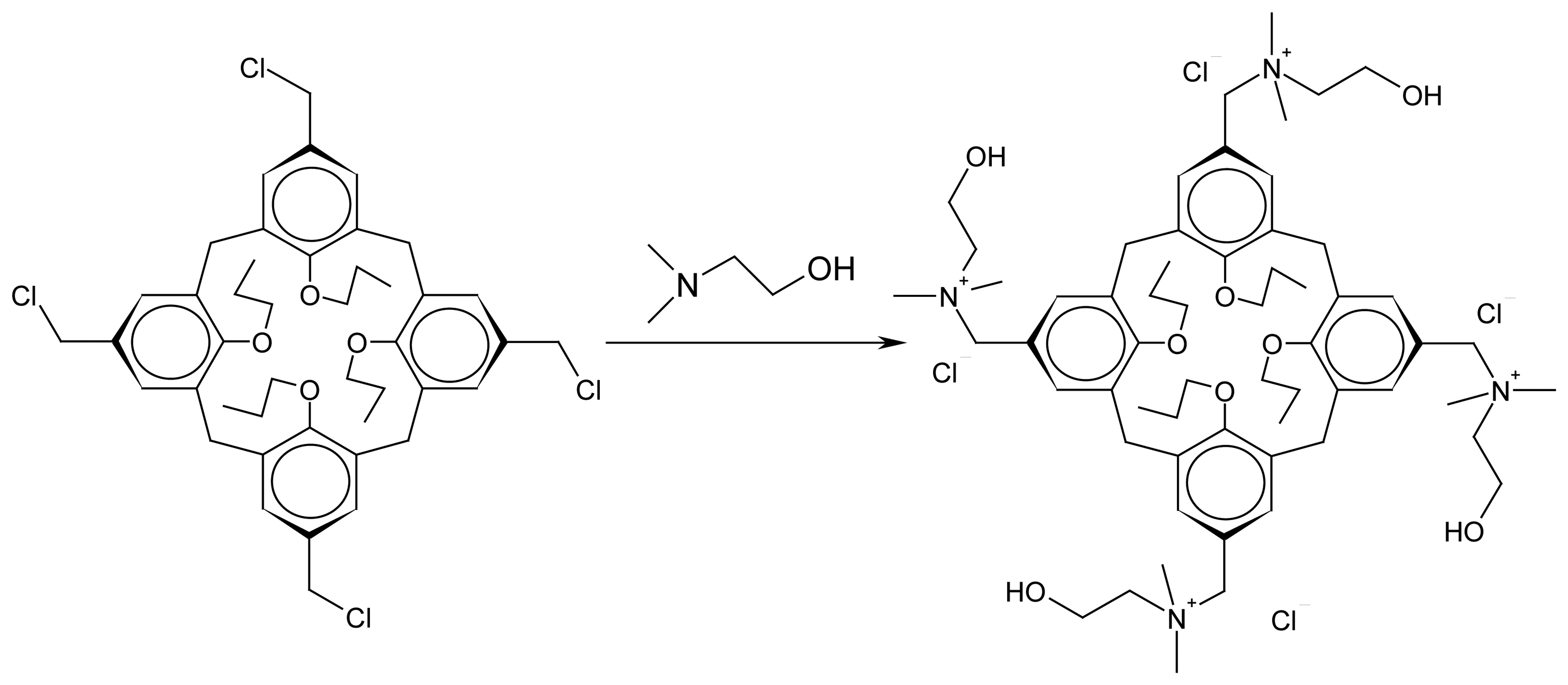
  18. Almi, M.; Arduini, A.; Casnati, A.; Pochini, A.; Ungaro, R. Chloromethylation of Calixarenes and Synthesis of New Water Soluble Macrocyclic Host. Tetrahedron 1989, 45, 2177–2182. [Google Scholar]
  19. Mchedlov-Petrossyan, N. O.; Kholin, Yu. V. Aggregation of Rhodamine B in Water. Russ. J. Appl. Chem. (English transl.) 2004, 27, 414–422, and references cited therein. [Google Scholar]
  20. Mchedlov-Petrossyan, N. O.; Vodolazkaya, N. A.; Reichardt, C. Unusual Findings on Studying Surfactant Solutions: Displacing Solvatochromic Pyridinium N-phenolate Towards Outlying Areas of Rod–Like Micelles? Colloids and Surfaces A 2002, 205, 215–229. [Google Scholar]
  21. Moller, J. V.; Kragh-Hanssen, U. Indicator Dyes and Probes of Electrostatic Potential Changes on Macromolecular Surfaces. Biochemistry 1975, 14, 2317–2322. [Google Scholar]
  22. Rozendorfova, J.; Cermakova, L. Spectrophotometric Study of the Interaction of Some Triphenylmethane Dyes and 1-Carbethoxypentadecyltrimethylammonium Bromide. Talanta 1980, 27, 705–708. [Google Scholar]
  23. Drummond, C.; Grieser, F.; Healy, T. W. Acid-Base Equilibria in Aqueous Micellar Solutions. J. Chem. Soc., Faraday Trans 1 1989, 85, 537–550. [Google Scholar]
  24. Funasaki, N. The Effect of the Solvent Property of the Surfactant Micelle on the Dissociation of Weak Electrolytes. Nippon Kagaku Kaishi 1976, 722–726. [Google Scholar]
  25. Fernandes, M.S.; Fromherz, P. Lipoid pH Indicators as Probes of Electrical Potential and Polarity in Micelles. J. Phys. Chem. 1977, 81, 1755–1761. [Google Scholar]
  26. Funasaki, N. Micellar Effects on the Kinetics and Equilibrium of Chemical Reactions in Salt Solutions. J. Phys. Chem. 1979, 83, 1998–2003. [Google Scholar]
  27. Mchedlov-Petrossyan, N. O.; Timiy, A. V.; Vodolazkaya, N. A. Binding of Sulfonephthalein Anions to the Micelles of an Anionic Surfactant. J. Mol. Liq. 2000, 87, 75–84. [Google Scholar]
  28. Mchedlov-Petrossyan, N. O.; Vodolazkaya, N. A.; Doroshenko, A. O. Ionic Equilibria of Fluorophores in Organized Solutions. The Influence of Micellar Microenvironment on Protolytic and Photophysical Properties of Rhodamine B. J. Fluoresc. 2003, 13, 235–248. [Google Scholar]
Figure1. Absorption spectra of R species of α-DNP (7.38×10⁻⁵ M) in water (1), in 2.5×10⁻³ M calix[4]arene solution (2), and in CTAB micellar solution (3); pH = 6.5–12.
Sensors 06 00962f1
Figure 2. Absorption spectra of BPB species: R2–, pH = 6 (1, 1′) and HR, pH = 0–1.0 (2, 2′) (5.0×10⁻⁶ M) in water (1, 2) and in 5×10⁻³ M calix[4]arene solutions (1′, 2′).
Sensors 06 00962f2
Figure 3. The dependence of the p K a a values on calix[4]arene concentration in aqueous media, at ionic strength 0.05 M (NaCl): BPB (1); α-DNP (2).
Sensors 06 00962f3
Figure 4. The dependence of p K a a values on CTAB concentrations: BPB, ionic strength: 0.1 M, KBr (1); α-DNP, ionic strength 0.05 M, KBr (2).
Sensors 06 00962f4
Figure 5. Emission spectra of DODR in aqueous solutions of calix[4]arene (2.5×10⁻³ M): HR+, pH = 1.2 (1) and R±, pH = 6 (2).
Sensors 06 00962f5
Figure 6. Emission spectra of DODR in aqueous CTAB solutions (0.01 M): HR+, pH = 0–0.5, (1) and R±, pH = 9.2 (2).
Sensors 06 00962f6
Figure 7. Absorption spectra of DODR species in aqueous solutions of calix[4]arene (2.5×10⁻³ M): HR+, pH = 1.2 (1) and R±, pH = 6 (2).
Sensors 06 00962f7
Figure 8. Absorption spectra of DODR species in aqueous CTAB solutions (0.01 M): HR+, pH = 0–0.5, (1) and R±, pH = 9.2 (2).
Sensors 06 00962f8
Chart 1. The water–soluble amphiphilic tetraalkylammonium tetrapropoxycalix[4]arene.
Sensors 06 00962f9
Chart 2. The acid–base equilibria of indicator dyes.
Sensors 06 00962f10aSensors 06 00962f10b
Chart 3. Cetyltrimethylammonium bromide.
Sensors 06 00962f11
Chart 4. The structure of tetraalkylammonium tetrapropoxycalixarene (HyperChem 6. MM+).
Sensors 06 00962f12
Chart 5. Molecular structure of BPB anion, HR, as obtained by AM1 method.
Sensors 06 00962f13
Table 1. The p K a w and p K a a values of dyes in water in 2.5×10⁻³ M calix[4]arene solutions, and in 0.01 M CTAB solutions; ionic strength 0.05 M (NaCl + buffer), 25°C.
Dye p K a w p K a a Δ p K a a p K a a Δ p K a a
in calixarene solutionin CTAB solution
α-DNF3.81±0.022.88±0.05–0.932.46±0.02–1.35
BPB4.05±0.022.59±0.02–1.462.83±0.02 a–1.22
DODR3.35±0.01 b2.58±0.03 c–0.772.49±0.07–0.86
aIn 3×10⁻³ M CTAB solutions;
bthis value refers to the water–soluble analog of DODR, rhodamine 19, with ethyl groups instead of n-octadecyl ones, and with methyl groups in 2 and 7 positions;
cthese solutions contain 7.8 mass % of ethanol.

