Multifunctional Nanotechnology-Enabled Sensors for Rapid Capture and Detection of Pathogens


1
Department of Chemistry and Biomolecular Science, Clarkson University, Potsdam, NY 13699-5810, USA
2
Applied Organic Chemistry Department, National Research Centre (NRC), El Bohouth st., Dokki, 12622-Giza, Egypt
*
Author to whom correspondence should be addressed.
Sensors 2017, 17(9), 2121; https://doi.org/10.3390/s17092121
Submission received: 12 August 2017
/
Revised: 4 September 2017
/
Accepted: 7 September 2017
/
Published: 15 September 2017
(This article belongs to the Special Issue Sensors for Toxic and Pathogen Detection)
Abstract
:Nanomaterial-based sensing approaches that incorporate different types of nanoparticles (NPs) and nanostructures in conjunction with natural or synthetic receptors as molecular recognition elements provide opportunities for the design of sensitive and selective assays for rapid detection of contaminants. This review summarizes recent advancements over the past ten years in the development of nanotechnology-enabled sensors and systems for capture and detection of pathogens. The most common types of nanostructures and NPs, their modification with receptor molecules and integration to produce viable sensing systems with biorecognition, amplification and signal readout are discussed. Examples of all-in-one systems that combine multifunctional properties for capture, separation, inactivation and detection are also provided. Current trends in the development of low-cost instrumentation for rapid assessment of food contamination are discussed as well as challenges for practical implementation and directions for future research.
1. Introduction
Continued interest in human health and food safety has driven the innovation in the development of technology for the rapid assessment of toxicity risks due to presence of harmful molecules and toxins affecting the quality of food products and the environment. The Centers for Disease Control and Prevention (CDC) in the USA estimates that approximately 48 million people are affected by diseases caused by bacteria, viruses and parasites [1]. Statistics worldwide indicate 600 million foodborne illnesses with 420,000 deaths in 2010, according to a report issued by WHO Foodborne Disease Burden Epidemiology Reference Group (FERG). Pathogenic bacteria are some of the most threatening organisms [2], with Salmonella spp., Campylobacter spp. and Escherichia coli (E. coli) as the primary pathogens responsible for most outbreaks in the US [3,4,5,6]. The large number of reported foodborne outbreaks and the economic and social implications require analytical methodologies that can provide rapid screening and identification of pathogen and toxins in a timely manner. Conventional analytical methods are often cumbersome and cannot be used directly in the field. Methods for detection of bacterial pathogens such as polymerase chain reaction (PCR) and plate counting are time and labor extensive and they usually require further enrichment and amplification [7]. Rapid and simple methods that can be used in the field with minimum reagents and power requirement are more desirable for rapid field screening and quantification of samples [8,9]. The basic properties of the different types of nanostructures enable use of these materials for pathogen detection and integration with biomolecules [10,11]. Besides their intensive use in the development of bioassays and sensors, several types of nanostructures fabricated from materials such as silver, copper, gold/silver-tellurium in various configurations including nanowires, nanotubes, nano- particles (NPs) and nanoarrays have been reported for their antimicrobial activity [12,13,14,15,16,17]. Thus, the development of sensors and smart labels as indicators of toxicity and multifunctional systems that combine capture, detection and inactivation functions has recently become an important area of research [18,19,20]. Such properties can be achieved by integrating nanosized materials with unique multifunctional properties [21]. The high surface-area-to-volume ratio and the nanosize properties can be tailored to change in response to a target making them attractive for designing multifunctional sensing systems [22]. For example, NPs such as silver (AgNPs) can be used to prevent bacterial infections, and in the same time enable detection and inactivation of bacteria [18]. These can be added to surfaces and coatings to create dual antimicrobial and sensing systems. To achieve selectivity, nanomaterials are conjugated with biological and molecular receptors that have the ability to bind and enrich the target and improve detection sensitivity. Commonly used are antibodies [23,24,25,26,27,28], enzymes [29], DNA [30,31,32,33], phages [34,35,36,37,38,39,40,41,42,43], biologically derived materials such as aptamers [19,44,45,46,47,48,49,50,51,52,53,54,55], synthetic antimicrobial polypeptides [56], recombinant antibodies [57] and biomimetic molecules like molecular imprinted polymers (MIPs) [58,59,60]. Nanomaterials have been used in sensing platforms to enhance sensor performance by providing the actual signal [25,27,50,51], for signal amplification [23,46,52,53,54,61,62] and labeling purposes [19,45,48,54] as well as for concentration and separation [26,34,48]. While a regulatory framework is being developed [63], there is an increased trend to implement nanomaterials in food applications, smart packaging and consumer products [64,65,66,67].
The aim of this review is to provide a critical overview of the different types of bio-functionalized nanomaterials that can be used to develop smart multifunctional sensors and labels for rapid capture, detection and screening of pathogens and toxins. We discuss their assembly in portable sensing platforms and provide examples of real world applications. Opportunities of these technologies as well as possible risks and challenges for implementation are discussed.
2. Nanotechnology for Detection of Pathogens and Toxins: Opportunities and Challenges
The combination of nanotechnology with biosciences, electronics and software engineering has enabled the development of novel systems that are capable of providing selective and specific information on the presence and amount of pathogens and toxins [22]. Nanomaterials-based sensing approaches provide opportunities for miniaturization, increased portability, large scale production and cost reduction for rapid measurement and screening applications [68]. A variety of systems have been designed to measure specific target analyses, indicate a total toxicity or provide a general product quality status during transport or storage, as well as a nutritional content. Others can be used for product authentication or detection of food adulteration. By carefully selecting sensing materials, additional capabilities can be achieved to increase shelf-life [66,67]. Examples of nano-based biotechnological approaches include:
- miniaturized portable instrumentation for field testing
- smart labels to indicate quality and safety
- delivery systems of active ingredients
- nano-barcodes or trackers for product traceability and authentication
Ongoing research is dedicated to develop affordable portable systems to move away from centralized laboratory and enable faster, high throughput and lower cost analysis. Several portable sensors currently exist that measure temperature and humidity [71,72,73] in the packaging of goods for product traceability. Advanced capabilities can be achieved by integrating materials with biologically selective receptors to achieve selectivity and expand detection capabilities to analysis of specific targets associated with toxicity or freshness status [74]. Nanomaterials of various compositions ranging from metal NPs to quantum dots and carbon-based nanostructures have been interfaced with virtually all types of biomolecule, e.g., antibodies, aptamers and enzymes [68]. Artificial receptors such as synthetic peptides and molecularly imprinted polymers have also been used. Optical and electrochemical sensors are the most widely used detection modalities due to their simple operation and portability, although sensors based on micro-cantilever detection, radiofrequency identification (RFID) and quartz crystal microbalance (QCM) are also reported [75]. Despite significant progress, the development of biosensing systems for food packaging applications is still in infancy. The following sections provide an overview of the current status of bio-functionalized nanostructured interfaces and representative sensing schemes for detection of pathogens and their applications.
3. Nanotechnology-Enabled Sensors and Sensing Systems for Detection of Pathogens
3.1. Aptamer-Based Nanosensing
Aptamers are short sequences of oligonucleotides or peptides synthesized by systematic evolution of ligands by exponential enrichment (SELEX) [76,77,78]. Due to their high specificity and affinity to a variety of targets aptamers have found many applications as bioreceptors in the development of bioanalytical assays and biosensors as a replacement for antibodies [53,76,79]. Table 1 provides a summary of the various sensing strategies involving aptamer recognition on nanomaterial supports.
Most materials used for designing aptasensors for pathogen detection are carbon-based (e.g., single or multi-wall carbon nanotubes (SWCNT [44], MWCNTs [52]), graphene oxide (GO)), metal NPs like Au [37], fluorescent quantum dots (QDs) [31,36] and magnetic beads [17]. Zelada-Guillén et al. [53] demonstrated potentiometric label free detection E. coli with a linearity response up to 104 CFU/mL using SWCNT functionalized with aptamers. The assembly of aptamers on carbon nanotube hybrids was also studied using molecular dynamics (MD) simulations [44]. DNA was observed to undergo a spontaneous conformational change enabling the hybrid to self-assemble via the π–π stacking interactions [80]. In presence of bacteria, the aptamer changes its conformation from the SWCNT sidewalls, in the region that separates the phosphate groups, largely ionized at pH 7.4, inducing a change in surface charge and surface potential (Figure 1). The approach can be used for potentiometric detection of bacteria [81]. A SWCNT based potentiometric aptasensor enabled selective detection and differentiation of different strains of bacteria such as E. coli CECT 675 as a nonpathogenic and pathogenic E. coli O157:H in milk and apple juice [52]. The aptasensor was connected to a pre-treatment separation system to remove the effect of matrix and control the ionic strength (Figure 2A).
The electrical properties of carbon-based nanomaterials have been used to construct a variety of electrochemical sensors for detection of pathogens. A polydimethylsiloxane (PDMS)/paper/glass hybrid microfluidic system integrated with aptamer-functionalized GO nano-biosensors enabled rapid one-step multiplex detection of pathogens based on fluorescence quenching (Figure 2B) [54]. A detection limit of 11 CFU/mL for Lactobacillus acidophilus was achieved with the microchip in 10 min. The system was extended to measure simultaneously Staphylococcus aureus and Salmonella enterica.
GO was used in conjunction with AuNPs to fabricate an impedimetric aptasensor S. aureus on a glassy carbon electrode (GCE) [46]. The aptamer was immobilized via thiol chemistry on AuNPs (Figure 3a). Bacteria were quantified in the concentration range from 10 to 106 CFU/mL with a detection limit of 10 CFU/mL (S/N = 3). Detection was also achieved by immobilizing different types of thiolated aptamers specific to Lactobacillus acidophilus, Salmonella typhimurium and Pseudomonas aeruginosa on a multispot gold-capped NPs array (MG-NPA) chip [51]. The chip was fabricated from a dielectric layer of a thin gold layer on silica NPs over a glass slide. Detection was achieved by measuring changes in the localized surface plasmon resonance (LSPR) upon the binding of bacteria (Figure 3b). In another work [47] an aptamer/graphene interdigitated gold piezoelectric sensor was fabricated using mercaptobenzenediazonium tetrafluoroborate (MBDT) attached to graphene on a gold surface through thiol chemistry.
Aptamers were immobilized on the surface of graphene through π–π stacking between aptamer bases and graphene. Upon the addition of S. aureus, the aptamer binds to its target (S. aureus) which causes detachment of the aptamer from the graphene surface causing a change in the oscillator frequency. Pathogen detection was completed within one hour. The sensor showed a linear relationship in concentrations ranging from 4.1 × 10 to 4.1 × 10⁵ CFU/mL S. aureus with a detection limit of 41 CFU/mL (Figure 3c). Similar concepts were explored using reduced GO and carboxyl-modified multi-walled carbon nanotubes (MWCNTs) electrochemically immobilized on the surface of a GCE and functionalized with an amino-modified aptamer specific for Salmonella [61]. When exposed to samples containing Salmonella, the anti-Salmonella aptamer on the electrode captures its target and the electron transfer is blocked, which results in a large increase in impedance. Salmonella was quantified in the range from 75 to 7.5 × 10⁵ CFU/mL with a detection limit of 25 CFU/mL.A popular strategy is to use AuNPs functionalized with aptamers and detect bacteria by colorimetric means by measuring aggregation/de-aggregation upon target binding [50]. Addition of target bacteria (E. coli O157:H7 or Salmonella typhimurium) to AuNPs functionalized with their aptamers induce NP aggregation in presence of salt. Bacteria are quantified by a shift in color from red to blue (Figure 4). Using this strategy, aptamer-gold NPs sensors were designed for E. coli O157:H7 and Salmonella typhimurium. The test was completed within 20 min or less in a concentration range close to 105 CFU/mL [50]. Colorimetric visualization of bacteria holds promise as an attractive method to design instrument free, portable and simple to perform analysis that can be quantified by the naked eye or with simple color measurement software. Bacteria measurements have been reported with a variety of gold nanostructures including nanocrystals [25], nanostars [82], nanorods [83] and various aptamer-modified AuNPs [26,28,84]. A GCE modified with an immuno-double-layer of AuNPs and chitosan was used for Bacillus cereus detection. The results showed a high sensitivity of 10.0 CFU/mL [62]. Other materials such as lipopolysaccharides (LPS)-binding aptamer on the surface of nanoscale polydiacetylene (PDA) vesicle were also reported [49].
Fluorescence detection was achieved with CdTe QDs as fluorescent markers coupled with aptamers for selective binding and molecular recognition [19,45]. Specific recognition of Vibrio parahaemolyticus and Salmonella typhimurium from complex mixtures including shrimp samples was achieved using this aptamer-modified QDs and flow cytometer [42]. Simultaneous detection of these two bacteria was further demonstrated by using fluorescence resonance energy transfer (FRET) from green-emitting QDs and red-emitting QDs as donors, and amorphous carbon NPs as acceptors [19]. In the absence of target, the fluorescence of QDs is quenched by the carbon NPs. When a target is present, quenching is suppressed and this emitted light is related linearly to the concentration of bacteria. The concentration of the two pathogens measured by this method was from 50 to 106 CFU/mL, with detection limits of 25 CFU/mL for V. parahaemolyticus, and 35 CFU/mL for S. typhimurium. Multiplexed analysis of Staphylococcus aureus, Vibrio parahemolyticus, and Salmonella typhimurium was demonstrated using rare earth upconversion NPs (UCNPs-NaYF4: Yb, Tm NaYF4: Yb, Ho, NaYF4: Yb, Er/Mn) as luminescence labels for aptamers.
For S. aureus, carboxylic acid-modified NaYF4: Yb, Tm UCNPs (UCNPsTm) were conjugated with amino-modified Apt1 through carbodiimide chemistry, Fe3O4 magnetic nanoparticles (MNPs) were conjugated with cDNA1, and then Apt1-UCNPsTm was conjugated with cDNA1-MNPs. Before incubation with S. aureus, the collected luminescence signal was for UCNPsTm-MNPs. After addition of S. aureus, Apt1 dissociated from UCNPsTm and attached to S. aureus. The emission peak was quenched as a result of the reduced concentration of UCNPsTm-MNPs signal. The three types of UCNPs resulted in different colors which allowed multiplex analysis of pathogenic bacteria [48] as shown in Figure 5c. Other examples of fluorescence methodologies and the use of fluorescence labels for pathogen detection have been reviewed [85,86].
NP-based aptasensors have been connected with immunomagnetic platforms for separation and detection. A selective aptasensor with immunomagnetic separation and electrochemical detection was developed using a dual aptamer system with an aptamer for S. aureus attached to AgNPs and a primary aptamer attached to magnetic beads (MB) [44]. The capture probe consisted of a biotinylated primary anti-S. aureus aptamer attached to streptavidin-modified MB while AgNPs conjugated to a secondary aptamer were used for signal quantification. Bacteria in the sample will attach to aptamer-MB. The aptamer-AgNP will then bind to the MB carrying the bacteria, which is then separated by an external magnetic field. Finally, differential pulse stripping voltammetry was used to measure the bound AgNPs (Figure 6) A detection limit of 1.0 CFU/mL and a dynamic range from 10 to 1 × 10⁶ CFU/mL were reported for S. aureus in a sandwich format by measuring the electrochemical signal of AgNPs using anodic stripping voltammetry. Additionally, a florescent aptasensor for simultaneous detection of the pathogens Vibrio parahaemolyticus and Salmonella typhimurium was developed using carbon dots [19,87]. Improved stability was reported with an electrochemical aptamer sensor developed on a nanostructured gold microelectrode [88] fabricated by electrodeposition of dendritic-like gold structures.
3.2. Immuno-Based Nanosensor Strategies
Conventional ELISA (enzyme-linked immunosorbent assay) assays utilize immunological reagents to detect bacteria [89]. Following similar principles, a variety of sensing platforms have been designed that incorporate antibodies (Ab) to improve portability, reduce analysis time and simplify detection [90]. Most nano-sensing platforms are based on AuNPs modified with Ab and detection is based on measurements of the surface plasmon resonance (SPR) or color change associated with aggregation/de-aggregation upon target binding. Figure 7 shows a SPR-based immunosensor for detection of E. coli K12 and Lactobacillus fermentium [23]. Antibodies specific for these bacteria were immobilized over a gold layer or AuNPs deposited atop the gold layer using 16-mercaptoundecanoic acid and carbodiimide coupling between the acid group on Au surface and the amine residue of the Ab. Measurements of the change in resonance angle and refractive index with different bacteria concentration provided a detection limit of 103 CFU/mL when AuNPs were used, as compared to 104 CFU/mL in the absence of NPs [23]. Singhet al. [27] have reported an immunosensor for detection of E. Coli using Au nanorods functionalized with E. coli Ab and two-photon Rayleigh scattering (TPRS) spectroscopy as a detection technique. In presence of E. coli O157:H7 bacterium, the modified nanorods bind to E. coli causing aggregation which resulted in an increase in the TPRS signal. The analysis took 15 min and the LOD was 50 CFU/mL.
A colorimetric AuNPs-based immunosensor assay for Giardia lamblia cysts was developed using Ab-functionalized NP probes [25]. To perform the assay, bacteria were first concentrated on a centrifuge filter and then incubated with the immunoprobes (Figure 8). Binding was confirmed by TEM imaging. Unbound probes were removed by filtration. The color change of AuNPs due to binding was detected by UV-spectroscopy by measuring the red-shift UV-absorbance which showed an increased absorbance at 550 nm as bacteria concentration increases. A linear concentration range up to 104 cells/mL was measured with a LOD of 1.088 × 10³ cells/mL. Other examples of AuNPs based detection are summarized in Table 2.
Setterington and Alocilja [26] designed an electrochemical immunosensor with magnetic separation for detection of Bacillus and E. coli O157:H7 using trifunctional NPs of immuno-magnetic/polyaniline core/shell (c/sNP). The NP system contains Abs as a specific bioreceptor for bacteria, a magnetic moiety to enhance separation and concentration and polyaniline as an electrical conductive material to enhance the conductivity for electrochemical measurements. The sensor was characterized by LODs of 40 and 6 CFU/mL for both bacteria types (Figure 9). Cyclic voltammetry and amperometry were used as detection techniques, showing a current decrease with increasing bacteria concentration.
Other works reported a low cost paper-based technology in which nitrocellulose paper was modified with immunological reagents against bacteria and AuNPs for detection. Li et al. [24] reported a multiplex paper-based immunosensor for detection of Pseudomonas aeruginosa and Staphylococcus aureus in which bacteria Abs were attached to AuNPs on nitrocellulose paper. The assay was developed as a portable strip reader and was able to detect 500–5000 CFU/mL. In other examples electrochemical immunosensors for Salmonella were designed using graphene quantum dots (GQDs) [91,92]. In other works, Deisingh and Thompson exploited the use of Raman spectroscopy on nano-engineered surfaces for bacterial detection in food and environmental analysis [93,94]. Other platforms use silica NPs as immobilization platforms for the detection of Escherichia coli [95]. Immobilization of the biosensing element on nanomaterials was shown to enhance the molecular recognition and increase the selectivity [68,96,97,98,99,100]. Three-dimensional modeling was used for nano-manipulations and predicting the selectivity towards different analyes [99,101,102]. The current development status demonstrates that immunosensors and aptasensors have great potential to improve performance of devices for pathogen detection and this approach can resolve a potentially large number of challenges in bioassays [103]. However, during the immobilization process, the lack of orientation of the antibodies or aptamers, which may result in random conjugation with the target of interest, are critical issues that still need to be addressed. Additional challenges are issues of specificity, some due to the presence of non-specific adsorption which require development of suitable materials and methods to improve selectivity, enable site-specific orientation of bio-receptors on surfaces and prevent non-specific adsorption.
3.3. Phage-Based Recognition
Bacteriophages or phages are viruses able to recognize and infect host bacteria producing a large number of virons and cause lysis to the host bacteria [104,105]. Phages are used to identify bacteria and differentiate between different types of bacteria strains [106]. They have a high specificity for their hosts, are able to differentiate between live and dead cells [42] and can be easily prepared at low cost [104,105]. These properties make phages good candidates as molecular recognition elements for designing biosensors for detecting bacteria. Table 3 summarizes the various types of phage based biosensors for bacteria and their transduction method reported in literature. Chen et al. [35] reported a multifunctional T7 bacteriophage-conjugated magnetic probe that was used to concentrate, separate and detect Escherichia coli (E. coli) from drinking water. The detection principle is shown in Figure 10. First, T7 bacteriophage was amino modified in order to attach to the carboxylic functionalized magnetic beads. Then, the T7 bacteriophage-conjugated magnetic probe was added to a sample containing E. coli and the E. coli-T7 bacteriophage-conjugated magnetic assembly was separated using a magnet. E. coli was lysed and β-galactosidase (β-gal) was released from bacterial cells. The signal was obtained by analyzing the catalytic process of β-gal to chlorophenol red-β-d-galactopyranoside (CRPG). The colorimetric signal was analyzed by UV spectrometry and a mobile camera. The method was able to detect E. coli at LOD of 1 × 10⁴ CFU/mL within 2.5 h. The specificity of the phage based magnetic probes toward E. coli was demonstrated against Salmonella enterica (S. enterica), Staphylococcus aureus (S. aureus), and Pseudomonas aeruginosa (P. aeruginosa). A concentration of 10 CFU/mL in drinking water was detected after 6 h pre-enrichment. Several other studies reported similar designs with optical transduction methods [35,36,43]. Quantitative details on these sensors are summarized in Table 3.
Olsen et al. 2006 [40] prepared a biosensor to detect Salmonella typhimurium by using physically adsorbed bacteriophage on a piezoelectric transducer. Upon bacteria binding, a decrease in resonance frequency occurs, allowing quantitative measurement of the bacteria host by the immobilized bacteriophage. Guntupalli et al. [38] studied the detection and differentiation between methicillin resistant (MRSA) and sensitive (MSSA) Staphylococcal species using QCM with dissipation (QCM-D). Detection was achieved using immobilized lytic phages on the QCM sensor.
The binding of bacteria to phage resulted in reduced frequency and increased dissipation energy. MRSA and MSSA strains were differentiated by exposure to penicillin-binding protein Ab after binding to phages. MRSA interacts with Abs due to their specificity, while MSSA didn’t. Another bacteriophage sensor was developed for detection of E. coli and MRSA using SPR detection [42]. T4 bacteriophage was covalently attached to gold surface for E. coli and a specific bacteriophage B14 was used for MRSA detection (Figure 11). BSA was added to prevent non-specific adsorption. Contact of bacteria with the phage initiated bacteria lysis within 20 min producing a concentration-dependent change in the SPR signal after 10 min.
In other bacteriophages-sensing designs, lysis products were quantified by electrochemical techniques with sensors based on amperometric [39] and impedimetric detection [41]. For example, the presence of β-d-Galactosidase enzyme in the lysis products was used to quantify E. coli. The enzymatic activity was measured amperometrically using p-aminophenyl-β-d-galactopyranoside (β-PAPG) as substrate and determining the product of the reaction, p-aminophenol through oxidation at a carbon electrode [39]. Direct impedance measurements of bacteria were accomplished by using phages as recognition probe without directly quantifying lysis components [38,40,41,42]. T4 phage was directly immobilized onto screen-printed carbon electrode microarrays using magnetic beads, to act as a specific probe [41] as shown in Figure 12.
The phage-modified beads were then mixed with the bacteria sample for 10 min, and the mixture was deposited onto to the phage-modified screen-printed electrode. A magnet was then placed under the electrode to attract the magnetic beads, along with the captured bacteria. The amount of bacteria captured by the phage was measured by impedance spectroscopy.
3.4. Molecularly Imprinted Polymers (MIP)
Molecularly imprinted polymers defined as artificial recognition elements are of growing interest for applications in several life science sectors involving the separation and detection of specific molecules [107,108]. These polymers have attractive properties such as high recognition capability, mechanical and chemical stability, easy preparation and low cost which make them superior over natural recognition reagents [107,109]. Sensing of pathogens is also possible using molecular imprinting that allows creation of specific recognition sites by polymerization of monomers in presence of a template molecule [110]. Removal of the template creates a shape memory cavity with binding properties that can serve as recognition sites for molecules with identical geometry to that of the imprint molecule [111,112]. The main advantages of this approach are the stability and low cost [111], and the ability of several analytes to be bounded to MIPs ranging from small to large molecules [58,59,60,113,114]. Target binding can be monitored using electrochemical, QCM or SPR methods.
A QCM-based MIP bacteria platform was reported using electrochemically polymerized polypyrrole (PPy) deposited as a thin film on gold-evaporated quartz crystal [59]. The bacteria was removed by applying lysozyme and 10% Triton X to disrupt the binding between the bacteria’s polysaccharide surface and the polymer and then overoxidized leaving a shape memory cavity for bacteria. Living bacteria were trapped vertically in the cavity (Figure 13) and quantified using QCM by measuring the decrease of oscillating frequency upon exposure to bacteria. The method showed high selectivity to Pseudomonas aeruginosa, Acinetobacter calcoaceticus, E. coli, and Serratia marcescens and was demonstrated in apple juice as a real sample. A detection limit of 103 CFU/mL and a linearity range from 103 to 109 CFU/mL was obtained within 3 min, without any pretreatment.
A MIP-based QCM and SPR detection [60] of E. coli was developed on a modified gold surface. QCM and SPR gold surfaces were modified with allyl mercaptan and N-methacryloyl-l-histidine methylester monomers that have some similarities with natural antibodies. Micro-contact imprinting of E. coli was achieved by UV-photo polymerization as shown in Figure 14. Bacteria were removed using lysozyme. The sensors showed short response times of 113 s for SPR and 56 s for QCM were used with apple juice as real sample. However, the LOD of both methods were relatively high of 1.54 × 10⁶ CFU/mL, 3.72 × 10⁵ CFU/mL with SPR and QCM respectively.
Jiang et al. [58] demonstrated detection of some types of Gram-negative bacteria by measuring their quorum signaling small organic molecules N-acyl-homoserine-lactones (AHLs) and templated magnetic Fe3O4 to facilitate separation. The MIP sensor was fabricated using 2,5-dimethyl-4-hydroxy-3(2H)-furanone (DMHF) selected as a template due to its chemical and size similarity to AHL. Measurements were run in a solution of Fe(CN)63−/4− using cyclic voltammetry (CV) and differential pulse voltammetry (DPV). The method enabled detection of AHL with a detection limit of 8 × 10⁻¹⁰ mol/L and a linear detection range from 2.5 × 10⁻⁹ mol/L to 1.0 × 10⁻⁷ mol/L. The design was successful in detecting Aeromonas hydrophila and Pseudomonas aeruginosa. Differentiation between strains with and without AHL was also demonstrated. A summary of MIP-based sensors for pathogen detection is provided in Table 4.
3.5. Antimicrobial Peptides
Naturally occurring antimicrobial peptides (AMPs) are part of the host’s innate immune system acting as a defensive mechanism against invasive species [115]. The antimicrobial activity is thought to originate from binding to the bacteria surface and disruption of the cell membrane [116]. Although most applications of AMPs are in the clinical field [117,118] a few studies have explored the recognition properties of AMPS for bacteria detection [119,120,121,122]. Recent work has demonstrated the capability of both natural and synthetic antimicrobial peptides (AMPs) to act as biorecognition elements for the detection and differentiation of bacteria. In most designs, impedance spectroscopy has been used to monitor binding of bacteria to electrode surfaces. AMPs provide high stability and good activity even under harsh environments [123,124]. Their main disadvantage is the lack of selectivity. As alternative to natural peptides, it is possible to rationally design synthetic peptides with improved binding characteristics. The advantages of the synthetic AMPs are the possibility to rationally design their structure, binding and recognition properties as well as their low cost production and high stability [123,124].
We have recently used synthetic engineered supramolecular AMPs to design an impedimetric biosensor for detection of bacterial pathogens [56]. The biosensor was developed on a AuNPs-functionalized electrode that was modified with synthetic AMPs through site specifically engineered amino acids which enabled oriented attachment of the AMPs. The peptides were synthesized from a beta-sheet-forming peptide, K2(QL)6K2 that showed antimicrobial activity against E. coli, Pseudomonas aeruginosa (P. aeruginosa), Staphylococcus aureus (S. aureus) and Staphylococcus epidermidis (S. epidermidis) [125,126].
To enable controlled binding, the peptides were modified with an external cysteine residue that allowed one step site-specific orientation to a gold surface through the affinity of cysteine for gold. The peptide structure and biosensor design are illustrated in Figure 15. EIS was used as transduction method to quantify the binding of bacteria and enable rapid and label free detection in a single step. This strategy could be used in the future to prepare sensor chips for high-throughput screening of bacteria [127]. The method can also be used to modify surfaces to impart antimicrobial activity for detection and prevention of biofilm formation.
Other mechanisms involve the use of proteases attached on magnetic NP surfaces. Alhogail et al. [128] reported a colorimetric assay for Listeria using a specific magnetic NPs-protease-gold sensing probe (Figure 16). Magnetic NPs were conjugated with a L. monocytogenes protease specific substrate which selectively cleaves the L. monocytogenes proteases. The substrate was linked to carboxylated magnetic NPs using carbadiimide chemistry, which were then deposited onto a gold sensor surface on paper forming black magnetic nanobeads. Detection was based on color change from black to golden upon the cleavage of the specific peptide sequence by Listeria protease. A LOD of 2.17 × 10² CFU/mL was reported for Listeria with high specificity against four different foodborne bacteria (E. coli, Salmonella, Shigella flexnerii and Staphylococcus aureus). The sensor showed functionality in artificially spiked milk and ground meat.
Other paper based sensors for bacteria were reported based on detection of enzyme activity. Jokerst et al. [129] and Adkins et al. [130] detected bacteria by measuring the change in color of the substrate due to enzyme evolution from the bacteria. Enzymes such as β-gal and β-glucuronidase (β-glucur) are both produced by E. coli, while β-glucosidase is produced by Enterococcus spp. Thus the enzymatic catalysis of p-nitrophenyl-β-d-glucuronide (PNP-glucer) into phenolic compound p-nitrophenol (PNP) by β-glucur is expected and can be measured as color change from colorless to yellow at pH > 7.18 [130].
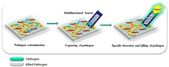
3.6. Multifunctional Platforms for Inactivation and Detection of Pathogens
The development of multifunctional platforms for packaging applications is an area of growing interest. Most nanocomposites used for packaging are based on AgNPs [64,131,132,133] but other materials like GO [134], polyelectrolyte multilayers [135], antimicrobial polymer nanocomposites [136] and natural antimicrobial agents [69] have also been reported. Platforms carrying antimicrobial activity could be interfaced in the future with biomolecular recognition and be used to detect and control foodborne pathogens.
Recent developments in pathogen detection are focused towards fabrication of integrated platforms that can perform multiple functions for simultaneous capture, detection and inactivation. Wang et al. [137] described a SERS multifunctional chip made of silicon wafer containing AgNPs modified with 4-mercaptophenylboronic acid (4-MPBA). The platform showed binding and detection capabilities for E. coli and S. aureus at a concentration range of 500–2000 CFU/mL (LOD was 200 CFU/mL) while also inactivating the pathogens on contact in human blood. Bacteria inactivation was enabled by the dissolved Ag+ ions released from the immobilized AgNPs. Multifunctional capabilities for capture, detection and inactivation were also reported with conjugated polyelectrolytes (CPs)-Ag nanostructures [18]. In this system, (Figure 17) detection and inactivation was achieved through the fluorescence and light-harvesting properties of CPs originating from their conducting polymer backbone which provide optical properties and the ability to generate reactive oxygen species (ROS), enhanced by the use of Ag as substrate.
Other types of multifunctional platforms that integrate separation and concertation of the sample typically based on magnetic particles functionalized with Ab or aptamers are now commonly used to capture and separate targeted analytes in the presence of an external magnetic field. For example, aptamer-functionalized Fe3O4 magnetic NPs were used to separate Staphylococcus aureus, Vibrio parahemolyticus, and Salmonella typhimurium from solution [48]. In another case, FeCo NPs were used to separate phage bounded bacteria from the unbound bacteria [34]. A screen printed carbon electrode modified with immuno-magnetic/polyaniline core/shell NPs was also developed for separation and detection of Bacillus cereus and E. coli with LODs of 40 CFU/mL and 6 CFU/mL, respectively using immunomagnetic separation and electrochemical detection [26].
Simultaneous detection, elimination, and inactivation of pathogenic bacteria was illustrated using vancomycin-functionalised AgNPs/3D-ZnO nanorod arrays [138]. In the system, three-dimensional ZnO nanorod arrays were used as detection platform, AgNPs were used as antibacterial agent, while vancomycin was used to selectively recognize pathogenic bacteria. Moreover, a hybrid of multifunctional system consisting of graphene oxide/AgNPs (GO-Ag NPs) was synthesized and applied for monitoring and disinfecting of gram-negative Escherichia coli as well as the gram-positive Staphylococcus aureus [139]. In another work, a multifunctional nanosystem based on synthesized core–shell fluorescent magnetic NPs (FMNPs) conjugated with gentamicin were able to capture and disrupt the cell wall of E. coli (1 × 10⁷ CFU mL−1 from 10 mL of solution) within 20 min [140].
4. Conclusions and Future Perspectives
Biosensors for pathogens detection are widely used. Nanomaterials can provide optical, catalytic, magnetic and antimicrobial properties for sensing applications. Therefore, the integration of nanotechnology in sensing platforms has provided significant enhancements in detection capabilities and functionality of these devices. On the other hand, multifunctional nanosystems have the potential to act simultaneously as a method for rapid microbial capture, detection, and decontamination. Thus, future developments are also expected in the development of smart labels to indicate food spoilage or presence of harmful toxins. Hence, several types of NPs and nanocomposites have been used in the packaging industry to inhibit bacterial growth and increase the shelf-life of foods. Systems with integrated detection, capture and inactivation capabilities could be developed in the future to design multifunctional platforms for food safety applications. Consequently, the introduction of nanosensors to food packaging to indicate contamination, detect microorganisms, toxicants, moisture or gases from food spoiling is expected to grow. An area of future development is to design food packaging equipped with smart and connected indicators and nanosensors that can be used as tracking devices for product identification, authenticity, traceability and anti-counterfeiting [141]. To enable rapid implementation of this technology in consumer products, this area would benefit from fundamental advances in the development of low cost and flexible nano-sensors suitable for roll-to-roll manufacturing for large scale production. The use of inexpensive materials such as paper or plastic and integration of all sensing reagents (including standards) into a portable compact unit is also desirable for future deployment and rapid implementation of these devices. Method validation, comparability, stability and inter-laboratory studies to evaluate performance are also needed to ensure robustness and accuracy of these devices for real world applications. Eventually, application for the detection of pathogenic organisms in complex matrices needs to be demonstated in real samples to move this technology into the market place.
Acknowledgments
This material is based upon work supported in part by the National Science Foundation under Grant No. 1561491. Any opinions, findings, and conclusions or recommendations expressed in this material are those of the author(s) and do not necessarily reflect the views of the National Science Foundation. S.A. and R.Y.A.H. acknowledge the National Academy of Sciences (NAS) for a NAS fellowship.
Conflicts of Interest
The authors declare no conflict of interest.
References
- CDC. Foodborne Illness: Frequently Asked Questions; Centers for Disease Control and Prevention: Atlanta, GA, USA, 2016. [Google Scholar]
- WHO. Global Burden of Foodborne Diseases; WHO Press: Geneva, Switzerland, 2015. [Google Scholar]
- Centers for Disease Control and Prevention. Surveillance for foodborne disease outbreaks—United States, 2009–2010. MMWR Morb. Mortal. Wkly. Rep. 2013, 62, 41. [Google Scholar]
- Centers for Disease Control and Prevention. Surveillance for Foodborne Disease Outbreaks, United States, 2011; Annual Report; US Department of Health and Human Services: Atlanta, GA, USA, 2014. [Google Scholar]
- Centers for Disease Control and Prevention. Surveillance for Foodborne Disease Outbreaks, United States, 2012; Annual Report; US Department of Health and Human Services: Atlanta, GA, USA, 2014. [Google Scholar]
- Centers for Disease Control and Prevention. Surveillance for Foodborne Disease Outbreaks, United States, 2013; Annual Report; US Department of Health and Human Services: Atlanta, GA, USA, 2015. [Google Scholar]
- De Boer, E.; Beumer, R.R. Methodology for detection and typing of foodborne microorganisms. Int. J. Food Microbiol. 1999, 50, 119–130. [Google Scholar] [CrossRef]
- Bülbül, G.; Hayat, A.; Andreescu, S. Portable Nanoparticle-Based Sensors for Food Safety Assessment. Sensors 2015, 15, 29826. [Google Scholar] [CrossRef] [PubMed]
- Sanvicens, N.; Pastells, C.; Pascual, N.; Marco, M.P. Nanoparticle-based biosensors for detection of pathogenic bacteria. TrAC Trends Anal. Chem. 2009, 28, 1243–1252. [Google Scholar] [CrossRef]
- Ray, P.C.; Khan, S.A.; Singh, A.K.; Senapati, D.; Fan, Z. Nanomaterials for targeted detection and photothermal killing of bacteria. Chem. Soc. Rev. 2012, 41, 3193–3209. [Google Scholar] [CrossRef] [PubMed]
- Chen, J.; Andler, S.M.; Goddard, J.M.; Nugen, S.R.; Rotello, V.M. Integrating recognition elements with nanomaterials for bacteria sensing. Chem. Soc. Rev. 2017, 46, 1272–1283. [Google Scholar] [CrossRef] [PubMed]
- Mocan, T.; Matea, C.T.; Pop, T.; Mosteanu, O.; Buzoianu, A.D.; Puia, C.; Iancu, C.; Mocan, L. Development of nanoparticle-based optical sensors for pathogenic bacterial detection. J. Nanobiotechnol. 2017, 15, 25. [Google Scholar] [CrossRef] [PubMed]
- Pyrgiotakis, G.; Vedantam, P.; Cirenza, C.; McDevitt, J.; Eleftheriadou, M.; Leonard, S.S.; Demokritou, P. Optimization of a nanotechnology based antimicrobial platform for food safety applications using Engineered Water Nanostructures (EWNS). Sci. Rep. 2016, 6, 21073. [Google Scholar] [CrossRef] [PubMed]
- Chang, H.Y.; Cang, J.; Roy, P.; Chang, H.T.; Huang, Y.C.; Huang, C.C. Synthesis and Antimicrobial Activity of Gold/Silver-Tellurium Nanostructures. ACS Appl. Mater. Interfaces 2014, 6, 8305–8312. [Google Scholar] [CrossRef] [PubMed]
- Ramani, M.; Ponnusamy, S.; Muthamizhchelvan, C.; Marsili, E. Amino acid-mediated synthesis of zinc oxide nanostructures and evaluation of their facet-dependent antimicrobial activity. Coll. Surf. B Biointerfaces 2014, 117, 233–239. [Google Scholar] [CrossRef] [PubMed]
- Mageshwari, K.; Sathyamoorthy, R. Flower-shaped CuO Nanostructures: Synthesis, Characterization and Antimicrobial Activity. J. Mater. Sci. Technol. 2013, 29, 909–914. [Google Scholar] [CrossRef]
- De Azeredo, H.M.C. Antimicrobial nanostructures in food packaging. Trends Food Sci. Technol. 2013, 30, 56–69. [Google Scholar] [CrossRef]
- Wang, X.Y.; Cui, Q.; Yao, C.; Li, S.; Zhang, P.; Sun, H.; Lv, F.; Liu, L.; Li, L.; Wang, S. Conjugated Polyelectrolyte-Silver Nanostructure Pair for Detection and Killing of Bacteria. Adv. Mater. Technol. 2017, 2. [Google Scholar] [CrossRef]
- Duan, N.; Wu, S.; Dai, S.; Miao, T.; Chen, J.; Wang, Z. Simultaneous detection of pathogenic bacteria using an aptamer based biosensor and dual fluorescence resonance energy transfer from quantum dots to carbon nanoparticles. Microchim. Acta 2015, 182, 917–923. [Google Scholar] [CrossRef]
- Vargas, C.A.; Wilhelm, A.A.; Williams, J.; Lucas, P.; Reynolds, K.A.; Riley, M.R. Integrated Capture and Spectroscopic Detection of Viruses. Appl. Environ. Microbiol. 2009, 75, 6431–6440. [Google Scholar] [CrossRef] [PubMed]
- Othman, A.; Karimi, A.; Andreescu, S. Functional nanostructures for enzyme based biosensors: Properties, fabrication and applications. J. Mater. Chem. B 2016, 4, 7178–7203. [Google Scholar] [CrossRef]
- Armentano, I.; Rescignano, N.; Fortunati, E.; Mattioli, S.; Morena, F.; Martino, S.; Torre, L.; Kenny, J.M. Chapter 5—Multifunctional nanostructured biopolymeric materials for therapeutic applications A2. In Nanostructures for Novel Therapy; Ficai, D., Grumezescu, A.M., Eds.; Elsevier: Amsterdam, The Netherlands, 2017; pp. 107–135. [Google Scholar]
- Baccar, H.; Mejri, M.B.; Hafaiedh, I.; Ktari, T.; Aouni, M.; Abdelghani, A. Surface plasmon resonance immunosensor for bacteria detection. Talanta 2010, 82, 810–814. [Google Scholar] [CrossRef] [PubMed]
- Celardo, I.; De Nicola, M.; Mandoli, C.; Pedersen, J.Z.; Traversa, E.; Ghibelli, L. Ce3+ Ions Determine Redox-Dependent Anti-apoptotic Effect of Cerium Oxide Nanoparticles. ACS Nano 2011, 5, 4537–4549. [Google Scholar] [CrossRef] [PubMed]
- Li, X.X.; Cao, C.; Han, S.J.; Sim, S.J. Detection of pathogen based on the catalytic growth of gold nanocrystals. Water Res. 2009, 43, 1425–1431. [Google Scholar] [CrossRef] [PubMed]
- Setterington, E.B.; Alocilja, E.C. Electrochemical Biosensor for Rapid and Sensitive Detection of Magnetically Extracted Bacterial Pathogens. Biosensors 2012, 2, 15. [Google Scholar] [CrossRef] [PubMed]
- Singh, A.K.; Senapati, D.; Wang, S.; Griffin, J.; Neely, A.; Naylor, K.M.; Varisli, B.; Kalluri, J.R.; Ray, P.C. Gold Nanorod Based Selective Identification of Escherichia coli Bacteria Using Two-Photon Rayleigh Scattering Spectroscopy. ACS Nano 2009, 3, 1906–1912. [Google Scholar] [CrossRef] [PubMed]
- Wu, W.; Li, J.; Pan, D.; Li, J.; Song, S.; Rong, M.; Li, Z.; Gao, J.; Lu, J. Gold Nanoparticle-Based Enzyme-Linked Antibody-Aptamer Sandwich Assay for Detection of Salmonella Typhimurium. ACS Appl. Mater. Interfaces 2014, 6, 16974–16981. [Google Scholar] [CrossRef] [PubMed]
- Ispas, C.R.; Crivat, G.; Andreescu, S. Review: Recent Developments in Enzyme-Based Biosensors for Biomedical Analysis. Anal. Lett. 2012, 45, 168–186. [Google Scholar] [CrossRef]
- Chua, A.; Yean, C.Y.; Ravichandran, M.; Lim, B.; Lalitha, P. A rapid DNA biosensor for the molecular diagnosis of infectious disease. Biosens. Bioelectron. 2011, 26, 3825–3831. [Google Scholar] [CrossRef] [PubMed]
- Liu, J.; Cheng, J.; Zhang, Y. Upconversion nanoparticle based LRET system for sensitive detection of MRSA DNA sequence. Biosens. Bioelectron. 2013, 43, 252–256. [Google Scholar] [CrossRef] [PubMed]
- Thiruppathiraja, C.; Kamatchiammal, S.; Adaikkappan, P.; Santhosh, D.J.; Alagar, M. Specific detection of Mycobacterium sp. genomic DNA using dual labeled gold nanoparticle based electrochemical biosensor. Anal. Biochem. 2011, 417, 73–79. [Google Scholar] [CrossRef] [PubMed]
- Walter, A.; Wu, J.; Flechsig, G.U.; Haake, D.A.; Wang, J. Redox cycling amplified electrochemical detection of DNA hybridization: Application to pathogen E. coli bacterial RNA. Anal. Chim. Acta 2011, 689, 29–33. [Google Scholar] [CrossRef] [PubMed]
- Chen, J. Bacteriophage-based nanoprobes for rapid bacteria separation. Nanoscale 2015, 7, 16230. [Google Scholar] [CrossRef] [PubMed]
- Chen, J.; Alcaine, S.D.; Jiang, Z.; Rotello, V.M.; Nugen, S.R. Detection of Escherichia coli in Drinking Water Using T7 Bacteriophage-Conjugated Magnetic Probe. Anal. Chem. 2015, 87, 8977–8984. [Google Scholar] [CrossRef] [PubMed]
- Derda, R.; Lockett, M.R.; Tang, S.K.; Fuller, R.C.; Maxwell, E.J.; Breiten, B.; Cuddemi, C.A.; Ozdogan, A.; Whitesides, G.M. Filter-Based Assay for Escherichia coli in Aqueous Samples Using Bacteriophage-Based Amplification. Anal. Chem. 2013, 85, 7213–7220. [Google Scholar] [CrossRef] [PubMed]
- Franche, N.; Vinay, M.; Ansaldi, M. Substrate-independent luminescent phage-based biosensor to specifically detect enteric bacteria such as E. coli. Environ. Sci. Pollut. Res. 2017, 24, 42–51. [Google Scholar] [CrossRef] [PubMed]
- Guntupalli, R.; Sorokulova, I.; Olsen, E.; Globa, L.; Pustovyy, O.; Vodyanoy, V. Biosensor for Detection of Antibiotic Resistant Staphylococcus Bacteria. J. Vis. Exp. 2013, 75, e50474. [Google Scholar] [CrossRef] [PubMed]
- Neufeld, T.; Schwartz-Mittelmann, A.; Biran, D.; Ron, E.Z.; Rishpon, J. Combined Phage Typing and Amperometric Detection of Released Enzymatic Activity for the Specific Identification and Quantification of Bacteria. Anal. Chem. 2003, 75, 580–585. [Google Scholar] [CrossRef] [PubMed]
- Olsen, E.V.; Sorokulova, I.B.; Petrenko, V.A.; Chen, I.H.; Barbaree, J.M.; Vodyanoy, V.J. Affinity-selected filamentous bacteriophage as a probe for acoustic wave biodetectors of Salmonella typhimurium. Biosens. Bioelectron. 2006, 21, 1434–1442. [Google Scholar] [CrossRef] [PubMed]
- Shabani, A.; Marquette, C.A.; Mandeville, R.; Lawrence, M.F. Magnetically-assisted impedimetric detection of bacteria using phage-modified carbon microarrays. Talanta 2013, 116, 1047–1053. [Google Scholar] [CrossRef] [PubMed]
- Tawil, N.; Sacher, E.; Mandeville, R.; Meunier, M. Surface plasmon resonance detection of E. coli and methicillin-resistant S. aureus using bacteriophages. Biosens. Bioelectron. 2012, 37, 24–29. [Google Scholar] [CrossRef] [PubMed]
- Vinay, M.; Franche, N.; Grégori, G.; Fantino, J.R.; Pouillot, F.; Ansaldi, M. Phage-Based Fluorescent Biosensor Prototypes to Specifically Detect Enteric Bacteria Such as E. coli and Salmonella enterica Typhimurium. PLoS ONE 2015, 10, e0131466. [Google Scholar] [CrossRef] [PubMed] [Green Version]
- Abbaspour, A.; Norouz-Sarvestani, F.; Noori, A.; Soltani, N. Aptamer-conjugated silver nanoparticles for electrochemical dual-aptamer-based sandwich detection of staphylococcus aureus. Biosens. Bioelectron. 2015, 68, 149–155. [Google Scholar] [CrossRef] [PubMed]
- Duan, N.; Wu, S.; Yu, Y.; Ma, X.; Xia, Y.; Chen, X.; Huang, Y.; Wang, Z. A dual-color flow cytometry protocol for the simultaneous detection of Vibrio parahaemolyticus and Salmonella typhimurium using aptamer conjugated quantum dots as labels. Anal. Chim. Acta 2013, 804, 151–158. [Google Scholar] [CrossRef] [PubMed]
- Jia, F.; Duan, N.; Wu, S.; Ma, X.; Xia, Y.; Wang, Z.; Wei, X. Impedimetric aptasensor for Staphylococcus aureus based on nanocomposite prepared from reduced graphene oxide and gold nanoparticles. Microchim. Acta 2014, 181, 967–974. [Google Scholar] [CrossRef]
- Lian, Y.; He, F.; Wang, H.; Tong, F. A new aptamer/graphene interdigitated gold electrode piezoelectric sensor for rapid and specific detection of Staphylococcus aureus. Biosens. Bioelectron. 2015, 65, 314–319. [Google Scholar] [CrossRef] [PubMed]
- Wu, S.; Duan, N.; Shi, Z.; Fang, C.; Wang, Z. Simultaneous Aptasensor for Multiplex Pathogenic Bacteria Detection Based on Multicolor Upconversion Nanoparticles Labels. Anal. Chem. 2014, 86, 3100–3107. [Google Scholar] [CrossRef] [PubMed]
- Wu, W.; Zhang, J.; Zheng, M.; Zhong, Y.; Yang, J.; Zhao, Y.; Wu, W.; Ye, W.; Wen, J.; Wang, Q.; Lu, J. Correction: An Aptamer-Based Biosensor for Colorimetric Detection of Escherichia coli O157:H7. PLoS ONE 2013, 8, e48999. [Google Scholar] [CrossRef]
- Wu, W.-H.; Li, M.; Wang, Y.; Ouyang, H.X.; Wang, L.; Li, C.X.; Cao, Y.C.; Meng, Q.H.; Lu, J.X. Aptasensors for rapid detection of Escherichia coli O157:H7 and Salmonella typhimurium. Nanoscale Res. Lett. 2012, 7, 658. [Google Scholar] [CrossRef] [PubMed]
- Yoo, S.M.; Kim, D.-K.; Lee, S.Y. Aptamer-functionalized localized surface plasmon resonance sensor for the multiplexed detection of different bacterial species. Talanta 2015, 132, 112–117. [Google Scholar] [CrossRef] [PubMed]
- Zelada-Guillén, G.A.; Bhosale, S.V.; Riu, J.; Rius, F.X. Real-Time Potentiometric Detection of Bacteria in Complex Samples. Anal. Chem. 2010, 82, 9254–9260. [Google Scholar] [CrossRef] [PubMed]
- Zelada-Guillén, G.A.; Riu, J.; Düzgün, A.; Rius, F.X. Immediate Detection of Living Bacteria at Ultralow Concentrations Using a Carbon Nanotube Based Potentiometric Aptasensor. Angew. Chem. Int. Ed. 2009, 48, 7334–7337. [Google Scholar] [CrossRef] [PubMed]
- Zuo, P. A PDMS/paper/glass hybrid microfluidic biochip integrated with aptamer-functionalized graphene oxide nano-biosensors for one-step multiplexed pathogen detection. Lab Chip 2013, 13, 3921. [Google Scholar] [CrossRef] [PubMed]
- Sharma, R.; Ragavan, K.V.; Thakur, M.S.; Raghavarao, K.S. Recent advances in nanoparticle based aptasensors for food contaminants. Biosens. Bioelectron. 2015, 74, 612–627. [Google Scholar] [CrossRef] [PubMed]
- Liu, X.; Marrakchi, M.; Xu, D.; Dong, H.; Andreescu, S. Biosensors based on modularly designed synthetic peptides for recognition, detection and live/dead differentiation of pathogenic bacteria. Biosens. Bioelectron. 2016, 80, 9–16. [Google Scholar] [CrossRef] [PubMed]
- Emanuel, P.A.; Dang, J.; Gebhardt, J.S.; Aldrich, J.; Garber, E.A.; Kulaga, H.; Stopa, P.; Valdes, J.J.; Dion-Schultz, A. Recombinant antibodies: A new reagent for biological agent detection. Biosens. Bioelectron. 2000, 14, 751–759. [Google Scholar] [CrossRef]
- Jiang, H.; Jiang, D.; Shao, J.; Sun, X. Magnetic molecularly imprinted polymer nanoparticles based electrochemical sensor for the measurement of Gram-negative bacterial quorum signaling molecules (N-acyl-homoserine-lactones). Biosens. Bioelectron. 2016, 75, 411–419. [Google Scholar] [CrossRef] [PubMed]
- Tokonami, S.; Nakadoi, Y.; Takahashi, M.; Ikemizu, M.; Kadoma, T.; Saimatsu, K.; Dung, L.Q.; Shiigi, H.; Nagaoka, T. Label-Free and Selective Bacteria Detection Using a Film with Transferred Bacterial Configuration. Anal. Chem. 2013, 85, 4925–4929. [Google Scholar] [CrossRef] [PubMed]
- Yilmaz, E.; Majidi, D.; Ozgur, E.; Denizli, A. Whole cell imprinting based Escherichia coli sensors: A study for SPR and QCM. Sens. Actuators B Chem. 2015, 209, 714–721. [Google Scholar] [CrossRef]
- Jia, F.; Duan, N.; Wu, S.; Dai, R.; Wang, Z.; Li, X. Impedimetric Salmonella aptasensor using a glassy carbon electrode modified with an electrodeposited composite consisting of reduced graphene oxide and carbon nanotubes. Microchim. Acta 2016, 183, 337–344. [Google Scholar] [CrossRef]
- Kang, X.; Pang, G.; Chen, Q.; Liang, X. Fabrication of Bacillus cereus electrochemical immunosensor based on double-layer gold nanoparticles and chitosan. Sens. Actuators B Chem. 2013, 177, 1010–1016. [Google Scholar] [CrossRef]
- Chau, C.F.; Wu, S.H.; Yen, G.C. The development of regulations for food nanotechnology. Trends Food Sci. Technol. 2007, 18, 269–280. [Google Scholar] [CrossRef]
- Ariyarathna, I.R.; Rajakaruna, R.M.P.I.; Karunaratne, D.N. The rise of inorganic nanomaterial implementation in food applications. Food Control 2017, 77, 251–259. [Google Scholar] [CrossRef]
- Cao, Y.; Li, J.; Liu, F.; Li, X.; Jiang, Q.; Cheng, S.; Gu, Y. Consideration of interaction between nanoparticles and food components for the safety assessment of nanoparticles following oral exposure: A review. Environ. Toxicol. Pharmacol. 2016, 46, 206–210. [Google Scholar] [CrossRef] [PubMed]
- Weiss, J.; Takhistov, P.; McClements, D.J. Functional materials in food nanotechnology. J. Food Sci. 2006, 71, R107–R116. [Google Scholar] [CrossRef]
- Fonseca, L.; Cane, C.; Mazzolai, B. Application of micro and nanotechnologies to food safety and quality monitoring. Meas. Control 2007, 40, 116–119. [Google Scholar] [CrossRef]
- Zhang, Y.; Wei, Q. The role of nanomaterials in electroanalytical biosensors: A mini review. J. Electroanal. Chem. 2016, 781, 401–409. [Google Scholar] [CrossRef]
- Irkin, R.; Esmer, O.K. Novel food packaging systems with natural antimicrobial agents. J. Food Sci. Technol. Mysore 2015, 52, 6095–6111. [Google Scholar] [CrossRef] [PubMed]
- Hoseinnejad, M.; Jafari, S.M.; Katouzian, I. Inorganic and metal nanoparticles and their antimicrobial activity in food packaging applications. Crit. Rev. Microbiol. 2017, 1–21. [Google Scholar] [CrossRef] [PubMed]
- Bibi, F.; Guillaume, C.; Gontard, N.; Sorli, B. A review: RFID technology having sensing aptitudes for food industry and their contribution to tracking and monitoring of food products. Trends Food Sci. Technol. 2017, 62, 91–103. [Google Scholar] [CrossRef]
- Karuppuswami, S.; Kaur, A.; Arangali, H.; Chahal, P.P. A Hybrid Magnetoelastic Wireless Sensor for Detection of Food Adulteration. IEEE Sens. J. 2017, 17, 1706–1714. [Google Scholar] [CrossRef]
- Mills, A.; Hawthorne, D.; Burns, L.; Hazafy, D. Novel temperature-activated humidity-sensitive optical sensor. Sens. Actuators B Chem. 2017, 240, 1009–1015. [Google Scholar] [CrossRef]
- Maloney, N.; Lukacs, G.; Jensen, J.; Hegner, M. Nanomechanical sensors for single microbial cell growth monitoring. Nanoscale 2014, 6, 8242–8249. [Google Scholar] [CrossRef] [PubMed]
- Wang, Y.; Duncan, T.V. Nanoscale sensors for assuring the safety of food products. Curr. Opin. Biotechnol. 2017, 44, 74–86. [Google Scholar] [CrossRef] [PubMed]
- Jayasena, S.D. Aptamers: An Emerging Class of Molecules That Rival Antibodies in Diagnostics. Clin. Chem. 1999, 45, 1628–1650. [Google Scholar] [PubMed]
- Tuerk, C.; Gold, L. Systematic evolution of ligands by exponential enrichment: RNA ligands to bacteriophage T4 DNA polymerase. Science 1990, 249, 505–510. [Google Scholar] [CrossRef] [PubMed]
- Zayats, M.; Huang, Y.; Gill, R.; Ma, C.A.; Willner, I. Label-Free and Reagentless Aptamer-Based Sensors for Small Molecules. J. Am. Chem. Soc. 2006, 128, 13666–13667. [Google Scholar] [CrossRef] [PubMed]
- O’Sullivan, C.K. Aptasensors—The future of biosensing? Anal. Bioanal. Chem. 2002, 372, 44–48. [Google Scholar] [CrossRef] [PubMed]
- Johnson, R.R.; Johnson, A.T.C.; Klein, M.L. Probing the Structure of DNA–Carbon Nanotube Hybrids with Molecular Dynamics. Nano Lett. 2008, 8, 69–75. [Google Scholar] [CrossRef] [PubMed]
- Hong, Y.; Brown, D.G. Electrostatic Behavior of the Charge-Regulated Bacterial Cell Surface. Langmuir 2008, 24, 5003–5009. [Google Scholar] [CrossRef] [PubMed]
- Verma, M.S. Branching and size of CTAB-coated gold nanostars control the colorimetric detection of bacteria. RSC Adv. 2014, 4, 10660. [Google Scholar] [CrossRef]
- Chen, J.; Jackson, A.A.; Rotello, V.M.; Nugen, S.R. Colorimetric Detection of Escherichia coli Based on the Enzyme-Induced Metallization of Gold Nanorods. Small 2016, 12, 2469–2475. [Google Scholar] [CrossRef] [PubMed]
- Richards, S.J. Discrimination between bacterial phenotypes using glyco-nanoparticles and the impact of polymer coating on detection readouts. J. Mater. Chem. 2014, 2, 1490. [Google Scholar] [CrossRef]
- Li, B.M.; Yu, Q.L.; Duan, Y.X. Fluorescent labels in biosensors for pathogen detection. Crit. Rev. Biotechnol. 2015, 35, 82–93. [Google Scholar] [CrossRef] [PubMed]
- Liao, Y.H.; Zhou, X.M.; Xing, D. Quantum Dots and Graphene Oxide Fluorescent Switch Based Multivariate Testing Strategy for Reliable Detection of Listeria monocytogenes. ACS Appl. Mater. Interfaces 2014, 6, 9988–9996. [Google Scholar] [CrossRef] [PubMed]
- Zuo, P.; Lu, X.; Sun, Z.; Guo, Y.; He, H. A review on syntheses, properties, characterization and bioanalytical applications of fluorescent carbon dots. Microchim. Acta 2016, 183, 519–542. [Google Scholar] [CrossRef]
- Liu, J.; Wagan, S.; Dávila Morris, M.; Taylor, J.; White, R.J. Achieving Reproducible Performance of Electrochemical, Folding Aptamer-Based Sensors on Microelectrodes: Challenges and Prospects. Anal. Chem. 2014, 86, 11417–11424. [Google Scholar] [CrossRef] [PubMed]
- Swanink, C.M.; Meis, J.F.; Rijs, A.J.; Donnelly, J.P.; Verweij, P.E. Specificity of a sandwich enzyme-linked immunosorbent assay for detecting Aspergillus galactomannan. J. Clin. Microbiol. 1997, 35, 257–260. [Google Scholar] [PubMed]
- Guner, A.; Çevik, E.; Şenel, M.; Alpsoy, L. An electrochemical immunosensor for sensitive detection of Escherichia coli O157:H7 by using chitosan, MWCNT, polypyrrole with gold nanoparticles hybrid sensing platform. Food Chem. 2017, 229, 358–365. [Google Scholar] [CrossRef] [PubMed]
- Ye, W.; Guo, J.; Bao, X.; Chen, T.; Weng, W.; Chen, S.; Yang, M. Rapid and Sensitive Detection of Bacteria Response to Antibiotics Using Nanoporous Membrane and Graphene Quantum Dot (GQDs)-Based Electrochemical Biosensors. Materials 2017, 10, 603. [Google Scholar] [CrossRef] [PubMed]
- Inbaraj, B.S.; Chen, B.H. Nanomaterial-based sensors for detection of foodborne bacterial pathogens and toxins as well as pork adulteration in meat products. J. Food Drug Anal. 2016, 24, 15–28. [Google Scholar] [CrossRef]
- Li, Y.S.; Church, J.S. Raman spectroscopy in the analysis of food and pharmaceutical nanomaterials. J. Food Drug Anal. 2014, 22, 29–48. [Google Scholar] [CrossRef] [PubMed]
- Deisingh, A.K.; Thompson, M. Biosensors for the detection of bacteria. Can. J. Microbiol. 2004, 50, 69–77. [Google Scholar] [CrossRef] [PubMed]
- Mathelié-Guinlet, M.; Gammoudi, I.; Beven, L.; Moroté, F.; Delville, M.H.; Grauby-Heywang, C.; Cohen-Bouhacina, T. Silica Nanoparticles Assisted Electrochemical Biosensor for the Detection and Degradation of Escherichia Coli Bacteria. Procedia Eng. 2016, 168, 1048–1051. [Google Scholar] [CrossRef]
- Wang, Z.; Yu, J.; Gui, R.; Jin, H.; Xia, Y. Carbon nanomaterials-based electrochemical aptasensors. Biosens. Bioelectron. 2016, 79, 136–149. [Google Scholar] [CrossRef] [PubMed]
- Velu, R.; Frost, N.; DeRosa, M.C. Linkage inversion assembled nano-aptasensors (LIANAs) for turn-on fluorescence detection. Chem. Commun. 2015, 51, 14346–14349. [Google Scholar] [CrossRef] [PubMed]
- Rowland, C.E.; Brown, C.W.; Delehanty, J.B.; Medintz, I.L. Nanomaterial-based sensors for the detection of biological threat agents. Mater.Today 2016, 19, 464–477. [Google Scholar] [CrossRef]
- Liu, Y.; Yu, J. Oriented immobilization of proteins on solid supports for use in biosensors and biochips: A review. Microchim. Acta 2016, 183, 1–19. [Google Scholar] [CrossRef]
- Wang, H.; Yang, R.; Yang, L.; Tan, W. Nucleic Acid Conjugated Nanomaterials for Enhanced Molecular Recognition. ACS Nano 2009, 3, 2451–2460. [Google Scholar] [CrossRef] [PubMed]
- Korayem, M.H.; Estaji, M.; Homayooni, A. Noncalssical multiscale modeling of ssDNA manipulation using a CNT-nanocarrier based on AFM. Coll. Surf. B Biointerfaces 2017, 158, 102–111. [Google Scholar] [CrossRef] [PubMed]
- Jeddi, I.; Saiz, L. Three-dimensional modeling of single stranded DNA hairpins for aptamer-based biosensors. Sci. Rep. 2017, 7, 1178. [Google Scholar] [CrossRef] [PubMed]
- Nasiri Khonsari, Y.; Sun, S. Recent trends in electrochemiluminescence aptasensors and their applications. Chem. Commun. 2017, 53, 9042–9054. [Google Scholar] [CrossRef] [PubMed]
- Guttman, B.; Raya, R.; Kutter, E. Basic phage biology. Bacteriophages Biol. Appl. 2005, 4. [Google Scholar] [CrossRef]
- Tawil, N. Bacteriophages: Biosensing tools for multi-drug resistant pathogens. Analyst 2014, 139, 1224. [Google Scholar] [CrossRef] [PubMed]
- Baggesen, D.L.; Wegener, H.C. Phage types of Salmonella enterica ssp. enterica serovar typhimurium isolated from production animals and humans in Denmark. Acta Vet. Scand. 1993, 35, 349–354. [Google Scholar]
- Yanez-Sedeno, P.; Campuzano, S.; Pingarron, J.M. Electrochemical sensors based on magnetic molecularly imprinted polymers: A review. Anal. Chim. Acta 2017, 960, 1–17. [Google Scholar] [CrossRef] [PubMed]
- Gama, M.R.; Bottoli, C.B. Molecularly imprinted polymers for bioanalytical sample preparation. J. Chromatogr. B Anal. Technol. Biomed. Life Sci. 2017, 1043, 107–121. [Google Scholar] [CrossRef] [PubMed]
- Tokonami, S.; Shiigi, H.; Nagaoka, T. Review: Micro- and nanosized molecularly imprinted polymers for high-throughput analytical applications. Anal. Chim. Acta 2009, 641, 7–13. [Google Scholar] [CrossRef] [PubMed]
- Idil, N.; Mattiasson, B. Imprinting of Microorganisms for Biosensor Applications. Sensors 2017, 17, 708. [Google Scholar] [CrossRef] [PubMed]
- Haupt, K.; Mosbach, K. Molecularly Imprinted Polymers and Their Use in Biomimetic Sensors. Chem. Rev. 2000, 100, 2495–2504. [Google Scholar] [CrossRef] [PubMed]
- Mayes, A.G.; Mosbach, K. Molecularly imprinted polymers: Useful materials for analytical chemistry? TrAC Trends Anal. Chem. 1997, 16, 321–332. [Google Scholar] [CrossRef]
- Cai, D.; Ren, L.; Zhao, H.; Xu, C.; Zhang, L.; Yu, Y.; Wang, H.; Lan, Y.; Roberts, M.F.; Chuang, J.H.; et al. A molecular-imprint nanosensor for ultrasensitive detection of proteins. Nat. Nanotechnol. 2010, 5, 597–601. [Google Scholar] [CrossRef] [PubMed]
- Whitcombe, M.J. The rational development of molecularly imprinted polymer-based sensors for protein detection. Chem. Soc. Rev. 2011, 40, 1547. [Google Scholar] [CrossRef] [PubMed]
- Reddy, K.V.R.; Yedery, R.D.; Aranha, C. Antimicrobial peptides: Premises and promises. Int. J. Antimicrob. Agents 2004, 24, 536–547. [Google Scholar] [CrossRef] [PubMed]
- Brogden, K.A. Antimicrobial peptides: Pore formers or metabolic inhibitors in bacteria? Nat. Rev. Microbiol. 2005, 3, 238–250. [Google Scholar] [CrossRef] [PubMed]
- Lupetti, A.; Welling, M.M.; Pauwels, E.K.; Nibbering, P.H. Radiolabelled antimicrobial peptides for infection detection. Lancet Infect. Dis. 2003, 3, 223–229. [Google Scholar] [CrossRef]
- Welling, M.M.; Lupetti, A.; Balter, H.S.; Lanzzeri, S.; Souto, B.; Rey, A.M.; Savio, E.O.; Paulusma-Annema, A.; Pauwels, E.K.; Nibbering, P.H. 99mTc-Labeled Antimicrobial Peptides for Detection of Bacterial and Candida albicans Infections. J. Nucl. Med. 2001, 42, 788–794. [Google Scholar] [PubMed]
- Kulagina, N.V.; Lassman, M.E.; Ligler, F.S.; Taitt, C.R. Antimicrobial Peptides for Detection of Bacteria in Biosensor Assays. Anal. Chem. 2005, 77, 6504–6508. [Google Scholar] [CrossRef] [PubMed]
- Yoo, J.H.; Woo, D.H.; Chang, M.S.; Chun, M.S. Microfluidic based biosensing for Escherichia coli detection by embedding antimicrobial peptide-labeled beads. Sens. Actuators B Chem. 2014, 191, 211–218. [Google Scholar] [CrossRef]
- Kulagina, N.V.; Shaffer, K.M.; Anderson, G.P.; Ligler, F.S.; Taitt, C.R. Antimicrobial peptide-based array for Escherichia coli and Salmonella screening. Anal. Chim. Acta 2006, 575, 9–15. [Google Scholar] [CrossRef] [PubMed]
- Mannoor, M.S.; Zhang, S.; Link, A.J.; McAlpine, M.C. Electrical detection of pathogenic bacteria via immobilized antimicrobial peptides. Proc. Natl. Acad. Sci. USA 2010, 107, 19207–19212. [Google Scholar] [CrossRef] [PubMed]
- Friedrich, C.; Scott, M.G.; Karunaratne, N.; Yan, H.; Hancock, R.E. Salt-Resistant Alpha-Helical Cationic Antimicrobial Peptides. Antimicrob. Agents Chemother. 1999, 43, 1542–1548. [Google Scholar] [PubMed]
- Rydlo, T.; Rotem, S.; Mor, A. Antibacterial Properties of Dermaseptin S4 Derivatives under Extreme Incubation Conditions. Antimicrob. Agents Chemother. 2006, 50, 490–497. [Google Scholar] [CrossRef] [PubMed]
- Yang, M.; Xu, D.; Jiang, L.; Zhang, L.; Dustin, D.; Lund, R.; Liu, L.; Dong, H. Filamentous supramolecular peptide-drug conjugates as highly efficient drug delivery vehicles. Chem. Commun. 2014, 50, 4827–4830. [Google Scholar] [CrossRef] [PubMed]
- Xu, D.; Jiang, L.; Singh, A.; Dustin, D.; Yang, M.; Liu, L.; Lund, R.; Sellati, T.J.; Dong, H. Designed supramolecular filamentous peptides: Balance of nanostructure, cytotoxicity and antimicrobial activity. Chem. Commun. 2015, 51, 1289–1292. [Google Scholar] [CrossRef] [PubMed]
- Wang, Z.-F.; Cheng, S.; Ge, S.L.; Wang, H.; Wang, Q.J.; He, P.G.; Fang, Y.Z. Ultrasensitive Detection of Bacteria by Microchip Electrophoresis Based on Multiple-Concentration Approaches Combining Chitosan Sweeping, Field-Amplified Sample Stacking, and Reversed-Field Stacking. Anal. Chem. 2012, 84, 1687–1694. [Google Scholar] [CrossRef] [PubMed]
- Alhogail, S.; Suaifan, G.A.R.Y.; Zourob, M. Rapid colorimetric sensing platform for the detection of Listeria monocytogenes foodborne pathogen. Biosens. Bioelectron. 2016, 86, 1061–1066. [Google Scholar] [CrossRef] [PubMed]
- Jokerst, J.C.; Adkins, J.A.; Bisha, B.; Mentele, M.M.; Goodridge, L.D.; Henry, C.S. Development of a Paper-Based Analytical Device for Colorimetric Detection of Select Foodborne Pathogens. Anal. Chem. 2012, 84, 2900–2907. [Google Scholar] [CrossRef] [PubMed]
- Adkins, J.A.; Boehle, K.; Friend, C.; Chamberlain, B.; Bisha, B.; Henry, C.S. Colorimetric and Electrochemical Bacteria Detection Using Printed Paper- and Transparency-Based Analytic Devices. Anal. Chem. 2017, 89, 3613–3621. [Google Scholar] [CrossRef] [PubMed]
- Tsai, T.T.; Huang, T.H.; Chang, C.J.; Ho, N.Y.; Tseng, Y.T.; Chen, C.F. Antibacterial cellulose paper made with silver-coated gold nanoparticles. Sci. Rep. 2017, 7, 3155. [Google Scholar] [CrossRef] [PubMed]
- Arfat, Y.A.; Ejaz, M.; Jacob, H.; Ahmed, J. Deciphering the potential of guar gum/Ag-Cu nanocomposite films as an active food packaging material. Carbohydr. Polym. 2017, 157, 65–71. [Google Scholar] [CrossRef] [PubMed]
- Tiimob, B.J.; Mwinyelle, G.; Abdela, W.; Samuel, T.; Jeelani, S.; Rangari, V.K. Nanoengineered Eggshell-Silver Tailored Copolyester Polymer Blend Film with Antimicrobial Properties. J. Agric. Food Chem. 2017, 65, 1967–1976. [Google Scholar] [CrossRef] [PubMed]
- Hu, W.B.; Peng, C.; Luo, W.; Lv, M.; Li, X.; Li, D.; Huang, Q.; Fan, C. Graphene-Based Antibacterial Paper. ACS Nano 2010, 4, 4317–4323. [Google Scholar] [CrossRef] [PubMed]
- Lichter, J.A.; Van Vliet, K.J.; Rubner, M.F. Design of Antibacterial Surfaces and Interfaces: Polyelectrolyte Multilayers as a Multifunctional Platform. Macromolecules 2009, 42, 8573–8586. [Google Scholar] [CrossRef]
- Emamifar, A. Applications of Antimicrobial Polymer Nanocomposites in Food Packaging. In Advances in Nanocomposite Technology; Hashim, A., Ed.; InTech; Available online: https://www.intechopen.com/books/advances-in-nanocomposite-technology/applications-of-antimicrobial-polymer-nanocomposites-in-food-packaging (accessed on 15 September 2017).
- Wang, H.Y.; Zhou, Y.; Jiang, X.; Sun, B.; Zhu, Y.; Wang, H.; Su, Y.; He, Y. Simultaneous Capture, Detection, and Inactivation of Bacteria as Enabled by a Surface-Enhanced Raman Scattering Multifunctional Chip. Angew. Chem.-Int. Ed. 2015, 54, 5132–5136. [Google Scholar] [CrossRef] [PubMed]
- Yang, Z.; Wang, Y.; Zhang, D. A novel multifunctional electrochemical platform for simultaneous detection, elimination, and inactivation of pathogenic bacteria based on the Vancomycin-functionalised AgNPs/3D-ZnO nanorod arrays. Biosens. Bioelectron. 2017, 98, 248–253. [Google Scholar] [CrossRef] [PubMed]
- Tam, L.T.; Dinh, N.X.; Van Cuong, N.; Mølhave, K.; Le, A. Graphene Oxide/Silver Nanohybrid as Multi-functional Material for Highly Efficient Bacterial Disinfection and Detectionof Organic Dye. J. Electron. Mater. 2016, 45, 5321–5333. [Google Scholar] [CrossRef]
- Chen, L.; Razavi, F.S.; Mumin, A.; Guo, X.; Sham, T.K.; Zhang, J. Multifunctional nanoparticles for rapid bacterial capture, detection, and decontamination. RSC Adv. 2013, 3, 2390–2397. [Google Scholar] [CrossRef]
- Kuswandi, B. Environmental friendly food nano-packaging. Environ. Chem. Lett. 2017, 15, 205–221. [Google Scholar] [CrossRef]
Figure 1.
(a) Possible conformations of the aptamers that are self-assembled on carbon nanotubes; (b) Schematic representation of the interaction between the target bacteria and the hybrid aptamer–SWCNT system (adapted with permission from [53]).
Figure 1.
(a) Possible conformations of the aptamers that are self-assembled on carbon nanotubes; (b) Schematic representation of the interaction between the target bacteria and the hybrid aptamer–SWCNT system (adapted with permission from [53]).

Figure 2.
(A) Biosensing measurements using a potentiometric SWCNP-based aptasensor connected to a sample pretreatment system to remove the matrix in real samples and detect microorganisms. From left to right: filtration of sample and matrix removal, washing with PBS, elution with PBS and potentiometric detection of bacteria recovered in eluate (adapted with permission from [43]. Copyright (2010) American Chemical Society; (B) PDMS/paper hybrid microfluidic system for one-step multiplexed pathogen detection using aptamer-functionalized GO biosensors. (a) Microfluidic biochip layout; (b,c) illustrate the principle of the one-step ‘turn-on’ detection approach based on the interaction among GO, aptamers and pathogens. Step 1: when an aptamer is linked to the GO surface, its fluorescence is quenched. Step 2: when the target pathogen is present, the target pathogen induces the aptamer to be liberated from GO and thereby restores its fluorescence for detection (adapted from [45] with permission of The Royal Society of Chemistry).
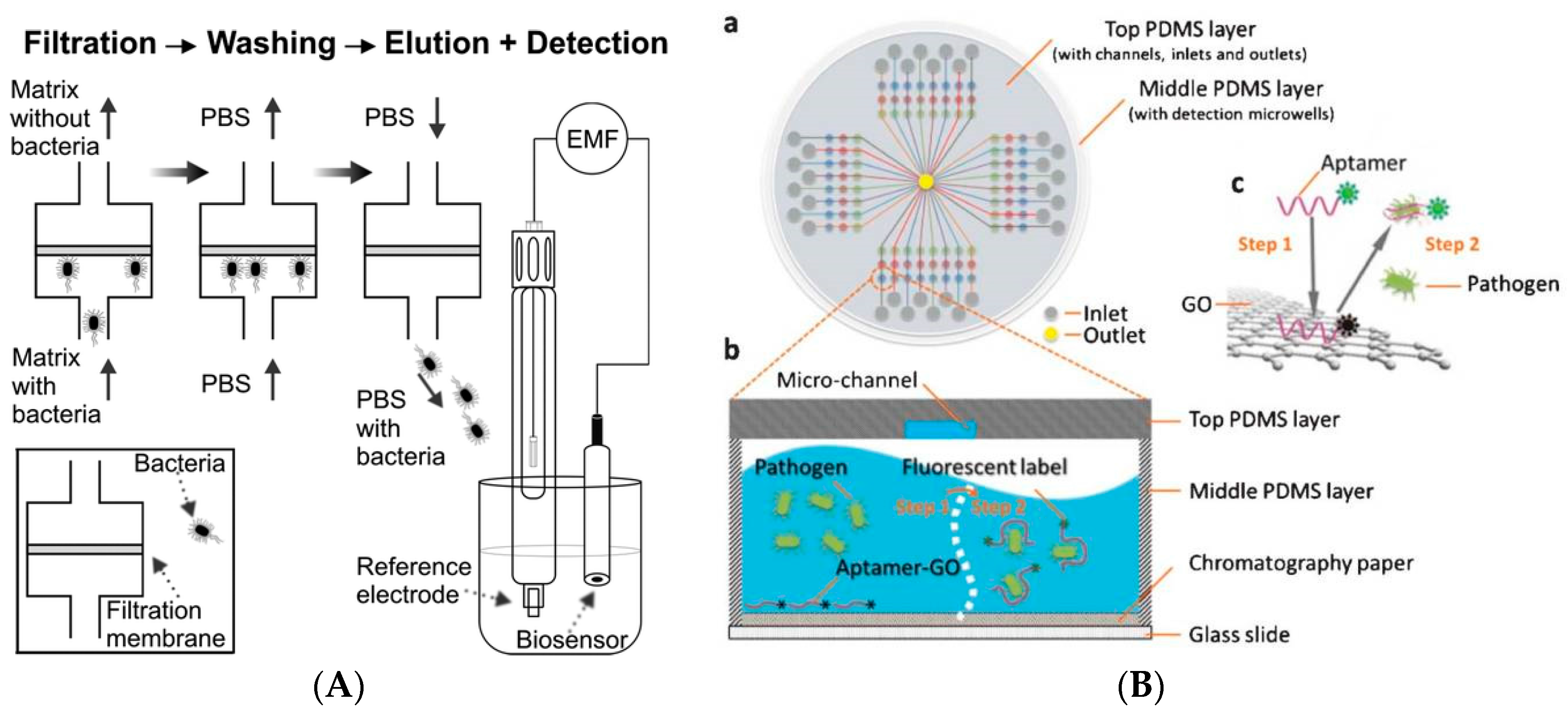
Figure 3.
(a) Schematic representation of the principle of impedance-based detection of S. aureus on a GCE-rGO-ssDNA-AuNPs-aptamer nanocomposite (adapted with permission from [37]); (b) Aptamer based multispot gold-capped NPs array (MG-NPA) chip containing a dielectric layer of a thin gold (Au) layer on silica (Si) NPs-absorbed glass slide (adapted with permission from [42]); (c) Measurements of S. aureuse on Au surface functionalized with grahene and aptamer with modification steps: (A) immobilization of mercaptobenzenediazonium tetrafluoroborate (MBDT) on grapheme; (B) Immobilization of graphene on Au; (C) immobilization of aptamer and (D) detachment of aptamers from graphene in the presence of S. aureus (adapted with permission from [38]).

Figure 4.
Colorimetric detection of bacteria using aptamers and AuNPs (adapted with permission from [41]).
Figure 4.
Colorimetric detection of bacteria using aptamers and AuNPs (adapted with permission from [41]).

Figure 5.
(a) Detection of Vibrio parahaemolyticus and Salmonella typhimurium using aptamer-functionalized QDs and flow cytometry (adapted with permission from [36]); (b) FRET based measurement b (adapted with permission from [31]); (c) shows multiplexed detection of three types of bacteria by three different aptamer-based upconversion rare earth NPs. The detection is based on the luminscence signals of free NPs after separation from bacteria mixture (adapted with permission from [39]. Copyright (2014) American Chemical Society).

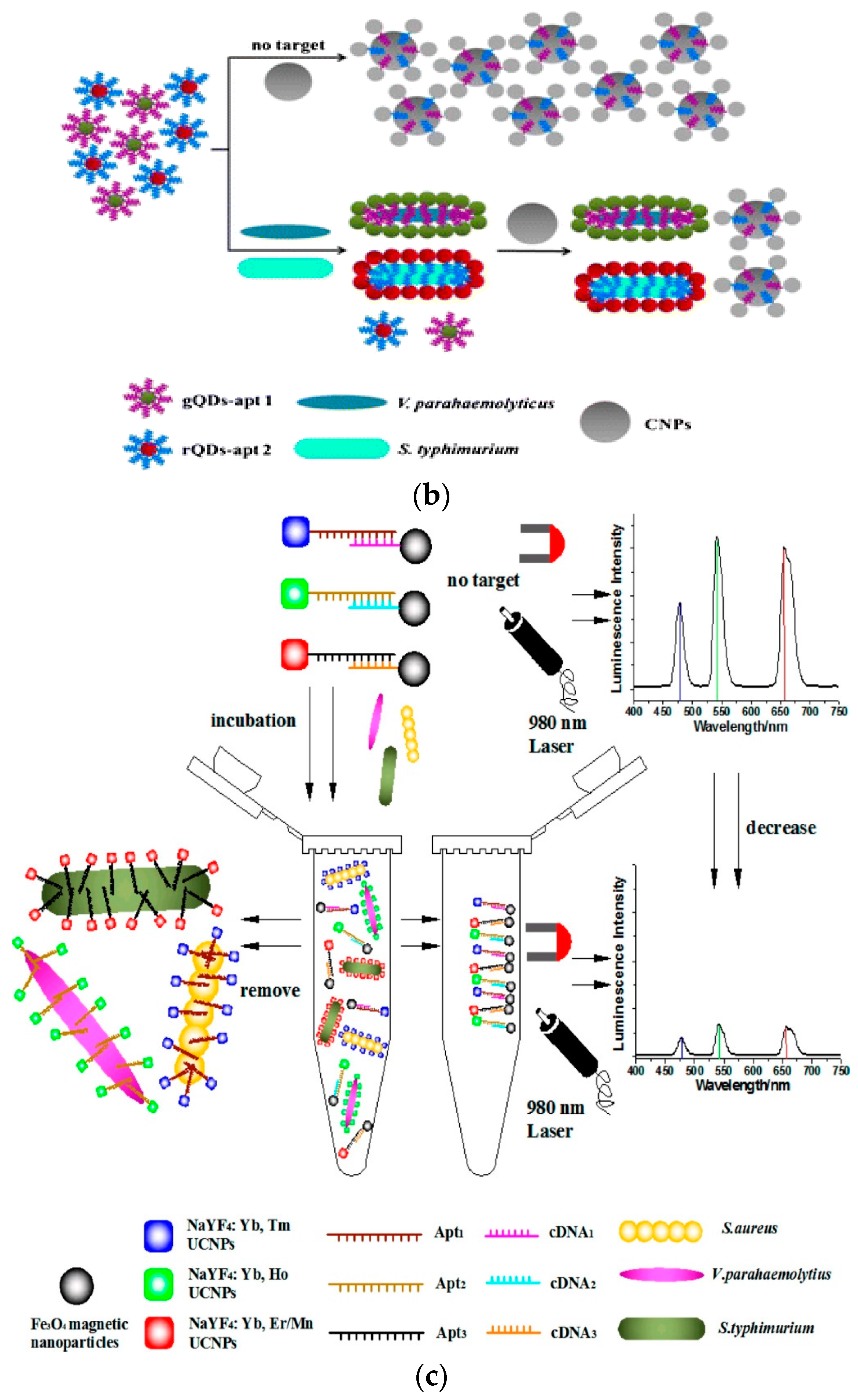
Figure 6.
Example of electrochemical aptamer-based sensor with AgNPs labels and magnetic separation for detection of S. aureus (adapted with permission from [35]).
Figure 6.
Example of electrochemical aptamer-based sensor with AgNPs labels and magnetic separation for detection of S. aureus (adapted with permission from [35]).
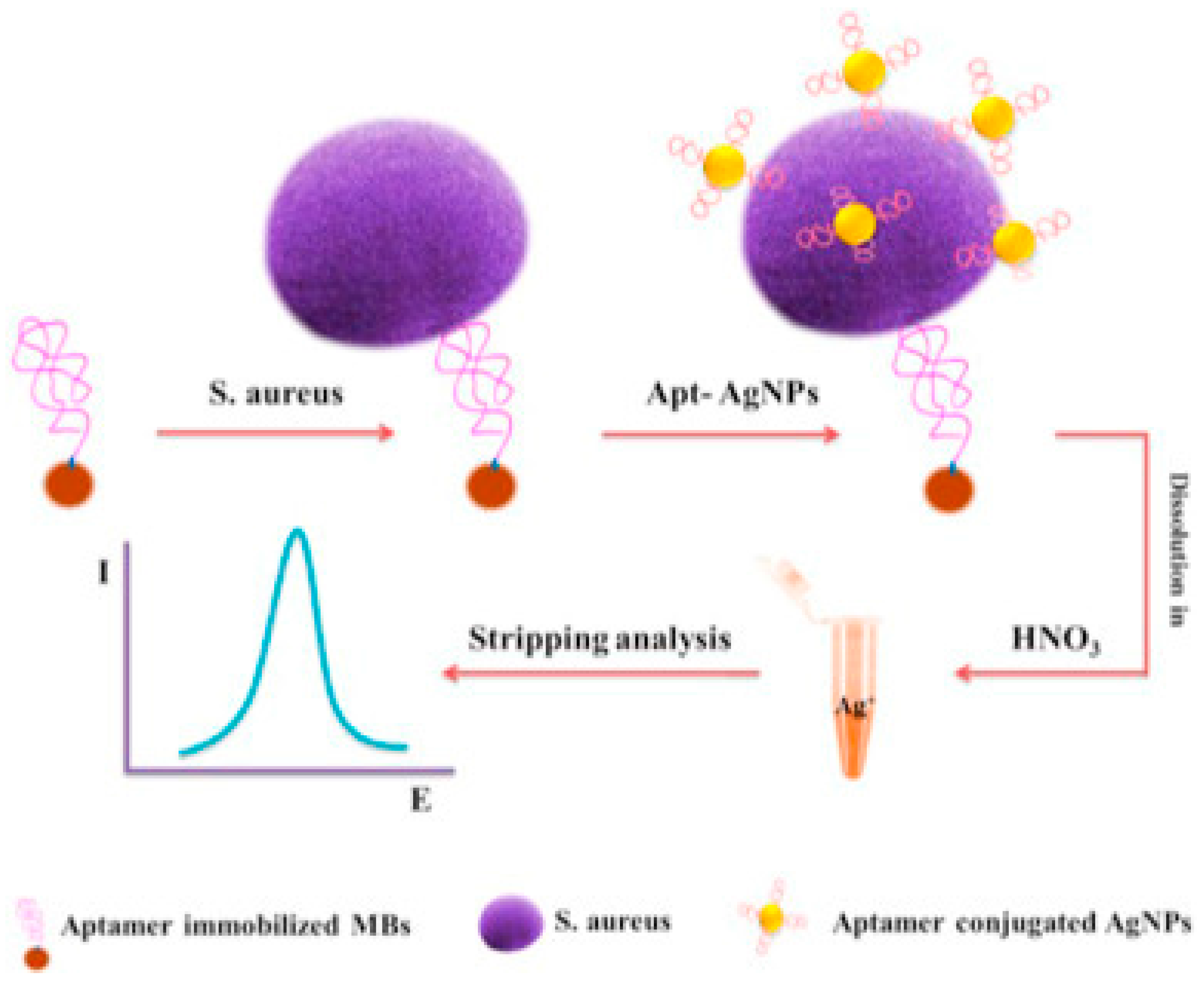
Figure 7.
Examples of E. coli immunosensors using: (a) AuNPs with SPR quantification (adapted with permission from [14]) and (b) Au nanorods and two-photon Rayleigh scattering (TPRS) spectroscopy as a detection technique. Adapted with permission from [18]. Copyright (2009) American Chemical Society.
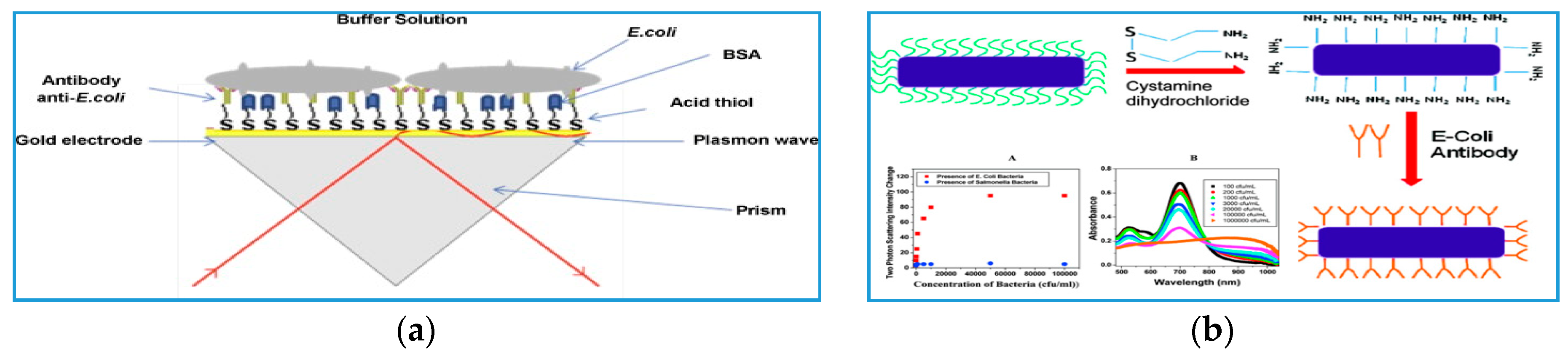
Figure 8.
AuNPs-based immunosensor for Giardia lamblia cysts detection. (Left) The sample is concentrated through a centrifuge filter, and then incubated with Ab-AuNPs immunoprobes. The binding is quantified as a color change of the AuNPs detected by UV-spectroscopy. The (Right) image shows TEM images of immunoprobes on the surface of Giardia lamblia cysts at a scale of (a) 500 nm and (b) 100 nm. Giardia lamblia cysts morphology is shown in the inset (adapted with permission from [16]).

Figure 9.
Schematic representation of the use of immuno-magnetic/polyaniline core/shell nanoparticle (c/sNP) with cyclic voltammerty for Bacillus and E. coli O157:H7 detection (adapted with permission from [17].
Figure 9.
Schematic representation of the use of immuno-magnetic/polyaniline core/shell nanoparticle (c/sNP) with cyclic voltammerty for Bacillus and E. coli O157:H7 detection (adapted with permission from [17].

Figure 10.
Schematic representation of the use of T7 bacteriophage-conjugated magnetic probe to detect Escherichia coli in drinking water (i) Introduction of T7 bacteriophage-conjugated magnetic probe to attack E. coli and separate it by the influence of magnet (ii) The explosion or lysis of E. coli and the release of T7 phages and β-gal; (iii) β-gal catalyzed CPRG hydrolysis to produce colorimetric signal (adapted with permission from [26]. Copyright (2015) American Chemical Society).

Figure 11.
(a) Concept of bacteriophage-based sensor for E. coli and MRSA using covalently attached T4 and BP14 bacteriophages and (b) The response of SPR upon attachment of phages, and then with E. coli. (adapted with permission from [42]).
Figure 11.
(a) Concept of bacteriophage-based sensor for E. coli and MRSA using covalently attached T4 and BP14 bacteriophages and (b) The response of SPR upon attachment of phages, and then with E. coli. (adapted with permission from [42]).
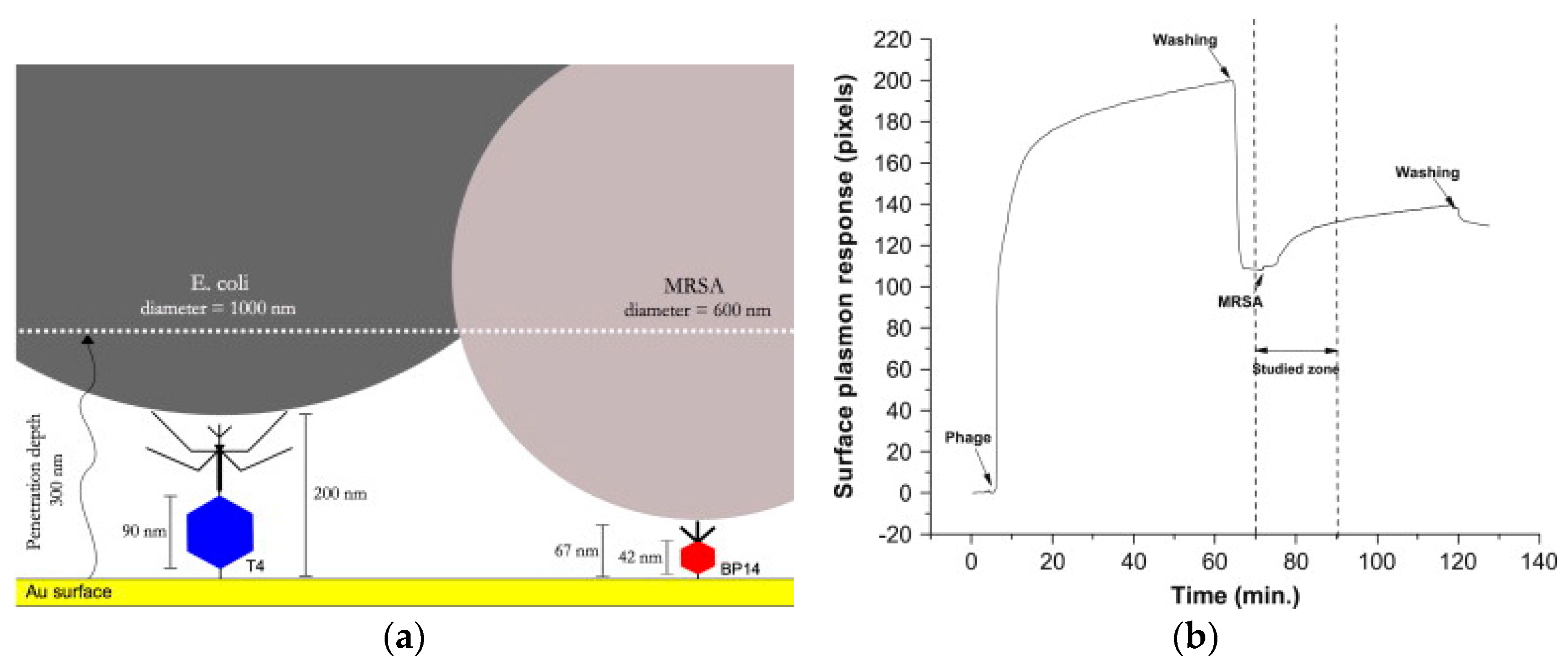
Figure 12.
Illustration of the screen-printed carbon electrode and its use for the EIS detection of magnetically separated E. coli K12 using immobilized bacteriophages (adapted with permission from [32]).
Figure 12.
Illustration of the screen-printed carbon electrode and its use for the EIS detection of magnetically separated E. coli K12 using immobilized bacteriophages (adapted with permission from [32]).

Figure 13.
(a) Illustration depicting imprinting Pseudomonas aeruginosa bacteria on a polypyrrole (PPy) film and (b) electrode configuration for label free detection with the PPy film (adapted with permission from [50]. Copyright (2013) American Chemical Society).
Figure 13.
(a) Illustration depicting imprinting Pseudomonas aeruginosa bacteria on a polypyrrole (PPy) film and (b) electrode configuration for label free detection with the PPy film (adapted with permission from [50]. Copyright (2013) American Chemical Society).
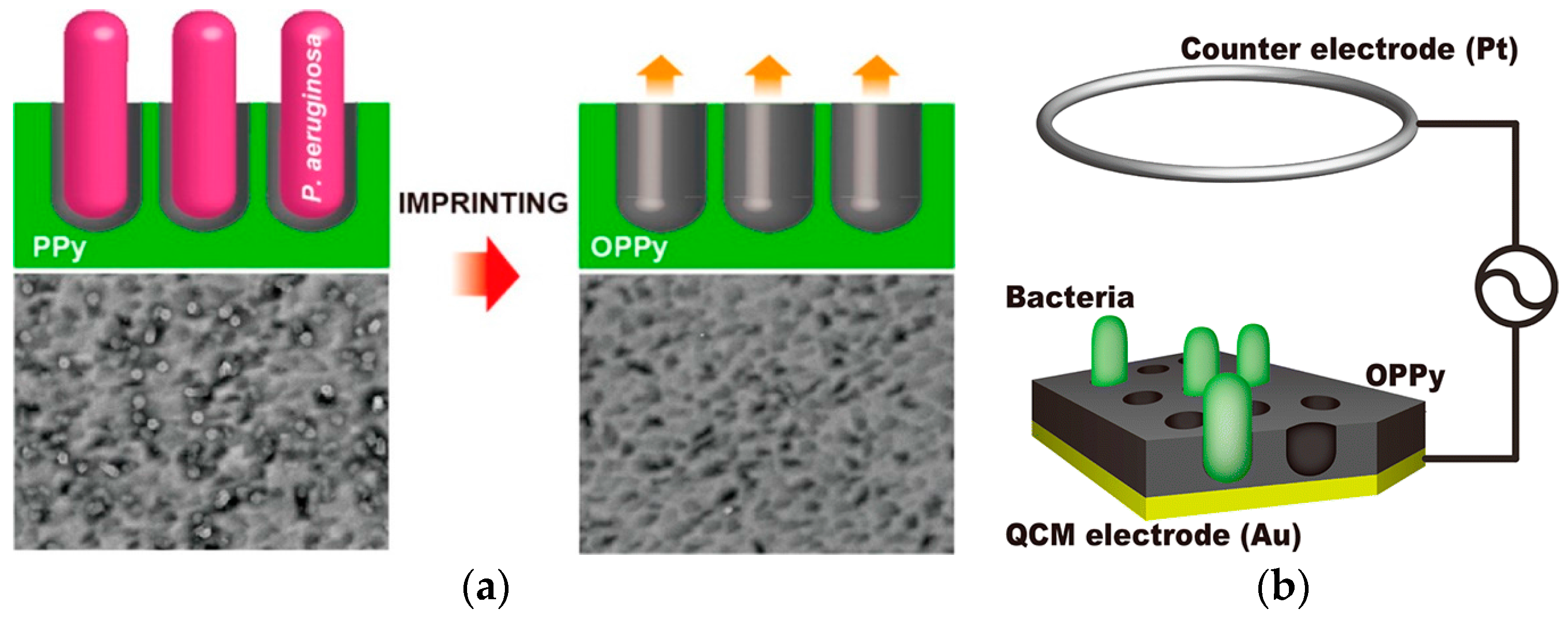
Figure 14.
Schematic representation of micro-contact imprinted SPR and QCM sensor surfaces (adapted with permission from [60]).
Figure 14.
Schematic representation of micro-contact imprinted SPR and QCM sensor surfaces (adapted with permission from [60]).

Figure 15.
(A) Impedimetric bacteria sensing platform using synthetic cysteine-modified AMP. (B) Sequence and orientation of the active peptide (WK3(QL)6K2G3C) on AuNPs functionalized electrode (adapted with permission from [56]).
Figure 15.
(A) Impedimetric bacteria sensing platform using synthetic cysteine-modified AMP. (B) Sequence and orientation of the active peptide (WK3(QL)6K2G3C) on AuNPs functionalized electrode (adapted with permission from [56]).

Figure 16.
Listeria biosensor using modified magnetic NP. (A) gold sputtered on paper (yellow) over a plastic strip with a magnet underneath to remove unbounded magnetic NPs after immobilization; (B) magnetic NPs with immobilized peptide sequence placed over the gold surface to mask the color; (C) Adding protease enzyme of L. monocytogenesis will cleave the peptide from the NPs resulting in dissociation of the magnetic beads complex, exposing the gold surface (adapted with permission from [128]).
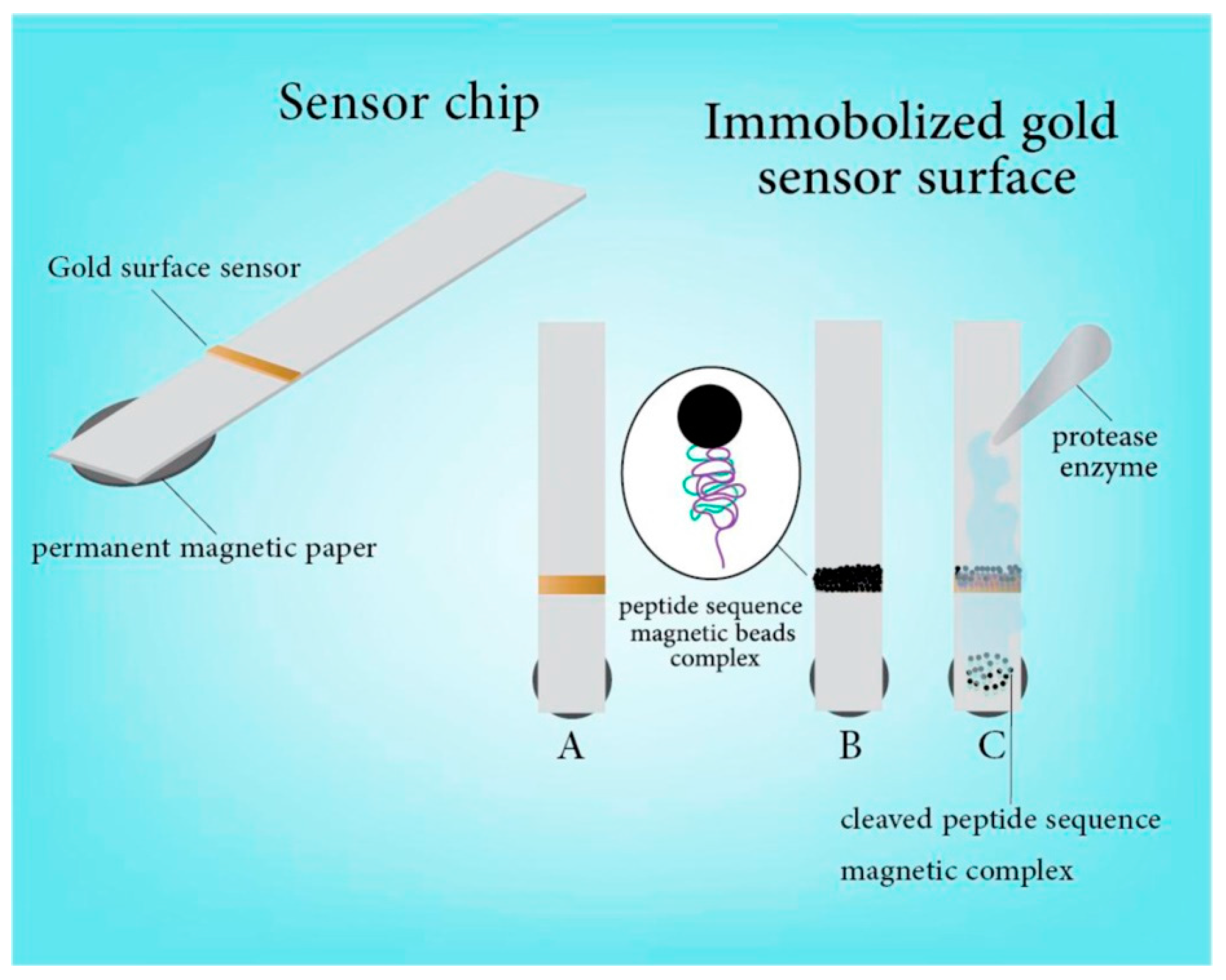
Figure 17.
(a) Example of multifunctional bioassay for detection and disinfection using adsorbed PLL-g-PEG brushes for preventing adsorption of proteins and bacteria (b) Operational principle for capture and detection of E. coli by fluorescently measuring the response of CP coupled with the plasmon-enhance fluorescence from the Ag nanostructure (c) Disinfection is achieved from reactive oxygen species produced by CP under white light irradiation (adapted with permission from [18]).


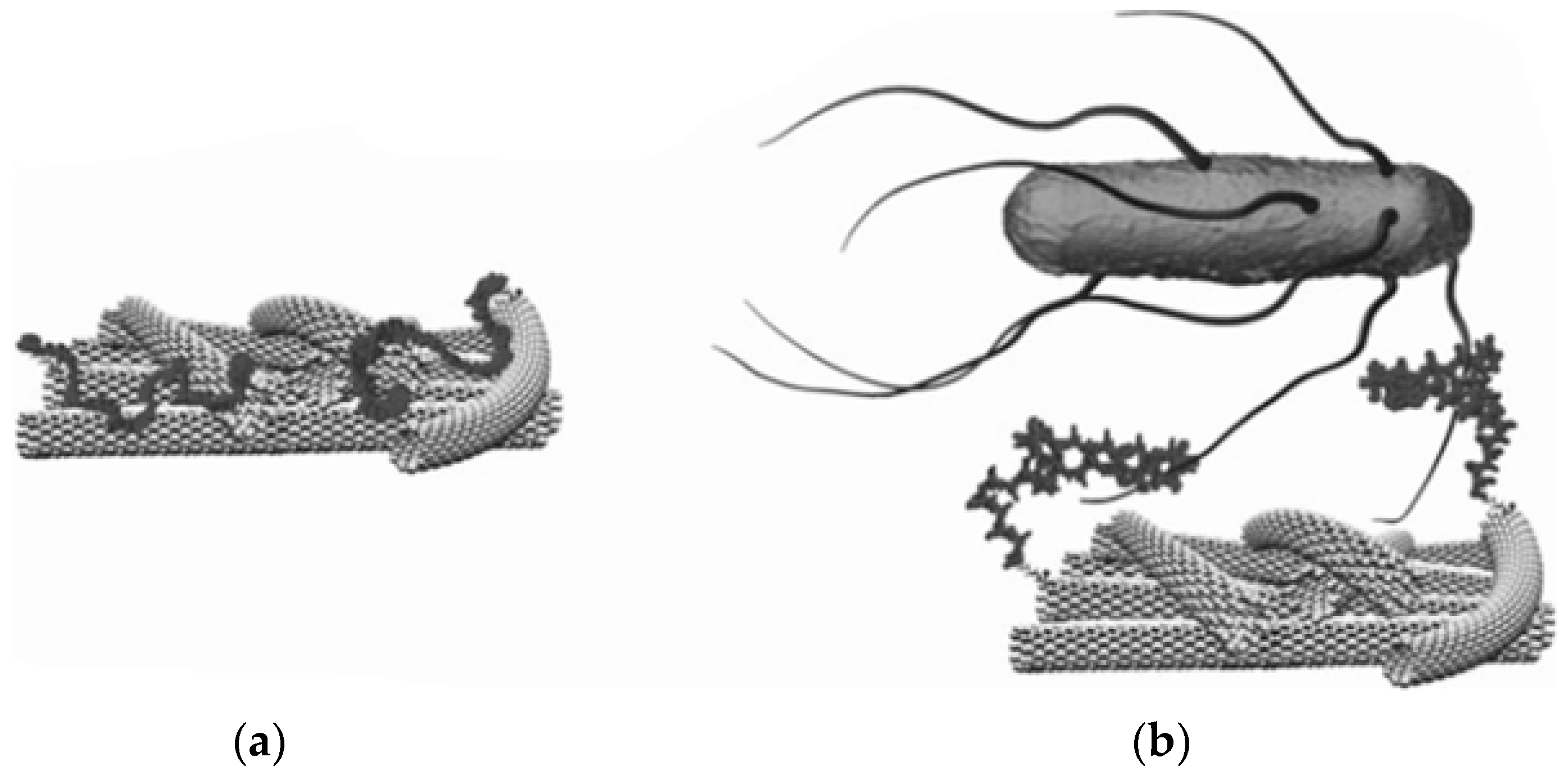
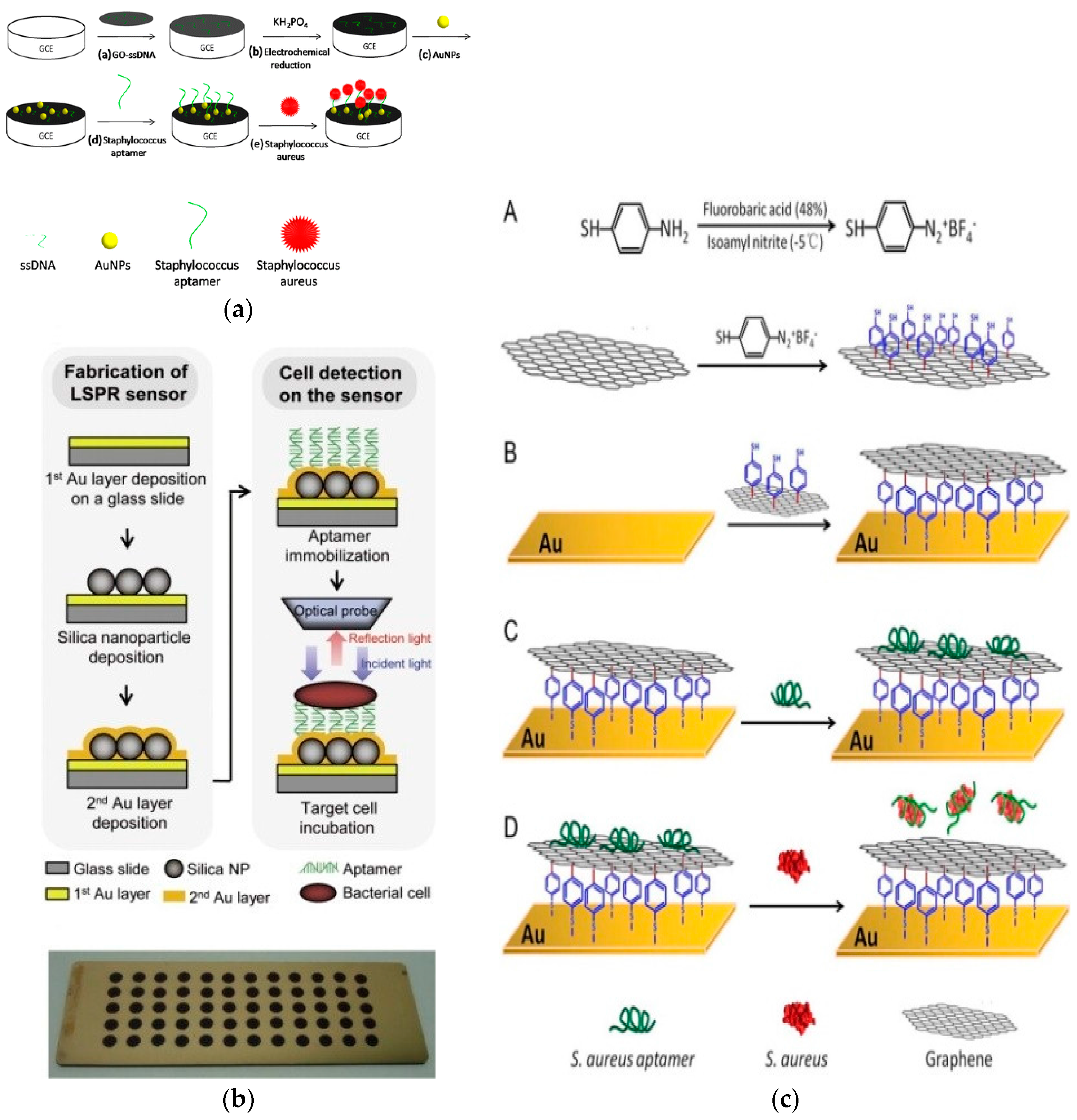
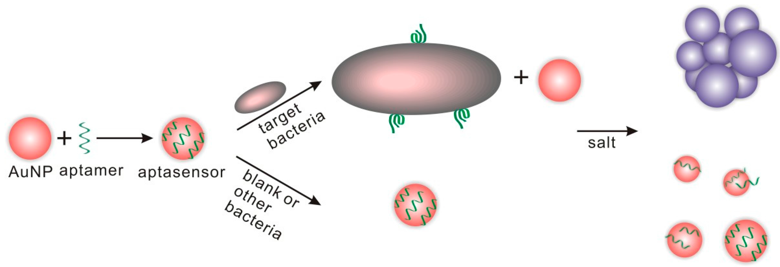
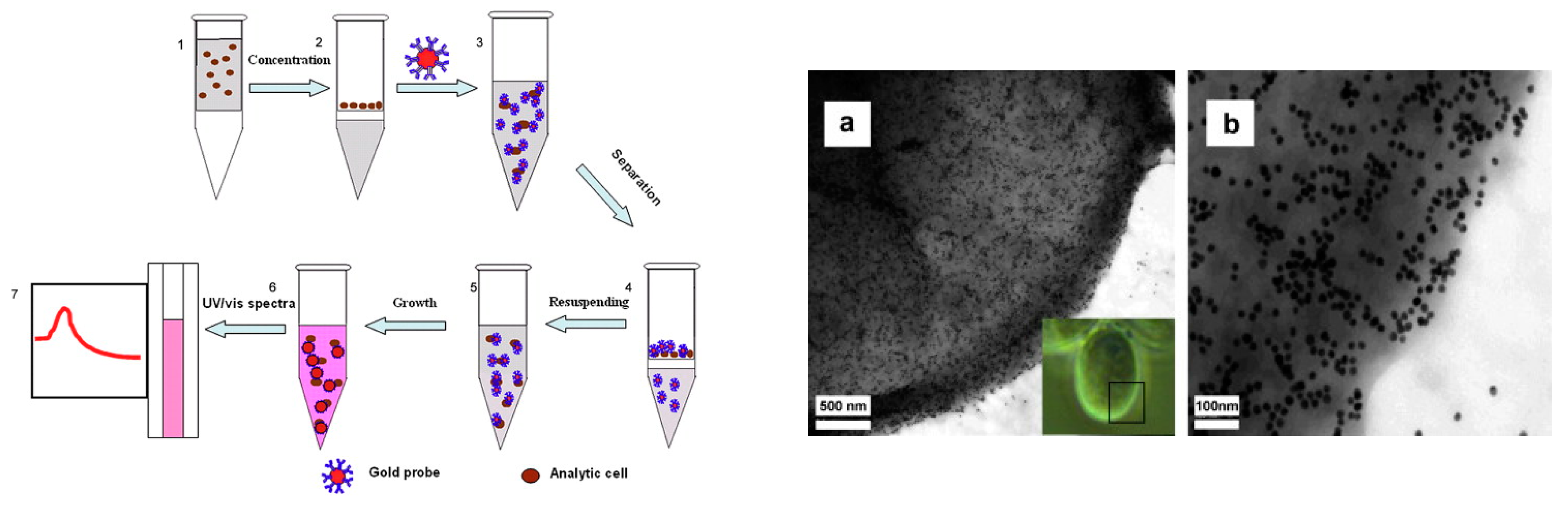
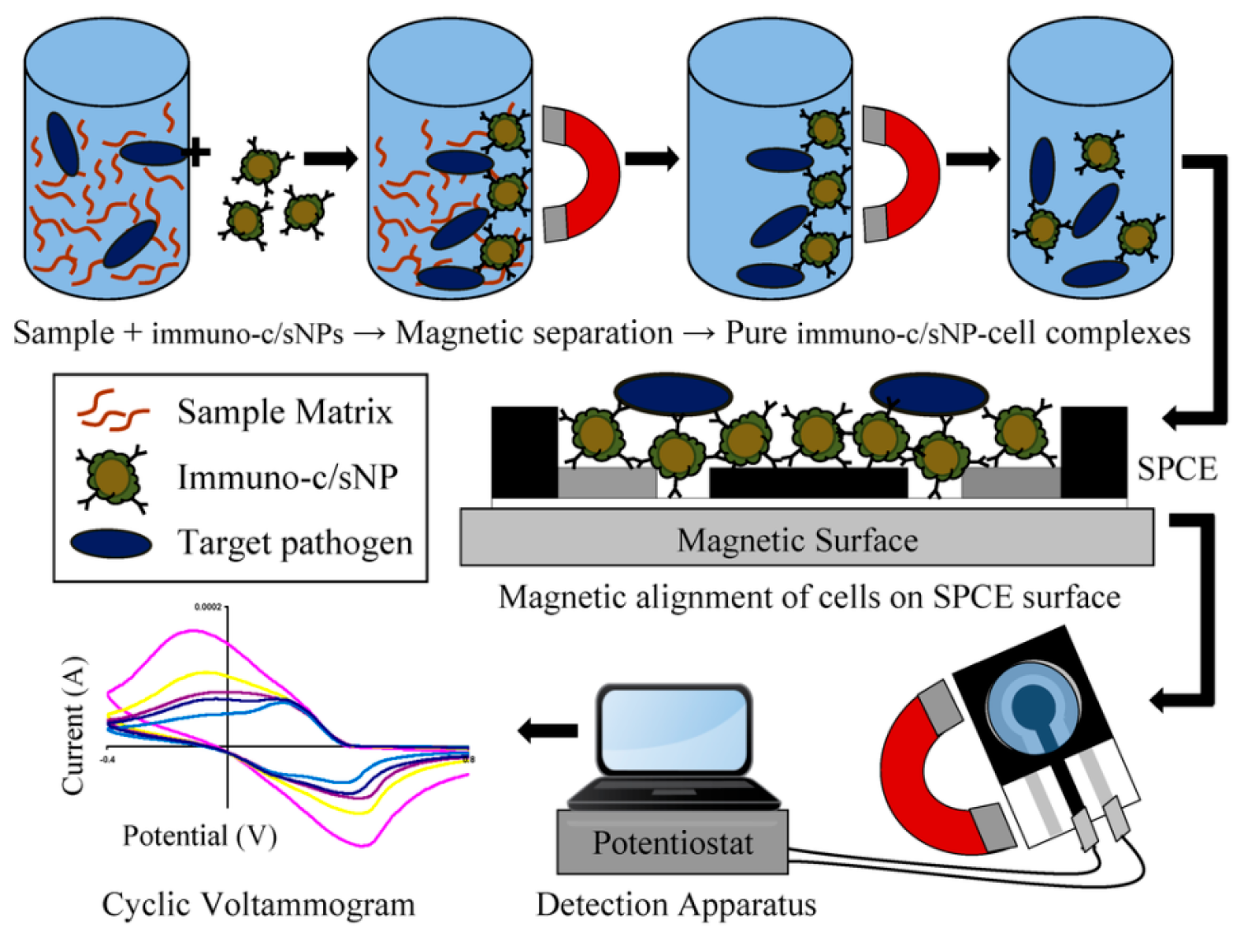
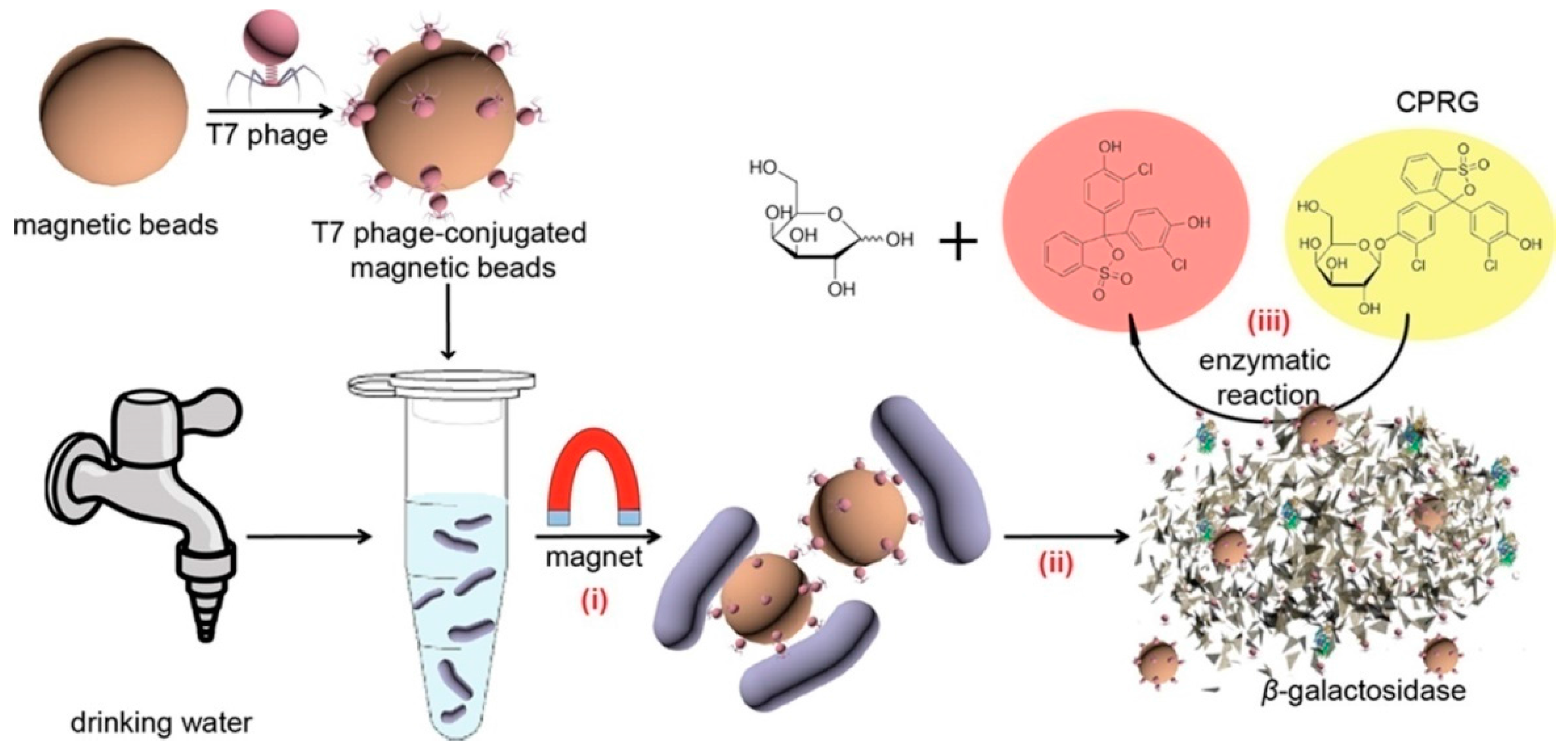
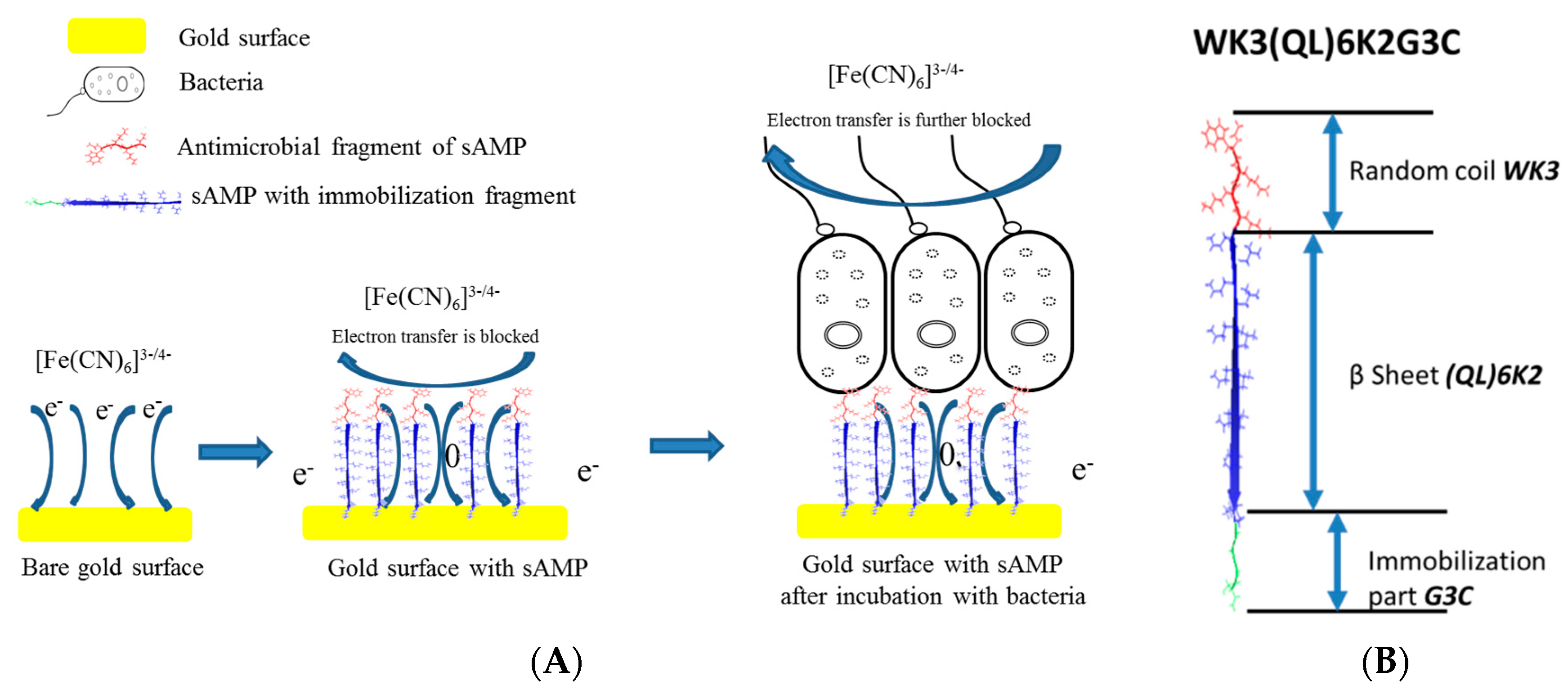
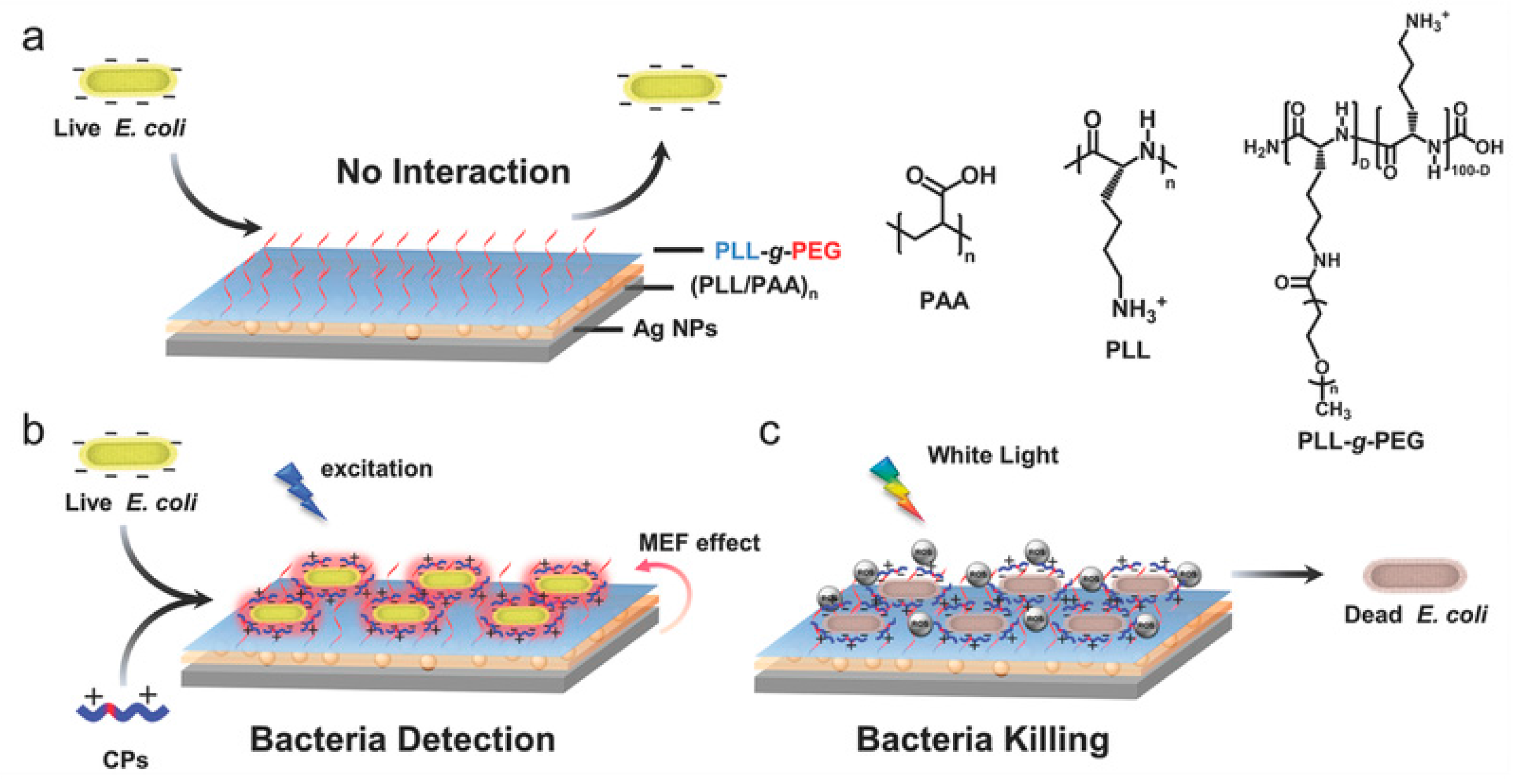
Table 1.
Aptamer-based biosensors for bacteria detection.
| Ref. | Target Bacteria | NPs Used in the Sensor | NPs Function | LOD | Real Sample | Time | Detection Method | Range |
|---|---|---|---|---|---|---|---|---|
| [53] | Salmonella Typhibacteria | SWCNT | Conductive support for aptamer where change in conformation occurs in presence of target bacteria | 1 CFU/mL | - | Few seconds | Potentiometric | 0.2–103 CFU/mL |
| [52] | E. coli CECT 675 as a nonpathogenic surrogate for pathogenic E. coli O157:H7 | SWCNT | Conductive support for aptamer where change in conformation occurs in presence of target bacteria | LOD 12 CFU in 2 mL of milk and 26 CFU/mL in apple juice | Milk and apple juice | Couple of minutes | Potentiometric | linear response of up to 104 CFU/mL |
| [50] | E. coli O157:H7 and Salmonella typhimurium | AuNPs | Color change due to target induced aggregation | 105 CFU/mL | 20 min or less | Optical/Colorimetric UV-Vis | ||
| [45] | Vibrio parahaemolyticus and Salmonella typhimurium | CDs | Fluorescent label | 5 × 10³ CFU/mL | Shrimp | Optical/Fluorescence | 3.8 × 10⁴–3.8 × 10⁷ CFU/mL | |
| [54] | Lactobacillus acidophilus, Staphylococcus aureus and Salmonella enterica | Gaphene oxide (GO) nanomaterial | Fluorescent signal adsorbent | 11.0 CFU/mL for Lactobacillus acidophilus 61.0 CFU/mL for S. enterica and 800.0 CFU/mL and S. aureus | 10 min | Optical/Fluorescence | 9.4–150.0 CFU/mL for Lactobacillus acidophilus 42.2–675.0 CFU/mL for S. enterica and 104–106 CFU/mL for S. aureus | |
| [46] | Staphylococcus aureus | AuNPs-reduced graphene oxide nanocomposite | Signal-amplification and support for aptamer | 10 CFU/mL | water and fish | 60 min | Electrochemical/impedance | 10–106 CFU/mL |
| [49] | E. coli O157:H7 | nanoscale polydiacetylene polymer (PDA ) | Generates color change | 104 CFU/mL | Clinical fecal specimens | 2 h | Optical/colorimetric UV-Vis | 104–108 CFU/mL |
| [51] | Lactobacillus acidophilus Salmonella typhimurium Pseudomonas aeruginosa | Au layer | The combination of gold and silicon NPs (MG-NP) forms a dilectric layer; attachment of biomolecule changes the peak extinction intensity | 30 CFU per assay | - | - | Optical/localized surface plasmon resonance LSPR | 109–104 CFU/mL |
| [48] | Staphylococcus aureus, Vibrio parahemolyticus, and Salmonella typhimurium | 1-Rare earth upconversion nanoparticles (UCNPs) (NaYF4: Yb, Tm NaYF4: Yb, Ho NaYF4: Yb, Er/Mn), 2-magnetic nanoparticles Fe2O3 | 1-luminescence labels for aptamers 2-separation and concentration | 25, 10, and 15 CFU/mL for S. aureus, V. parahemolyticus, and S. typhimurium, respectively | Milk and shrimp | - | Optical/luminescence | 50–106 CFU/mL |
| [19] | Vibrio parahaemolyticus and Salmonella typhimurium | 1-QDs 2-novel amorphous carbon nanoparticles (CNPs) | 1-Fluorescence emitter 2-Fluoresence acceptor | 25 CFU/mL for V. parahaemolyticus, and 35CFU/mL for S. typhimurium | Chicken and shrimps | - | Optical/dual fluorescence resonance energy transfer (FRET) | 50–106 CFU/mL |
| [47] | Staphylococcus aureus | graphene to interdigital gold electrodes connected to a series electrode piezoelectric quartz crystal | - | 41 CFU/mL | Milk | 60 min | Mechanical/series electrode piezoelectric quartz crystal SPQC | 4.1 × 101–4.1 × 10⁵ CFU/mL |
| [44] | Staphylococcus aureus (S. aureus) | AgNPs | Origin of electrochemical signal | 1.0 CFU/mL | Real water | - | Electrochemical/stripping voltammetry | 10–1 × 10⁶ CFU/mL |
| [28] | Salmonella enterica serovar Typhimurium | antibodies -horseradish peroxidase-gold nanoparticles | Amplification of color | 1 × 10³ CFU/mL | milk | <3 h | Optical | 1 × 10³–1 × 10⁸ CFU/mL |
| [61] | Salmonella | multi-walled carbon nanotubes (MWCNTs) | Signal-amplification and a support material for the bioreceptor (aptamer) | 25 CFU/mL | chicken | 60 min | Amperometric: Cyclic voltammetry and impedimetric | 75–7.5 × 10⁵ CFU⋅mL−1 |
Table 2.
Immuno-based biosensors for bacteria detection.
| Ref. | Target Bacteria | NPs | NPs Function | LOD | Real Sample | Time | Detection Method | Range |
|---|---|---|---|---|---|---|---|---|
| [23] | E. coli K12 (gram negative) and Lactobacillus fermentium (gram positive) | AuNPs | amplifying the SPR signal | 104 CFU/mL and 103 CFU/mL in presence of Au NPs | - | 1 min | SPR | 105–107 CFU/mL |
| [24] | Pseudomonas aeruginosa and Staphylococcus aureus | AuNPs | Signaling- origin of color | - | Sputum | 5 min | Visually and Optical Density at 600 nm (OD600) | 500–5000 CFU/mL |
| [25] | Giardia lamblia cysts | AuNPs | Signaling- origin of color | 1.088 × 10³ cells mL−1 | - | - | UV-Vis | 103–104 cells/mL |
| [26] | Bacillus and E. coli O157:H7 | magnetic/polyaniline core/shell nanoparticle (c/sNP) | Separation and electrical conductive based material | 40 CFU/mL and 6 CFU/mL | - | ~1 h | Amperometric: Cyclic voltammetry | 100–102 CFU/mL |
| [27] | E. coli O157:H7 | Au nanorods | Signaling- origin of color | 50 CFU/mL | - | 15 min | two-photon Rayleigh scattering (TPRS) | 50–2100 CFU/mL |
| [62] | Bacillus cereus | AuNPs | Increase sensitivity and stability | 10.0 CFU/mL | Milk | - | Amperometric: Cyclic voltammetry | 5.0 × 101–5.0 × 10⁴ CFU/mL |
Table 3.
Phage-based biosensors for bacteria detection.
| Ref. | Phage | Target Bacteria | LOD | Sample | Time | Detection Method | Range |
|---|---|---|---|---|---|---|---|
| [35] | T7 | E. coli | 10 CFU/mL | Drinking water | 2.5 h | Optical/colorimetric | - |
| [36] | M13KE phage | E. coli K12 | 5 CFU/L | Water | overnight | Colorimetric-culture based assay | - |
| 50 CFU/L water (or 5 CFU/mL orange juice and skim milk) | Water, orange juice and skim milk | <4 h | Colorimetric-solution based assay | - | |||
| [34] | T7 | E. coli K12 | - | - | - | Bacteria culture | 102–107 CFU/mL |
| [43] | Engineered HK620 | E. coli TD2158 and Salmonella | 10 bacteria/mL | Sea water | 1 h | Optical/Fluorescence | |
| [37] | Engineered HK620 and HK97 | E. coli | 104 bacteria/mL | - | 1.5 h | luminescence | - |
| [39] | virulent phage-typing (λ vir) | E. coli (K-12, MG1655) | 1 CFU/100 mL | - | 6–8 h | Electrochemical/amperometric | 102–105 with extended incubation time and 105–109 without time extension |
| [41] | T4 | E. coli K12 | 103 CFU/mL | Milk | - | Electrochemical/impedimetric | 103–108 CFU/mL |
| [40] | Filamentous phage (clone E2—displaying foreign peptide VTPPTQHQ | Salmonella typhimurium | 102 cells/mL | - | <180 s | Mechanical/QCM | 101–107 cells/mL |
| [38] | S. aureus bacteriophage | Staphylococcal and methicillin resistant (MRSA) and sensitive (MSSA) S. aureus species | 104 CFU/mL surface plasmon resonance | - | 16 min | Mechanical/QCM | - |
| [42] | T4 and BP14 phage was used to detect MRSA | E. coli O157:H7 and methicillin-resistant Staphylococcus aureus (MRSA) | 103 CFU/mL | 20 min | Optical/SPR | - |
Table 4.
MIP-based biosensors for bacteria detection.
| Ref. | MIP | Target Bacteria | NPs Used in the Sensor | NPs Function | LOD | Real Sample | Time | Detection Method | Range |
|---|---|---|---|---|---|---|---|---|---|
| [59] | Polypyrrole (PPy) | Pseudomonas aeruginosa | - | - | 103 CFU/mL | Apple juice | 3 min | Mechanical/QCM | 103 to 109 CFU/mL |
| [60] | - | E. coli | - | - | 1.54 × 10⁶ CFU/mL, 3.72 × 10⁵ CFU/mL with SPR and QCM | Apple juice | 113 s for SPR 56 s for QCM respectively, while respectively. | 1-Optical/SPR 2-Mechanical/QCM | 5.13 × 10⁶ CFU/mL, 1.24 × 10⁶ CFU/mL with SPR and QCM |
| [58] | 2,5-dimethyl-4-hydroxy-3(2H)-furanone (DMHF) | Aeromonas hydrophila and Pseudomonas aeruginosa | Magnetic Fe3O4@SiO2–NH2 (MNPs) | Faciliate separation | AHL LOD 8 × 10⁻¹⁰ mol L−1 | Bacteria supernatant spiked samples | - | Electrochemical/ Differential Pulse Voltammetry (DPV) | 2.5 × 10⁻⁹–1.0 × 10⁻⁷ mol/L |
© 2017 by the authors. Licensee MDPI, Basel, Switzerland. This article is an open access article distributed under the terms and conditions of the Creative Commons Attribution (CC BY) license (http://creativecommons.org/licenses/by/4.0/).
Share and Cite
MDPI and ACS Style
Mustafa, F.; Hassan, R.Y.A.; Andreescu, S. Multifunctional Nanotechnology-Enabled Sensors for Rapid Capture and Detection of Pathogens. Sensors 2017, 17, 2121. https://doi.org/10.3390/s17092121
AMA Style
Mustafa F, Hassan RYA, Andreescu S. Multifunctional Nanotechnology-Enabled Sensors for Rapid Capture and Detection of Pathogens. Sensors. 2017; 17(9):2121. https://doi.org/10.3390/s17092121
Chicago/Turabian StyleMustafa, Fatima, Rabeay Y. A. Hassan, and Silvana Andreescu. 2017. "Multifunctional Nanotechnology-Enabled Sensors for Rapid Capture and Detection of Pathogens" Sensors 17, no. 9: 2121. https://doi.org/10.3390/s17092121
Note that from the first issue of 2016, this journal uses article numbers instead of page numbers. See further details here.