An Exonuclease I-Based Quencher-Free Fluorescent Method Using DNA Hairpin Probes for Rapid Detection of MicroRNA

State Key Laboratory of Medical Genetics & School of Life Sciences, Central South University, Changsha 410013, China
*
Authors to whom correspondence should be addressed.
Sensors 2017, 17(4), 760; https://doi.org/10.3390/s17040760
Submission received: 3 February 2017
/
Revised: 26 March 2017
/
Accepted: 31 March 2017
/
Published: 3 April 2017
(This article belongs to the Special Issue Genosensing)
Abstract
:MicroRNAs (miRNAs) act as biomarkers for the diagnosis of a variety of cancers. Since the currently used methods for miRNA detection have limitations, simple, sensitive, and cost-effective methods for the detection of miRNA are required. This work demonstrates a facile, quencher-free, fluorescence-based analytical method for cost-effective and sensitive detection of miRNA using a super 2-aminopurine (2-AP)-labeled hairpin probe (HP) and exonuclease I activity. Specifically, the fluorescence of 2-AP is strongly quenched when it is incorporated within DNA. In the presence of a target miRNA, HP attains an open conformation by hybridizing with the target miRNA to form a double-stranded structure with a protruding 3′-terminus. Next, the digestion of the protruding 3′-terminus is triggered by exonuclease I, during which 2-AP is released free in solution from the DNA, thereby increasing fluorescence. This method is highly sensitive, with a detection limit of 0.5 nM—10 times lower than a previously reported quencher-free fluorescence method. Furthermore, this method has potential applications in clinical diagnosis and biomedical research.
1. Introduction
MicroRNAs (miRNAs) are a class of small (19–23 nucleotides long), single-stranded, endogenous, non-coding RNAs that commonly exist in plant, animal, and virus genomes, and were first discovered in the 1990s [1,2,3]. It has been demonstrated that miRNAs play key regulatory roles in a series of critical biological processes, such as metabolism, differentiation, apoptosis, and immunological response [4,5]. Since abnormal miRNA expression is associated with a variety of cancers, such as cancers of the breast, prostate, liver, lung, and colon, miRNAs have drawn considerable attention as putative biomarkers and therapeutic targets for cancer treatment [6,7,8]. Therefore, devising rapid and sensitive methods for the detection of miRNAs is important.
Several traditional mainstream techniques for miRNA detection and quantitation are Northern blotting, quantitative reverse transcription polymerase chain reaction (qRT-PCR), and microarray analysis; however, each of these methods have limitations which hinder their wide and convenient application [9,10,11]. Northern blotting is widely used in miRNA detection and quantitation, although it is time-consuming and requires the use of radiolabels and large sample size [12,13]. qRT-PCR is a highly sensitive method; however, the methodology requires tedious preparation and highly-skilled personnel [12]. Microarray is a high-throughput method, but the analysis platform is not suitable for quantitating rare miRNAs, owing to its low sensitivity [13]. Hence, pursuits for alternative analytical methods that circumvent the above limitations have received increasing attention. Recently, a variety of device-based analytical strategies and methods-based approaches have been developed to quantitatively analyze miRNAs. Device-based analytical strategies mainly include electrochemical sensors and MEMS [14,15]. Methods-based approaches mainly include fluorescence, colorimetric, and luminescence assays [16,17,18,19,20,21,22,23,24,25,26,27,28]. Among these, the fluorometric method is an excellent choice for the quantitative detection of miRNAs due to its high sensitivity and convenience of operation. Santangelo and co-workers have reported a facile method for the detection of miRNA using dual fluorescence resonance energy transfer (FRET) molecular beacons [29]. Ryoo et al. have developed a method to detect miRNA based on nano graphene oxide (PANGO—peptide nucleic acid and nano graphene oxide) [30]. Yin et al. have developed a novel method to detect miRNAs based on duplex-specific nuclease signal amplification [31]. Zhang et al. have designed a strategy for the detection of miRNAs by a catalyst–oligomer-mediated enzymatic amplification-based fluorescence biosensor, which is a highly-sensitive method [32]. Although these strategies are usually rapid and highly sensitive, they still utilize expensive dual labeled probes or tedious preparations. Therefore, developing a facile, sensitive, and low-cost analytical method to detect miRNAs is still an open challenge.
2-Aminopurine (2-AP)—whose fluorescence is strongly quenched when incorporated within DNA—has been used as a fluorescent analogue of adenine for many years [33]. 2-AP is often used as a quencher-free fluorescent probe for the study of protein–DNA interactions and DNA flipping [34,35]. The quenching of 2-AP fluorescence results from its stacking interaction with adjacent bases of DNA without the involvement of any extra quenchers [36,37]. Exonuclease I (exo I) belongs to the exonuclease family that can degrade single-stranded DNA in a 3′→5′ direction and release deoxyribonucleoside 5′-monophosphates in a step-wise manner. Here, we developed a facile quencher-free fluorescence-based analytical strategy for sensitive and low-cost detection of miRNAs using a super 2-AP-labeled hairpin probe (HP) and exo I activity. By taking advantage of the super quencher-free fluorescence-labeled 2-AP probe, we provide a facile, low-cost, and sensitive analytical method for quantitating miRNA [38,39].
2. Materials and Methods
2.1. Reagents
miRNA-21 (5′-UAGCUUAUCAGACUGAUGUUGA-3′), miR-141 (5′-UAACAC UGUCUGGUAAAGAUGG-3′), and let-7d (5′-AGAGGUAGUAGGUUGCAUAGUU-3′) synthesized by TaKaRa Biotechnology Co., Ltd. (Dalian, China) were purified by high-performance liquid chromatography (HPLC). Hairpin probe (HP: 5′-GCG TCG TCA ACA TCA GTC TGA TAA GCT ACG /2-amp/CGC-3′), which were purified by HLPC, were obtained from Sangon Biotechnology Co., Ltd. (Shanghai, China). Exonuclease I, 10× exonuclease I buffer (670 mM Glycine-KOH, 10 mM Dithiothreitol (DTT), 67 mM MgCl2, pH 9.5), recombinant RNase inhibitor (RRI), and RNase-free water were obtained from TaKaRa Biotechnology Co., Ltd. (Dalian, China). Human serum samples were collected from the third Xiangya hospital of Central South University (Changsha, China). All the reagents were used without further purification. All the nucleic acids used in this study were dissolved in RNase-free water and stored at −20 °C until further use.
2.2. Fluorescence Measurement
All fluorescence measurements were carried out on an F2700 (Hitachi, Japan) with excitation at 310 nm and emission at 365 nm for 2-AP. The excitation slits and emission slits were both set at 5.0 nm.
2.3. Detection of miRNA
Two samples were prepared: sample A contained only 200 nM HP, whereas sample B contained 200 nM HP and 200 nM miRNA-21. Next, 20 U RRI and 20 U/mL exo I were added to samples A and B, respectively. Assays were carried out in 1× exonuclease I buffer (67 mM Glycine-KOH, 1 mM DTT, 6 mM MgCl2, pH 9.5). RNase-free water was used in all the reactions. Thereafter, samples A and B were incubated at 37 °C for approximately 30 min before taking measurements.
2.4. Optimization of Analysis Conditions
Certain critical reactions were optimized with respect to the time of reaction and the concentrations of HP and exo I that were used. The HP concentration range used was 50 to 400 μM, and the exo I concentration range was 3 to 40 U/mL; the reaction time range was 5 to 40 min.
2.5. Quantitative Analysis of miRNA
HP (200 nM), 20 U RRI, 20 U/mL exo I, and different amounts of miRNA-21 were assembled in 100 μL of 1× exo I buffer and incubated at 37 °C for about 30 min before taking measurements. miRNA-21 concentration in the reaction varied from 0 to 200 nM.
2.6. Application of the Method in Biological Assays
Enzymes and proteins were removed from human serum by calefaction, following which the serum was diluted 1:10 with phosphate-buffered saline (PBS) buffer, and then spiked with miRNA at three concentrations (50, 100, and 200 nM miRNA-21) and analyzed.
3. Results and Discussion
3.1. Designing the Strategy
The working principle of the quencher-free fluorescence-based method for miRNA detection is illustrated in Figure 1. The reaction system consists of HP, exo I, and miRNA. Exo I specifically degrades single-stranded DNA (ssDNA) in a 3′→5′ direction, and has no activity on double-stranded DNA (dsDNA). The HP has two domains: a 2-AP labeled stem without overhanging nucleotides, which protects the HP from exo I digestion and a loop sequence which is complementary to the target miRNA. In the absence of the target miRNA, 2-AP is incorporated within HP, and its fluorescence is quenched. Once the target miRNA hybridizes with the HP, the latter attains an open conformation and forms a double-stranded structure with a protruding 3′-terminus, which triggers the digestion of the protruding 3′-terminus by exo I. During the digestion, 2-AP is separated from the DNA strands and released free in solution, thereby increasing fluorescence. Thus, the fluorescence intensity observed at 365 nm is directly proportional to the concentration of miRNA present, allowing quantitative detection of the target miRNA.
3.2. Validation of miRNA Detection with the Method
We evaluated the feasibility of the proposed method for miRNA detection by using two samples. Figure 2 depicts the fluorescence emission spectra in the presence and absence of target miRNA. The fluorescence intensity was weak in the absence of target miRNA (curve A). When the target miRNA was added to reaction solution, an increase in fluorescence intensity was observed (curve B), which confirmed that our assay can actually detect miRNAs.
3.3. Optimization of Experimental Conditions
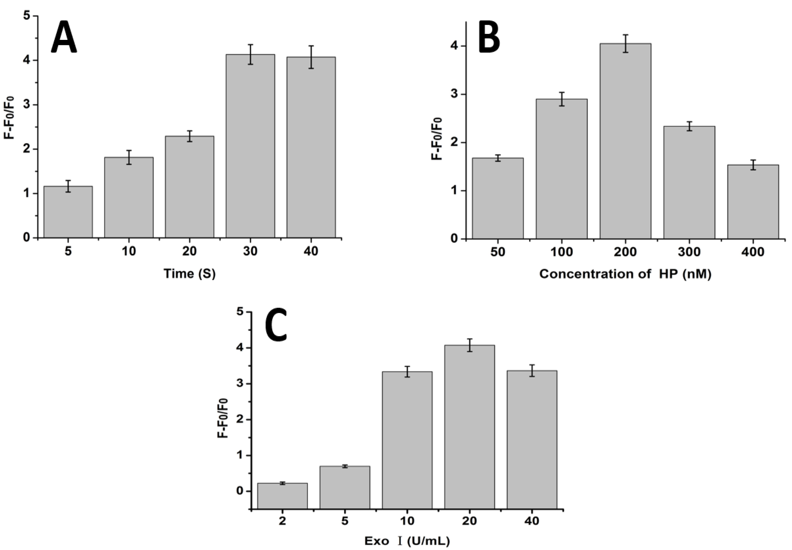
Since reaction time and concentrations of HP and exo I can influence the performance of the proposed method, we first optimized these conditions to obtain the best result. After a series of experiments, we found that 30 min reaction time, 200 nM HP, and 20 U/mL exo I were the optimal reaction conditions (Figure 3). Accordingly, these conditions were used in the subsequent experiments.
3.4. Quantitative Analysis of miRNA
Various concentrations of target miRNA (miRNA-21) (0, 0.5, 1, 5, 20, 50, 100, 200, 400 nM) were added to the reaction sample to evaluate the sensitivity of the proposed method under the optimized conditions. The corresponding fluorescence intensity at 365 nm dynamically increased with increase in target miRNA concentration (Figure 4A). Figure 4B shows the relationship between fluorescence intensity and the concentration of target miRNA. The linear regression equation was F = 2.8402C + 198.96 (C, nM) with an R2 of 0.996, where F was the fluorescence intensity at 365 nm and C was the target miRNA concentration. The limit of detection (LOD) of the proposed strategy was estimated to be 0.5 nM, which was 10 times lower than a previously reported quencher-free fluorescence method (5 nM) [40].
3.5. Selectivity of the miRNA Detection Assay
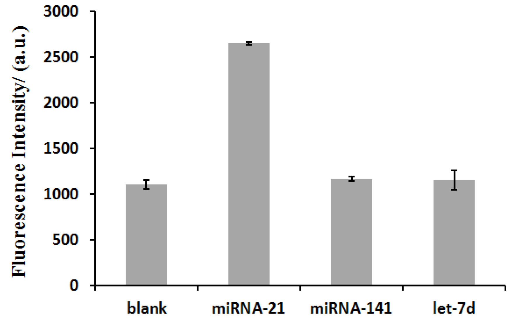
The selectivity of this miRNA biosensor was investigated by using 800 nM of two mismatched miRNA sequences—namely, miRNA-141 and let-7d. As shown in Figure 5, none of these mismatched miRNA sequences could trigger a significant increase in fluorescence intensity except the target miRNA, miRNA-21 (200 nM). These results underline the ability of our method to distinguish between perfectly matched target miRNAs and mismatched miRNAs, suggesting that it can be used for miRNA detection.
3.6. Application of the Method in Biological Assays
A literature survey revealed that miRNA-21 concentration in human serum is about 35 fM, which is lower than the detection limit of the proposed method [41]. To demonstrate the feasibility of the approach in biological matrices, the human serum samples were spiked with miRNA at three concentrations (50, 100, and 200 nM miRNA-21), and analyzed. The analytical results are shown in Table 1. The fluorescence recovery rates for the different concentrations of target miRNA were 89% for 50 nM, 104.4% for 100 nM, and 99.4% for 200 nM. This indicates that the method may be used for analysis of miRNA in real biological samples.
4. Conclusions
In summary, we have successfully demonstrated a user-friendly, quencher-free fluorescence-based method for sensitive miRNA detection based on an exonuclease I-catalyzed DNA degradation reaction. Firstly, the LOD of the quencher-free fluorescence method was estimated to be 0.5 nM, which is 10 times lower than a previously reported quencher-free fluorescence method. Secondly, by taking advantage of the quencher-free fluorescent 2-AP, we developed a low-cost and sensitive assay. Thirdly, the total reaction time in the proposed method is only 30 min, which is much shorter than those in most existing methods. We envision that our quencher-free fluorescence-based strategy for quantitative analysis of miRNA may have potential applications in clinical diagnosis and biomedical research.
Acknowledgments
This work was supported by National Natural Science Foundation of China (No. 21205142), The Research Innovation Program for Graduates of Central South University (2016zzts580).
Author Contributions
H.L. and C.M. conceived and designed the experiments; H.L. performed the experiments; H.L., K.W., M.C. and L.Z. analyzed the data; C.M. and J.W. contributed reagents/materials/analysis tools; H.L. and C.M. wrote the paper.
Conflicts of Interest
The authors declare no conflict of interest.
References
- Li, J.; Tan, S.; Kooger, R.; Zhang, C.; Zhang, Y. MicroRNAs as novel biological targets for detection and regulation. Chem. Soc. Rev. 2014, 43, 506–517. [Google Scholar] [CrossRef] [PubMed]
- Tian, T.; Wang, J.Q.; Zhou, X. A review: MicroRNA detection methods. Org. Biomol. Chem. 2015, 13, 2226–2238. [Google Scholar] [CrossRef] [PubMed]
- Cullen, B.R. Viral and cellular messenger RNA targets of viral microRNAs. Nature 2009, 457, 421–425. [Google Scholar] [CrossRef] [PubMed]
- Aviñó, A.; Huertas, C.S.; Lechuga, L.M.; Eritja, R. Sensitive and label-free detection of miRNA-145 by triplex formation. Anal. Bioanal. Chem. 2016, 408, 885–893. [Google Scholar] [CrossRef] [PubMed]
- Ma, C.B.; Yeung, E.S.; Qi, S.D.; Han, R. Highly sensitive detection of microRNA by chemiluminescence based on enzymatic polymerization. Anal. Bioanal. Chem. 2012, 402, 2217–2220. [Google Scholar] [CrossRef] [PubMed]
- Serpico, D.; Molino, L.; Cosimo, S.D. MicroRNAs in breast cancer development and treatment. Cancer Treat. Rev. 2014, 40, 595–604. [Google Scholar] [CrossRef] [PubMed]
- Mishra, S.; Srivastava, A.K.; Suman, S.; Kumar, V.; Shukla, Y. Circulating miRNAs revealed as surrogate molecular signatures for the early detection of breast cancer. Cancer Lett. 2015, 369, 67–75. [Google Scholar] [CrossRef] [PubMed]
- Lu, J.; Getz, G.; Miska, E.A.; Saavedra, E.A.; Lamb, J.; Peck, D.; Cordero, A.S.; Ebert, B.L.; Mak, R.H.; Ferrando, A.A.; et al. MicroRNA expression profiles classify human cancers. Nature 2005, 435, 834–838. [Google Scholar] [CrossRef] [PubMed]
- Chen, C.; Ridzon, D.A.; Broomer, A.J.; Zhou, Z.; Lee, D.H.; Nguyen, J.T.; Barbisin, M.; Xu, N.L.; Mahuvakar, V.R.; Andersen, M.R.; et al. Real-time quantification of microRNAs by stem-loop RT-PCR. Nucleic Acids Res. 2005, 33, 179–188. [Google Scholar] [CrossRef] [PubMed]
- Roy, S.; Soh, J.H.; Ying, J.Y. A microarray platform for detecting disease-specific circulating miRNA in human serum. Biosens. Bioelectron. 2016, 75, 238–246. [Google Scholar] [CrossRef] [PubMed]
- Válóczi, A.; Hornyik, C.; Varga, N.; Burgyán, J.; Kauppinen1, S.; Havelda, Z. Sensitive and specific detection of microRNAs by northern blot analysis using LNA-modified oligonucleotide probes. Nucleic Acids Res. 2004, 32, 175–183. [Google Scholar] [CrossRef] [PubMed]
- Xu, Y.; Wang, Y.; Liu, S.; Yu, J.H.; Wang, H.Z.; Guo, Y.N.; Huang, J. Ultrasensitive and rapid detection of miRNA with three-way junction structure-based trigger-assisted exponential enzymatic amplification. Biosens. Bioelectron. 2016, 81, 236–241. [Google Scholar] [CrossRef] [PubMed]
- Lu, Z.; Tang, H.; Wu, D.; Xia, Y.; Wu, M.; Yi, X.; Li, H.; Wang, J. Amplified voltammetric detection of miRNA from serum samples of glioma patients via combination of conducting magnetic microbeads and ferrocene-capped gold nanoparticle/streptavidin conjugates. Biosens. Bioelectron. 2016, 86, 502–507. [Google Scholar] [CrossRef] [PubMed]
- Lu, Y.; Zhang, Z.; Xu, Z.; Yin, H.; Ai, S. MicroRNA-21 detection based on molecular switching by amperometry. New J. Chem. 2012, 36, 1985–1991. [Google Scholar]
- Johnson, B.; Mutharasan, R. Sample preparation-free, real-time detection of microRNA in human serum using piezoelectric cantilever biosensors at attomole level. Anal. Chem. 2012, 84, 10426–10436. [Google Scholar] [CrossRef] [PubMed]
- Xi, Q.; Zhou, D.; Kan, Y.; Ge, J.; Wu, Z.; Yu, R.; Jiang, J. Highly sensitive and selective strategy for microRNA detection based on WS2 nanosheet mediated fluorescence quenching and duplex-specific nuclease signal amplification. Anal. Chem. 2014, 86, 1361–1365. [Google Scholar] [CrossRef] [PubMed]
- Cui, L.; Lin, X.; Lin, N.; Song, Y.; Zhu, Z.; Chen, Z.; Yang, C. Graphene oxide-protected DNA probes for multiplex microRNA analysis in complex biological samples based on a cyclic enzymatic amplification method. Chem. Commun. 2012, 48, 194–196. [Google Scholar] [CrossRef] [PubMed]
- Lin, X.; Zhang, C.; Huang, Y.; Zhu, Z.; Chen, X.; Yang, C. Backbone-modified molecular beacons for highly sensitive and selective detection of microRNAs based on duplex specific nuclease signal amplification. Chem. Commun. 2013, 49, 7243–7245. [Google Scholar] [CrossRef] [PubMed]
- Li, Y.; Zhang, J.; Zhao, J.; Zhao, L.; Cheng, Y.; Li, Z. A simple molecular beacon with duplex-specific nuclease amplification for detection of microRNA. Analyst 2016, 14, 1071–1076. [Google Scholar] [CrossRef] [PubMed]
- Xie, Y.; Lin, X.; Huang, Y.; Zhi, R.P.Z.; Zhou, L.; Yang, C. Highly sensitive and selective detection of miRNA: DNase I-assisted target recycling using DNA probes protected by polydopamine nanospheres. Chem. Commun. 2015, 51, 2156–2158. [Google Scholar] [CrossRef] [PubMed]
- Hou, T.; Li, W.; Liu, X.; Li, F. Label-Free and Enzyme-Free homogeneous electrochemical biosensing strategy based on hybridization chain reaction: A facile, sensitive, and highly specific microRNA assay. Anal. Chem. 2015, 87, 11368–11374. [Google Scholar] [CrossRef] [PubMed]
- Xu, F.; Shi, H.; He, X.; Wang, K.; He, D.; Guo, Q.; Qing, Z.; Yan, L.; Ye, X.; Li, D.; et al. Concatemeric dsDNA-templated copper nanoparticles strategy with improved sensitivity and stability based on rolling circle replication and its application in microRNA detection. Anal. Chem. 2014, 86, 6976–6982. [Google Scholar] [CrossRef] [PubMed]
- Li, D.D.; Cheng, W.; Yan, Y.R.; Zhang, Y.; Yin, Y.B.; Ju, H.X.; Ding, S.J. A colorimetric biosensor for detection of attomolar microRNA with a functional nucleic acid-based amplification machine. Talanta 2016, 146, 470–476. [Google Scholar] [CrossRef] [PubMed]
- Wu, H.; Liu, Y.L.; Wang, H.Y.; Wu, J.; Zhu, F.F.; Zou, P. Label-free and enzyme-free colorimetric detection of microRNA by catalyzed hairpin assembly coupled with hybridization chain reaction. Biosens. Bioelectron. 2016, 81, 303–308. [Google Scholar] [CrossRef] [PubMed]
- Wu, X.Y.; Chai, Y.Q.; Yuan, R.; Su, H.L.; Han, J. A novel label-free electrochemical microRNA biosensor using Pd nanoparticles as enhancer and linker. Analyst 2013, 138, 1060–1066. [Google Scholar] [CrossRef] [PubMed]
- Campuzano, S.; Pedrero, M.; Pingarrón, J.M. Electrochemical genosensors for the detection of cancer-related miRNAs. Anal. Bioanal. Chem. 2014, 406, 27–33. [Google Scholar] [CrossRef] [PubMed]
- Chen, Y.; Xiang, Y.; Yuan, R.; Chai, Y.Q. Intercalation of quantum dots as the new signal acquisition and amplification platform for sensitive electrochemiluminescent detection of microRNA. Anal. Chim. Acta 2015, 891, 130–135. [Google Scholar] [CrossRef] [PubMed]
- Gao, Z.; Deng, H.; Shen, W.; Ren, Y. A label-free biosensor for electrochemical detection of femtomolar miRNAs. Anal. Chem. 2013, 85, 1624–1630. [Google Scholar] [CrossRef] [PubMed]
- Santangelo, P.J.; Nix, B.; Tsourkas, A.; Bao, G. Dual FRET molecular beacons for mRNA detection in living cells. Nucleic Acids Res. 2004, 32, 112–118. [Google Scholar] [CrossRef] [PubMed]
- Ryoo, S.R.; Lee, J.; Yeo, J.; Na, H.K.; Kim, Y.K.; Jang, H.; Lee, J.H.; Han, S.W.; Lee, Y.; Kim, V.N.; et al. Quantitative and multiplexed microRNA sensing in living cells based on peptide nucleic acid and nano graphene oxide (PANGO). ACS Nano 2013, 7, 5882–5891. [Google Scholar] [CrossRef] [PubMed]
- Yin, B.C.; Liu, Y.Q.; Ye, B.C. One-Step, multiplexed fluorescence detection of microRNAs based on duplex-specific nuclease signal amplification. J. Am. Chem. Soc. 2012, 134, 5064–5067. [Google Scholar] [CrossRef] [PubMed]
- Zhang, K.; Wang, K.; Zhu, X.; Xie, M.H. A one-pot strategy for the sensitive detection of miRNA by catalyst–oligomer-mediated enzymatic amplification-based fluorescence biosensor. Sens. Actuators B Chem. 2016, 223, 586–590. [Google Scholar] [CrossRef]
- Liu, H.S.; Ma, C.B.; Zhou, M.J.; Chen, H.C.; He, H.L.; Wang, K.M. Quencher-free fluorescence strategy for detection of DNA methyltransferase activity based on exonuclease III-assisted signal amplification. Anal. Bioanal. Chem. 2016, 408, 8111–8116. [Google Scholar] [CrossRef] [PubMed]
- Jones, A.C.; Neely, R.K. 2-Aminopurine as a fluorescent probe of DNA conformation and the DNA-enzyme interface. Rev. Biophys. 2015, 48, 244–279. [Google Scholar] [CrossRef] [PubMed]
- Holz, B.; Klimasauskas, S.; Serva1, S.; Weinhold, E. 2-Aminopurine as a fluorescent probe for DNA base flipping by methyltransferases. Nucleic Acids Res. 1998, 26, 1076–1083. [Google Scholar] [CrossRef] [PubMed]
- Somsen, J.G.; Hoek, V.A.; Amerongen, V.H. Fluorescence quenching of 2-aminopurine in dinucleotides. Chem. Phys. Lett. 2005, 402, 61–65. [Google Scholar] [CrossRef]
- Zhu, G.; Liang, L.; Zhang, C. Quencher-free fluorescent method for homogeneously sensitive detection of microRNAs in human lung tissues. Anal. Chem. 2014, 86, 11410–11416. [Google Scholar] [CrossRef] [PubMed]
- Johnson, B.; Mutharasan, R. Biosensor-based microRNA detection: Techniques, design, performance, and challenges. Analyst 2014, 139, 1576–1588. [Google Scholar] [CrossRef] [PubMed]
- Hunt, E.; Broyles, D.; Head, T.; Deo, S. MicroRNA Detection: Current Technology and Research Strategies. Annu. Rev. Anal. Chem. 2015, 8, 217–237. [Google Scholar] [CrossRef] [PubMed]
- Liao, R.; He, K.; Chen, C.Y.; Chen, X.M.; Cai, C.Q. Double-strand displacement biosensor and quencher-free fluorescence strategy for rapid detection of microRNA. Anal. Chem. 2016, 88, 4254–4258. [Google Scholar] [CrossRef] [PubMed]
- Shuai, H.L.; Huang, K.J.; Chen, Y.X.; Fang, L.X.; Jia, M.P. Au nanoparticles/hollow molybdenum disulfide microcubes based biosensor for microRNA-21 detection coupled with duplex-specific nuclease and enzyme signal amplification. Biosens. Bioelectron. 2017, 89, 989–997. [Google Scholar] [CrossRef] [PubMed]
Figure 1.
Schematic showing the principle of the quencher-free fluorescence-based method for the detection of microRNA. Exo I: Exonuclease I.
Figure 1.
Schematic showing the principle of the quencher-free fluorescence-based method for the detection of microRNA. Exo I: Exonuclease I.
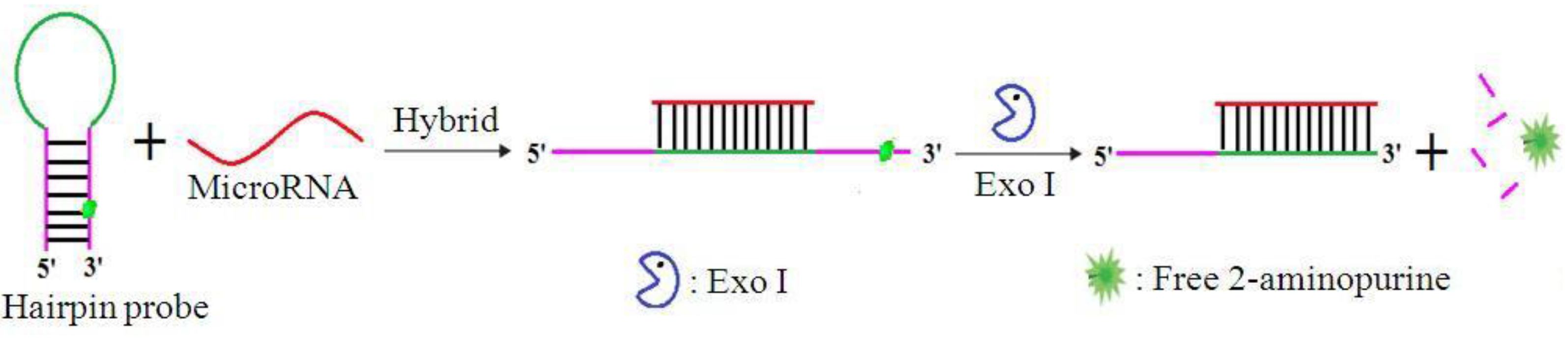
Figure 2.
Validation of microRNA (miRNA) detection by the proposed method. Fluorescence emission spectra obtained when (A) no target miRNA and (B) 200 nM target miRNA were used in the reaction.
Figure 2.
Validation of microRNA (miRNA) detection by the proposed method. Fluorescence emission spectra obtained when (A) no target miRNA and (B) 200 nM target miRNA were used in the reaction.
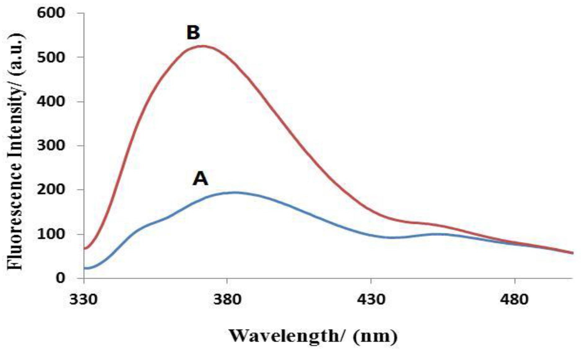
Figure 3.
Optimization of miRNA detection conditions. (A) Bar graph showing the relationship between reaction time and (F0-F)/F0. Each sample contained 200 nM HP, 200 nM miRNA, and 20 U/mL exo I. (B) Bar graph showing the relationship between the concentration of HP and (F0-F)/F0. In each sample, 200 nM miRNA and 20 U/mL exo I were present. (C) Bar graph showing the relationship between the concentration of exo I and (F0-F)/F0. In each sample, 200 nM HP and 200 nM miRNA were present. Error bars were estimated from three replicate measurements.

Figure 4.
(A) Fluorescence emission spectra in the presence of increasing amounts of miRNA (0, 0.5, 1, 5, 20, 50, 100, 200, 400 nM). (B) Plot showing changes in fluorescence intensity with miRNA concentration. Inset: graph showing linear relationship of fluorescence intensity with miRNA concentration at low concentration range; 200 nM HP and 20 U/mL exo I were used. Error bars were estimated from three replicate measurements.
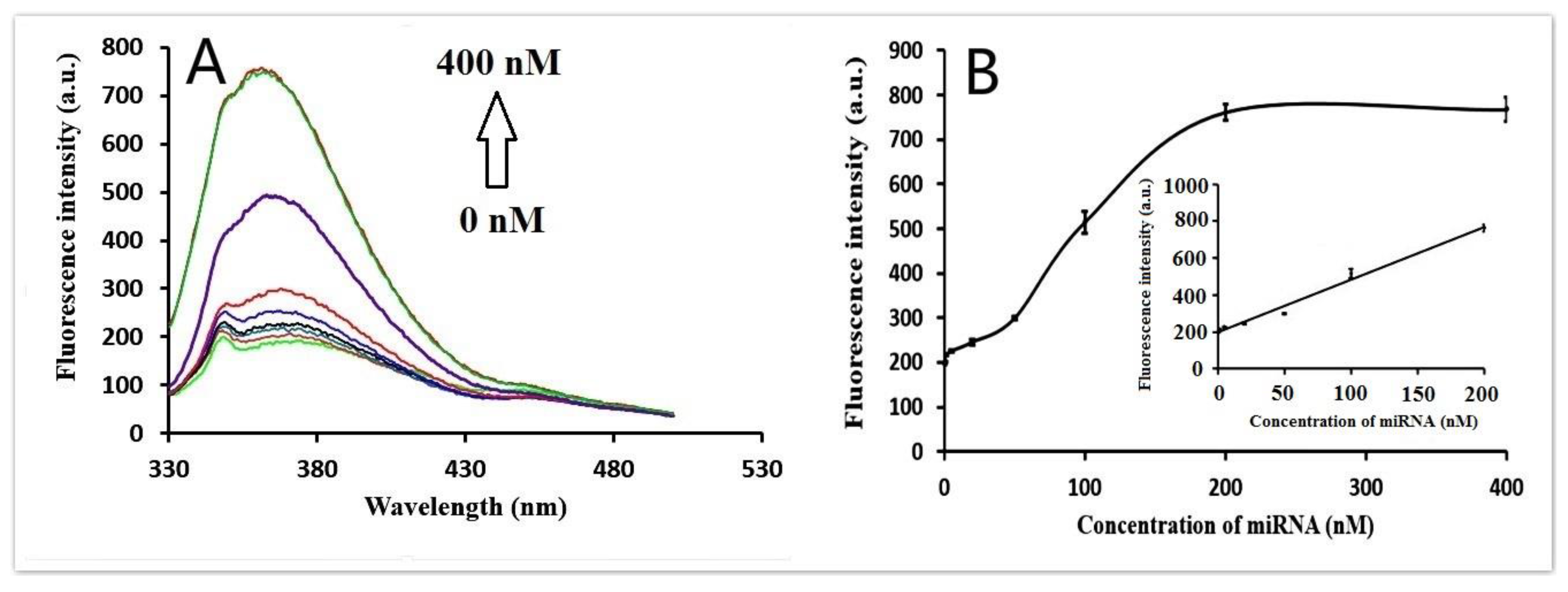
Figure 5.
The proposed method is highly selective for target miRNA detection. Respectively 800 nM of miRNA-141 and let-7d were used in the presence of 200 nM HP and 20 U/mL exo I. The concentration of miRNA-21 was 200 nM. “Blank” is the sample without miRNA. Error bars were estimated from three replicate measurements.


Table 1.
Recovery of miRNA in diluted human serum using the proposed method.
| Sample | Initial Concentration of miRNA-21 (nM) | Concentration of Recovered miRNA-21 (nM) | Recovery (%) |
|---|---|---|---|
| 1 | 50 | 44.5 | 89 |
| 2 | 100 | 104.4 | 104.4 |
| 3 | 200 | 198.8 | 99.4 |
© 2017 by the authors. Licensee MDPI, Basel, Switzerland. This article is an open access article distributed under the terms and conditions of the Creative Commons Attribution (CC BY) license (http://creativecommons.org/licenses/by/4.0/).
Share and Cite
MDPI and ACS Style
Ma, C.; Liu, H.; Wu, K.; Chen, M.; Zheng, L.; Wang, J. An Exonuclease I-Based Quencher-Free Fluorescent Method Using DNA Hairpin Probes for Rapid Detection of MicroRNA. Sensors 2017, 17, 760. https://doi.org/10.3390/s17040760
AMA Style
Ma C, Liu H, Wu K, Chen M, Zheng L, Wang J. An Exonuclease I-Based Quencher-Free Fluorescent Method Using DNA Hairpin Probes for Rapid Detection of MicroRNA. Sensors. 2017; 17(4):760. https://doi.org/10.3390/s17040760
Chicago/Turabian StyleMa, Changbei, Haisheng Liu, Kefeng Wu, Mingjian Chen, Liyang Zheng, and Jun Wang. 2017. "An Exonuclease I-Based Quencher-Free Fluorescent Method Using DNA Hairpin Probes for Rapid Detection of MicroRNA" Sensors 17, no. 4: 760. https://doi.org/10.3390/s17040760
Note that from the first issue of 2016, this journal uses article numbers instead of page numbers. See further details here.