Next Article in Journal
Design and Application of a High Sensitivity Piezoresistive Pressure Sensor for Low Pressure Conditions
Next Article in Special Issue
Fluorescence-Based Bioassays for the Detection and Evaluation of Food Materials
Previous Article in Journal
High-Temperature Dielectric Properties of Aluminum Nitride Ceramic for Wireless Passive Sensing Applications
Previous Article in Special Issue
Analysis of a Lipid/Polymer Membrane for Bitterness Sensing with a Preconditioning Process
 
 
Erratum published on 25 April 2017, see Sensors 2017, 17(5), 945.
Article

Novel PCR Assays Complement Laser Biosensor-Based Method and Facilitate Listeria Species Detection from Food

1
Molecular Food Microbiology Laboratory, Department of Food Science, Purdue University, West Lafayette, IN 47907, USA
2
Department of Food Science and Technology, College of Agriculture and Life Sciences, Chonbuk National University, Jeonbuk 561756, Korea
3
Department of Comparative Pathobiology, Purdue University, West Lafayette, IN 47907, USA
*
Author to whom correspondence should be addressed.
Present address: CROUS de Dijon, Dijon Cedex 21012, France.
These authors contributed equally to this study.
Sensors 2015, 15(9), 22672-22691; https://doi.org/10.3390/s150922672
Submission received: 4 June 2015 / Accepted: 1 September 2015 / Published: 8 September 2015
(This article belongs to the Special Issue Sensors for Food Safety and Quality)

Abstract

:
The goal of this study was to develop the Listeria species-specific PCR assays based on a house-keeping gene (lmo1634) encoding alcohol acetaldehyde dehydrogenase (Aad), previously designated as Listeria adhesion protein (LAP), and compare results with a label-free light scattering sensor, BARDOT (bacterial rapid detection using optical scattering technology). PCR primer sets targeting the lap genes from the species of Listeria sensu stricto were designed and tested with 47 Listeria and 8 non-Listeria strains. The resulting PCR primer sets detected either all species of Listeria sensu stricto or individual L. innocua, L. ivanovii and L. seeligeri, L. welshimeri, and L. marthii without producing any amplified products from other bacteria tested. The PCR assays with Listeria sensu stricto-specific primers also successfully detected all species of Listeria sensu stricto and/or Listeria innocua from mixed culture-inoculated food samples, and each bacterium in food was verified by using the light scattering sensor that generated unique scatter signature for each species of Listeria tested. The PCR assays based on the house-keeping gene aad (lap) can be used for detection of either all species of Listeria sensu stricto or certain individual Listeria species in a mixture from food with a detection limit of about 104 CFU/mL.

Graphical Abstract

1. Introduction

Listeria monocytogenes, a foodborne pathogen, causes fatal systemic infection in immunocompromised hosts including the elderly, infants, pregnant women and their fetuses, HIV infected patients, and patients with malignancy receiving chemotherapy. Alcohol acetaldehyde dehydrogenase (Aad) in L. monocytogenes is a house-keeping enzyme and is involved in bacterial adhesion and paracellular translocation through epithelial barrier during intestinal phase of listeriosis [1,2,3,4]. Such a housekeeping enzyme with moonlighting function in prokaryotes plays an important role in pathogenesis [5,6]. The Aad (Lmo1634) is also known as Listeria adhesion protein (LAP) and its homolog is present in all species of Listeria sensu stricto (i.e., in the narrow or strict sense) also known as archetypal Listeria species (L. monocytogenes, L. ivanovii, L. seeligeri, L. welshimeri, L. innocua, and L. marthii) [1,7,8]. Whereas, L. floridensis, L. aquatic, L. cornellensis, L. riparia, L. grandensis, L. booriae, L. rocourtiae, L. newyorkensis, L. weihenstephanensis, L. fleischmannii and L. grayi [8,9] are considered atypical (sensu lato: in the broad sense) and these group are phylogenetically divergent from the species of Listeria sensu stricto [9,10,11,12].
L. monocytogenes is pathogenic to humans and is responsible for fatal outbreaks involving ready-to-eat meat, dairy, fish, fruits, and vegetables [13]. It was responsible for 57 cases (22 fatalities) from consumption of tainted meat products in Canada [14], 27 cases (8 fatalities) from Quargel sour milk curd cheese [15], 147 cases (33 fatalities) from cantaloupe [16], and most recently in 2015, 35 cases (7 deaths) from caramel apple [17] and 10 cases (3 deaths) from ice cream [18]. The case-fatality rate for listeriosis is 20%–30% [19]. Under the United States Food and Drug Administration (FDA) definition of Current Good Manufacturing Practice, cGMP [21 CFR 110.5(a)], it is mandatory to monitor food for adulterations [21 U.S.C 342(a)] including all poisonous or deleterious substances, which may render food injurious to health. The FDA recommends initial rapid screening of frozen or refrigerated ready-to-eat (RTE) food products for Listeria species rather than the lengthy specific test for L. monocytogenes [20].
In this study, species of Listeria sensu stricto-specific PCR primer sets targeting lap (aad), a house-keeping gene were developed that detected all species of Listeria sensu stricto tested. House-keeping genes are integral and essential for bacterial metabolic function and survival [21], thus they provide an attractive target for detection. This molecular assay based on lap could be used as a screening tool to address the needs of food safety and the regulatory agency. These PCR primer sets were further used to detect Listeria species from inoculated food samples. In addition, the light scattering sensor, BARDOT (bacterial rapid detection using optical scattering technology) [22,23,24] was also employed to verify the presence of L. monocytogenes and L. innocua from a mixed culture (Listeria plus Lactobacillus casei and Escherichia coli O157:H7) inoculated food samples. In BARDOT, a red-diode laser (635 nm; 1 mW; 1 mm diameter) passes through the center of a bacterial colony on an agar plate and generates a 2-dimensional forward scatter fingerprint of each colony within 3–5 s [23]. Organism-specific features are extracted from scatter patterns and are used to identify unknown bacteria using the scatter image library [25]. Scatter image libraries for the thirteen serotypes of L. monocytogenes (1/2a, 1/2b, 1/2c, 3a, 3b, 3a, 4a, 4b, 4ab, 4c, 4d, 4e and 7) were also developed for the BARDOT-based detection in future studies.

2. Experimental Section

2.1. Bacterial Cultures, Growth and Ribotyping

All bacterial cultures (Table S1) used in this study are from our collection. All cultures were stored at −80 °C as 10% frozen glycerol stocks, and fresh cultures were obtained by propagating in Brain Heart Infusion broth (BHI) or Tryptic soy broth with 0.6% yeast extract (TSB-YE) at 37 °C for 16–18 h, with the exception of L. rocourtiae, which was grown at 32 °C. The bacterial cultures were plated on Brain Heart Infusion Agar (BHIA), Luria-Bertani Agar (LBA) to capture the colony scatter patterns. The majority of ribopattern information for cultures was obtained from a previous study from our lab [26]. Additional cultures were ribotyped using the automated Riboprinter® (Qualicon, Inc., Wilmington, DE, USA) as described in our previousstudy [27]. For the food sample study, Fraser Broth (FB) containing 10 mL antimicrobial supplement (25 mg acriflavin, 20 mg nalidixic acid and 500 mg ammonium ferric citrate per liter) was used. Dehydrated media or media components were purchased from BD (Sparks, MD, USA) and FB was purchased from Acumedia (Neogen, Lansing, MI, USA).

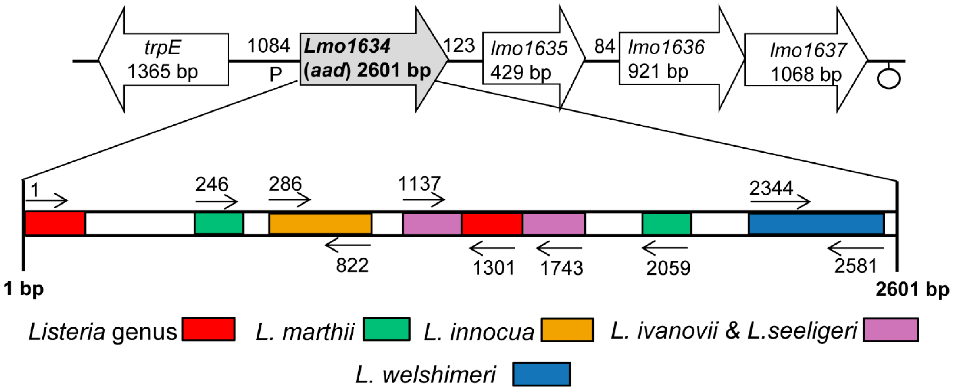
2.2. Design of Lap Gene-Specific Primer Sets for Listeria Species

The lap sequences in L. monocytogenes F4244 (Acc. No. AY561824), L. innocua F4248 (Acc. No. AY561825), L. welshimeri ATCC35897 (Acc. No. AY561828), L. seeligeri SE31 (Acc. No. AY561827) and L. ivanovii SE98 (Acc. No. AY561826) were reported previously [1]. In addition, the complete sequences of the lap gene from L. monocytogenes EGD (Acc. No. NC_003210), L. innocua CLIP11262 (Acc. No. NC003212) and L. marthii (Acc. No. NZ_CM001047) [28] were obtained from NCBI GenBank [29]. To identify a species-specific DNA sequence region, the MultAlin [30] program was used to align and compare the sequences of the lap gene. The scheme for Listeria genus/species-specific primer binding sites on the lap gene are represented in Figure 1.
Two conserved sequence regions (1–54 and 1294–1401) were found in different Listeria species (L. monocytogenes EGD, F4244; L. innocua CLIP11262, F4248; L. welshimeri; L. ivanovii; L. seeligeri and L. marthii), and these regions were used to design the species of Listeria sensu stricto-specific primer set, ELAP-F2 and LIS-R1 (Table 1). Other Listeria species-specific primer sets were developed based on the rule that the 3′-end of primer should be unique to the target species. L. innocua-specific primers are designated as Inn-F1 and Inn-R1, and L. welshimeri-specific primers are named as Wel-F1 and Wel-R1. A primer set IvaSee-F1 and IvaSee-R1 was specific for both L. ivanovii and L. seeligeri as they represent close genetic relatedness [31]. In addition, primers Mar-F1 and Mar-R3 were specific for L. marthii. Specific primers for either L. monocytogenes, L. ivanovii or L. seeligeri could not be obtained, possibly due to their highly conserved lap gene sequence motifs [1].
Figure 1. Schematic representation of the lap (aad) gene-specific primer binding sites for PCR-based detection of Listeria. Open block arrow represents genes flanking the lap. Abbreviations represents (similar proteins to): trpE, anthranilate synthase alpha subunit; lmo1634, alcohol-acetaldehyde dehydrogenase (LAP); lmo1635, unknown protein; lmo1636, ABC transporter (ATP-binding protein); lmo1637, membrane protein. The Listeria lap primers were specific for all species of Listeria sensu stricto tested, but not the atypical listeriae; L. grayi and L. rocourtiae. Colored boxes indicate primer binding sites for Listeria species (see Table 1 for PCR product size).
Sensors 15 22672 g001
Table 1. Sequences of species-specific primers based on Lap sequence used in this study.
PrimerSequence aLocation in Lap GeneProduct Size (bp)Specificity
ELAP-F15′CGGTCCCCGGGTACCATGGCAATTAAAGAAAATGCGGCC3′1–13011301Listeria spp. (except L. grayi, L. rocourtiae)
LIS-R15′TTTGTGATACAGAGTTTTTACC3′
Inn-F15′GGAGTTATTAACGAAGATACT3′286–822536L. innocua
Inn-R15′TTCTGCTTTTACTTCTTTAGCA3′
IvaSee-F15′AAGCTGCAGTTATTCATTCC3′1137–1743606L. ivanovii,L. seeligeri
IvaSee-R15′ATCTAAGAATTTTTGTTTTAGT3′
Wel-F15′TTCTCGTATTATCGGTTTACCA3′2344–2581237L. welshimeri
Wel-R15′GCTTCAAGATAGATTTCTTTCAA3′
Mar-F15′AGAATATATTTGGAACAGCATC3′246–20591813L. marthii
Mar-R15′GTTCGATTGCACGGATGGAAAG3′
a Underlining indicates artificial nucleotide addition sites; translation start codon is indicated in bold.

2.3. PCR Conditions, Primers and DNA Extraction

For PCR, 100 ng of template DNA, 25 pmol of each primer, 0.2 μL of GoTaq polymerase (5 U/μL stock; Promega), 1× GoTaq flexi colored buffer (5x stock, Promega), 2 mmol/L MgCl2 (25 mmol/L stock, Promega), and 200 µM of dNTPs (10 mmol/L stock, Promega) were mixed for a 25 µL final volume. PCR amplification was done using a thermocycler (GeneAmp PCR System 9700, Applied Biosystems) as follows: Hot start at 95 °C for 5 min; 30 cycles with denaturation at 95 °C for 1 min, annealing at 54 °C for 1 min, and extension at 72 °C for 1.5 min; final extension at 72 °C for 10 min. The amplified DNA was resolved in 1.2% agarose gel and visualized by ethidium bromide staining with a ChemiDoc XRS gel documentation system (Bio-Rad). The species of Listeria sensu stricto-specific primer set and individual Listeria species-specific primer sets (Table 1) were used for identification of each Listeria species in a pure culture and in the model foods. List of primers and their binding locations on lap gene is presented in Table 1. To further verify the lap-gene specific PCR results, two sets of cell wall hydrolase; CWH or p60 (iap) gene-specific primers, Lis1B/MonoA and Lis1B/Ino2 [32], were applied to verify L. monocytogenes and L. innocua cultures, respectively. The sequence of Lis1B, MonoA and Ino2 primers are 5′-TTATACGCGACCGAAGCCAAC-3′, 5′-CAAACTGCTAACACAGCTACT-3′ and 5′-ACTAGCACTCCAGTTGTTAAAC-3′, respectively.
The genomic DNA from reference cultures or enriched food samples were extracted with DNeasy Tissue Kit (Qiagen) following manufacturer’s protocol. Briefly, the cultures were pretreated with lysozyme solution (10 mg/mL in TE buffer (pH 7.0) containing 10 mmol/L Tris-Cl, pH 7.0 and 1 mmol/L EDTA) at 37 °C for 30 min prior to cell lysis. The total DNA was also extracted following the published protocol [33]. The concentration and purity of genomic DNA was determined using NanoDrop 2000C (Thermo Scientific, Franklin, MA, USA).

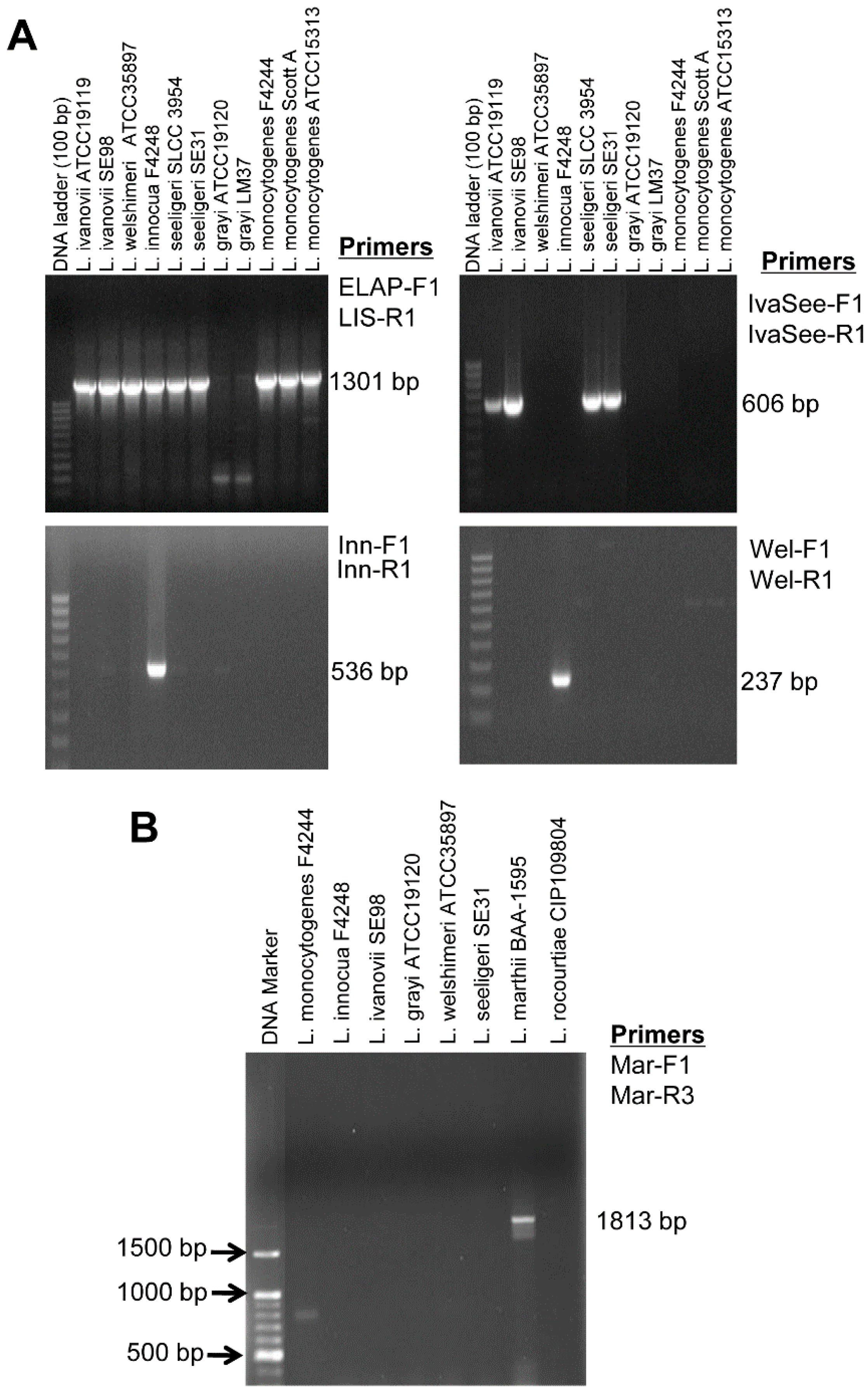
2.4. Specificity and Sensitivity of Lap Gene Primers for Listeria Detection

A total of 55 Listeria (n = 47) and non-Listeria (n = 8) cultures were tested to determine the specificity of lap gene primer sets for the species of Listeria sensu stricto or individual species: L. monocytogenes (n = 13), L. ivanovii (n = 12), L. innocua (n = 10), L. seeligeri (n = 5), L. welshimeri (n = 3), L. grayi (n = 2), L. marthii (n = 1) and L. rocourtiae (n = 1). Non-Listeria cultures included Enterobacter aerogenes, Serratia marcescens, Hafnia alvei, Lactobacillus casei, Lactobacillus acidophilus, Bacillus cereus, Escherichia coli O157:H7, and Salmonella enterica serovar Typhimurium (Table S1).
To determine the sensitivity (limit of detection) of the lap gene primer sets for detection of Listeria species, pure cultures of L. monocytogenes F4244 and L. innocua F4248 cells were plated on modified oxford (MOX) agar for enumeration. Simultaneously, total DNA was extracted in 200 µL of PBST (20 mM phosphate buffered saline (PBS), pH 7.2, with 0.05% Tween 20) from 1 mL of pure cultures using the boiling method [33]. A relationship between the number of cells and the corresponding DNA/genomic equivalents was established before performing the lap gene-based PCR to establish the sensitivity of the reaction. Overnight (16 h) grown cells of L. monocytogenes F4244 (8.02 ± 0.11 log10 CFU/mL) yielded 96.1 ± 2.1 ng/µL DNA, and 1 ng DNA was equivalent to 5.5 log10genomic equivalent (GE), whereas L. innocua F4248 (8.15 ± 0.24 log10 CFU/mL) yielded 98.4 ± 0.6 ng/µL DNA, and 1 ng DNA was equivalent to 5.5 log10 GE. PCR was performed with the diluted DNA and “GE was calculated. The genome size of L. monocytogenes and L. innocua 2.9 and 3.0 Mbp, “respectively, thus yielded 5.5 log10 GE for 1 ng of DNA. The amplified PCR products obtained from different cell concentrations were quantified using the NIH ImageJ tool, an image processing and analysis software [34].

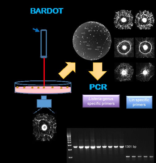
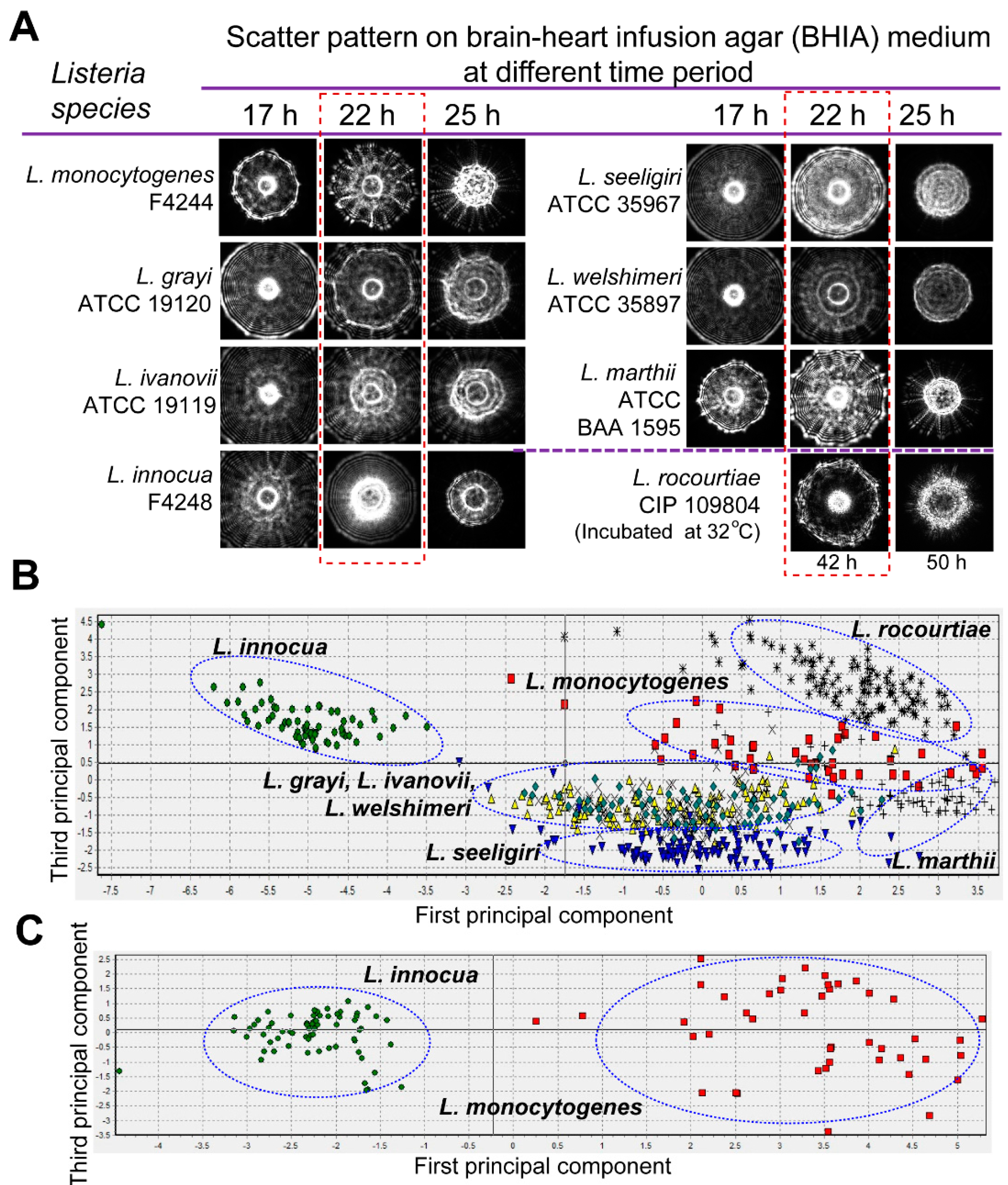
2.5. Laser Optical Sensor and Scatter Image Analysis

Laser optical sensor, also designated BARDOT, works on the biophysical principles (refraction, diffraction, interference) of forward light scattering. An external design of BARDOT and its internal scheme has been described previously [23,35]. The detection time (sample-to-result) for the BARDOT-based detection of Listeria spp. colonies on BHI agar plate (BHIA) is about 22 h except for L. rocourtiae, which took about 48 h to generate the colony scatter pattern.
To find the optimal incubation time that generates maximal scatter features and distinguishing scatter patterns, a time-lapse study was performed to capture the scatter pattern of Listeria species at 17, 22 and 25 h. Scatter patterns were acquired for Listeria colonies after plating on BHIA, and each colony (~1 mm diameter) contained about 2.5 × 10⁸ Listeria cells. The scatter patterns were captured when the colony size reached close to 1 mm in diameter. A total of 1,884 scatter images from pure cultures of eight Listeria species were captured on BHIA, where 677 scatter patterns were used to build the scatter image library and the rest of the scatter patterns were generated to find the optimal incubation time [24]. The scatter image library of eight Listeria species (L. monocytogenes, L. innocua, L. grayi, L. seeligeri, L. welshimeri, L. marthii, L. ivanovii, and L. rocourtiae) consisted of an average of 80 scatter patterns per species. This Listeria species library was used to differentiate L. monocytogenes and L. innocua inoculated in the food sample. Another scatter image library of L. monocytogenes and L. innocua (110 scatter images) was also built to specifically differentiate the two species. The scatter images were further processed and analyzed using a built-in image analysis software [25]. This analysis generated the cross validation matrix for the principal component analysis of the scatter images of the Listeria species. Scatter image libraries for 13 serovars of L. monocytogenes were also generated after growth on BHIA or LB agar (LBA) plates.

2.6. Detection and Identification of Listeria in Artificially Inoculated Food Samples

Two types of food samples, ready-to-eat hotdogs (franks) and cantaloupes, were procured from a local grocery store (West Lafayette, IN, USA). To test the application of designed primer sets, food samples were artificially inoculated and tested in three independent experimental replicates. Twenty-five grams of each hotdog and cantaloupe rinds (each piece was about 2 × 3 cm) were artificially inoculated with 100 CFU of single culture (L. monocytogenes F4244 or L. innocua F4248) or 100 CFU of a mixed culture (50 CFU of L. monocytogenes F4244 and 50 CFU of L. innocua F4248). Since we did not find any background microbial load in hotdogs, samples were inoculated with Lactobacillus casei (100 CFU/25g) and Escherichia coli (100 CFU/25g) as background contaminants. Four sets of food samples: (i) food alone; food inoculated with (ii) L. monocytogenes; (iii) L. innocua; and (iv) L. monocytogenes and L. innocua, were enriched in FB according to the USDA-FSIS protocol [36]. All inoculated food samples (25 ± 2 g) were enriched in 225 ± 2.5 mL of FB at 37 °C in a shaking incubator (140 rpm) for 24 h. One milliliter of enriched broth was used for the DNA extraction (as described before) and plated on MOX agar plates for enumeration and BHIA for identification by BARDOT [23,37]. Briefly, for BARDOT analysis, FB enriched samples were decimally diluted in 20 mmol/L PBS (pH 7.2), plated on BHIA, and incubated at 37 °C for 24 h or until the colony diameter reached 1.1 ± 0.2 mm. The scatter patterns of colonies were compared with the scatter pattern library of Listeria species for identification [23,24].

3. Results

3.1. Specificity and Sensitivity of Lap Gene Primers for Listeria Detection

A total of 55 different Listeria (n = 47) and non-Listeria (n = 8) cultures were analyzed (Table S1). When the general Listeria primer set, ELAP-F2 and LIS-R1, was used, all the tested species of Listeria sensu stricto produced a 1301 bp band (Figure 2A, Table S1). When the L. innocua-specific primer set, Inn-F1 and Inn-R1, was used, a 536 bp band was amplified only in the L. innocua strains (Table S1) Likewise, the IvaSee-F1 and IvaSee-R1 primer set produced L. ivanovii and L. seeligeri-specific 606 bp band. The primer set, Wel-F1 and Wel-R1, generated 237 bp band only in the L. welshimeri strains, and a L. marthii-specific primer set, Mar-F1 and Mar-R3, produced a 1813 bp band without showing any amplified products from the other Listeria species (Figure 2B, Table S1).
Figure 2. Representative agarose gel showing PCR amplification of selected Listeria species by using species-specific primers. (A) PCR results based on the species of Listeria sensu stricto-specific, and L. innocua-, L. ivanovii- and L. seeligeri- and L. welshimeri-specific primers. (B) PCR result using L. marthii-specific primers.
Sensors 15 22672 g002
These results demonstrated that the general Listeria primer set (ELAP-F2 and LIS-R1) could detect all the tested species of Listeria sensu stricto. The IvaSee-F1 and IvaSee-R1 were able to differentiate either L. ivanovii or L. seeligeri from other Listeria species or non-Listeria organisms without giving any false-positive results. L. ivanovii and L. seeligeri contain virulence gene sequences in their genome similar to L. monocytogenes [31], and exhibited high sequence homology in lap [1].
The L. innocua-specific primers (Inn-F1 and Inn-R1) were highly specific and did not give any PCR products with other Listeria species, including L. monocytogenes strains. This primer set could be used to detect L. innocua as a mixed culture with L. monocytogenes. Since these two species are usually found together in food and other ecological habitats, the presence of L. innocua could be used as an indicator for L. monocytogenes [38,39,40,41].
Use of the L. welshimeri-specific primer set, Wel-F1 and Wel-R1, successfully produced a PCR product in all four L. welshimeri strains tested. Since we could not design L. monocytogenes, L. ivanovii or L. seeligeri-specific primer sets, PCR assays for detection of these individual species were not possible with the primers sets used in this study.
Since 2010, eleven new Listeria species (a total of 17 species) were added to the genus Listeria; L. marthii [42], L. rocourtiae [11], L. weihenstephanensis [12], L. fleischmannii [10,43], L. floridensis, L. aquatic, L. cornellensis, L. riparia, L. grandensis [8], L. booriae, and L. newyorkensis [9]. The presence of a lap homologue in L. marthii [7] has been reported and the resulting primer set (Mar-F1 and Mar-R3) is specific, but we were unable to obtain any lap gene based primers for L. rocourtiae, L. weihenstephanensis, L. fleischmannii and L. grayi. These are considered atypical and are phylogenetically divergent from the species of Listeria sensu stricto within the genus Listeria [1,11,43]. L. grayi and L. rocourtiae did not give any amplification with the species of Listeria sensu-stricto-specific primer set indicating the possible sequence heterogeneity in the lap sequence in these atypical listeriae (Table S1).
The specificity of all primer sets was examined with eight non-Listeria cultures and none of them yielded any PCR product (Table S1). We were even able to identify four mislabeled microorganisms: Two with general Listeria primer set, one of each with L. innocua-specific and L. welshimeri-specific primer set (Table S1). Ribotyping identified them as L. monocytogenes DUP-1035 and DUP-1039, L. welshimeri DUP-1074 and L. innocua DUP-1009 (Table S1).
The detection limit (sensitivity) of PCR with the species of Listeria sensu stricto-specific primer (ELAP-R1/LIS-R1) was 4.5 log10 genome equivalents for both L. monocytogenes and L. innocua (Figure 3). PCR for the DNA sensitivity was performed with the total DNA extracted from 1 mL culture of L. monocytogenes F4244 (8.02 ± 0.11 log10 CFU/mL) and L. innocua F4248 (8.15 ± 0.24 log10 CFU/mL) that also indirectly depicted the PCR sensitivity for the bacterial cell number. One milliliter cultures of L. monocytogenes and L. innocua yielded 96.1 ± 2.1 ng/µL and 98.4 ± 0.6 ng/µL of DNA concentrations, respectively. The genome equivalents were calculated from the genome size (L. monocytogenes size is 2.9 × 10⁶ bp and L. innocua is 3.0 × 10⁶ bp), and the molecular weight of nucleotide (1 bp = 650 Da).
Figure 3. Sensitivity of lap gene-based Listeria species sensu stricto-specific primer (ELAP-F1/LIS-R1) tested against L. monocytogenes F4244 and L. innocua F4248. Agarose gel showing amplifications with the primers for different concentrations of template DNA of L. monocytogenes and L. innocua in reaction volume of 25 µL. PCR products in the gel were quantified using the NIH ImageJ image processing and analysis software.
Sensors 15 22672 g003

3.2. Scatter Image Library of Listeria Species and Serovars
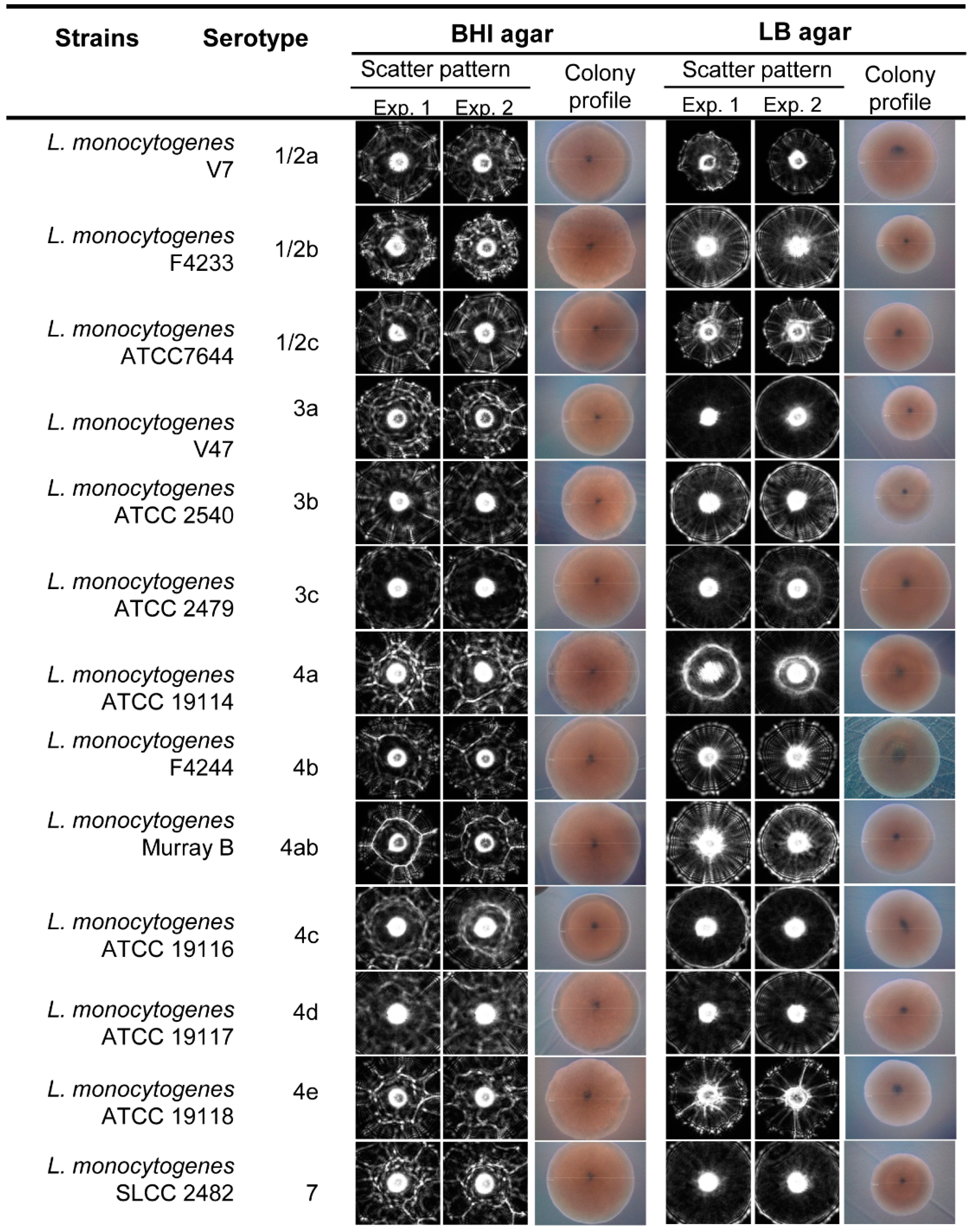
The light scattering sensor (BARDOT) generated distinguishing forward scattering patterns for colonies of Listeria species on BHIA. Time-lapse measurement of the scatter patterns indicated that Listeria species generated scatter patterns with maximal differential scatter features at 22 h of incubation (Figure 4A). Principal component analysis performed on the basis of cross validation matrix revealed that L. innocua, L. rocourtiae, L. monocytogenes, L. marthii, and L. seeligeri can be grouped separately based on the differences in the scatter patterns (Figure 4B) with 100%, 100%, 97.7%, 95.2% and 94.9%, positive predictive value (PPV), also known as classification accuracy, respectively (Table S2). However, L. grayi, L. ivanovii, and L. welshimeri could not be differentiated based on the scatter patterns on BHIA. Application of the L. monocytogenes and L. innocua-specific image libraries, generated even higher PPVs of 100% for both of the species, and they grouped separately in the principal component analysis (Figure 4C). These image libraries were also used to match scatter images of L. monocytogenes and L. innocua that were obtained from artificially inoculated food samples mentioned below in the result section.
Figure 4. Optical scatter patterns of Listeria species and image analysis. (A) Colony scatter patterns were captured using BARDOT at different incubation times for eight Listeria species on BHI agar plates. Rectangular selection with broken line depicts the optimal incubation time (22 h) that yielded differentiating scatter images when the colony size was 1.1 ± 0.2 mm diameter; (B) Principal component analysis of the eight Listeria species used to build the scatter image library. Blue oval selections indicate grouping of the Listeria species; (C) Principal component analysis of L. monocytogenes and L. innocua colony scatter images that were used to build a two-species scatter image library. The blue oval selections indicate grouping of each Listeria species.
Sensors 15 22672 g004
In this study we also tested the capabilities of the laser sensor to differentiate L. monocytogenes at the serovar level after growth on BHI and LB agar plates (Figure 5). Differences at the serovar level were observed after analysis using the cross validation matrix, where a high PPV average was observed on LB agar compared to the BHI agar media, 90.1% and 82.9%, respectively (Table 2). Scatter pattern analysis for the thirteen serotypes underscores the feasible application of the laser optical sensor to generate a scatter image library with differentiating scatter patterns for L. monocytogenes serotypes that can be used for screening and detection of L. monocytogenes at the serovar level.
Figure 5. Representative scatter images of colonies of L. monocytogenes serotypes grown on BHI and LB agar. Between 50 and 100 colony scatter images for each serovar were collected from each experiment. Colony profiles were measured under phase contrast microscope with 10× objective when the colony size was 1.1 ± 0.2 mm diameter on BHI and LB agar after 21–23 h and 25–27 h of incubation, respectively.
Sensors 15 22672 g005
Table 2. Positive predictive value (PPV, precision rate) for the scatter images of colonies obtained from thirteen L. monocytogenes serotypes grown on BHIA and LBA media.
StrainsSerotype% Average Positive Predictive Value (PPV ± SD)
BHILB
L. monocytogenes V71/2a81.8 ± 2.396.2 ± 1.9
L. monocytogenes F42331/2b89.4 ± 1.196.8 ± 2.1
L. monocytogenes ATCC76441/2c74.5 ± 3.281.3 ± 3.8
L. monocytogenes V473a80.8 ± 1.877.6 ± 5.6
L. monocytogenes ATCC 25403b74.2 ± 2.297.6 ± 1.7
L. monocytogenes ATCC 24793c99.8 ± 0.985.2 ± 2.3
L. monocytogenes ATCC 91144a93.2 ± 1.398.0 ± 1.8
L. monocytogenes F42444b46.2 ± 3.588.8 ± 2.1
L. monocytogenes Murray B4ab92.8 ± 2.985.4 ± 3.2
L. monocytogenes ATCC 191164c98.8 ± 1.093.6 ± 2.8
L. monocytogenes ATCC 191174d99.0 ± 0.588.2 ± 3.1
L. monocytogenes ATCC 191184e65.4 ± 5.692.6 ± 1.7
L. monocytogenes SLCC 2482782.4 ± 2.490.2 ± 3.1
Average precision rate82.9 ± 2.290.1 ± 2.7

3.3. Detection and Verification of Listeria from Food Samples
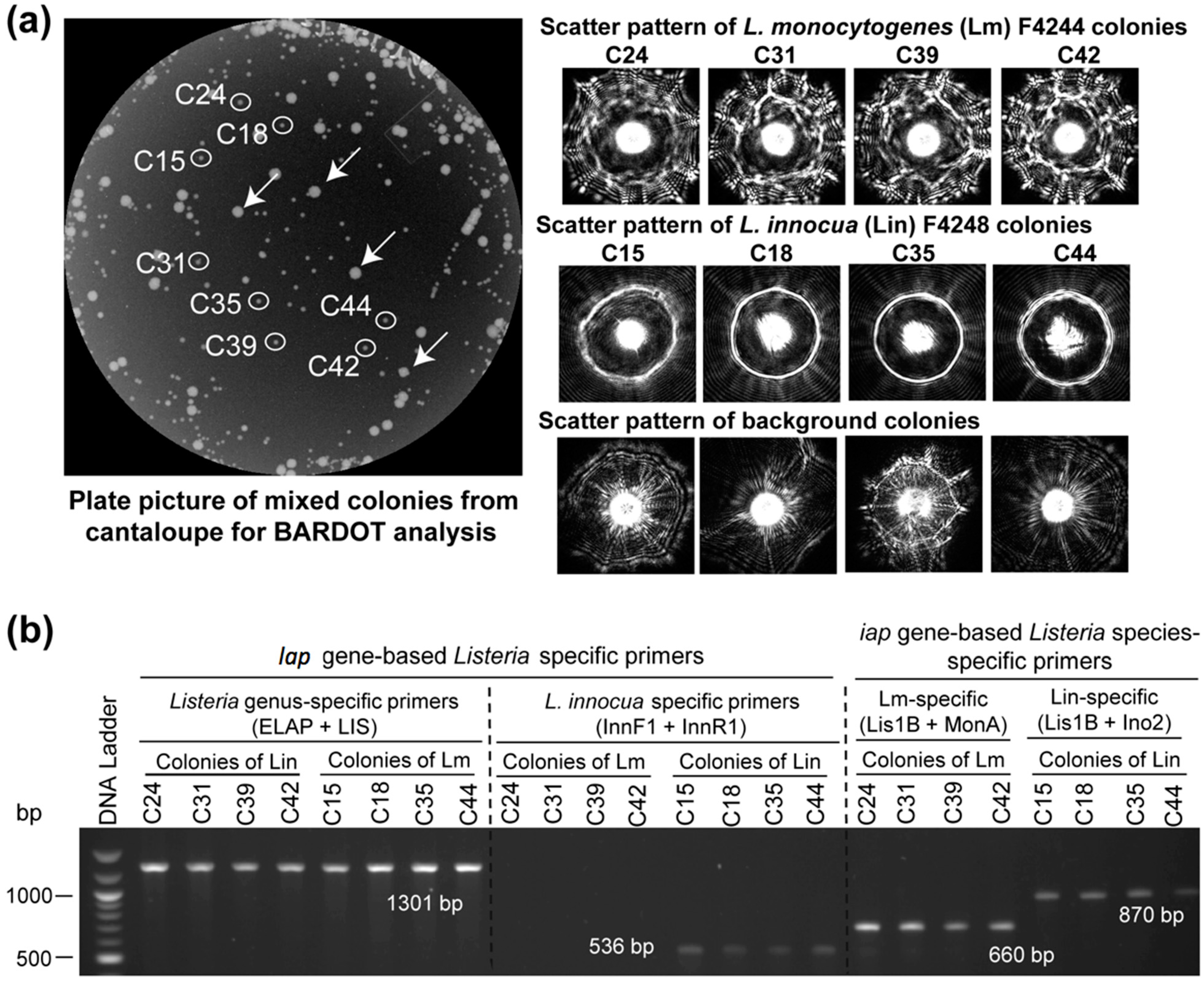
The ability of lap gene-specific primer sets to detect L. monocytogenes and L. innocua from inoculated food samples were verified (Figure S1, Table 3). Since a lap gene-based L. monocytogenes specific primer set could not be designed, we used the combination of species of Listeria sensu stricto-specific and L. innocua specific primer sets to detect Listeria from food. The food samples that revealed positive amplification with ELAP-F1 & LIS-R, but did not show any amplification with the L. innocua-specific primer sets (Inn-F1 & Inn-R1), were considered to contain any Listeria spp. other than the L. innocua (Table 3, Figure S1). Samples with positive amplification for both the primer sets (ELAP-F1 & LIS-R1 and with Inn-F1 & Inn-R1) corroborated the presence of L. innocua in the food sample (Table 3). Background microbial colonies obtained from the un-inoculated cantaloupe did not result in any positive amplification (Figure 6). This highlights the specificity and applicability of lap gene-specific primers for detection of Listeria species even in food samples with background microbiota. We further verified these results by analyzing the enriched food samples by BARDOT.
In our previous study, BARDOT generated distinct signature scatter patterns for the colonies of L. monocytogenes or L. innocua in mixed culture [23]. The distinctive scatter patterns generated with BARDOT facilitated accurate identification of L. monocytogenes or L. innocua or both in food samples after matching the scatter patterns with the respective image libraries (Figure 6). Colonies # C42, C41, C43, C34, and C29 originated from L. monocytogenes and L. innocua-inoculated hotdog sample on BHIA were identified as L. innocua, while colonies # C28, C25, C23, and C5 were identified as L. monocytogenes after comparing scatter images with the library (Figure 6A). These colonies were initially identified as Listeria spp. by PCR with the primer set (ELAP-F1/LIS-R1) designed in this study (Figure 6B). Further, these colonies were also confirmed at the species level (L. monocytogenes and L. innocua) using the primers for the iap gene [32,44]. The scatter patterns of L. innocua, when matched with the libraries of Listeria species as well as L. monocytogenes and L. innocua, generated 100% match with scatter image library. L. monocytogenes colonies from the artificially inoculated hotdog sample revealed low PPV (<80%) when matched with the libraries of Listeria species; however, the same L. monocytogenes colony scatter pattern generated a high PPV (>90%) when matched with the L. monocytogenes and L. innocua library. The low PPV of L. monocytogenes with the Listeria species library could be attributed to the overlapping pattern of L. monocytogenes with the scatter pattern of other Listeria species. BARDOT-based identification of L. monocytogenes and L. innocua colonies along with PCR analysis with the lap and iap gene-specific primers resulted in 100% and 100% identification, respectively, for both the Listeria species.
Figure 6. Detection and verification of L. monocytogenes (Lm) and L. innocua (Lin) in mixture from inoculated cantaloupe samples with BARDOT and PCR. (A) Enriched cantaloupe samples containing L. monocytogenes and L. innocua were plated on BHI agar and colony scatter patterns were obtained. Scatter patterns were matched against the BARDOT scatter image library for Listeria identification. The white arrows indicate background bacterial colonies from the cantaloupe; (B) BARDOT identified colonies were picked and tested with the primer sets specific for species of Listeria sensu stricto (ELAP-F1/LIS-R1), L. monocytogenes (Lis1B/MonA), and L. innocua (InnF1/InnR1, Lis1B/Ino2) for verification of colonies.
Sensors 15 22672 g006
Table 3. Listeria detection using lap gene-specific primers in food systems.
Treatment aInoculation (CFU/25g)Enrichment Time (h) in Fraser Broth at 37 °CPCR b
HotdogCantaloupe
ELAP-F1/LIS-R1Inn-F1/Inn-R1ELAP-F1/LIS-R1Inn-F1/Inn-R1
Uninoculated024
L. innocua (Lin)10024++++
L. monocytogenes (Lm)10024++
Lin and Lmc10024++++
Lb. casei10024
E. coli10024
a Three independent experiments were performed for each food sample; b DNA extracted from broth enrichment following the published protocol [33] were amplified with the lap gene specific primers for species of Listeria sensu stricto (ELAP-F1/LIS-R1) and L. innocua (Inn-F1/InnR1); c Food samples were inoculated with 50 CFU each of L. monocytogenes (Lm) and L. innocua (Lin) in 25 g of food sample.

4. Discussion

This study reports the application feasibility of primer sets designed from a gene encoding the house-keeping alcohol acetaldehyde dehydrogenase (aad), also known as Listeria adhesion protein (lap), for detection of Listeria at the genus and species level. This highly conserved house-keeping enzyme is involved in the pathogenesis of virulent Listeria but not avirulent Listeria species [1,45] thus providing an attractive target for Listeria detection. The aad (lap) sequence is conserved (97%–98% homology) in the species of Listeria sensu stricto and yielded a primer set (ELAP-F1 and LIS-R1) that detected these Listeria sensu stricto (archetypal) species, -but not the atypical (Listeria sensu lato) listeriae: L. grayi and L. rocourtiae. Even though both L. grayi and L. rocourtiae possess a lap (aad) homolog, they did not give any amplification with the species of Listeria sensu stricto-specific primer set indicating a possible sequence heterogeneity in the lap gene in these atypical listeriae. Indeed, lap gene sequence comparison between L. monocytogenes F4244 (AY561824) and L. grayi DSM 20601 (NZ_GL538352.1) or L. rocourtiae FSL F6-920 c6 (NZ_AODK01000006.1) revealed only 80% homology. The other newly isolated Listeria species were not tested in the PCR assay, but we anticipate negative results for these species since they are genetically similar to the two atypical listeriae tested in this study [8,9]. Furthermore, a minor variation in lap sequences (97%–98% similarity) among the different species of Listeria sensu stricto [1] yielded highly specific primer sets for L. innocua, L. welshimeri, L. marthii, and L. ivanovii and L. seeligeri together, but none for L. monocytogenes (Figure 1, Table S1). These could be useful for specific identification at the species level. Listeria sensu stricto-specific and other species-specific sets of primers also did not amplify any non-listerial bacteria tested with pure cultures or in a food matrix (Tables S1 and 3), highlighting the specificity of the lap gene-specific primer sets for Listeria spp. The detection limit of primer sets with the diluted template DNA revealed an indirect detection limit of about 4.5 log10 genome equivalents for this assay. These primer sets could be used to detect and identify Listeria species during screening of frozen or refrigerated ready-to-eat (RTE) food products as recommended by the FDA [20]. Application of PCR-based assay targeting gene encoding house-keeping enzyme (cell wall hydrolase; CWH or p60 encoded by iap) in Listeria spp. was reported earlier, in which species-specific primer sets successfully detected each Listeria spp.; L. monocytogenes, L. ivanovii, L. innocua, L. seeligeri, L. welshimeri within the genus, except L. grayi [32]. Similarly, PCR assay targeting genes encoding aminopeptidase and fibronectin-binding protein were also used for rapid detection of L. monocytogenes [46,47].
We developed the light scattering sensor, BARDOT, through collaborative efforts with engineers at the Center for Food Safety Engineering at Purdue University [48]. We have successfully used BARDOT to differentiate and detect L. monocytogenes, L. ivanovii, L. innocua, L. seeligeri, L. welshimeri and L. grayi. The BARDOT system was also successfully applied to differentiate L. monocytogenes from other pathogens (Salmonella enterica serovar Enteritidis and Typhimurium, E. coli O157:H7) based on the scatter patterns from artificially inoculated ready-to-eat hotdog, shredded beef, raw ground beef and chicken, frozen and fresh spinach, and fresh tomato [37]. Recently, we have also optimized the BARDOT-based method for detection and screening of several additional foodborne pathogens including Bacillus spp. [49], Campylobacter spp. [50], Salmonella enterica serovars [24], Shiga-toxigenic E. coli [35], and Vibrio spp. [51]. In this study, we used BARDOT to differentiate the species of Listeria when grown on BHIA. On BHIA, BARDOT successfully differentiated L. monocytogenes, L. innocua, L. rocourtiae, L. marthii, and L. seeligeri; however, it did not yield satisfactory differential patterns of L. grayi, L. ivanovii, and L. welshimeri (Figure 4B). In our previous report we have shown that the BHIA and modified Oxford agar prepared without ferric ammonium citrate were able to successfully differentiate the colonies of L. monocytogenes, L. ivanovii, L. innocua, L. seeligeri, L. welshimeri and L. grayi, and colonies of L. monocytogenes from L. innocua, respectively, based on scatter signature patterns [23,44]. These findings reaffirm the media-dependent generation of scatter signatures for bacterial identification.
L. monocytogenes is the primary human pathogen in the genus Listeria and among the 13 serotypes, serotype 1/2a and 4b are responsible for ~75% of all L. monocytogenes related outbreaks. Here we have shown that BARDOT can differentiate L. monocytogenes serovars 1/2a and 4b with high accuracy on LBA with 96.2% ± 1.9% and 88.8% ± 2.1% PPV, respectively. Observed differences in the scatter pattern of different serovars could be attributed to the O (somatic) antigens expressed on the surface of L. monocytogenes [52]. Furthermore, metabolic activity and genomic differences between different species or serotypes of Listeria can also contribute to the differential scatter patterns [37]. The genome size for serotype 1/2a (L. monocytogenes F6854) is 2.97 × 10⁶ bp, with a total of 3028 genes, of which 2963 are protein coding genes. Serotype 4b (L. monocytogenes F2365) has a genome size of 2.91 × 10⁶ bp, with a total of 2933 genes, of which 2848 are protein coding genes. Thus, a difference of 115 protein coding genes in serotype 1/2a to that of 4b could be crucial in generating differential scatter patterns for these two serotypes. In a comparative whole genome sequencing study, it was found that 83 genes were restricted to 1/2a serotype and 51 genes were restricted to 4b serotype [53].

5. Conclusions

In summary, lap gene based Listeria sensu stricto and individual species-specific primers successfully detected all tested species of Listeria sensu stricto (archetypal) while some limitations for individual species level detection. The PCR based assays with the species of Listeria sensu stricto-specific primer sets based on lap and iap genes also successfully detected L. monocytogenes and L. innocua from mixed culture-inoculated food samples, and each bacterium in food was verified by the light scattering sensor that generated unique scatter signature for each species of Listeria. These data emphasize that the BARDOT system could be used to identify Listeria spp. on agar plates from a mixed cultures and may serve as a complimentary tool when testing samples with nucleic acid-based molecular methods.

Acknowledgments

This research was supported through a cooperative agreement with the Agricultural Research Service of the U.S. Department of Agriculture project number 1935-42000-072-02G and the Center for Food Safety Engineering at Purdue University. We thank Valerie Ryan and Taylor Bailey for critical reading of the manuscript.

Author Contributions

Arun K. Bhunia and Kwang-Pyo Kim conceived the idea. Kwang-Pyo Kim, Atul K. Singh, Xingian Bai, and Lena Leprun performed the experiments. Kwang-Pyo Kim, Atul K. Singh and Arun K. Bhunia analyzed the results. Kwang-Pyo Kim, Atul K. Singh and Arun K. Bhunia wrote the manuscript.

Conflicts of Interest

The authors declare no conflict of interest.

References

  1. Jagadeesan, B.; Koo, O.K.; Kim, K.P.; Burkholder, K.M.; Mishra, K.K.; Aroonnual, A.; Bhunia, A.K. LAP, an alcohol acetaldehyde dehydrogenase enzyme in Listeria promotes bacterial adhesion to enterocyte-like Caco-2 cells only in pathogenic species. Microbiology 2010, 156, 2782–2795. [Google Scholar] [CrossRef]
  2. Jagadeesan, B.; Fleishman Littlejohn, A.E.; Amalaradjou, M.A. R.; Singh, A.K.; Mishra, K.K.; La, D.; Kihara, D.; Bhunia, A.K. N-Terminal Gly224–Gly411 domain in Listeria adhesion protein interacts with host receptor Hsp60. PLoS ONE 2011, 6. [Google Scholar] [CrossRef]
  3. Burkholder, K.M.; Bhunia, A.K. Listeria monocytogenes uses Listeria adhesion protein (LAP) to promote bacterial transepithelial translocation, and induces expression of LAP receptor Hsp60. Infect. Immun. 2010, 78, 5062–5073. [Google Scholar] [CrossRef]
  4. Kim, K.P.; Jagadeesan, B.; Burkholder, K.M.; Jaradat, Z.W.; Wampler, J.L.; Lathrop, A.A.; Morgan, M.T.; Bhunia, A.K. Adhesion characteristics of Listeria adhesion protein (LAP)-expressing Escherichia coli to Caco-2 cells and of recombinant LAP to eukaryotic receptor Hsp60 as examined in a surface plasmon resonance sensor. FEMS Microbiol. Lett. 2006, 256, 324–332. [Google Scholar] [CrossRef]
  5. Henderson, B.; Martin, A. Bacterial moonlighting proteins and bacterial virulence. Curr. Top. Microbiol. Immunol. 2013, 358, 155–213. [Google Scholar]
  6. Burkholder, K.M.; Bhunia, A.K. Listeria monocytogenes and Host Hsp60—An invasive pairing. In Moonlighting Cell Stress Proteins in Microbial Infections, Heat Shok Proteins; Henderson, B., Ed.; Springer Science+Business Media: Dordrecht, Germany, 2013; pp. 267–282. [Google Scholar]
  7. Den Bakker, H.C.; Bundrant, B.N.; Fortes, E.D.; Orsi, R.H.; Wiedmann, M. A population genetics-based and phylogenetic approach to understanding the evolution of virulence in the genus Listeria. Appl. Environ. Microbiol. 2010, 76, 6085–6100. [Google Scholar] [CrossRef]
  8. Den Bakker, H.C.; Warchocki, S.; Wright, E.M.; Allred, A.F.; Ahlstrom, C.; Manuel, C.S.; Stasiewicz, M.J.; Burrell, A.; Roof, S.; Strawn, L.K.; et al. Listeria floridensis sp nov., Listeria aquatica sp nov., Listeria cornellensis sp nov., Listeria riparia sp nov and Listeria grandensis sp nov., from agricultural and natural environments. Int. J. Syst. Evol. Microbiol. 2014, 64, 1882–1889. [Google Scholar] [CrossRef]
  9. Weller, D.; Andrus, A.; Wiedmann, M.; den Bakker, H.C. Listeria booriae sp nov and Listeria newyorkensis sp nov., from food processing environments in the USA. Int. J. Syst. Evol. Microbiol. 2015, 65, 286–292. [Google Scholar] [CrossRef]
  10. Bertsch, D.; Rau, J.; Eugster, M.R.; Haug, M.C.; Lawson, P.A.; Lacroix, C.; Meile, L. Listeria fleischmannii sp nov., isolated from cheese. Int. J. Syst. Evol. Microbiol. 2013, 63, 526–532. [Google Scholar] [CrossRef]
  11. Leclercq, A.; Clermont, D.; Bizet, C.; Grimont, P.A.D.; le Fleche-Mateos, A.; Roche, S.M.; Buchrieser, C.; Cadet-Daniel, V.; le Monnier, A.; Lecuit, M.; et al. Listeria rocourtiae sp. nov. Int. J. Syst. Evol. Microbiol. 2010, 60, 2210–2214. [Google Scholar] [CrossRef]
  12. Halter, E.L.; Neuhaus, K.; Scherer, S. Listeria weihenstephanensis sp. nov., isolated from the water plant Lemna trisulca taken from a freshwater pond. Int. J. Syst. Evol. Microbiol. 2013, 63, 641–647. [Google Scholar] [CrossRef]
  13. Silk, B.J.; Date, K.A.; Jackson, K.A.; Pouillot, R.; Holt, K.G.; Graves, L.M.; Ong, K.L.; Hurd, S.; Meyer, R.; Marcus, R.; et al. Invasive listeriosis in the foodborne diseases active surveillance network (FoodNet), 2004–2009: Further targeted prevention needed for higher-risk groups. Clin. Infect. Dis. 2012, 54, S396–S404. [Google Scholar] [CrossRef]
  14. Anonymous. 2008 Listeriosis Outbreak in Ontario Epidemiologic Summary. Available online: http://www.health.gov.on.ca/en/public/publications/disease/docs/listeriosis_outbreak_epi_sum.pdf (accessed on 26 May 2015).
  15. Fretz, R.; Pichler, J.; Sagel, U.; Much, P.; Ruppitsch, W.; Pietzka, A.T.; Stöger, A.; Huhulescu, S.; Heuberger, S.; Appl, G.; et al. Update: Multinational listeriosis outbreak due to “Quargel”, a sour milk curd cheese, caused by two different L. monocytogenes serotype 1/2a strains, 2009–2010. Eurosurveillance 2010, 15, 1–2. [Google Scholar]
  16. CDC. Multistate outbreak of listeriosis associated with Jensen Farms cantaloupe—United States, August–September 2011. MMWR. Morb. Mortal. Weekly Rep. 2011, 60, 1357–1358. [Google Scholar]
  17. Anonymous. Multistate Outbreak of Listeriosis Linked to Commercially Produced, Prepackaged Caramel Apples Made from Bidart Bros. Apples (Final Update). Available online: http://www.cdc.gov/listeria/outbreaks/caramel-apples-12-14/ (accessed on 2 September 2015).
  18. Anonymous. Multistate Outbreak of Listeriosis Linked to Blue Bell Creameries Products (Final Update). Available online: http://www.cdc.gov/listeria/outbreaks/ice-cream-03-15/ (accessed on 2 September 2015).
  19. Wang, S.; Orsi, R.H. Listeria. In Foodborne Infections and Intoxications, 4th ed.; Morris, J.G.J., Potter, M.E., Eds.; Elsevier: New York, NY, USA, 2013; pp. 199–216. [Google Scholar]
  20. FDA. Guidance for industry: Control of Listeria monocytogenes in refrigerated or frozen ready-to-eat foods; Draft guidance. In US Department of Health and Human Services: Center for Food Safety and Applied Nutrition; Rockville, MD, USA, 2008. [Google Scholar]
  21. Gil, R.; Silva, F.J.; Pereto, J.; Moya, A. Determination of the core of a minimal bacterial gene set. Microbiol. Mol. Biol. Rev. 2004, 68, 518–537. [Google Scholar] [CrossRef]
  22. Bae, E.; Banada, P.P.; Huff, K.; Bhunia, A.K.; Robinson, J.P.; Hirleman, E.D. Biophysical modeling of forward scattering from bacterial colonies using scalar diffraction theory. Appl. Opt. 2007, 46, 3639–3648. [Google Scholar] [CrossRef]
  23. Banada, P.P.; Guo, S.; Bayraktar, B.; Bae, E.; Rajwa, B.; Robinson, J.P.; Hirleman, E.D.; Bhunia, A.K. Optical forward-scattering for detection of Listeria monocytogenes and other Listeria species. Biosens. Bioelectron. 2007, 22, 1664–1671. [Google Scholar] [CrossRef]
  24. Singh, A.K.; Bettasso, A.M.; Bae, E.; Rajwa, B.; Dundar, M.M.; Forster, M.D.; Liu, L.; Barrett, B.; Lovchik, J.; Robinson, J.P.; et al. Laser optical sensor, a label-free on-plate Salmonella enterica colony detection tool. mBio 2014, 5. [Google Scholar] [CrossRef]
  25. Ahmed, W.M.; Bayraktar, B.; Bhunia, A.K.; Hirleman, E.D.; Robinson, J.P.; Rajwa, B. Classification of bacterial contamination using image processing and distributed computing. IEEE J. Biomed. Health Inform. 2013, 17, 232–239. [Google Scholar] [CrossRef]
  26. Lathrop, A.A.; Jaradat, Z.W.; Haley, T.; Bhunia, A.K. Characterization and application of a Listeria monocytogenes reactive monoclonal antibody C11E9 in a resonant mirror biosensor. J. Immunol. Methods 2003, 281, 119–128. [Google Scholar] [CrossRef]
  27. Gray, K.M.; Bhunia, A.K. Specific detection of cytopathogenic Listeria monocytogenes using a two-step method of immunoseparation and cytotoxicity analysis. J. Microbiol. Methods 2005, 60, 259–268. [Google Scholar] [CrossRef]
  28. Den Bakker, H.C.; Cummings, C.A.; Ferreira, V.; Vatta, P.; Orsi, R.H.; Degoricija, L.; Barker, M.; Petrauskene, O.; Furtado, M.R.; Wiedmann, M. Comparative genomics of the bacterial genus Listeria: Genome evolution is characterized by limited gene acquisition and limited gene loss. BMC Genomics 2010, 11. [Google Scholar] [CrossRef]
  29. Glaser, P.; Frangeul, L.; Buchrieser, C.; Rusniok, C.; Amend, A.; Baquero, F.; Berche, P.; Bloecker, H.; Brandt, P.; Chakraborty, T.; et al. Comparative genomics of Listeria species. Science 2001, 294, 849–852. [Google Scholar]
  30. Corpet, F. Multiple sequence alignment with hierarchical clustering. Nucleic Acids Res. 1988, 16, 10881–10890. [Google Scholar] [CrossRef]
  31. Gouin, E.; Mengaud, J.; Cossart, P. The virulence gene cluster of Listeria monocytogenes is also present in Listeria ivanovii, an animal pathogen, and Listeria seeligeri, a nonpathogenic species. Infect. Immun. 1994, 62, 3550–3553. [Google Scholar]
  32. Bubert, A.; Hein, I.; Rauch, M.; Lehner, A.; Yoon, B.; Goebel, W.; Wagner, M. Detection and differentiation of Listeria spp. by a single reaction based on multiplex PCR. Appl. Environ. Microbiol. 1999, 65, 4688–4692. [Google Scholar]
  33. Longhi, C.; Maffeo, A.; Penta, M.; Petrone, G.; Seganti, L.; Conte, M.P. Detection of Listeria monocytogenes in Italian-style soft cheeses. J. Appl. Microbiol. 2003, 94, 879–885. [Google Scholar] [CrossRef]
  34. Schneider, C.A.; Rasband, W.S.; Eliceiri, K.W. NIH Image to ImageJ: 25 years of image analysis. Nat. Method 2012, 9, 671–675. [Google Scholar] [CrossRef]
  35. Tang, Y.; Kim, H.; Singh, A.K.; Aroonnual, A.; Bae, E.; Rajwa, B.; Fratamico, P.M.; Bhunia, A.K. Light scattering sensor for direct identification of colonies of Escherichia coli serogroups O26, O45, O103, O111, O121, O145 and O157. PLoS ONE 2014, 9. [Google Scholar] [CrossRef]
  36. Isolation and Identification of Listeria monocytogenes from Red Meat, Poultry and Egg Products, and Environmental Samples; USDA-FSIS: Washington, DC, USA, 2013; Volume MLG 8.09, p. 21.
  37. Banada, P.P.; Huff, K.; Bae, E.; Rajwa, B.; Aroonnual, A.; Bayraktar, B.; Adil, A.; Robinson, J.P.; Hirleman, E.D.; Bhunia, A.K. Label-free detection of multiple bacterial pathogens using light-scattering sensor. Biosens. Bioelectron. 2009, 24, 1685–1692. [Google Scholar] [CrossRef]
  38. Curiale, M.S.; Sons, T.; Fanning, L.; Lepper, W.; McIver, D.; Garramone, S.; Mozola, M. Deoxyribonucleic acid hybridization method for the detection of Listeria in dairy products, seafoods, and meats: Collaborative study. J. AOAC Int. 1994, 77, 602–617. [Google Scholar]
  39. Rocourt, J.; Cossart, P. Listeria Monocytogenes; ASM Press: Washington, DC, USA, 1997. [Google Scholar]
  40. Besse, N.G.; Barre, L.; Buhariwalla, C.; Vignaud, M.L.; Khamissi, E.; Decourseulles, E.; Nirsimloo, M.; Chelly, M.; Kalmokoff, M. The overgrowth of Listeria monocytogenes by other Listeria spp. in food samples undergoing enrichment cultivation has a nutritional basis. Int. J. Food Microbiol. 2010, 136, 345–351. [Google Scholar] [CrossRef]
  41. Ryser, E.T. Foodborne listeriosis. In Listeria, Listeriosis, and Food Safety; Ryser, E.T., Marth, E.H., Eds.; Marcel Decker: New York, NY, USA, 1999; pp. 299–358. [Google Scholar]
  42. Graves, L.M.; Helsel, L.O.; Steigerwalt, A.G.; Morey, R.E.; Daneshvar, M.I.; Roof, S.E.; Orsi, R.H.; Fortes, E.D.; Milillo, S.R.; den Bakker, H.C.; et al. Listeria marthii sp. nov., isolated from the natural environment, Finger Lakes National Forest. Int. J. Syst. Evol. Microbiol. 2010, 60, 1280–1288. [Google Scholar] [CrossRef]
  43. Den Bakker, H.C.; Manuel, C.S.; Fortes, E.D.; Wiedmann, M.; Nightingale, K.K. Genome sequencing identifies Listeria fleischmannii subsp coloradonensis subsp nov., isolated from a ranch. Int. J. Syst. Evol. Microbiol. 2013, 63, 3257–3268. [Google Scholar] [CrossRef]
  44. Koo, O.K.; Aroonnual, A.; Bhunia, A.K. Human heat-shock protein 60 receptor-coated paramagnetic beads show improved capture of Listeria monocytogenes in the presence of other Listeria in food. J. Appl. Microbiol. 2011, 111, 93–104. [Google Scholar] [CrossRef]
  45. Kim, H.; Bhunia, A.K. Secreted Listeria adhesion protein (Lap) influences Lap-mediated Listeria monocytogenes paracellular translocation through epithelial barrier. Gut Pathog. 2013, 5. [Google Scholar] [CrossRef]
  46. Winters, D.K.; Maloney, T.P.; Johnson, M.G. Rapid detection of Listeria monocytogenes by a PCR assay specific for an aminopeptidase. Mol. Cell. Probes 1999, 13, 127–131. [Google Scholar] [CrossRef]
  47. Gilot, P.; Content, J. Specific identification of Listeria welshimeri and Listeria monocytogenes by PCR assays targeting a gene encoding a fibronectin-binding protein. J. Clin. Microbiol. 2002, 40, 698–703. [Google Scholar] [CrossRef]
  48. Bhunia, A.K.; Bae, E.; Rajwa, B.; Robinson, J.P.; Hirleman, E.D. Utilization of optical forward scatter image biological database: Foodborne pathogen colony differentiation and detection. In Omics, Microbial Modeling and Technologies for Foodborne Pathogens; Yan, X., Juneja, V.K., Fratamico, P.M., Smith, J.L, Eds.; Lancaster, PA, USA, 2012; pp. 553–578. [Google Scholar]
  49. Singh, A.K.; Sun, X.; Bai, X.; Kim, H.; Abdalhaseib, M.U.; Bae, E.; Bhunia, A.K. Label-free, non-invasive light scattering sensor for rapid screening of Bacillus colonies. J. Microbiol. Methods 2015, 109, 56–66. [Google Scholar] [CrossRef]
  50. He, Y.; Reed, S.; Bhunia, A.K.; Gehring, A.; Nguyen, L.H.; Irwin, P.L. Rapid identification and classification of Campylobacter spp. using laser optical scattering technology. Food Microbiol. 2015, 47, 28–35. [Google Scholar] [CrossRef]
  51. Huff, K.; Aroonnual, A.; Littlejohn, A.E.F.; Rajwa, B.; Bae, E.; Banada, P.P.; Patsekin, V.; Hirleman, E.D.; Robinson, J.P.; Richards, G.P.; et al. Light-scattering sensor for real-time identification of Vibrio parahaemolyticus, Vibrio vulnificus and Vibrio cholerae colonies on solid agar plate. Microb. Biotechnol. 2012, 5, 607–620. [Google Scholar] [CrossRef]
  52. Liu, D. Identification, subtyping and virulence determination of Listeria monocytogenes, an important foodborne pathogen. J. Med. Microbiol. 2006, 55, 645–659. [Google Scholar] [CrossRef]
  53. Nelson, K.E.; Fouts, D.E.; Mongodin, E.F.; Ravel, J.; de Boy, R.T.; Kolonay, J.F.; Rasko, D.A.; Angiuoli, S.V.; Gill, S.R.; Paulsen, I.T.; et al. Whole genome comparisons of serotype 4b and 1/2a strains of the food-borne pathogen Listeria monocytogenes reveal new insights into the core genome components of this species. Nucleic Acids Res. 2004, 32, 2386–2395. [Google Scholar] [CrossRef]

