N-[2-(1H-Indol-3-yl)-1-(5-thioxo-4,5-dihydro-1,3,4-oxadiazol-2-yl)ethyl]-4-methylbenzenesulfonamide

Department of Chemistry, Mangalore University, Mangalagangothri, Mangaluru 574 199, Karnataka, India
*
Author to whom correspondence should be addressed.
Molbank 2018, 2018(3), M1008; https://doi.org/10.3390/M1008
Submission received: 10 July 2018
/
Revised: 24 July 2018
/
Accepted: 24 July 2018
/
Published: 25 July 2018
(This article belongs to the Special Issue Heteroatom Rich Organic Heterocycles)
Abstract
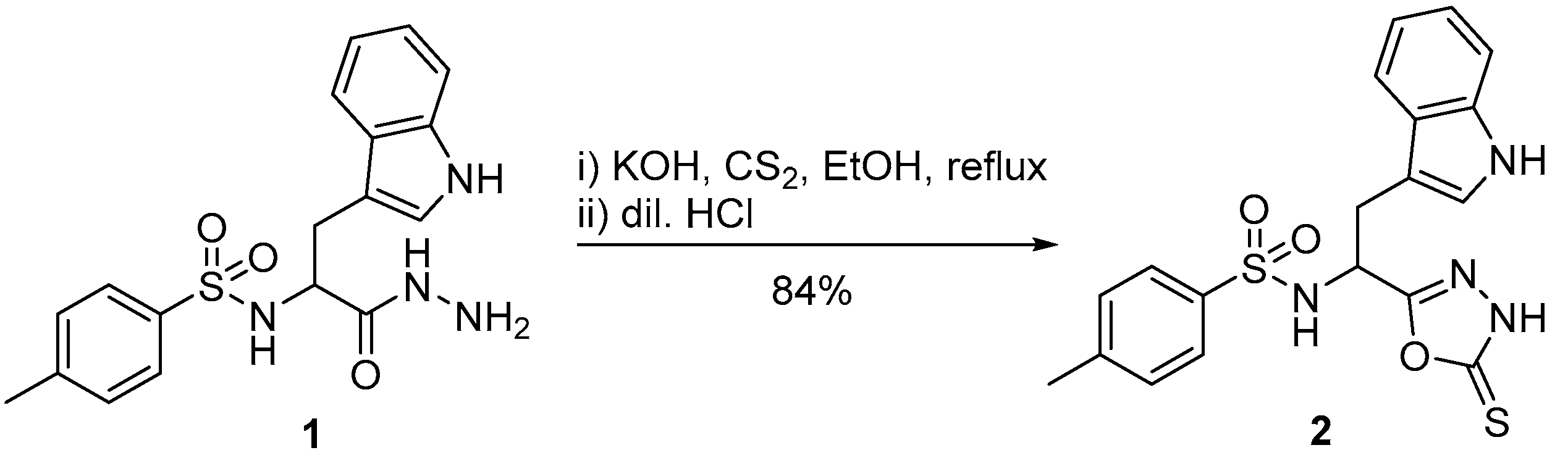
:N-[1-Hydrazinyl-3-(1H-indol-3-yl)-1-oxopropan-2-yl]-4-methylbenzenesulfonamide (1) on cyclization with carbon disulfide in ethanolic potassium hydroxide affords N-[2-(1H-indol-3-yl)-1-(5-thioxo-4,5-dihydro-1,3,4-oxadiazol-2-yl)ethyl]-4-methylbenzenesulfonamide (2) in 84% yield. The structure of compound 2 was supported by mass spectrometry, FT-IR and 1H- and 13C-NMR spectroscopy. To investigate the potential of compound 2 to act as antitubercular agent, it was docked against the enoyl reductase (InhA) enzyme of Mycobacterium tuberculosis. The docking pose and non-covalent interactions gave insights on its plausible inhibitory action.
1. Introduction
Oxadiazoles are privileged heterocyclic scaffolds in the field of medicinal chemistry due to their varied biological properties. Of special note are the 1,3,4-oxadiazoles due to their vast biological applications. They are reported to possess antibacterial [1], antifungal [2], antiviral [3], antitubercular [4], anticancer [5], antiallergic [6], anti-inflammatory [7], anticonvulsant [8], spasmolytic and hypotensive [9] properties. In view of the potential of the indole conjugated 1,3,4-oxadiazole containing moiety as an antitubercular entity [10], design and synthesis of the title compound 2 in which both the pharmacophoric scaffolds were co-existing was carried out.
Tuberculosis (TB) is one of the major health problems worldwide caused by Mycobacterium tuberculosis and is difficult to treat [11]. NADH–dependent enoyl–acyl carrier protein reductase enzyme from Mycobacterium tuberculosis (MtInhA), involved in the biosynthesis of its most resistant cell wall is a valid target for the design of antitubercular therapeutics [12]. Herein, we report the synthesis and in silico evaluation of the title compound 2 as a potential antitubercular agent.
2. Results
Initially, N-[1-hydrazinyl-3-(1H-indol-3-yl)-1-oxopropan-2-yl]-4-methylbenzenesulfonamide (1) was prepared according to the literature where by tosyl tryptophan methyl ester was treated with hydrazine hydrate to get the hydrazide 1 [13]. The hydrazide 1 was then treated with carbon disulfide and ethanolic potassium hydroxide at room temperature (1 h) and then at reflux (2 h), followed by acidification with dil. HCl to give the title compound 2 as a cream colored solid in 84% yield (Scheme 1). The compound was characterized by elemental analyses, mass spectrometry, FT-IR, 1H- and 13C-NMR spectroscopy (Supplementary Materials).
Molecular Docking
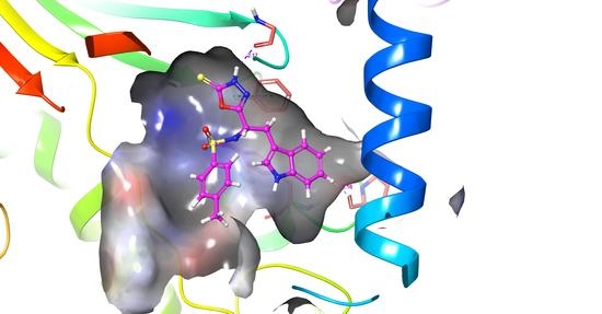
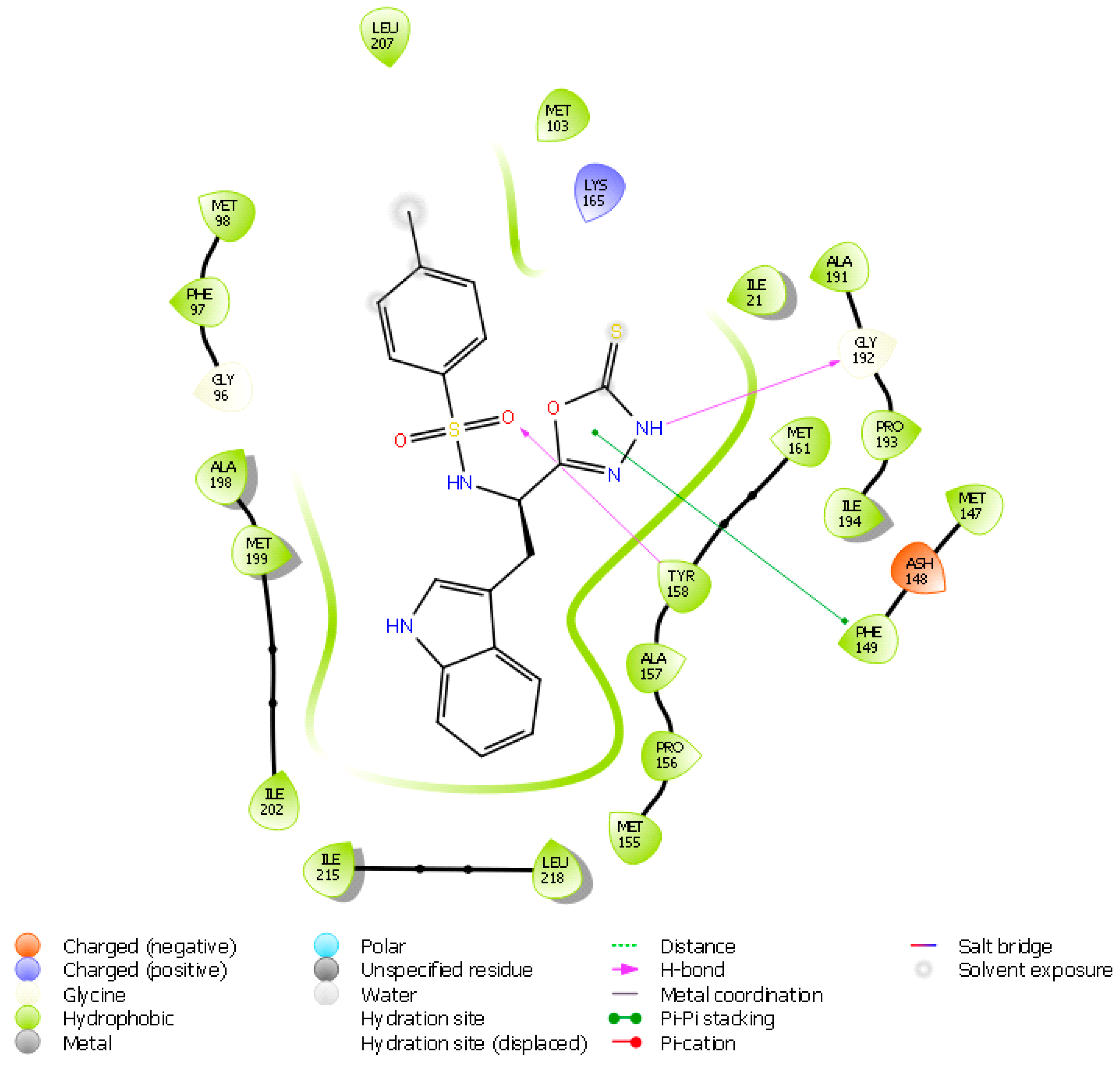
To get an insight into the plausible mechanistic inhibitory action of the title compound 2 on the enoyl reductase (InhA) enzyme of Mycobacterium tuberculosis, the molecule was docked against the active site of the enzyme. The molecule displayed several non-covalent interactions such as hydrogen bonds, π-π stacking and hydrophobic interactions as shown in the Figure 1 and Figure 2, with a docking score of −9.323 kcal/mol.
3. Discussion
The FT-IR spectrum of the title compound 2 showed the N-H stretching frequency at 3394 cm−1. A sharp band was observed at 1466 cm−1 corresponding to the C=S stretching. The asymmetric and symmetric stretching bands for the sulfonyl moiety appeared at 1336 and 1159 cm−1, respectively.
The 1H-NMR spectrum exhibited all the characteristic protons of tryptophan moiety as well as the tosyl group. The indole N-H proton appeared as a doublet with a coupling constant of 1.6 Hz at a chemical shift value of 10.84 ppm. The sulfonamide N-H proton resonated at 7.78 ppm as a doublet with a coupling constant of 7.6 Hz. The aromatic protons appeared in the region 6.96–7.53. The C-α proton appeared as a multiplet at 4.69–4.75 ppm. The CH2 protons appeared as a multiplet in the region 3.19–3.29 ppm due to itsdiasteriotopic nature and the methyl protons resonated at 2.38 ppm. In the 13C-NMR spectrum, the C=S carbon appeared at 168.6 and the C-5 of oxadiazoleappeared at 165.9 ppm. The remaining carbon signals appeared in the corresponding aromatic and aliphatic regions as expected. The mass spectrum further confirmed the formation of the title compound 2 due to its molecular ion peak appearing at m/z value of 415.0 [MH+] in positive ionization mode.
The N-3 of the oxadiazole makes a hydrogen bond as a donor with Gly 192. Whereas, the sulfonyl oxygen receives a hydrogen bond from Tyr 158. The π-electrons of the oxadiazole interact with those of the Phe 149 residue. Apart from this, the molecule makes hydrophobic interactions with the residues surrounding the binding pocket, such as Ile 21, Gly 96, Phe 97, Met 98, Met 103, Met 147, Phe 149, Met 155, Pro 156, Ala 157, Tyr 158, Met 161, Ala 191, Gly 192, Pro 193, Ile 194, Ala 198, Met 199, Ile 202, Leu 207, Ile 215 and Leu 218. All these interactions together enablethe molecule to have a high affinity towards the enzyme and inhibit the same with a docking score of −9.323 kcal/mol comparable with that of the reference inhibitor which is −9.088 kcal/mol.
4. Materials and Methods
All the precursor materials were purchased from Sigma-Aldrich (Bengaluru, India) and Spectrochem Pvt. Ltd. (Bengaluru, India). All the solvents and reagents were purchased from the commercial vendors (Mangaluru, India) in the appropriate grades and were used as such without purification. All the reactions were carried out with a calcium chloride guard tube. Melting points were determined by an open capillary method, which are uncorrected. FT-IR spectra were recorded on a Shimadzu FT-IR 157 Spectrometer. NMR spectra were measured in BrukerAvance II-400 spectrometer operating at 400 MHz for 1H and at 100 MHz for 13C nuclei respectively, using tetramethylsilane (TMS) as an internal standard. The chemical shifts (δ) and the coupling constants (J) are reported in parts per million (ppm) and inHertz, respectively. Mass spectra were recorded in Agilent Technology LC-mass spectrometer with ESI ionization in positive mode. Elemental analysis was performed using CHNS ElementarVario EL III. Purity of the compounds was checked by thin layer chromatography (TLC) on a silica coated aluminium sheet (silica gel 60F254) using ethyl acetate and hexane mixtures and visualized under UV at 254 nm.The docking of the ligands to the protein structural model was carried out using the Glide module in Schrödinger. Using the receptor grid generation panel in Glide, the grid was generated and using the ligand-docking panel in Glide, the molecular docking was carried out. The structures of the ligands were drawn using the 2D sketcher and then optimized with LigPrepto generate the energy optimized 3D structures. Ionization states at a pH range of 7.0 ± 2 were generated using Epikmodule with rest of the default options. Protein structure of mycobacterial enoyl reductase (PDB ID: 4TZK) protein structure was pre-processed using Protein preparation wizard, during which the missing hydrogens and amino acid side chains were added and any erroneous bond orders were corrected. The structure was optimized throughout to generate the hydrogen bond network and finally minimized to release any possible strains in the structure. The optimized protein structure was used for the receptor grid generation under the glide module wherein, the grid was generated for the protein structure by choosing the centroid of the bound inhibitor as the centroid of the grid. Successively, the prepared ligands were docked to protein grid generated on the receptor grid generation module under extra precision (XP) mode. The protein-ligand complexes after docking were further visualized and analyzed for the ligand fitting and interactions.
N-[1-Hydrazinyl-3-(1H-indol-3-yl)-1-oxopropan-2-yl]-4-methylbenzenesulfonamide (1) was prepared according to the literature [13].
N-[2-(1H-Indol-3-yl)-1-(5-thioxo-4,5-dihydro-1,3,4-oxadiazol-2-yl)ethyl]-4-methylbenzenesulfonamide (2): To a stirred solution of potassium hydroxide (0.56 g, 10 mmol) in ethyl alcohol (30 mL) at ca. 25 °C, was added N-(1-hydrazinyl-3-(1H-indol-3-yl)-1-oxopropan-2-yl)-4-methylbenzenesulfonamide (1) (3.72 g, 10 mmol) in one portion. To the resulting clear solution was added carbon disulfide (0.73 mL, 12 mmol) dropwise and stirred for 1 h. After 1 h, the reaction mixture was heated at reflux for an additional 2 h. On completion (TLC), the reaction mixture was then cooled to ca. 25 °C, quenched ontocrushed ice and acidified with dil. HCl in cold condition to obtain the title compound 2 (3.48 g, 84%) as a cream-colored solid, m.p. 182–184 °C (EtOH); FT IR (ATR, υmax, cm−1): 3394 (N-H), 1615 (C=N), 1597 (C=C), 1466 (C=S), 1336 (S=O), 1159 (S=O); δH(400 MHz, DMSO-d6) 10.84 (d, 1H, N-H, J 1.6), 8.78 (d, 1H, N-H, J 7.6), 7.52 (d, 2H, Ar-H, J 8.4), 7.34 (d, 1H, Ar-H, J 8.4), 7.26–7.29 (m, 3H, Ar-H), 7.07–7.11 (m, 1H, Ar-H), 7.05 (d, 1H, Ar-H, J 2.4), 6.96–7.00 (m, 1H, Ar-H), 4.69–4.75 (m, 1H, C-H), 3.19–3.29 (m, 2H, CH2), 2.38 (s, 3H, CH3); δC (100 MHz, DMSO-d6) 168.6, 165.9, 163.2, 142.9, 137.1, 136.0, 129.3, 126.6, 126.2, 124.1, 121.0, 118.6, 117.5, 111.5, 107.9, 50.0, 29.0, 20.9; ESI-MS (m/z): 415.0 [MH+]; Anal. calcd. for C19H18N4O3S2: C, 55.06; H, 4.38; N, 13.52. Found: C, 55.02; H, 4.35; N, 13.50%.
5. Conclusions
The 1,3,4-oxadiazole-2-thione 2 was successfully synthesized from tosyl protected tryptophan methyl ester by a simple method and well characterized by the spectroscopic techniques. Docking the title molecule into the binding site of the mycobacterial enoyl reductase (InhA) enzyme revealed the formation of hydrogen bonds and a π-π stacking interaction with a superior docking score to that of the reported inhibitor exemplified the potency of the compound to have promising antitubercular activity.
Supplementary Materials
The following are available online, Figure S1: FT-IR spectrum, Figure S2: Mass spectrum, Figures S3–S5: 1H-NMR spectrum, Figure S6: 13C-NMR Spectrum.
Author contributions
Experiments, Writing-Original Draft Preparation and Formal Analysis, N.P.; Supervision, B.P.
Funding
This research was funded by DST through fellowship.
Acknowledgments
The author N.P. thanks, Department of Science and Technology (DST), New Delhi for providing INSPIRE Fellowship. The authors are thankful to B. K. Sarojini, Mangalore University for providing the docking facility. The authors thank SAIF-Cochin for the spectral data.
Conflicts of Interest
The authors have no conflicts of interest.
References
- Adil, A.O.; Mebrouk, K.; Sarah, A. 1,3,4-Oxadiazole, 1,3,4-thiadiazole and 1,2,4-triazole derivatives as potential antibacterial agents. Arab. J. Chem. 2014. [Google Scholar] [CrossRef]
- Ates, O.; Kocabalkanli, A.; Cesur, N.; Otuk, G. Synthesis andantimicrobial activity of some 5-aryl-2-[(N,N-disubstitutedthiocarbamoylthio)acylamino]-1,3,4-oxadiazoles. Il Farmaco 1998, 53, 541–544. [Google Scholar] [CrossRef]
- Li, Z.; Zhan, P.; Liu, X. 1,3,4-Oxadiazole: A Privileged Structure in Antiviral Agents. Mini-Rev. Med. Chem. 2011, 11, 1130–1142. [Google Scholar] [CrossRef] [PubMed]
- Maria, A.M.; Isabel, C.G.V.; Julie, V.E.; Richard, K.G.; Candice, S.M.; Eva, M.M.; Aaron, K.; Yulia, O.; Lindsay, F.; Anisa, G.; et al. Synthesis and biological evaluation of aryl-oxadiazoles as inhibitors of Mycobacterium tuberculosis. Bioorg. Med. Chem. Lett. 2018, 28, 1758–1764. [Google Scholar]
- Ahmed, S.A.; Hamdy, M.A.; Nadia, M.M.; Mahmoud, A.E. Novel 5-(2-hydroxyphenyl)-3-substituted-2,3-dihydro-1,3,4-oxadiazole-2-thione derivatives: Promising anticancer agents. Bioorg. Med. Chem. 2006, 14, 1236–1246. [Google Scholar]
- Musser, J.H.; Brown, R.E.; Love, B.; Bailey, K.; Jones, H.; Kahen, R.; Huang, F.; Khandwala, A.; Leibowitz, M.; Sonnino-Goldman, P.; et al. Synthesis of 2-(2,3-dihydro-2-oxo-1,3,4-oxadiazol-5-yl) benzo heterocycles. A novel series of orally activeantiallergic agents. J. Med. Chem. 1984, 27, 121–125. [Google Scholar] [CrossRef] [PubMed]
- Sawhney, S.N.; Sharma, P.K. Synthesis and anti-inflammatory activity of some 3-heterocycle-1,2-benzisothiazoles. Bioorg. Med. Chem. Lett. 1993, 3, 1551–1554. [Google Scholar] [CrossRef]
- Zarghi, A.; Tabatabai, S.A.; Faizi, M.; Ahadian, A.; Navabi, P.; Zanganesh, V.; Shafiee, A. Synthesis and anticonvulsant activity of new 2-substituted-5-(2-benzyloxyphenyl)-1,3,4-oxadiazoles. Bioorg. Med. Chem. Lett. 2005, 15, 1863–1865. [Google Scholar] [CrossRef] [PubMed]
- Mishra, P.; Joshi, G.K.; Shakya, A.K.; Agarwal, R.K.; Patnaik, G.K. Pharmacological screening of few new 2-(substitutedacetyl)amino-5-alkyl-1,3,4-oxadiazoles. Ind. J. Physiol. Pharmacol. 1992, 36, 247–250. [Google Scholar]
- Desai, N.C.; Hardik, S.; Amit, T.; Kandarp, B.; Laxman, N.; Vijay, M.K.; Prakash, C.J.; Dhiman, S. Synthesis, biological evaluation and molecular docking study ofsome novel indole and pyridine based 1,3,4-oxadiazole derivatives aspotential antitubercular agents. Bioorg. Med. Chem. Lett. 2016, 26, 1776–1783. [Google Scholar] [CrossRef] [PubMed]
- Espinal, M.A. The global situation of MDR-TB. Tuberculosis 2003, 83, 44–51. [Google Scholar] [CrossRef]
- Goldberg, D.E.; Siliciano, R.F.; Jacobs, W.R. Outwitting evolution: fightingdrug-resistant TB, malaria, and HIV. Cell 2012, 148, 1271–1283. [Google Scholar] [CrossRef] [PubMed]
- Tashfeen, A.; Shahid, H.; Khalid, M.K.; Muhammad, I.C. Syntheses, urease inhibition, and antimicrobial studies of some chiral3-substituted-4-amino-5-thioxo-1H,4H-1,2,4-triazoles. Med. Chem. 2008, 4, 539–543. [Google Scholar]
Scheme 1.
Synthesis of N-[2-(1H-indol-3-yl)-1-(5-thioxo-4,5-dihydro-1,3,4-oxadiazol-2-yl)ethyl]-4-methylbenzenesulfonamide (2).
Scheme 1.
Synthesis of N-[2-(1H-indol-3-yl)-1-(5-thioxo-4,5-dihydro-1,3,4-oxadiazol-2-yl)ethyl]-4-methylbenzenesulfonamide (2).

Figure 1.
Dockingpose of the title compound 2 against mycobacterial enoyl reductase (InhA).

Figure 2.
Ligand interaction diagram of the title compound 2 with mycobacterial enoyl reductase (InhA).
Figure 2.
Ligand interaction diagram of the title compound 2 with mycobacterial enoyl reductase (InhA).

© 2018 by the authors. Licensee MDPI, Basel, Switzerland. This article is an open access article distributed under the terms and conditions of the Creative Commons Attribution (CC BY) license (http://creativecommons.org/licenses/by/4.0/).
Share and Cite
MDPI and ACS Style
Purushotham, N.; Poojary, B. N-[2-(1H-Indol-3-yl)-1-(5-thioxo-4,5-dihydro-1,3,4-oxadiazol-2-yl)ethyl]-4-methylbenzenesulfonamide. Molbank 2018, 2018, M1008. https://doi.org/10.3390/M1008
AMA Style
Purushotham N, Poojary B. N-[2-(1H-Indol-3-yl)-1-(5-thioxo-4,5-dihydro-1,3,4-oxadiazol-2-yl)ethyl]-4-methylbenzenesulfonamide. Molbank. 2018; 2018(3):M1008. https://doi.org/10.3390/M1008
Chicago/Turabian StylePurushotham, Nikil, and Boja Poojary. 2018. "N-[2-(1H-Indol-3-yl)-1-(5-thioxo-4,5-dihydro-1,3,4-oxadiazol-2-yl)ethyl]-4-methylbenzenesulfonamide" Molbank 2018, no. 3: M1008. https://doi.org/10.3390/M1008
Note that from the first issue of 2016, this journal uses article numbers instead of page numbers. See further details here.