Communication

Solubilization of Single-walled Carbon Nanotubes with Single- stranded DNA Generated from Asymmetric PCR

1
Department of Biochemistry, Nanjing University, Nanjing 210093, P. R. China
2
Shanghai Institute of Applied Physics, Chinese Academy of Sciences, Shanghai 201800, P. R. China
3
The People’s Hospital of Jiangsu Province, Nanjing 210029, P. R. China
*
Authors to whom correspondence should be addressed.
Int. J. Mol. Sci. 2007, 8(7), 705-713; https://doi.org/10.3390/i8070705
Submission received: 31 May 2007 / Revised: 29 June 2007 / Accepted: 13 July 2007 / Published: 23 July 2007
(This article belongs to the Special Issue Nucleic Acid Derivatives in Emerging Technologies)

Abstract

:
Carbon nanotubes (CNTs) can be effectively dispersed and functionalized by wrapping with long single-stranded DNA (ssDNA) synthesized by asymmetric PCR. The ssDNA-CNTs attached on surface of glass carbon electrode made it possible for electrochemical analysis and sensing, which was demonstrated by reduction of H2O2 on hemoglobin/ssDNA-CNTs modified electrodes. This research showed the potential application of DNA-functionalised CNTs in construction of future electrochemical biosensors.

1. Introduction

Carbon nanotubes (CNTs) have intrigued great research interest due to their excellent thermal [1], electrical [2] and mechanical [3] properties. However, CNTs tend to form bundles in solution, which is one of the barricades for its application [46]. Therefore, there is great demand to effectively solubilize CNTs in order to realize wide application of this interesting nanomaterial. A variety of approaches have been proposed to address this issue [79], and it appears that DNA polymer offers a great opportunity to solubilize CNT. Dwyer et al. reported that the use of amino-terminated DNA strands in functionalizing the open ends and defect sites of oxidatively prepared single-walled carbon nanotubes, which was an important first step in realizing a DNA-guided self-assembly process for carbon nanotubes [7]. O’ Connell et al. reported that SWCNTs had been solubilized in water by non-covelent association with linear polymers mostly with polyvinyl pyrrolidone (PVP) and polystyrene sulfonate (PSS) [8]. Zhu et.al. described about the charged particles which could be employed in the dispersion of SWNTs in water, up to single nanotube level [9]. Zheng and co-workers first reported that chemically synthesized DNA oligonucleotides (several tens bases) could disperse and sort single-walled carbon nanotubes (SWCNTs) [10,11], which was soon recognized to be a promising approach to solubilizing CNTs. In spite of its effectiveness, mass-production of oligonucleotide-dispersed CNTs are still limited by the high cost of chemical synthesis of oligonucleotides. In addition, only oligonucleotides with specific sequences could effectively wrap SWCNTs, which further limits its wide application.
In order to overcome these barricades, Brittany et al proposed a PCR-based approach, which produced a large amount of genomic DNA by using polymerase-based DNA amplification. They showed that long genomic DNA, in its single-stranded form, could interact with SWCNTs and serve as an effective dispersion reagent. Nevertheless, the normal PCR protocol generates double-stranded (ds-) rather than singled-stranded (ss-) DNA. Thus they employed a complicated approach that involved thiolated primers and gold nanoparticle-based separation [12]. More recently, Li and coworker developed a novel strategy that avoided using gold nanoparticles. They employed an isothermal amplification, rolling circle amplification(RCA), rather than PCR, to amplify DNA [13,14]. RCA is known to produce only ssDNA, thus obviating the necessity to separate PCR-generated dsDNA. In spite of its simplicity, the use of RCA-based amplification is limited by the synthesis of circular DNA, which is still commercially unavailable.
In this study, we employed a convenient and effective approach to obtain large amounts of ssDNA by using asymmetric PCR. A commercially available plasmid, pUC19, was employed as the template, and asymmetric PCR protocol was performed, which generated amplified ssDNA rather than dsDNA. It was demonstrated that long ssDNAs amplified from asymmetric PCR could easily wrap and make SWCNT dispersible (Scheme 1). We also demonstrated that the DNA/SWCNT complex could facilitate the electron transfer reaction of hemoglobin (Hb) at the electrode surface, showing this new material’s great potential in construction of novel electrochemical biosensors [15].

2. Results and Discussion

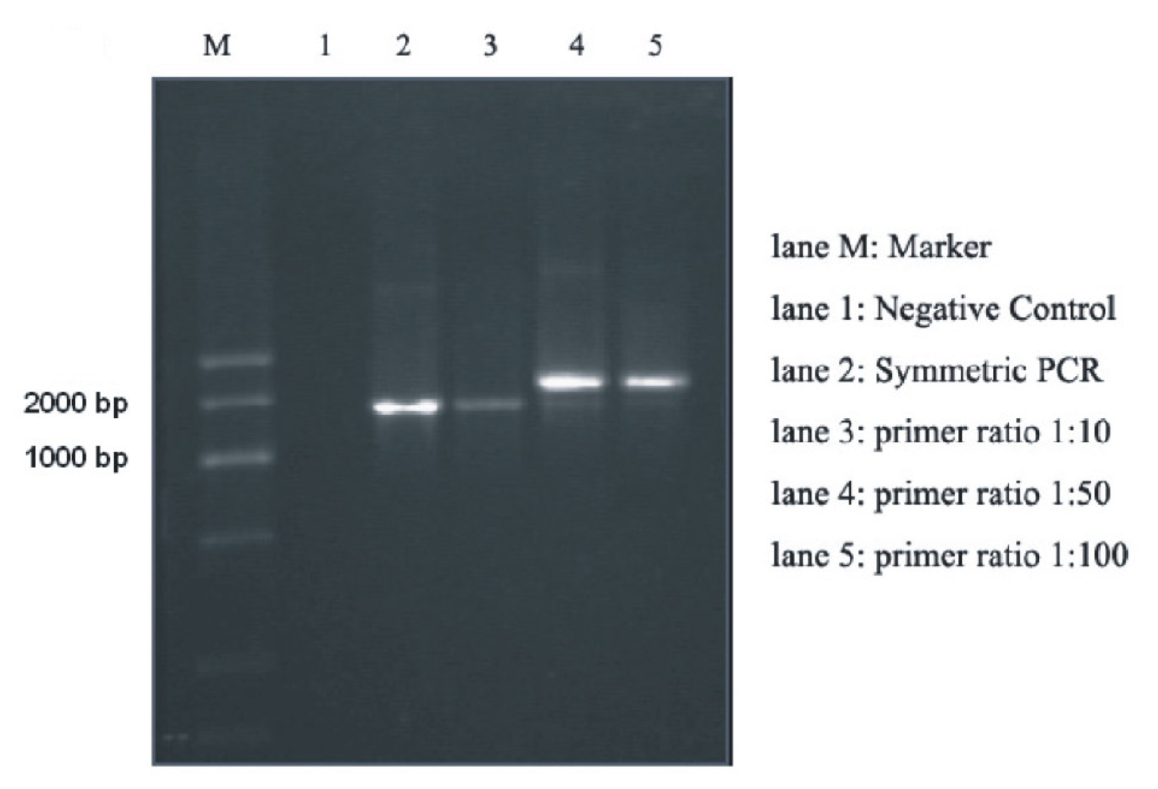
2.1. Amplification of DNA Templates Using Asymmetric PCR

We first tested whether the 1888-base ssDNA could be amplified through the asymmetric PCR protocol. Figure 1 shows the electrophoresis patterns of negative control (lane 1), symmetric PCR products (lane 2) and asymmetric PCR products with different primer ratios (lanes 3, 4 and 5). The concentration of reverse primer AS2395 solutions in the experiment was fixed to 10 μM, while the forward primer S525′s concentration ranged from 0.1 to 10 μM, in order to investigate the optimal ratio of primers that could produce most amounts of ssDNA. The primer ratios in lane 1 and 2 were 1:1 while no templates were added to lane 1′s mastermix. Compared to the blank result of negative control, the obvious band in lane 1 coincides with our previous expectation. As for the asymmetric ones, the results in lane 3, 4 and 5 represents the primer ratio of 1:10, 1:50 and 1:100, respectively. The band in lane 3 was parallel with that in lane 2, though a bit dimmer, suggesting that the majority of products in this ratio are still double stranded. There were two bands observed in each lane of 4 and 5. The main product bands in both lanes are on upward side of symmetric PCR results, corresponding to the principal that ssDNA moves slower than the double-strand ones in agarose electrophoresis. And the lower blurry band can be assigned as the dsDNA amplified in several cycles at the beginning. These results obviously indicate that the PCR products prepared according to our protocol are indeed single-stranded.

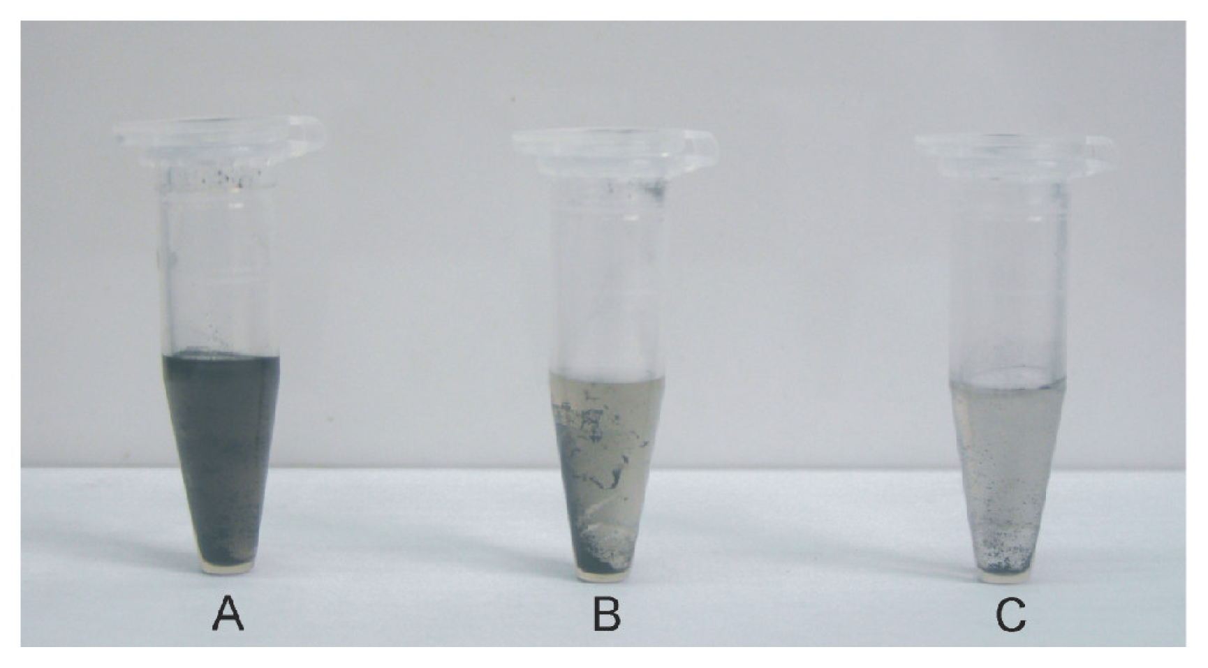
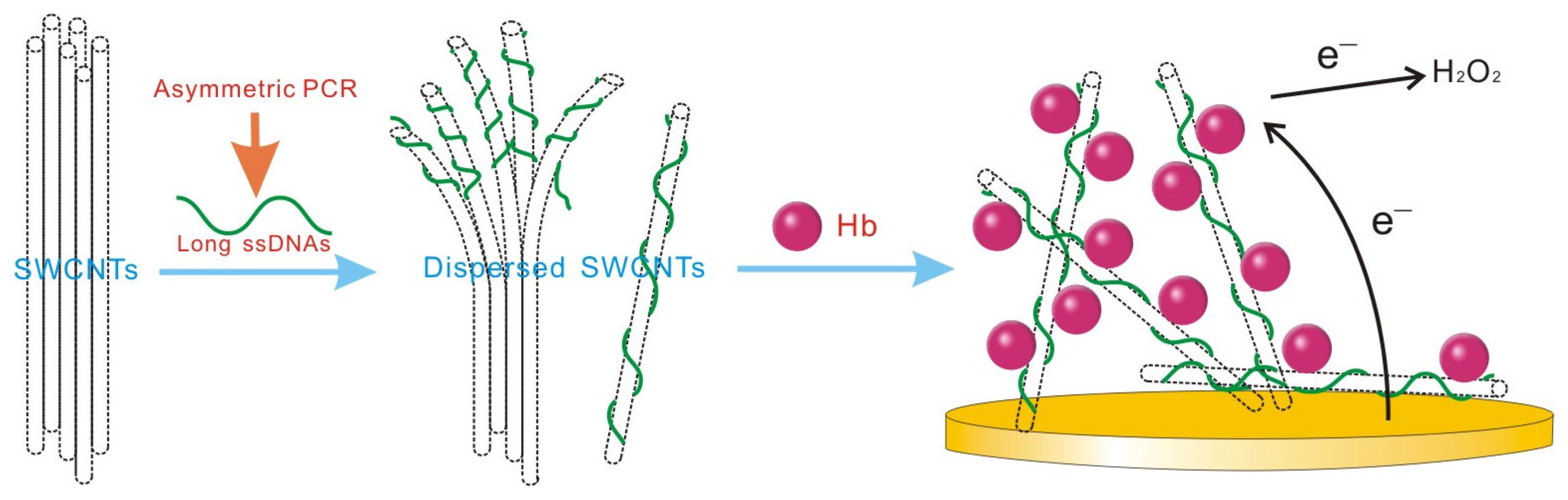
2.2. Solubilization of DNA-SWCNTs

As expected, in the absence of DNA, SWCNTs were not soluble in water (Figure 2C) because they aligned parallel to each other and pack into bundles due to strong intertube van der Waals attraction [5]. The dsDNA amplified from symmetric PCR didn’t disperse the CNTs into solutions as well (Figure 2B). By contrast, SWCNTs were readily dispersed in the presence of the asymmetric PCR product (Figure 2A) and, after centrifugation at 3000 rpm for 30 min, the supernatant could sustain in a stable state without apparent deposition for at least 1 week. We ascribe the solubilization effect of asymmetric PCR amplicons to the DNA base stacking on the SWCNT surface, leaving highly charged phosphate backbones exposed to water [11]. As a result, wrapping of SWCNTs with a hydrophilic shell readily disperses them in water.
It was found that the ssDNA-SWCNTs hybrid could firmly attach onto a glass carbon substrate and form a uniform film. This is due to the high dispersity of ssDNA-SWCNTs. The strains among all of the nanotubes to form the film are equivalent in all directions, since they are individually suspended in water. If the nanotubes are not individually suspended in water, the asymmetric strain from nanotubes will result in uneven film, which cannot adhere tightly to the surface of glass-based materials and will easily fall off when put into solution [16].

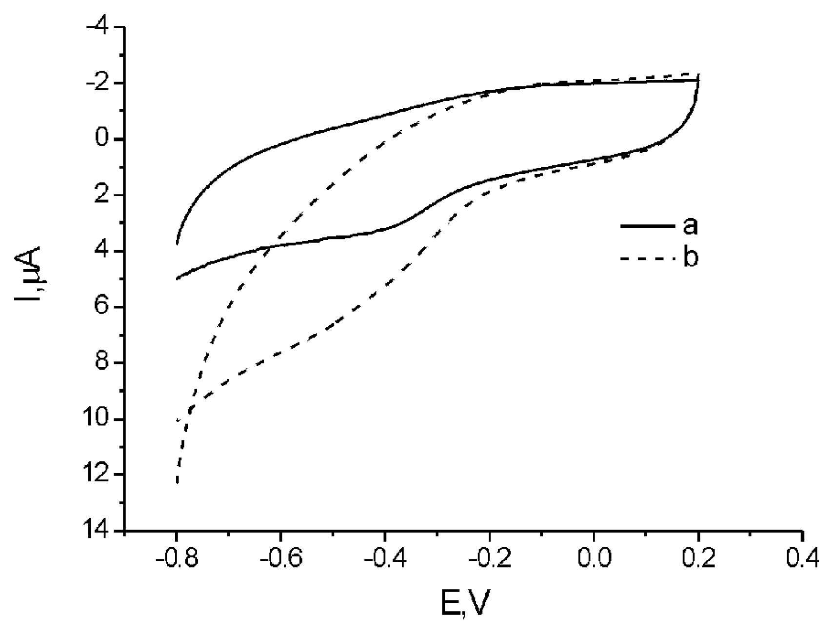
2.3. Electrochemical investigation of DNA-SWCNTs

Immobilization of protein molecules in biocompatible films or matrix, especially in DNA films, have attracted much attention since Nassar and Rusling reported DNA can enhance electron transfer between electrode and heme proteins in myoglobin–DNA films[17]. As is well known, Hb is very difficult to perform direct electrochemistry due to its large and complex structure [18] while incorporation of Hb in the DNA based films can enhance the Hb electron transfer rate due to its three dimensional structure and its potential conductive nature[19]. Here, we use the DNA/SWCNTs as the electron relay. As our previous study has shown, DNA single strand can be a good electron transfer mediator for proteins on surface of electrodes[20]. And in this part, ssDNA is used not only to disperse SWCNTs in solution but to help getting amplified protein electron transfer reactions on electrodes as well. Fig. 3a displays the cyclic voltammograms obtained at an Hb/ssDNA-SWCNTs film modified electrode in a 0.1 M phosphate buffer solution (PBS) at pH 7.0. A pair of redox peaks can be observed attributing to the redox reaction of Hb at the electrode. The cathodic and anodic peak potentials of Hb are located at −0.380 V and −0.190 V, respectively. The formal potential (E0′) is calculated to be −0.285V, which is similar to the previous reports[21]. Compared with the cathodic peak, the anodic peak of Hb is not obvious, which suggests that the ferrous Hb (reduced form of Hb) on the electrode surface is only partially converted to ferric Hb (oxidized form of Hb). It clearly demonstrates that although the electron transfer between Hb and SWCNT electrode performs better than that with bare electrode, it is still not quick enough, which is due to the fact that SWCNTs is a mixture of metallic and semi-conducting ones, usually in ratio of 1:2. Obviously, most part of SWCNTs can not perform satisfactory function of electron relay between Hb and electrode because of their semi-conducting properties. We suppose that electron transfer rate between Hb in the ssDNA-SWCNTs and electrode might be greatly improved if most SWCNTs is metallic. Though the main function of Hb in vivo is to transport oxygen, it also has potential peroxidase activity to catalyze the reduction of some small molecules, such as H2O2[2226]. Therefore, H2O2 is chosen as a model to interrogate such peroxidase function of Hb in ssDNA-SWCNTs. Fig. 3b shows the cyclic voltammograms of Hb/ssDNA-SWCNTs modified electrode in 0.1 M PBS at pH 7.0 containing 7.96×10⁻⁴ M H2O2. It can be obviously observed that the reduction peak of the protein increases largely, while, the anodic peak of serum responsive element (SRE) Hb is totally disappeared. This is attributed to the oxidation of Fe(II) to Fe(III) of SRE Hb by H2O2. It demonstrates that peroxidase activity of Hb is well maintained in ssDNA-SWCNTs while voltammograms of electrodes modified with proteins without catalytic ability remained unchanged along with the elevation of H2O2 concentration. It also implies that Hb/ssDNA-SWCNTs modified electrode may be used as a biosensor for H2O2.

3. Experimental Section

pUC19 in this experiment was purchased from SABC at the concentration of 0.8 μg/μl, which was diluted by 1000 times before being used as the template of asymmetric PCR. All of the PCR reagents (10×buffer, MgCl2, dNTP and taq DNA polymerase) were from Bio-Basic’s PCR kit. Single-walled carbon nanotubes used in the study were obtained from Shenzhen Nanotech Port Co. Ltd, while pig hemoglobin from Sigma. The sequences of the two primers were as follows: 5′-CGCAAGCATAAAGTGTAAAG-3′ (S525); 5′-AGAAACGCTGGTGAAAGT-3′ (AS2395). They were designed to obtain an amplicon of 1888 bp length. PCR reaction was applied on PTC-200 recycler (MJ Research Incorporated). The electrochemical measurements were performed on a CHI electrochemical analyzer (CH Instruments Inc). For each PCR experiment, 100 μl of mastermix was prepared. It contained 10 μl of 10×PCR buffer (200 mM Tris-HCl, pH 8.4, 500 mM KCl), 8 μl of 25 mM MgCl2, 10 μl of 10 mM dNTPs, 5 μl of forward primer ranging from 0.1~10 μM, 5 μl of 10 μM reverse primer, 10 μl of 5 μg/μl bovine serum albumin (BSA), 10 μl of 1 ng/μl DNA template(Puc19), 0.5 μl of 5 units/μl Taq DNA polymerase, and 41.5 μl autoclaved double-deionized water.
The mastermix was subjected to the following thermal cycling profile: initial denaturation at 95 °C for 2 min, 40 cycles at 95 °C for 30 s, at 48 °C for 40 s, at 72 °C for 2 min, and a final extension at 72 °C for 10 min. To confirm the fidelity of the reaction, PCR products were loaded in a 1% agarose gel prepared with TBE buffer (44.5 mM Tris, 44.5 mM boric acid, and 1 mM EDTA) containing 0.5 μg/ml ethidium bromide and electrophoresed at 80 V for 1.5 h. Then, the sample was visualized by UV transillumination and its size was compared with a known DNA Marker G (300~2500 bp, Biobasic Inc.).
The capability of the PCR product to disperse SWCNTs was examined. In a typical experiment, the DNA (20 μl) and SWCNTs (0.3 mg, 50~70 %, 1.2~1.5 nm in diameter and 2~5 mm in length) were mixed in milli-Q water to a final volume of 500 μl. The mixture was sonicated for 10 min. After sonication, the sample was centrifuged for 30 min at 3000 rpm. As control experiments, blank samples without DNA and solutions of dsDNA that had been produced by common symmetric PCR of the same primers were also sonicated and centrifuged. The resulting supernatant was then examined using electrochemical analyzer to investigate its biological activity on surface of glass-carbon electrode.
Glass-carbon electrodes (3 mm in diameter, CH Instruments Inc.) were first polished on microcloth (Buehler) with Gamma micropolish deagglomerated alumina suspension (0.05 μm) for 5 min. These electrodes were then sonicated in ethanol and Milli-Q water for 5 min, respectively. After drying with nitrogen, 20 μl mixed solution of Hb (8 mg/ml) and ssDNA-SWCNT in the volume ratio of 1:1 was spread evenly onto its surface and allowed to dry at room temperature (about 30 min). Finally it was thoroughly rinsed with Milli-Q water and was ready for use.
Electrochemical experiments were carried out in a 3 ml cell using a glass-carbon working electrode, a Pt counter electrode, and an Ag/AgCl reference electrode (immersed in a 3 M NaCl filling solution saturated with AgCl). Cyclic voltammetry (CV) was carried out at a scan rate of 50 mV/s. A 0.1 M phosphate buffer solution (KH2PO4 + Na2HPO4, pH7) was used as the supporting electrolyte. All electrochemical experiments were conducted under nitrogen atmosphere at the ambient temperature 25 ± 1 °C. The GC electrode was coated by casting 20 μl of supernatant of the ssDNA-SWCNT and dried before measurement. Prior to the electrochemical experiments, the test buffer solution was first bubbled thoroughly with high-purity nitrogen for at least 10 min. Then a stream of nitrogen was blown gently across the surface of the solution in order to maintain an anaerobic solution throughout the experiment. Cyclic voltammetry was carried out in the scan range of −0.8~0.2 V. All the experiments were performed at a temperature of 20 ± 0.5 °C.
Figure 1. Electrophoresis patterns of symmetric and asymmetric PCR products in 1% agarose gel. Lane M, DNA Marker G (300~2500bp); lane 1, negative control; lane 2, 1888-bp amplified using 10 μM S525 and 10 μM AS2395 as primers; lane 3 asymmetrically amplified using 1 μM S525 and 10 μM AS2395; lane 4 asymmetrically amplified using 0.2 μM S525 and 10 μM AS2395; lane 5 asymmetrically amplified using 0.1 μM S525 and 10 μM AS2395.
Ijms 08 00705f1
Figure 2. Photograph of SWCNT–water mixture in the absence (C), presence of the symmetric PCR product (B) and asymmetric PCR product (A) after centrifugation at 3000 rpm for 30 min.
Ijms 08 00705f2
Figure 3. Cyclic voltammetric detection at ssDNA-SWCNTelectrode before (a) and after (b) adding of H2O2 in 0.1 M PBS, pH 7.0
Ijms 08 00705f3
Scheme 1. Single-walled carbon nanotubes (SWCNTs) are dispersed by long single-strand DNA (ssDNA) generated from asymmetric PCR.
Ijms 08 00705f4

Acknowledgements

We thank the financial support from National Natural Science Foundation (60537030 and 20404016), Ministry of Science and Technology (2006CB933000, 2007CB936000), Science and Technology Commission of Shanghai Municipality (0652nm006, 0652nm016, 06ZR14106, Shanghai Rising-Star Program) and Chinese Academy of Sciences.

References and Notes

  1. Berber, S.; Kwon, Y.K.; Tomanek, D. Unusually high thermal conductivity of carbon nanotubes. Phys. Rev. Lett 2000, 84, 4613–4616. [Google Scholar]
  2. Wei, B.Q.; Vajtai, R.; Zhang, Z.J.; Ramanath, G.; Ajayan, P.M. Carbon nanotube-magnesium oxide cube networks. J. Nanosci. Nanotechno 2001, 1, 35–38. [Google Scholar]
  3. Guo, W.; Guo, Y.; Gao, H.; Zheng, Q.; Zhong, W. Energy dissipation in gigahertz oscillators from multiwalled carbon nanotubes. Phys. Rev. Lett 2003, 91, 125501. [Google Scholar]
  4. Giordani, S.; Bergin, S.D.; Nicolosi, V.; Lebedkin, S.; Kappes, M.M.; Blau, W.J.; Coleman, J.N. Debundling of single-walled nanotubes by dilution: observation of large populations of individual nanotubes in amide solvent dispersions. J. Phys. Chem. B 2006, 110, 15708–15718. [Google Scholar]
  5. Bandyopadhyaya, R.; Nativ-Roth, E.; Regev, O.; Yerushalmi-Rozen, R. Stabilization of Individual Carbon Nanotubes in Aqueous Solutions. Nano Lett 2002, 2, 25. [Google Scholar]
  6. Shvartzman-Cohen, R.; Levi-Kalisman, Y.; Nativ-Roth, E.; Yerushalmi-Rozen, R. Generic approach for dispersing single-walled carbon nanotubes: the strength of a weak interaction. Langmuir 2004, 20, 6085–6088. [Google Scholar]
  7. Dwyer, C.; Guthold, M.; Falvo, M.; Washburn, S.; Superfine, R.; Erie, D. DNA-functionalized single-walled carbon nanotubes. Nanotechnology 2002, 13, 601–604. [Google Scholar]
  8. O’Connell, M.; Peter, B.; Ericson, L.; Huffman, C.; Wang, Y.; Haroz, E.; Kuper, C. Reversible water-solubilization of single-walled carbon nanotubes by polymer wrapping. Chem. Phys. Lett 2001, 342, 265–271. [Google Scholar]
  9. Zhu, J.; Yudasaka, M.; Zhang, M.; Iijima, S. Dispersing Carbon Nanotubes in Water: A Noncovalent and Nonorganic Way. J. Phys. Chem. B 2004, 108, 11317–11320. [Google Scholar]
  10. Zheng, M.; Jagota, A.; Strano, M.S.; Santos, A.P.; Barone, P.; Chou, S.G.; Diner, B.A.; Dresselhaus, M.S.; McLean, R.S.; Onoa, G.B.; et al. Structure-based carbon nanotube sorting by sequence-dependent DNA assembly. Science 2003, 302, 1545–1548. [Google Scholar]
  11. Zheng, M.; Jagota, A.; Semke, E.D.; Diner, B.A.; McLean, R.S.; Lustig, S.R.; Richardson, R.E.; Tassi, N.G. DNA-assisted dispersion and separation of carbon nanotubes. Nat. Mater 2003, 2, 338–342. [Google Scholar]
  12. Gigliotti, B.; Sakizzie, B.; Bethune, D.S.; Shelby, R.M.; Cha, J.N. Sequence-Independent Helical Wrapping of Single-Walled Carbon Nanotubes by Long Genomic DNA. Nano Lett. 2006, 6(2), 159–164. [Google Scholar]
  13. Zhao, W.A.; Gao, Y.; Brook, M.A.; Li, Y.F. Wrapping single-walled carbon nanotubes with long single-stranded DNA molecules produced by rolling circle amplification. Chem. Commun. 2006, 3582–3584. [Google Scholar]
  14. Zhao, W.; Gao, Y.; Kandadai, S.A.; Brook, M.A.; Li, Y. DNA polymerization on gold nanoparticles through rolling circle amplification: towards novel scaffolds for three-dimensional periodic nanoassemblies. Angew. Chem.( Int. Edit.) 2006, 45, 2409–2413. [Google Scholar]
  15. Keren, K.; Berman, R.S.; Buchstab, E.; Sivan, U.; Braun, E. DNA-templated carbon nanotube field-effect transistor. Science 2003, 302, 1380–1382. [Google Scholar]
  16. Hu, C.; Zhang, Y.; Bao, G.; Zhang, Y.; Liu, M.; Wang, Z.L. DNA functionalized single-walled carbon nanotubes for electrochemical detection. J. Phys. Chem 2005, 109, 20072–20076. [Google Scholar]
  17. Nassar, A.E.F.; Rusling, J.F. Electron Transfer between Electrodes and Heme Proteins in Protein-DNA Films. J. Am. Chem. Soc 1996, 118, 3043–3044. [Google Scholar]
  18. Ryan, M.D.; Bowden, E.F.; Chambers, J.Q. Dynamic electrochemistry: methodology and application. Anal. Chem 1994, 66, 360R–427R. [Google Scholar]
  19. Tong, Z.Q.; Yuan, R.; Chai, Y.Q.; Chen, S.H.; Xie, Y. Amperometric biosensor for hydrogen peroxide based on Hemoglobin/DNA/Poly-2,6-pyridinediamine modified gold electrode. Thin Solid Films 2007, in press. [Google Scholar]
  20. Lao, R.; Wang, L.; Wan, Y.; Zhang, J.; Song, S.; Zhang, Z.; Fan, C.; Lin, H. Interactions between Cytochrome c and DNA Strands Self-Assembled at Gold Electrode. Int. J. Mol. Sci 2007, 8, 136–144. [Google Scholar]
  21. Zhou, Y.; Li, Z.; Hu, N.; Zeng, Y.; Rusling, J.F. Layer-by-Layer Assembly of Ultrathin Films of Hemoglobin and Clay Nanoparticles with Electrochemical and Catalytic Activity. Langmuir 2002, 18, 8573–8579. [Google Scholar]
  22. Liu, X.J.; Xu, Y.; Ma, X.; Li, G. A third-generation hydrogen peroxide biosensor fabricated with hemoglobin and Triton X-100. Sensor. Actuat. B 2005, 106, 284–288. [Google Scholar]
  23. Zhou, H.; Gan, X.; Wang, J.; Zhu, X.; Li, G. Hemoglobin-based hydrogen peroxide biosensor tuned by the photovoltaic effect of nano titanium dioxide. Anal. Chem 2005, 77, 6102–6104. [Google Scholar]
  24. Ma, X.; Liu, X.; Xiao, H.; Li, G. Direct electrochemistry and electrocatalysis of hemoglobin in poly-3-hydroxybutyrate membrane. Biosens. Bioelectron 2005, 20, 1836–1842. [Google Scholar]
  25. Fan, C.; Wang, H.; Sun, S.; Zhu, D.; Wagner, G.; Li, G. Electron-transfer reactivity and enzymatic activity of hemoglobin in a SP Sephadex membrane. Anal. Chem 2001, 73, 2850–2854. [Google Scholar]
  26. Huang, H.; Hu, N.; Zeng, Y.; Zhou, G. Electrochemistry and electrocatalysis with heme proteins in chitosan biopolymer films. Anal. Biochem 2002, 308, 141–151. [Google Scholar]

