One New Phenolic Compound from Castanea mollissima Shells and its Suppression of HepatomaCell Proliferation and Inflammation by Inhibiting NF-κB Pathway

Abstract
:1. Introduction
2. Results
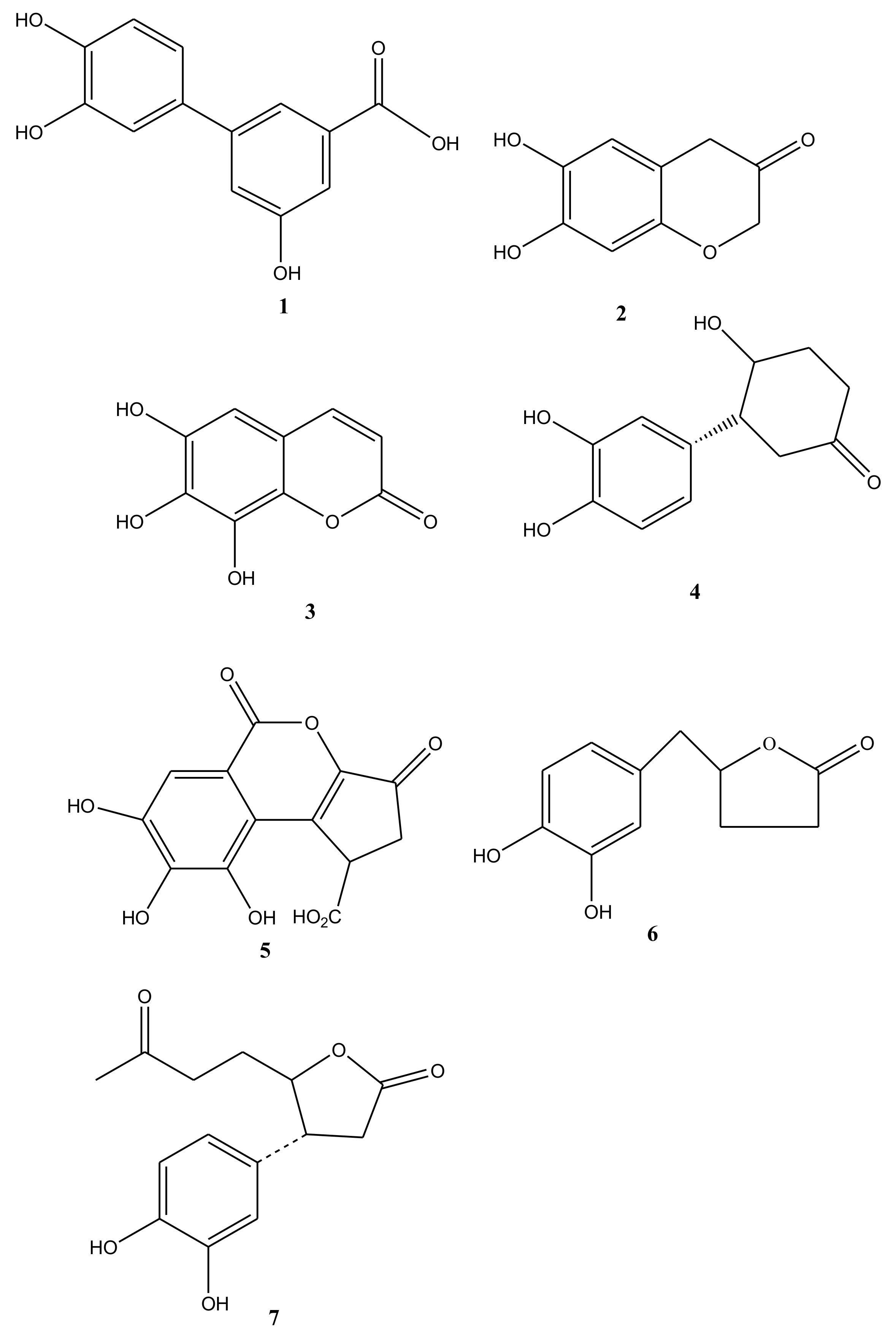
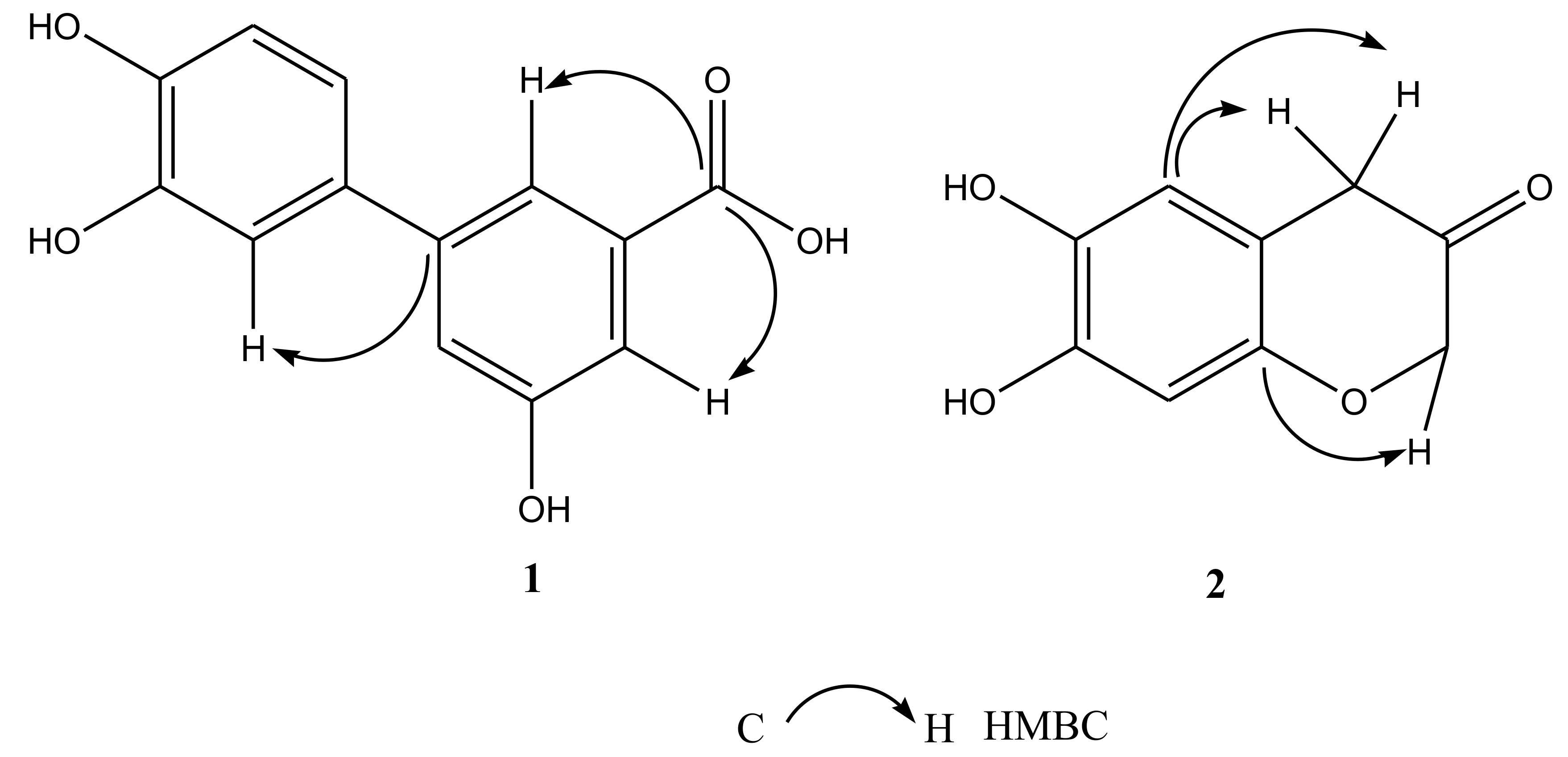
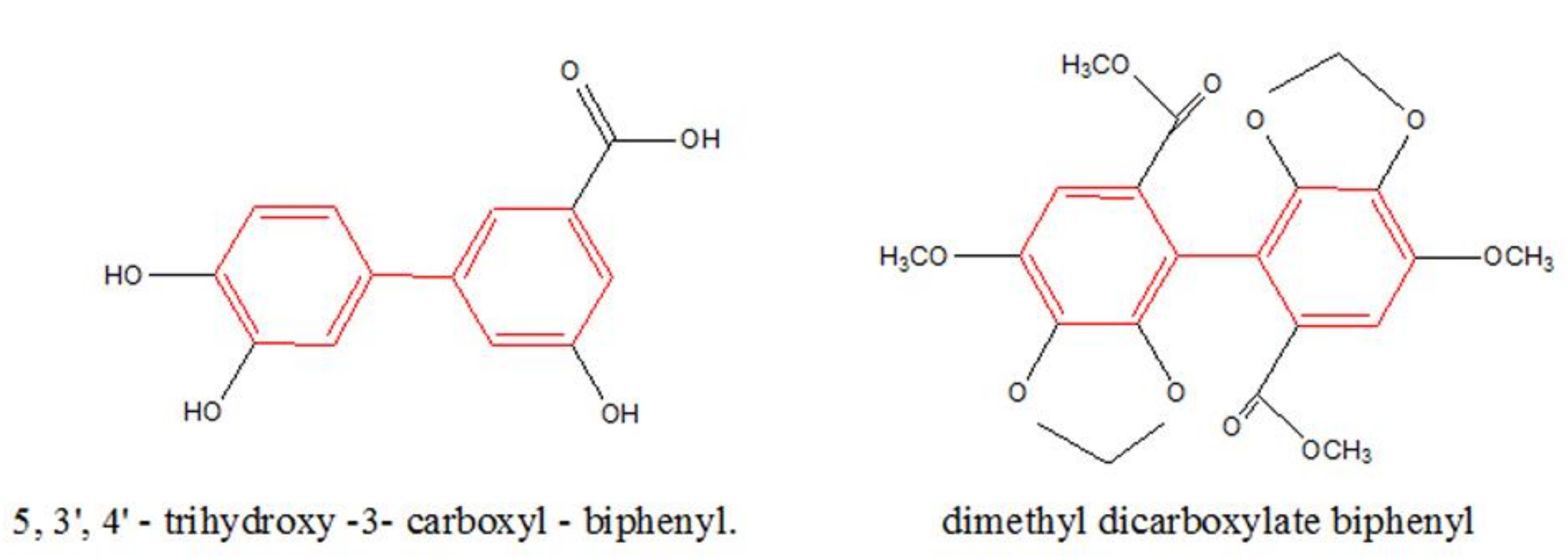
2.1. Chemistry
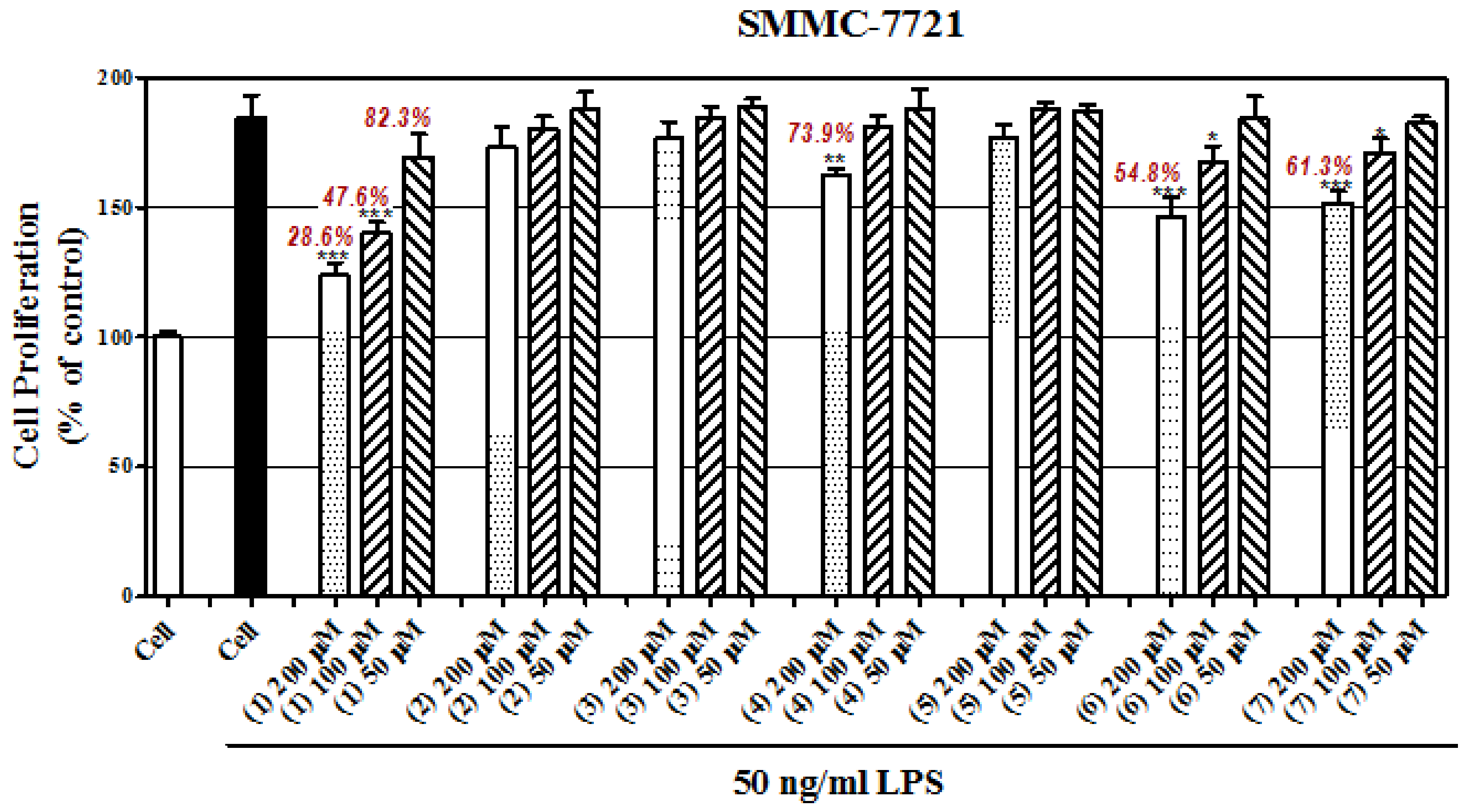
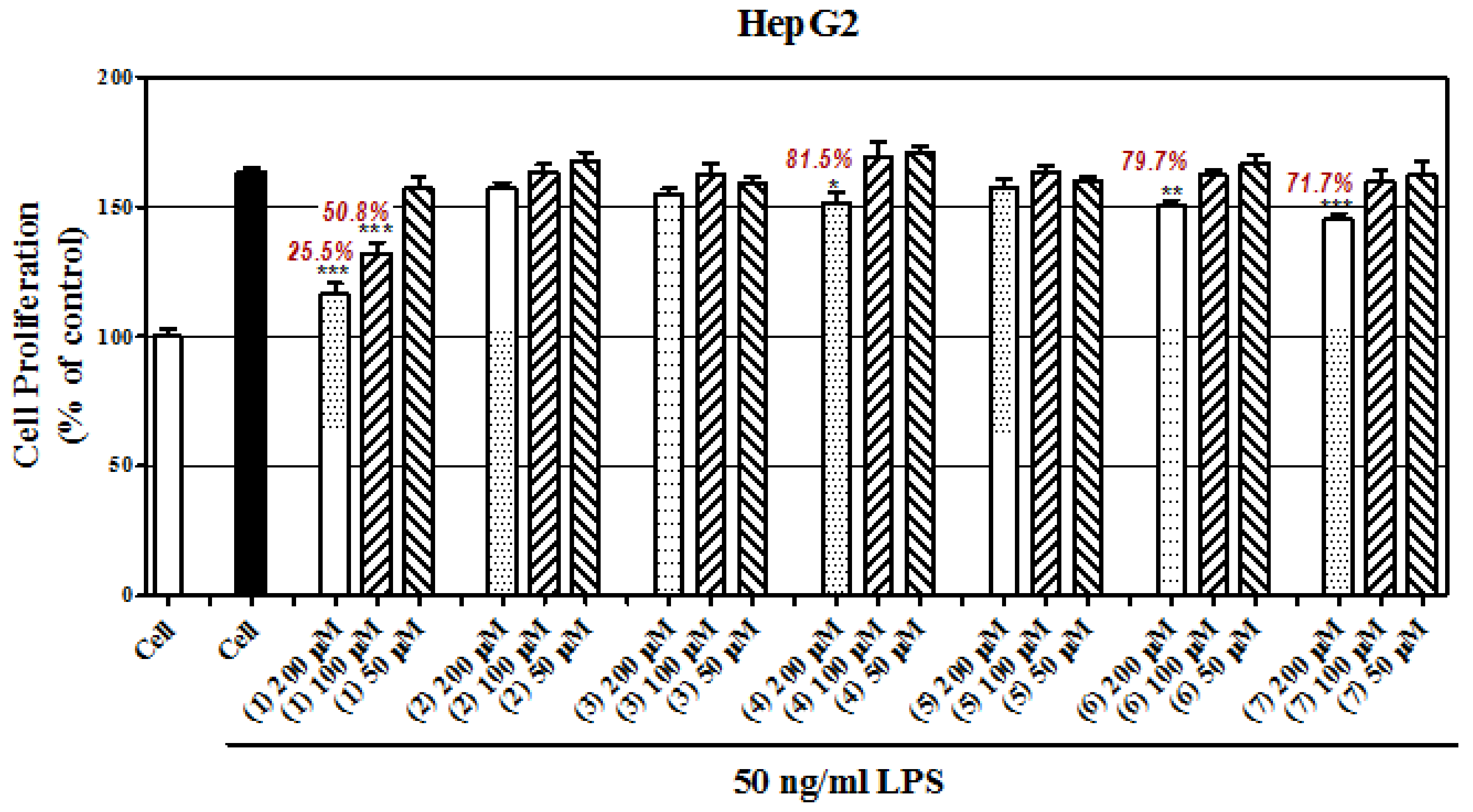
2.2. Antiproliferative Effect of These Compounds
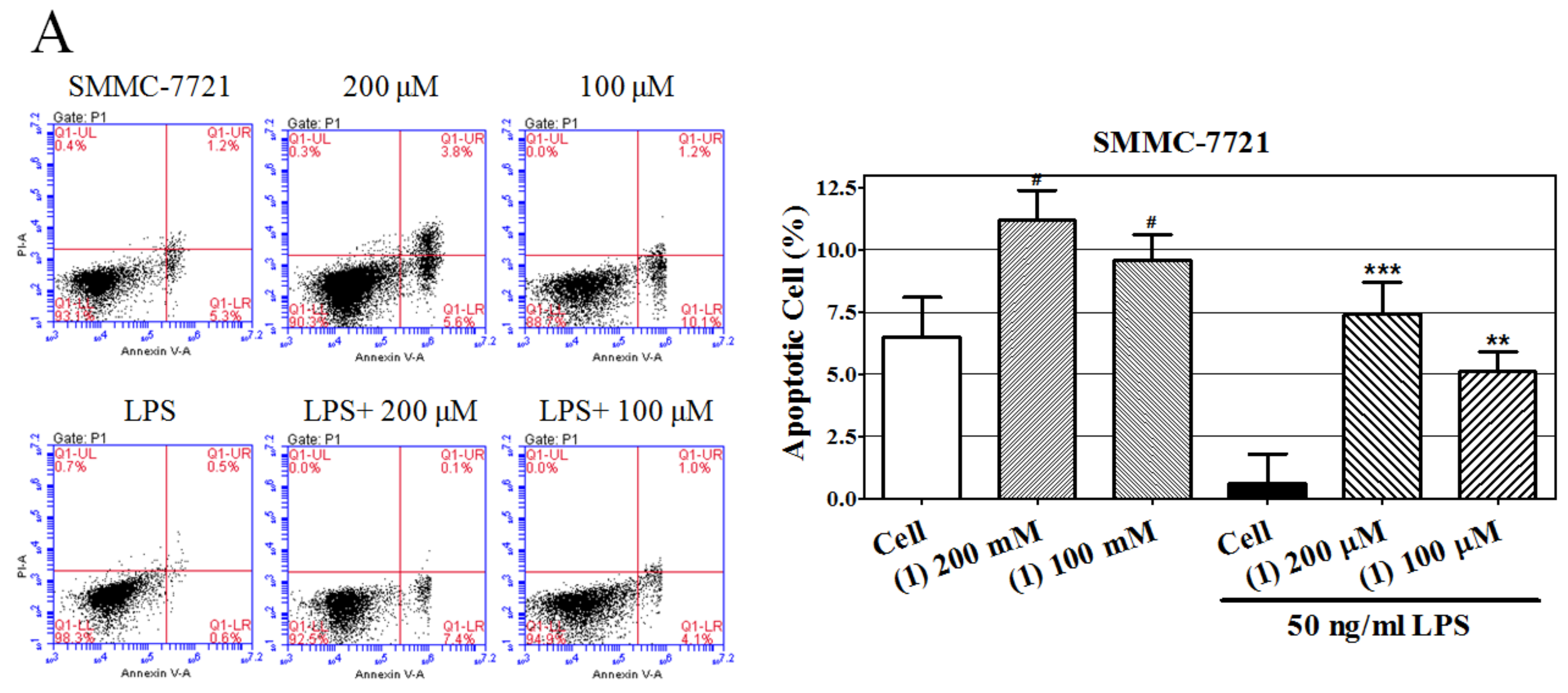
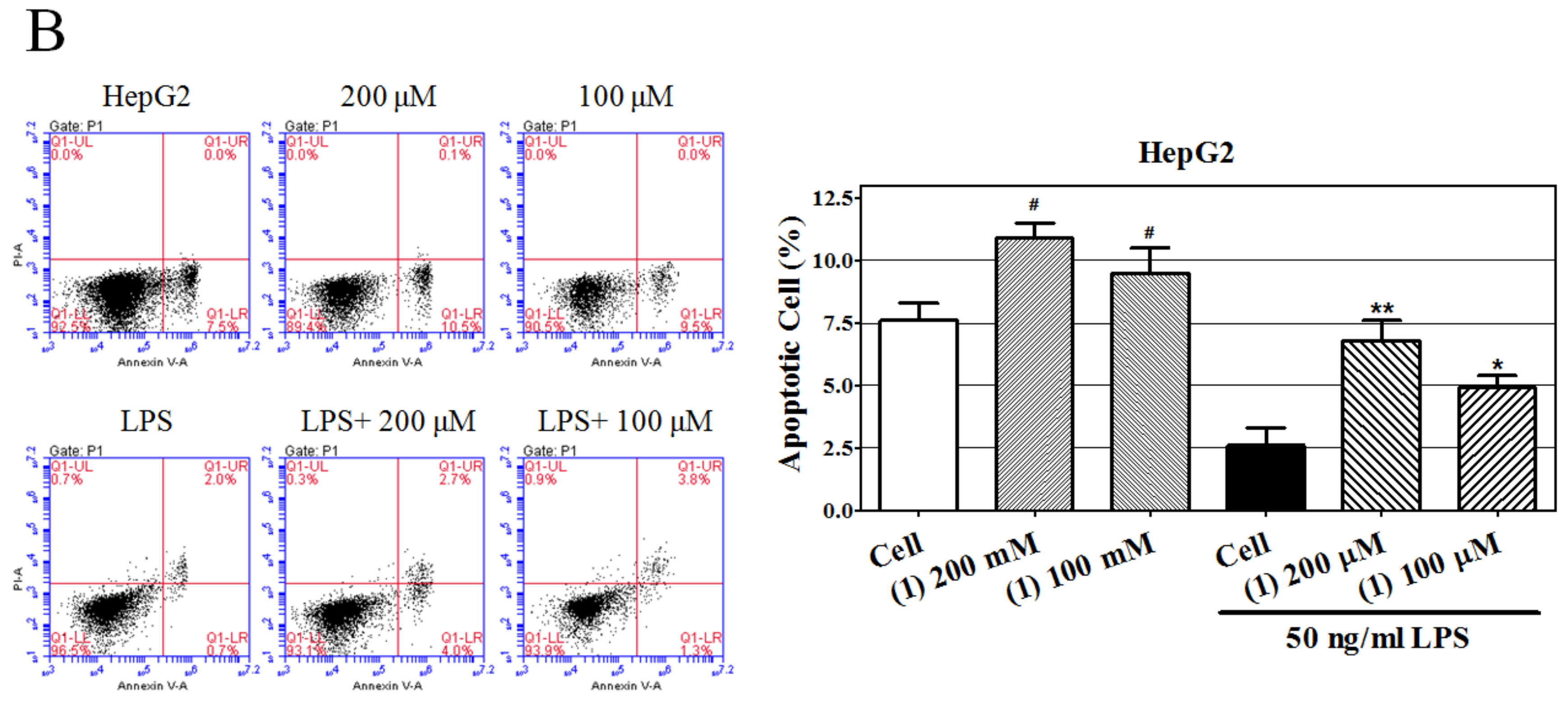
2.3. Castanol B(1)-Induced Cellular Apoptosis
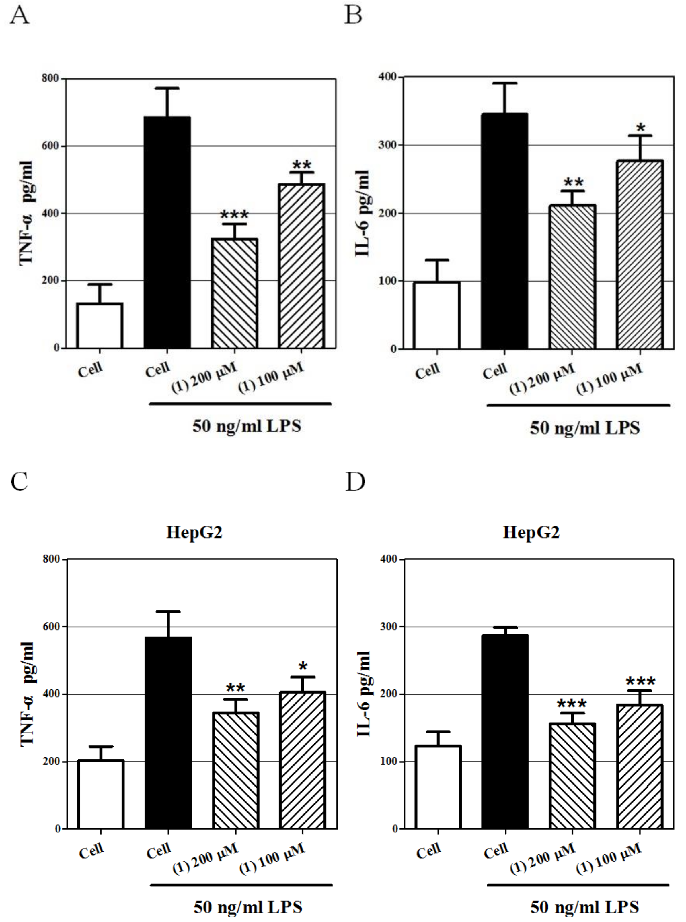
2.4. Effect of Castanol B (1) on LPS-Induced TNF-α and IL-6 Secretion in Hepatoma Cells
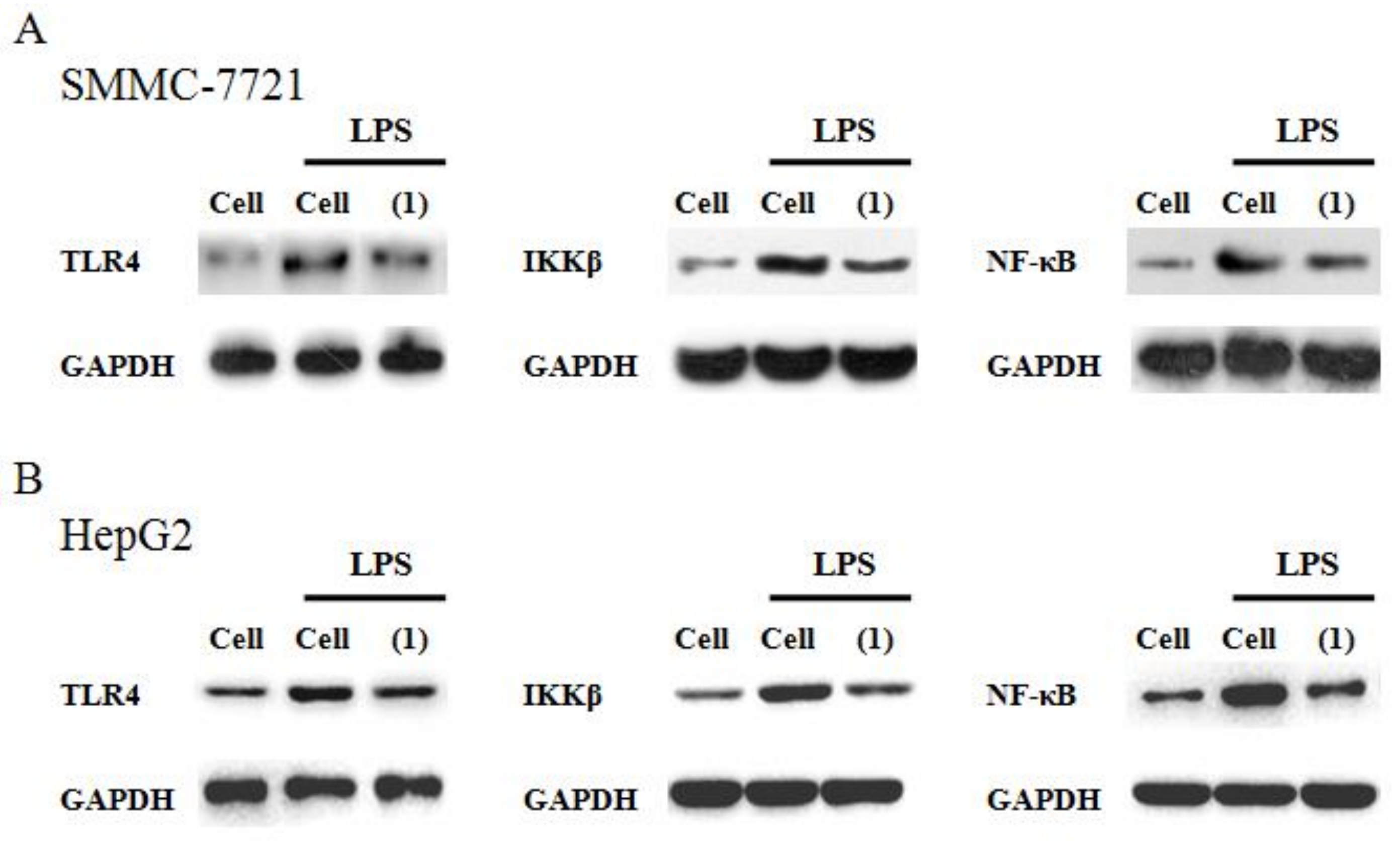
2.5. Effect Castanol B (1) on the TLR4–NF-κBSignal Pathway
3. Discussion
4. Materials and Methods
4.1. General
4.2. Plant Material
4.3. Water-Soluble Extract of CMS and Compound Isolation
4.4. Castanol B (1)
4.5. 7,8,dihydroxy-2H- chromen-3-one (2)
4.6. MTT Assay
4.7. Cell Apoptosis Assay
4.8. Cytokine Assays
4.9. Western Blotting
5. Statistical Analysis
6. Conclusions
Reference
Author Contributions
Funding
Acknowledgments
Conflicts of Interest
References
- Jia, L.; Xi, F.; Wang, N.; Jing, L.L.; Kong, D.Y. Chemical Constituents of Castanea mollissima Blume Shell. Chin. J. Pharm. 2010, 41, 98–102. [Google Scholar]
- FAOSTAT. Country Rank in the World, by Commodity: China. 1936–1944. Available online: http://www.fao.org/faostat/en/#rankings/countries_by_commodity (accessed on 23 October 2018).
- You, T.T.; Zhou, S.K.; Wen, J.L.; Ma, C.; Xu, F. Chemical Composition, Properties, and Antimicrobial Activity of the Water-Soluble Pigments from Castanea mollissima Shells. J. Agric. Food. 2014, 62, 1936–1944. [Google Scholar] [CrossRef] [PubMed]
- Zhang, H.; Ke, J.; Shao, T.; Li, J.; Duan, Y.; He, Y.; Zhang, C.; Chen, G.; Sun, G.; Sun, X. Cytotoxic effects of procyanidins from Castanea mollissimaBl. shell on human hepatoma G2 cells in vitro. Food. Chem. Toxicol. 2014, 64, 166–176. [Google Scholar] [CrossRef] [PubMed]
- Hauer, H.; Germer, S.; Elsaber, J.; Ritter, T. Benzopyranones and Their Sulfate Esters from Pelargonium sidoides. Planta Med. 2010, 76, 350–352. [Google Scholar] [CrossRef] [PubMed]
- Li, X.C.; Yang, L.X.; Wang, H.Q.; Chen, R.Y. Phenolic compounds from the aqueous extract of Acacia catechu. Chin. Chem. Letter. 2011, 22, 1331–1334. [Google Scholar] [CrossRef]
- Nawwar, M.A.M.; Hussein, S.A.M.; Merfort, I. NMR Spectral analysis of polyphenols from Puntica Granatum. Phytochemistry 1994, 36, 793–798. [Google Scholar] [CrossRef]
- Sun, Y.N.; Li, W.; Song, S.B.; Yan, X.T.; Zhao, Y.; Reum Jo, A.; Jong, S.K.; Kim, Y.H. A new phenolic derivative with soluble epoxide hydrolase and nuclear factor-kappaB inhibitory activity from the aqueous extract of Acacia catechu. Nat. Prod. Res. 2016, 30, 2085–2092. [Google Scholar] [CrossRef]
- Sun, H.; Liu, G.T. Chemopreventive effect of dimethyl dicarboxylate biphenyl on malignant transformation of WB-F344 rat liver epithelial cells. Acta Pharmacol. Sin. 2005, 26, 1339–1344. [Google Scholar] [CrossRef]
- Jin, J.; Sun, H.; Wei, H.; Liu, G. The anti-hepatitis drug DDB chemosensitizes multidrug resistant cancer cells in vitro and in vivo by inhibiting P-gp and enhancing apoptosis. Investig. New Drugs 2007, 25, 95–105. [Google Scholar] [CrossRef]
- Sun, H.; Liu, G.T. Inhibitory effect of dimethyl dicarboxylate biphenyl on invasionof humanhepatocellular carcinoma cell line MHCC97-H With high metastasis potential and its mechanisms. Chin. J. Cancer 2006, 25, 1464–1469. [Google Scholar]
- Mantovani, A.; Allavena, P.; Sica, A.; Balkwill, F. Cancer-related inflammation. Nature 2008, 454, 436–444. [Google Scholar] [CrossRef] [PubMed]
- He, G.; Karin, M. NF-κB and STAT3–key players in liver inflammation and cancer. Cell Res. 2011, 21, 159–168. [Google Scholar] [CrossRef] [PubMed]
- Wang, L.; Zhu, R.; Huang, Z.; Li, H.; Zhu, H. Lipopolysaccharide-induced toll-like receptor 4 signaling in cancer cells promotes cell survival and proliferation in hepatocellular carcinoma. Dig. Dis. Sci. 2013, 58, 2223–2236. [Google Scholar] [CrossRef]
- Li, H.; Li, Y.; Liu, D.; Liu, J. LPS promotes epithelial-mesenchymal transition and activation of TLR4/JNK signaling. Tumour Biol. 2014, 35, 10429–10435. [Google Scholar] [CrossRef] [PubMed]
- Jing, Y.Y.; Han, Z.P.; Sun, K.; Zhang, S.S.; Hou, J.; Liu, Y. Toll-like receptor 4 signaling promotes epithelial-mesenchymal transition in human hepatocellular carcinoma induced by lipopolysaccharide. BMC Med. 2012, 10, 98. [Google Scholar] [CrossRef] [PubMed]
- Drucker, C.; Parzefall, W.; Teufelhofer, O.; Grusch, M.; Ellinger, A.; Schulte-Hermann, R.; Grasl-Kraupp, B. Non-parenchymal liver cells support the growth advantage in the first stages of hepatocarcinogenesis. Carcinogenesis 2006, 27, 152–161. [Google Scholar] [CrossRef]
- Wang, Y.; Tu, Q.; Yan, W.; Xiao, D.; Zeng, Z.; Ouyang, Y.; Huang, L.; Cai, J.; Zeng, X.; Chen, Y.J.; et al. CXC195 suppresses proliferation and inflammatory response in LPS-induced human hepatocellular carcinoma cells via regulating TLR4-MyD88-TAK1-mediated NF-κB and MAPK pathway. Biochem. Biophys. Res. Commun. 2015, 456, 373–379. [Google Scholar] [CrossRef]
- He, M.; Zhang, W.; Dong, Y.; Wang, L.; Fang, T.; Tang, W.; Lv, B.; Chen, G.; Yang, B.; Huang, P.; et al. Pro-inflammation NF-κB signaling triggers a positive feedback via enhancing cholesterol accumulation in liver cancer cells. J. Exp. Clin. Cancer Res. 2017, 36, 15. [Google Scholar] [CrossRef]
- Quoilin, C.; Mouithys-Mickalad, A.; Duranteau, J.; Gallez, B.; Hoebeke, M. Endotoxin-induced basal respiration alterations of renal HK-2 cells: A sign of pathologic metabolism down-regulation. Biochem. Biophys. Res. Commun. 2012, 423, 350–354. [Google Scholar] [CrossRef]
- Roman-Blas, J.A.; Jimenez, S.A. NF-kappaB as a potential therapeutic target in osteoarthritis and rheumatoid arthritis. Osteoarthritis Cartilage 2006, 14, 839–848. [Google Scholar] [CrossRef]
- Blackwell, N.M.; Sembi, P.; Newson, J.S.; Lawrence, T.; Gilroy, D.W.; Kabouridis, P.S. Reduced infiltration and increased apoptosis of leukocytes at sites of inflammation by systemic administration of a membrane-permeable IkappaBalpha repressor. Arthritis Rheum 2004, 50, 2675–2684. [Google Scholar] [CrossRef] [PubMed]
- Hayden, M.S.; Ghosh, S. Shared principles in NF-kappaB signaling. Cell 2008, 132, 344–362. [Google Scholar] [CrossRef] [PubMed]


| No. | 1 | 2 |
|---|---|---|
| 1 | 132.2 | |
| 2 | 118.6 | 73.6 |
| 3 | 116.9 | 206.7 |
| 4 | 113.9 | 34.8 |
| 5 | 157.5 | 125.8 |
| 6 | 116.9 | 111.2 |
| 7 | 169.9 | 154.6 |
| 8 | 148.0 | |
| 9 | 108.2 | |
| 10 | 148. | |
| 1’ | 145.1 | |
| 2’ | 113.5 | |
| 3’ | 145.2 | |
| 4’ | 142.6 | |
| 5’ | 115.3 | |
| 6’ | 118.0 |
© 2019 by the authors. Licensee MDPI, Basel, Switzerland. This article is an open access article distributed under the terms and conditions of the Creative Commons Attribution (CC BY) license (http://creativecommons.org/licenses/by/4.0/).
Share and Cite
Wu, F.; Yao, X.; Xu, J.; Wu, Y.; Yang, Y.; Jin, Y.; Xie, H.; Liu, Y.; Yang, Y.; Zheng, X. One New Phenolic Compound from Castanea mollissima Shells and its Suppression of HepatomaCell Proliferation and Inflammation by Inhibiting NF-κB Pathway. Int. J. Mol. Sci. 2019, 20, 466. https://doi.org/10.3390/ijms20030466
Wu F, Yao X, Xu J, Wu Y, Yang Y, Jin Y, Xie H, Liu Y, Yang Y, Zheng X. One New Phenolic Compound from Castanea mollissima Shells and its Suppression of HepatomaCell Proliferation and Inflammation by Inhibiting NF-κB Pathway. International Journal of Molecular Sciences. 2019; 20(3):466. https://doi.org/10.3390/ijms20030466
Chicago/Turabian StyleWu, Fei, Xuan Yao, Jian Xu, Yue Wu, Yuejun Yang, Yu Jin, Huifang Xie, Yuancai Liu, Yifu Yang, and Xiangwei Zheng. 2019. "One New Phenolic Compound from Castanea mollissima Shells and its Suppression of HepatomaCell Proliferation and Inflammation by Inhibiting NF-κB Pathway" International Journal of Molecular Sciences 20, no. 3: 466. https://doi.org/10.3390/ijms20030466