Cytotoxicity of Plant-Mediated Synthesis of Metallic Nanoparticles: A Systematic Review

 ,
,
Abstract
:1. Introduction
2. Results
2.1. In Vitro Studies
2.2. In Vivo Studies
2.3. Safety of Plant-Mediated Synthesis of Metallic Nanoparticles
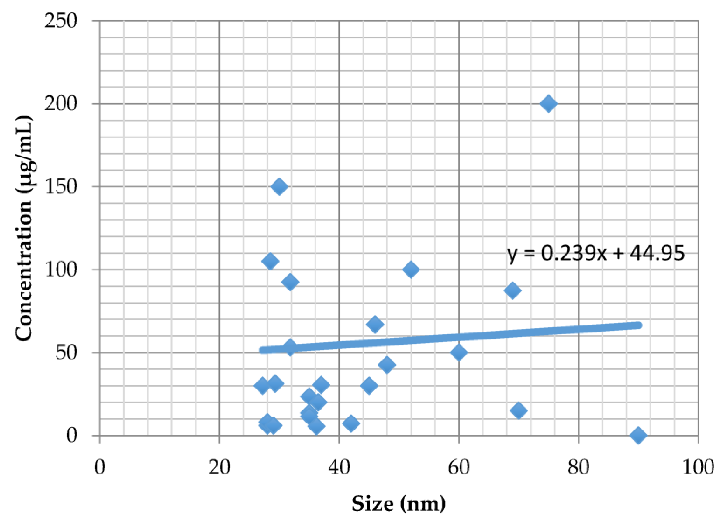
2.4. Size and Cytotoxicity
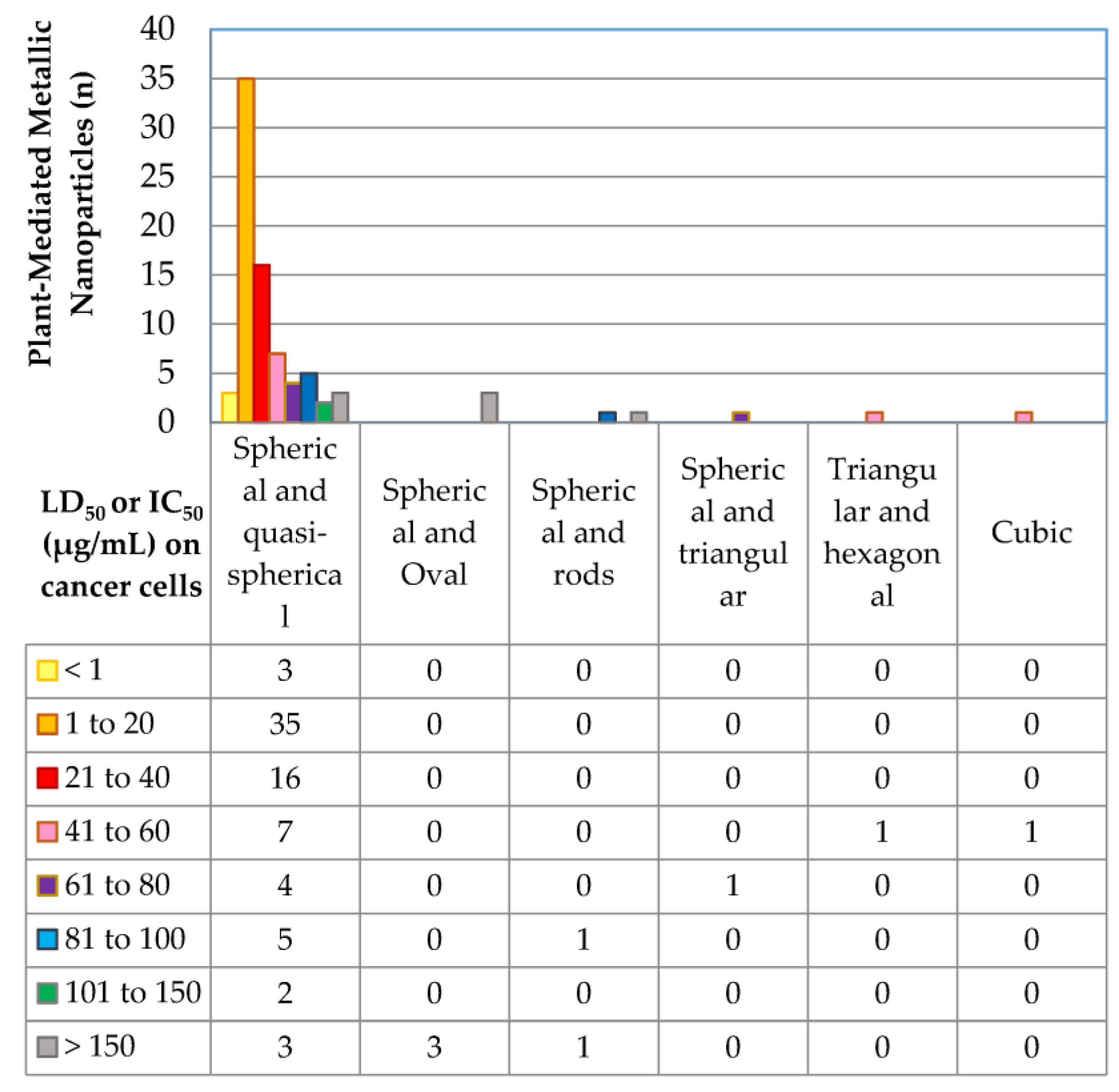
2.5. Morphology and Cytotoxicity
3. Discussion
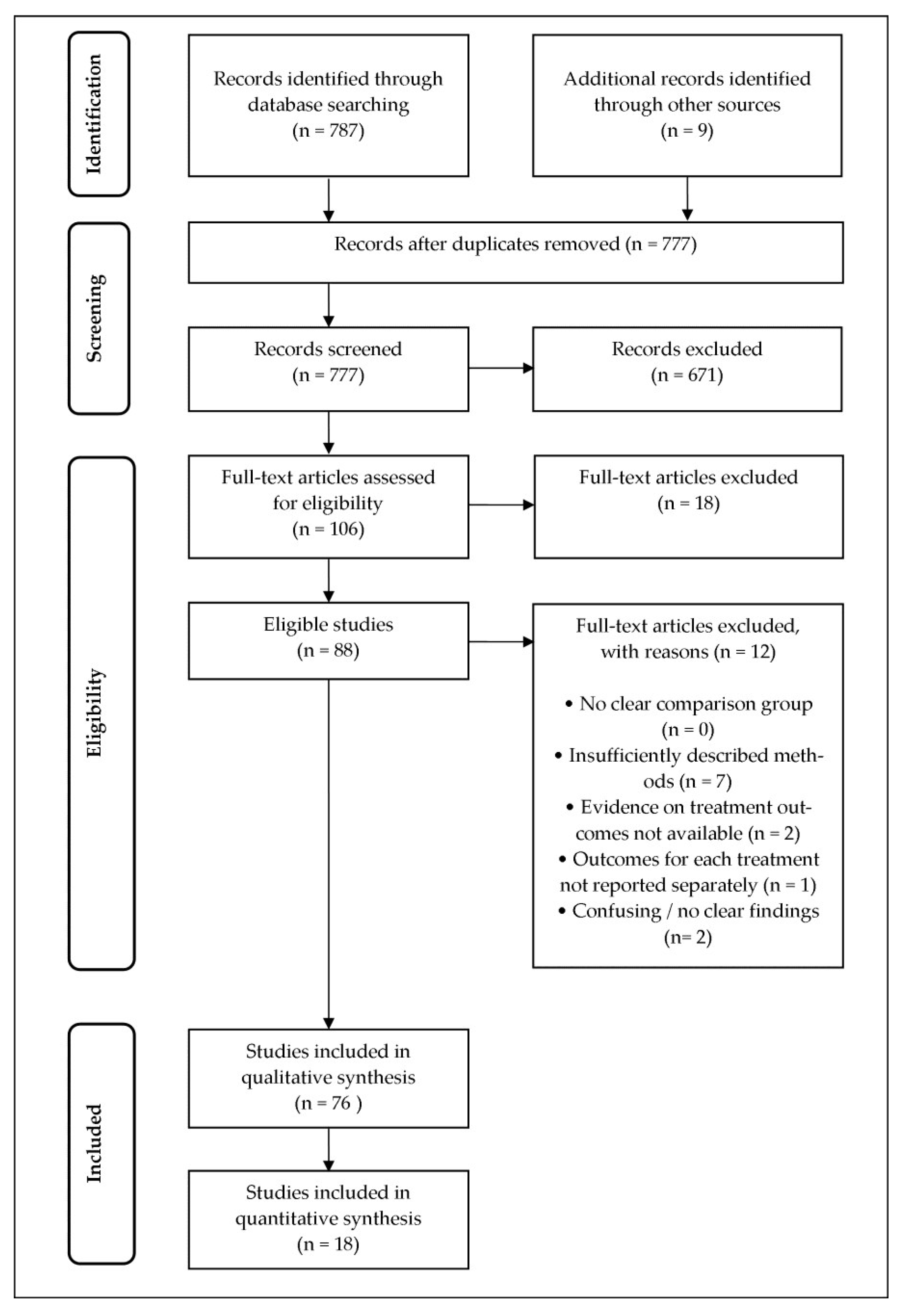
4. Materials and Methods
4.1. Search Strategy
4.2. Study Selection
5. Conclusions
Author Contributions
Acknowledgments
Conflicts of Interest
References
- Hay, S.I.; Jayaraman, S.P.; Truelsen, T.; Sorensen, R.J.; Millear, A.; Giussani, G.; Beghi, E. GBD 2015 Disease and Injury Incidence and Prevalence Collaborators. Global, regional, and national incidence, prevalence, and years lived with disability for 310 diseases and injuries, 1990–2015: A systematic analysis for the Global Burden of Disease Study 2015. Lancet 2017, 389, 1545–1602. [Google Scholar]
- Thakkar, K.N.; Mhatre, S.S.; Parikh, R.Y. Biological synthesis of metallic nanoparticles. Nanomed. Nanotechnol. Biol. Med. 2010, 6, 257–262. [Google Scholar] [CrossRef] [PubMed]
- Ahmad, N.; Sharma, S.; Alam, M.K.; Singh, V.; Shamsi, S.; Mehta, B.; Fatma, A. Rapid synthesis of silver nanoparticles using dried medicinal plant of basil. Colloids Surf. B Biointerfaces 2010, 81, 81–86. [Google Scholar] [CrossRef] [PubMed]
- Mohanpuria, P.; Rana, N.K.; Yadav, S.K. Biosynthesis of nanoparticles: Technological concepts and future applications. J. Nanopart. Res. 2008, 10, 507–517. [Google Scholar] [CrossRef]
- Kuppurangan, G.; Karuppasamy, B.; Nagarajan, K.; Sekar, R.K.; Viswaprakash, N.; Ramasamy, T. Biogenic synthesis and spectroscopic characterization of silver nanoparticles using leaf extract of Indoneesiella echioides: In vitro assessment on antioxidant, antimicrobial and cytotoxicity potential. Appl. Nanosci. 2016, 6, 973–982. [Google Scholar] [CrossRef]
- Selvarani, S. Anti-Cancer Activity of Silver Nanoparticle Synthesized from Stem Extract of Ocimum Kilimandscharicum against Hep-G2, Liver Cancer Cell Line. J. Nanotechnol. Nanosci. 2015, 1, 100103. [Google Scholar]
- Mata, R.; Nakkala, J.R.; Sadras, S.R. Catalytic and biological activities of green silver nanoparticles synthesized from Plumeria alba (frangipani) flower extract. Mater. Sci. Eng. C 2015, 51, 216–225. [Google Scholar] [CrossRef] [PubMed]
- Mishra, P.; Ray, S.; Sinha, S.; Das, B.; Khan, M.I.; Behera, S.K.; Yun, S.-I.; Tripathy, S.K.; Mishra, A. Facile bio-synthesis of gold nanoparticles by using extract of Hibiscus sabdariffa and evaluation of its cytotoxicity against U87 glioblastoma cells under hyperglycemic condition. Biochem. Eng. J. 2016, 105, 264–272. [Google Scholar]
- Dipankar, C.; Murugan, S. The green synthesis, characterization and evaluation of the biological activities of silver nanoparticles synthesized from Iresine herbstii leaf aqueous extracts. Colloids Surf. B Biointerfaces 2012, 98, 112–119. [Google Scholar] [CrossRef] [PubMed]
- Bupesh, G.; Manikandan, E.; Thanigaiarul, K.; Magesh, S.; Senthilkumar, V.; Tamilarasan, S.; Pandian, K.; Gurib-Fakim, A.; Maaza, M. Enhanced antibacterial, anticancer activity from Terminalia chebula medicinal plant rapid extract by phytosynthesis of silver nanoparticles core-shell structures. J. Nanomed. Nanotechnol. 2016, 7, 2. [Google Scholar]
- Mittal, A.K.; Tripathy, D.; Choudhary, A.; Aili, P.K.; Chatterjee, A.; Singh, I.P.; Banerjee, U.C. Bio-synthesis of silver nanoparticles using Potentilla fulgens Wall. ex Hook. and its therapeutic evaluation as anticancer and antimicrobial agent. Mater. Sci. Eng. C 2015, 53, 120–127. [Google Scholar]
- Mukundan, D.; Mohankumar, R.; Vasanthakumari, R. Green Synthesis of Silver Nanoparticles Using Leaves Extract of Bauhinia Tomentosa Linn and its Invitro Anticancer Potential. Mater. Today Proc. 2015, 2, 4309–4316. [Google Scholar] [CrossRef]
- Spector, R. Progress in the search for ideal drugs. Pharmacology 2002, 64, 1–7. [Google Scholar] [CrossRef] [PubMed]
- Parveen, A.; Rao, S. Cytotoxicity and genotoxicity of biosynthesized gold and silver nanoparticles on human cancer cell lines. J. Clust. Sci. 2015, 26, 775–788. [Google Scholar] [CrossRef]
- Nazeema, T.H.; Sugannya, P.K. Synthesis and characterization of silver nanoparticle from two medicinal plants and its anticancer property. Int. J. Res. Eng. Technol. 2014, 2, 49–56. [Google Scholar]
- Valodkar, M.; Jadeja, R.N.; Thounaojam, M.C.; Devkar, R.V.; Thakore, S. In vitro toxicity study of plant latex capped silver nanoparticles in human lung carcinoma cells. Mater. Sci. Eng. C 2011, 31, 1723–1728. [Google Scholar] [CrossRef]
- Manju, S.; Malaikozhundan, B.; Vijayakumar, S.; Shanthi, S.; Jaishabanu, A.; Ekambaram, P.; Vaseeharan, B. Antibacterial, antibiofilm and cytotoxic effects of Nigella sativa essential oil coated gold nanoparticles. Microb. Pathog. 2016, 91, 129–135. [Google Scholar] [CrossRef] [PubMed]
- Nakkala, J.R.; Mata, R.; Gupta, A.K.; Sadras, S.R. Biological activities of green silver nanoparticles synthesized with Acorous calamus rhizome extract. Eur. J. Med. Chem. 2014, 85, 784–794. [Google Scholar] [CrossRef] [PubMed]
- Venkatesan, B.; Subramanian, V.; Tumala, A.; Vellaichamy, E. Rapid synthesis of biocompatible silver nanoparticles using aqueous extract of Rosa damascena petals and evaluation of their anticancer activity. Asian Pac. J. Trop. Med. 2014, 7, S294–S300. [Google Scholar] [CrossRef]
- Sankar, R.; Karthik, A.; Prabu, A.; Karthik, S.; Shivashangari, K.S.; Ravikumar, V. Origanum vulgare mediated biosynthesis of silver nanoparticles for its antibacterial and anticancer activity. Colloids Surf. B Biointerfaces 2013, 108, 80–84. [Google Scholar] [CrossRef] [PubMed]
- Kanipandian, N.; Thirumurugan, R. A feasible approach to phyto-mediated synthesis of silver nanoparticles using industrial crop Gossypium hirsutum (cotton) extract as stabilizing agent and assessment of its in vitro biomedical potential. Ind. Crops Prod. 2014, 55, 1–10. [Google Scholar] [CrossRef]
- Sathishkumar, M.; Pavagadhi, S.; Mahadevan, A.; Balasubramanian, R. Biosynthesis of gold nanoparticles and related cytotoxicity evaluation using A549 cells. Ecotoxicol. Environ. Saf. 2015, 114, 232–240. [Google Scholar] [CrossRef] [PubMed]
- Kumar, B.; Smita, K.; Cumbal, L.; Camacho, J.; Hernández-Gallegos, E.; de Guadalupe Chávez-López, M.; Grijalva, M.; Andrade, K. One pot phytosynthesis of gold nanoparticles using Genipa americana fruit extract and its biological applications. Mater. Sci. Eng. C 2016, 62, 725–731. [Google Scholar] [CrossRef] [PubMed]
- Ashokkumar, T.; Prabhu, D.; Geetha, R.; Govindaraju, K.; Manikandan, R.; Arulvasu, C.; Singaravelu, G. Apoptosis in liver cancer (HepG2) cells induced by functionalized gold nanoparticles. Colloids Surf. B Biointerfaces 2014, 123, 549–556. [Google Scholar] [CrossRef] [PubMed]
- Inbathamizh, L.; Ponnu, T.M.; Mary, E.J. In vitro evaluation of antioxidant and anticancer potential of Morinda pubescens synthesized silver nanoparticles. J. Pharm. Res. 2013, 6, 32–38. [Google Scholar] [CrossRef]
- Patil, M.P.; Ngabire, D.; Thi, H.H.P.; Kim, M.-D.; Kim, G.-D. Eco-friendly synthesis of gold nanoparticles and evaluation of their cytotoxic activity on cancer cells. J. Clust. Sci. 2016, 28, 119–132. [Google Scholar] [CrossRef]
- Durai, P.; Chinnasamy, A.; Gajendran, B.; Ramar, M.; Pappu, S.; Kasivelu, G.; Thirunavukkarasu, A. Synthesis and characterization of silver nanoparticles using crystal compound of sodium para-hydroxybenzoate tetrahydrate isolated from Vitex negundo L leaves and its apoptotic effect on human colon cancer cell lines. Eur. J. Med. Chem. 2014, 84, 90–99. [Google Scholar] [CrossRef] [PubMed]
- Prabhu, D.; Arulvasu, C.; Babu, G.; Manikandan, R.; Srinivasan, P. Biologically synthesized green silver nanoparticles from leaf extract of Vitex negundo L. induce growth-inhibitory effect on human colon cancer cell line HCT15. Process Biochem. 2013, 48, 317–324. [Google Scholar]
- Kuppusamy, P.; Ichwan, S.J.; Al-Zikri, P.N.H.; Suriyah, W.H.; Soundharrajan, I.; Govindan, N.; Maniam, G.P.; Yusoff, M.M. In vitro anticancer activity of Au, Ag nanoparticles synthesized using Commelina nudiflora L. aqueous extract against HCT-116 colon cancer cells. Biol. Trace Elem. Res. 2016, 173, 297–305. [Google Scholar] [PubMed]
- Sengani, M.; Devi Rajeswari, V. Cytotoxicity and apoptotic effect of biogenic silver nanoparticles on human colorectal cell line HT-29. Res. J. Biotechnol. 2016, 11, 9. [Google Scholar]
- Ghozali, S.; Vuanghao, L.; Ahmad, N. Biosynthesis and Characterization of Silver Nanoparticles using Catharanthus roseus Leaf Extract and its Proliferative Effects on Cancer Cell Lines. J. Nanomed. Nanotechnol. 2015, 6, 1. [Google Scholar]
- Mata, R.; Nakkala, J.R.; Sadras, S.R. Polyphenol stabilized colloidal gold nanoparticles from Abutilon indicum leaf extract induce apoptosis in HT-29 colon cancer cells. Colloids Surf. B Biointerfaces 2016, 143, 499–510. [Google Scholar] [CrossRef] [PubMed]
- Premasudha, P.; Venkataramana, M.; Abirami, M.; Vanathi, P.; Krishna, K.; Rajendran, R. Biological synthesis and characterization of silver nanoparticles using Eclipta alba leaf extract and evaluation of its cytotoxic and antimicrobial potential. Bull. Mater. Sci. 2015, 38, 965–973. [Google Scholar] [CrossRef]
- Mata, R.; Nakkala, J.R.; Sadras, S.R. Biogenic silver nanoparticles from Abutilon indicum: Their antioxidant, antibacterial and cytotoxic effects in vitro. Colloids Surf. B Biointerfaces 2015, 128, 276–286. [Google Scholar] [CrossRef] [PubMed]
- Potara, M.; Bawaskar, M.; Simon, T.; Gaikwad, S.; Licarete, E.; Ingle, A.; Banciu, M.; Vulpoi, A.; Astilean, S.; Rai, M. Biosynthesized silver nanoparticles performing as biogenic SERS-nanotags for investigation of C26 colon carcinoma cells. Colloids Surf. B Biointerfaces 2015, 133, 296–303. [Google Scholar] [CrossRef] [PubMed]
- Gopinath, V.; Priyadarshini, S.; MubarakAli, D.; Loke, M.F.; Thajuddin, N.; Alharbi, N.S.; Yadavalli, T.; Alagiri, M.; Vadivelu, J. Anti-Helicobacter pylori, cytotoxicity and catalytic activity of biosynthesized gold nanoparticles: Multifaceted application. Arabian J. Chem. 2016. [CrossRef]
- Rashidipour, M.; Heydari, R. Biosynthesis of silver nanoparticles using extract of olive leaf: Synthesis and in vitro cytotoxic effect on MCF-7 cells. J. Nanostruct. Chem. 2014, 4, 1–6. [Google Scholar] [CrossRef]
- Sathishkumar, G.; Gobinath, C.; Wilson, A.; Sivaramakrishnan, S. Dendrophthoe falcata (Lf) Ettingsh (Neem mistletoe): A potent bioresource to fabricate silver nanoparticles for anticancer effect against human breast cancer cells (MCF-7). Spectrochim. Acta Part A 2014, 128, 285–290. [Google Scholar] [CrossRef] [PubMed]
- Reddy, N.J.; Vali, D.N.; Rani, M.; Rani, S.S. Evaluation of antioxidant, antibacterial and cytotoxic effects of green synthesized silver nanoparticles by Piper longum fruit. Mater. Sci. Eng. C 2014, 34, 115–122. [Google Scholar] [CrossRef] [PubMed]
- Farah, M.A.; Ali, M.A.; Chen, S.M.; Li, Y.; Al-Hemaid, F.M.; Abou-Tarboush, F.M.; Al-Anazi, K.M.; Lee, J. Silver nanoparticles synthesized from Adenium obesum leaf extract induced DNA damage, apoptosis and autophagy via generation of reactive oxygen species. Colloids Surf. B Biointerfaces 2016, 141, 158–169. [Google Scholar] [CrossRef] [PubMed]
- Remya, R.; Rajasree, S.R.; Aranganathan, L.; Suman, T. An investigation on cytotoxic effect of bioactive AgNPs synthesized using Cassia fistula flower extract on breast cancer cell MCF-7. Biotechnol. Rep. 2015, 8, 110–115. [Google Scholar] [CrossRef] [PubMed]
- Suganya, K.U.; Govindaraju, K.; Kumar, V.G.; Karthick, V.; Parthasarathy, K. Pectin mediated gold nanoparticles induces apoptosis in mammary adenocarcinoma cell lines. Int. J. Biol. Macromol. 2016, 93, 1030–1040. [Google Scholar] [CrossRef] [PubMed]
- Gajendran, B.; Chinnasamy, A.; Durai, P.; Raman, J.; Ramar, M. Biosynthesis and characterization of silver nanoparticles from Datura inoxia and its apoptotic effect on human breast cancer cell line MCF7. Mater. Lett. 2014, 122, 98–102. [Google Scholar] [CrossRef]
- Jeyaraj, M.; Sathishkumar, G.; Sivanandhan, G.; MubarakAli, D.; Rajesh, M.; Arun, R.; Kapildev, G.; Manickavasagam, M.; Thajuddin, N.; Premkumar, K.; et al. Biogenic silver nanoparticles for cancer treatment: An experimental report. Colloids Surf. B Biointerfaces 2013, 106, 86–92. [Google Scholar] [CrossRef] [PubMed]
- Ramar, M.; Manikandan, B.; Marimuthu, P.N.; Raman, T.; Mahalingam, A.; Subramanian, P.; Karthick, S.; Munusamy, A. Synthesis of silver nanoparticles using Solanum trilobatum fruits extract and its antibacterial, cytotoxic activity against human breast cancer cell line MCF 7. Spectrochim. Acta Part A 2015, 140, 223–228. [Google Scholar] [CrossRef] [PubMed]
- Sathishkumar, P.; Preethi, J.; Vijayan, R.; Yusoff, A.R.M.; Ameen, F.; Suresh, S.; Balagurunathan, R.; Palvannan, T. Anti-acne, anti-dandruff and anti-breast cancer efficacy of green synthesised silver nanoparticles using Coriandrum sativum leaf extract. J. Photochem. Photobiol. B Biol. 2016, 163, 69–76. [Google Scholar] [CrossRef] [PubMed]
- Sathishkumar, P.; Vennila, K.; Jayakumar, R.; Yusoff, A.R.M.; Hadibarata, T.; Palvannan, T. Phyto-synthesis of silver nanoparticles using Alternanthera tenella leaf extract: An effective inhibitor for the migration of human breast adenocarcinoma (MCF-7) cells. Bioprocess Biosyst. Eng. 2016, 39, 651–659. [Google Scholar] [CrossRef] [PubMed]
- Vivek, R.; Thangam, R.; Muthuchelian, K.; Gunasekaran, P.; Kaveri, K.; Kannan, S. Green biosynthesis of silver nanoparticles from Annona squamosa leaf extract and its in vitro cytotoxic effect on MCF-7 cells. Process Biochem. 2012, 47, 2405–2410. [Google Scholar] [CrossRef]
- Priya, M.K.; Iyer, P.R. Anticancer studies of the synthesized gold nanoparticles against MCF 7 breast cancer cell lines. Appl. Nanosci. 2015, 5, 443–448. [Google Scholar] [CrossRef]
- Lokina, S.; Stephen, A.; Kaviyarasan, V.; Arulvasu, C.; Narayanan, V. Cytotoxicity and antimicrobial activities of green synthesized silver nanoparticles. Eur. J. Med. Chem. 2014, 76, 256–263. [Google Scholar] [CrossRef] [PubMed]
- Kathiravan, V.; Ravi, S.; Ashokkumar, S. Synthesis of silver nanoparticles from Melia dubia leaf extract and their in vitro anticancer activity. Spectrochim. Acta Part A 2014, 130, 116–121. [Google Scholar] [CrossRef] [PubMed]
- Balasubramani, G.; Ramkumar, R.; Krishnaveni, N.; Pazhanimuthu, A.; Natarajan, T.; Sowmiya, R.; Perumal, P. Structural characterization, antioxidant and anticancer properties of gold nanoparticles synthesized from leaf extract (decoction) of Antigonon leptopus Hook. & Arn. J. Trace Elem. Med. Biol. 2015, 30, 83–89. [Google Scholar] [PubMed]
- Shittu, O.K.; Stephen, D.I. In vitro Membranous activity of Biosynthesized Gold Nanoparticle from Aqueous Leave Extract of Nelsonia canescens. Eur. J. Med. Plants 2016, 15, 1–8. [Google Scholar] [CrossRef]
- Aadil, K.R.; Barapatre, A.; Meena, A.S.; Jha, H. Hydrogen peroxide sensing and cytotoxicity activity of Acacia lignin stabilized silver nanoparticles. Int. J. Biol. Macromol. 2016, 82, 39–47. [Google Scholar] [CrossRef] [PubMed]
- Rajendran, R.; Ganesan, N.; Balu, S.K.; Alagar, S.; Thandavamoorthy, P.; Thiruvengadam, D. Green synthesis, characterization, antimicrobial and cytotoxic effects of silver nanoparticles using Origanum heracleoticum L. Leaf extract. Int. J. Pharm. Pharm. Sci. 2015, 7, 288–293. [Google Scholar]
- Jeyaraj, M.; Arun, R.; Sathishkumar, G.; MubarakAli, D.; Rajesh, M.; Sivanandhan, G.; Kapildev, G.; Manickavasagam, M.; Thajuddin, N.; Ganapathi, A. An evidence on G2/M arrest, DNA damage and caspase mediated apoptotic effect of biosynthesized gold nanoparticles on human cervical carcinoma cells (HeLa). Mater. Res. Bull. 2014, 52, 15–24. [Google Scholar] [CrossRef]
- Mahendran, G.; Kumari, B.R. Biological activities of silver nanoparticles from Nothapodytes nimmoniana (Graham) Mabb. fruit extracts. Food Sci. Hum. Wellness 2016, 5, 207–218. [Google Scholar] [CrossRef]
- Rajkuberan, C.; Prabukumar, S.; Sathishkumar, G.; Wilson, A.; Ravindran, K.; Sivaramakrishnan, S. Facile synthesis of silver nanoparticles using Euphorbia antiquorum L. latex extract and evaluation of their biomedical perspectives as anticancer agents. J. Saudi Chem. Soc. 2016, 21, 911–919. [Google Scholar]
- Balasubramani, G.; Ramkumar, R.; Raja, R.K.; Aiswarya, D.; Rajthilak, C.; Perumal, P. Albizia amara Roxb. Mediated Gold Nanoparticles and Evaluation of Their Antioxidant, Antibacterial and Cytotoxic Properties. J. Clust. Sci. 2017, 28, 259–275. [Google Scholar]
- Lokina, S.; Suresh, R.; Giribabu, K.; Stephen, A.; Sundaram, R.L.; Narayanan, V. Spectroscopic investigations, antimicrobial, and cytotoxic activity of green synthesized gold nanoparticles. Spectrochim. Acta Part A 2014, 129, 484–490. [Google Scholar] [CrossRef] [PubMed]
- Chanthini, A.B.; Balasubramani, G.; Ramkumar, R.; Sowmiya, R.; Balakumaran, M.D.; Kalaichelvan, P.T.; Perumal, P. Structural characterization, antioxidant and in vitro cytotoxic properties of seagrass, Cymodocea serrulata (R. Br.) Asch. & Magnus mediated silver nanoparticles. J. Photochem. Photobiol. B Biol. 2015, 153, 145–152. [Google Scholar]
- Sukirtha, R.; Priyanka, K.M.; Antony, J.J.; Kamalakkannan, S.; Thangam, R.; Gunasekaran, P.; Krishnan, M.; Achiraman, S. Cytotoxic effect of Green synthesized silver nanoparticles using Melia azedarach against in vitro HeLa cell lines and lymphoma mice model. Process Biochem. 2012, 47, 273–279. [Google Scholar] [CrossRef]
- Sulaiman, G.M.; Mohammed, W.H.; Marzoog, T.R.; Al-Amiery, A.A.A.; Kadhum, A.A.H.; Mohamad, A.B. Green synthesis, antimicrobial and cytotoxic effects of silver nanoparticles using Eucalyptus chapmaniana leaves extract. Asian Pac. J. Trop. Biomed. 2013, 3, 58–63. [Google Scholar] [CrossRef]
- Geetha, R.; Ashokkumar, T.; Tamilselvan, S.; Govindaraju, K.; Sadiq, M.; Singaravelu, G. Green synthesis of gold nanoparticles and their anticancer activity. Cancer Nanotechnol. 2013, 4, 91. [Google Scholar] [CrossRef] [PubMed]
- Mollick, M.M.R.; Rana, D.; Dash, S.K.; Chattopadhyay, S.; Bhowmick, B.; Maity, D.; Mondal, D.; Pattanayak, S.; Roy, S.; Chakraborty, M.; et al. Studies on green synthesized silver nanoparticles using Abelmoschus esculentus (L.) pulp extract having anticancer (in vitro) and antimicrobial applications. Arabian J. Chem. 2015. [Google Scholar] [CrossRef] [Green Version]
- Pattanayak, S.; Mollick, M.M.R.; Maity, D.; Chakraborty, S.; Dash, S.K.; Chattopadhyay, S.; Roy, S.; Chattopadhyay, D.; Chakraborty, M. Butea monosperma bark extract mediated green synthesis of silver nanoparticles: Characterization and biomedical applications. J. Saudi Chem. Soc. 2015. [CrossRef]
- Nayak, D.; Ashe, S.; Rauta, P.R.; Kumari, M.; Nayak, B. Bark extract mediated green synthesis of silver nanoparticles: Evaluation of antimicrobial activity and antiproliferative response against osteosarcoma. Mater. Sci. Eng. C 2016, 58, 44–52. [Google Scholar] [CrossRef] [PubMed]
- Karuppaiya, P.; Satheeshkumar, E.; Chao, W.-T.; Kao, L.-Y.; Chen, E.C.-F.; Tsay, H.-S. Anti-metastatic activity of biologically synthesized gold nanoparticles on human fibrosarcoma cell line HT-1080. Colloids Surf. B Biointerfaces 2013, 110, 163–170. [Google Scholar] [CrossRef] [PubMed]
- Rosarin, F.S.; Arulmozhi, V.; Nagarajan, S.; Mirunalini, S. Antiproliferative effect of silver nanoparticles synthesized using amla on Hep2 cell line. Asian Pac. J. Trop. Med. 2013, 6, 1–10. [Google Scholar] [CrossRef]
- Jacob, S.J.P.; Finub, J.; Narayanan, A. Synthesis of silver nanoparticles using Piper longum leaf extracts and its cytotoxic activity against Hep-2 cell line. Colloids Surf. B Biointerfaces 2012, 91, 212–214. [Google Scholar] [CrossRef] [PubMed]
- Velammal, S.P.; Devi, T.A.; Amaladhas, T.P. Antioxidant, antimicrobial and cytotoxic activities of silver and gold nanoparticles synthesized using Plumbago zeylanica bark. J. Nanostruct. Chem. 2016, 6, 247–260. [Google Scholar] [CrossRef]
- Chowdhury, N.R.; Macgregor-Ramiasa, M.; Zilm, P.; Majewski, P.; Vasilev, K. ‘Chocolate’ silver nanoparticles: Synthesis, antibacterial activity and cytotoxicity. J. Colloid Interface Sci. 2016, 482, 151–158. [Google Scholar] [CrossRef] [PubMed]
- Yang, N.; Weihong, L.; Hao, L. Biosynthesis of Au nanoparticles using agricultural waste mango peel extract and its in vitro cytotoxic effect on two normal cells. Mater. Lett. 2014, 134, 67–70. [Google Scholar] [CrossRef]
- Kumar, S.D.; Singaravelu, G.; Murugan, K.; Ajithkumar, S.; Sivashanmugam, K.; Nicoletti, M.; Benelli, G. Aegiceras corniculatum-Mediated Green Synthesis of Silver Nanoparticles: Biophysical Characterization and Cytotoxicity on Vero Cells. J. Clust. Sci. 2017, 28, 277–285. [Google Scholar] [CrossRef]
- Kalpana, D.; Pichiah, P.T.; Sankarganesh, A.; Park, W.S.; Lee, S.M.; Wahab, R.; Cha, Y.S.; Lee, Y.S. Biogenesis of gold nanoparticles using plant powders and assessment of in vitro cytotoxicity in 3T3-L1 cell line. J. Pharm. Innov. 2013, 8, 265–275. [Google Scholar] [CrossRef]
- Gogoi, N.; Babu, P.J.; Mahanta, C.; Bora, U. Green synthesis and characterization of silver nanoparticles using alcoholic flower extract of Nyctanthes arbortristis and in vitro investigation of their antibacterial and cytotoxic activities. Mater. Sci. Eng. C 2015, 46, 463–469. [Google Scholar] [CrossRef] [PubMed]
- Krishnaraj, C.; Harper, S.L.; Yun, S.-I. In Vivo toxicological assessment of biologically synthesized silver nanoparticles in adult Zebrafish (Danio rerio). J. Hazard. Mater. 2016, 301, 480–491. [Google Scholar] [CrossRef] [PubMed]
- Pan, Y.; Neuss, S.; Leifert, A.; Fischler, M.; Wen, F.; Simon, U.; Schmid, G.; Brandau, W.; Jahnen-Dechent, W. Size-dependent cytotoxicity of gold nanoparticles. Small 2007, 3, 1941–1949. [Google Scholar] [CrossRef] [PubMed]
- Yu, L.X.; Jiang, W.; Zhang, X.; Lionberger, R.; Makhlouf, F.; Schuirmann, D.J.; Muldowney, L.; Chen, M.L.; Davit, B.; Conner, D.; et al. Novel bioequivalence approach for narrow therapeutic index drugs. Clin. Pharmacol. Ther. 2015, 97, 286–291. [Google Scholar] [CrossRef] [PubMed]
- Muller, P.Y.; Milton, M.N. The determination and interpretation of the therapeutic index in drug development. Nat. Rev. Drug Discov. 2012, 11, 751–761. [Google Scholar] [CrossRef] [PubMed]
- Huang, X.; Teng, X.; Chen, D.; Tang, F.; He, J. The effect of the shape of mesoporous silica nanoparticles on cellular uptake and cell function. Biomaterials 2010, 31, 438–448. [Google Scholar] [CrossRef] [PubMed]
- Nel, A.; Xia, T.; Mädler, L.; Li, N. Toxic potential of materials at the nanolevel. Science 2006, 311, 622–627. [Google Scholar] [CrossRef] [PubMed]
- Yakop, F.; Abd Ghafar, S.A.; Yong, Y.K.; Saiful Yazan, L.; Mohamad Hanafiah, R.; Lim, V.; Eshak, Z. Silver nanoparticles Clinacanthus nutans leaves extract induced apoptosis towards oral squamous cell carcinoma cell lines. Artif. Cells Nanomed. Biotechnol. 2018. [CrossRef] [PubMed]
- Moher, D.; Shamseer, L.; Clarke, M.; Ghersi, D.; Liberati, A.; Petticrew, M.; Shekelle, P.; Stewart, L.A. Preferred reporting items for systematic review and meta-analysis protocols (PRISMA-P) 2015 statement. Syst. Rev. 2015, 4, 1. [Google Scholar] [CrossRef] [PubMed] [Green Version]
- Kim, S.Y.; Park, J.E.; Lee, Y.J.; Seo, H.-J.; Sheen, S.-S.; Hahn, S.; Jang, B.-H.; Son, H.-J. Testing a tool for assessing the risk of bias for nonrandomized studies showed moderate reliability and promising validity. J. Clin. Epidemiol. 2013, 66, 408–414. [Google Scholar] [CrossRef] [PubMed]
- Clark, M.A.; Finkel, R.; Rey, J.A.; Whalen, K. Lippincott’s Illustrated Reviews: Pharmacology; Lippincott Williams & Wilkins: Philadelphia, PA, USA, 2012. [Google Scholar]
 Plant metallic nanoparticles;
  Trendline.
Plant metallic nanoparticles; Trendline.
Trendline.
Plant metallic nanoparticles; Trendline.
 Plant metallic nanoparticles;
  Trendline.
Plant metallic nanoparticles; Trendline.
Trendline.
Plant metallic nanoparticles; Trendline.


| Cancer Cell Line | LD50 or IC50 | Cell Death | Exposure Duration | Response Relationship | Metallic Nanoparticle | Plant Used | Plant Part | Mechanism of Action | Ref. | Year | |
|---|---|---|---|---|---|---|---|---|---|---|---|
| Lung | A549 | NA | Complete cell death at 10 µg/mL | 4 h | Dose-dependent | Ag | Cassia auriculata | Leaf | Not studied | [14] | 2015 |
| 10 µg/mL | Complete cell death 30 µg/mL | 4 h | Dose-dependent | Au | Cassia auriculata | Leaf | Not studied | ||||
| 13.5 µg/mL | >80% cell death at >40 µg/mL | 4 h | Dose-dependent | Ag | Jatropha gossypifolia | Stem | Not studied | [15] | 2014 | ||
| 19.5 µg/mL | >80% cell death at >40 µg/mL | 4 h | Dose-dependent | Ag | Jatropha curcus | Stem | Not studied | ||||
| 20 µg/mL | NA | 24 h | Dose-dependent | Ag | Euphorbia nivulia | Latex | Apoptosis | [16] | 2011 | ||
| 28.125 µg/mL | NA | 24 h | Dose-dependent | Ag | Bauhinia tomentosa (Kanchini) | Leaf | Not studied | [12] | 2015 | ||
| 28.37 µg/mL | NA | 24 h | Dose-dependent | Au | Nigella sativa | Essential oil from seed | Not studied | [17] | 2016 | ||
| 53.2 µg/mL | NA | 24 h | Time and Dose-dependent | Ag | Acorous calamus | Rhizome | Apoptosis | [18] | 2014 | ||
| 80 µg/mL | NA | 24 h | Dose-dependent | Ag | Rosa damascena | Flower petal | Not studied | [19] | 2014 | ||
| 100 µg/mL | <20% at 500 µg/mL | 36 h | Dose-dependent | Ag | Origanum vulgare | Leaf | Reduce cell proliferation, increase ROS, DNA fragmentation, apoptosis | [20] | 2013 | ||
| 30 µg/mL | NA | 48 h | Dose-dependent | Ag | Indoneesiella echioides | Leaf | Not studied | [5] | 2016 | ||
| 32.1 µg/mL | NA | 48 h | Time and Dose-dependent | Ag | Acorous calamus | Rhizome | Apoptosis | [18] | 2014 | ||
| 40 µg/mL | NA | 48 h | Dose-dependent | Ag | Gossypium hirsutum | Leaf | Inhibit cell proliferation, induce loss of cell membrane integrity, apoptosis | [21] | 2014 | ||
| NA | 80.2% at 200 nM | 48 h | Dose-dependent | Au | Illicum verum(Star Anise) | Deseeded pod | Apoptosis | [22] | 2015 | ||
| NA | <10% cell death in 0.01–20 µM | 48 h | Non-cytotoxic | Au | Genipa americana | Fruit | Not studied | [23] | 2016 | ||
| Liver | HepG2 | 6 µg/mL | NA | 24 h | Time and Dose-dependent | Au | Cajanus cajan | Seed coat | Apoptosis | [24] | 2014 |
| NA | ≈80% cell death at 2 µg/mL | 48 h | |||||||||
| 49.5 µg/mL | >80% cell death at 100 µg/mL | 48 h | Dose-dependent | Ag | Oocimum kilimandscharicum | Stem | Not studied | [6] | 2015 | ||
| NA (GI50 = 93.75 µg/mL) | 16.39% cell death at 1 mg/mL | 48 h | Dose-dependent | Ag | Morinda pubescens | Leaf | Not studied | [25] | 2013 | ||
| Hep3B | 150 µg/mL | 20% cell death at 200 µg/mL | 24 h | Dose-dependent | Au | Rhus chinensis | Plant gall | Not studied | [26] | 2016 | |
| Colorectal | HCT15 | 8 µg/mL | NA | 24 h | Time and Dose-dependent | Ag | Vitex negundo | Leaf | Inhibit proliferation, cell cycle arrest, apoptosis | [27] | 2014 |
| 4 µg/mL | NA | 48 h | 2014 | ||||||||
| 20 µg/mL | NA | 48 h | Dose-dependent | Ag | Vitex negundo | Leaf | Apoptosis, cell cycle arrest | [28] | 2013 | ||
| HCT116 | 100 µg/mL | >60% cell death at 400 µg/mL | 24 h | Dose-dependent | Ag | Commelina nudiflora | Not stated | Apoptosis | [29] | 2016 | |
| 200 µg/mL | >70% cell death at 400 µg/mL | 24 h | Dose-dependent | Au | Commelina nudiflora | Not stated | Apoptosis | ||||
| HT29 | 30 µg/mL | NA | 12 h | Time and Dose-dependent | Ag | Couroupita guainensis | Leaf | Not studied | [30] | 2016 | |
| 6 µg/mL | NA | 24 h | Time and Dose-dependent | Ag | Vitex negundo | Leaf | Inhibit proliferation, cell cycle arrest, apoptosis | [27] | 2014 | ||
| 15 µg/mL | NA | 24 h | Time and Dose-dependent | Ag | Terminalia chebula | Fruit | Not studied | [10] | 2016 | ||
| 23.44 µg/mL | NA | 24 h | Dose-dependent | Ag | Catharanthus roseus | Leaf | Not studied | [31] | 2015 | ||
| 25 µg/mL | >75% cell death at 40 µg/mL | 24 h | Time and Dose-dependent | Ag | Couroupita guainensis | Leaf | Not studied | [30] | 2016 | ||
| 210 µg/mL | NA | 24 h | Dose-dependent | Au | Abutilon indicum | Leaf | DNA damage, arrest cell cycle, apoptosis | [32] | 2016 | ||
| 2 µg/mL | NA | 48 h | Time and Dose-dependent | Ag | Vitex negundo | Leaf | Inhibit proliferation, cell cycle arrest, apoptosis | [27] | 2014 | ||
| 10 µg/mL | NA | 48 h | Time and Dose-dependent | Ag | Terminalia chebula | Fruit | Not studied | [10] | 2016 | ||
| 20 µg/mL | >75% cell death at 40 µg/mL | 48 h | Time and Dose-dependent | Ag | Couroupita guainensis | Leaf | Not studied | [30] | 2016 | ||
| 39.06 µg/mL | NA | 48 h | Dose-dependent | Ag | Catharanthus roseus | Leaf | Not studied | [31] | 2015 | ||
| 180 µg/mL | NA | 48 h | Dose-dependent | Au | Abutilon indicum | Leaf | DNA damage, arrest cell cycle, apoptosis | [32] | 2016 | ||
| 46.88 µg/mL | NA | 72 h | Dose-dependent | Ag | Catharanthus roseus | Leaf | Not studied | [31] | 2015 | ||
| Caco-2 | 10 µM | ≈80% cell death at 50 µM | 48 h | Dose-dependent | Ag | Eclipta alba | Leaf | Not studied | [33] | 2015 | |
| Colo 205 | 4 µg/mL | NA | 24 h | Time and Dose-dependent | Ag | Abutilon inducum | Leaf | DNA damage, arrest cell cycle, apoptosis | [32] | 2016 | |
| 5.5 µg/mL | NA | 24 h | Time and Dose-dependent | Ag | Plumeria alba | Flower petal | Apoptosis | [7] | 2015 | ||
| 3 µg/mL | NA | 48 h | Time and Dose-dependent | Ag | Abutilon inducum | Leaf | DNA damage, arrest cell cycle, apoptosis | [34] | 2015 | ||
| 4.5 µg/mL | NA | 48 h | Time and Dose-dependent | Ag | Plumeria alba | Flower petal | Apoptosis | [7] | 2015 | ||
| C26 (murine) | NA | <20% at 6 µg/mL and >80% at 8 µg/mL | 24 h | Dose-dependent | Ag | Azadirachta indica | Leaf | Not studied | [35] | 2015 | |
| Stomach | AGS | NA | <30% cell death in 3.125 to 200 µg/mL for all duration | 8, 16, 24 h | Minimally Dose-dependent | Au | Tribulus terrestris | Fruit | Apoptosis | [36] | 2016 |
| MKN 28 | 150 µg/mL | 80% at 200 µg/mL | 24 h | Dose-dependent | Au | Rhus chinensis | Plant gall | Not studied | [26] | 2016 | |
| Breast | MCF7 | 0.024 µg/mL | NA | 24 h | Dose-dependent | Ag | Oleo europaea | Leaf | Not studied | [37] | 2014 |
| 4.91 µg/mL | NA | 24 h | Dose-dependent | Ag | Potentilla fulgens | Root | Apoptosis | [11] | 2015 | ||
| 5 µg/mL | NA | 24 h | Dose-dependent | Ag | Dendrophthoe falcata | Leaf | Not studied | [38] | 2014 | ||
| 67 µg/mL | NA | 24 h | Time and Dose-dependent | Ag | Piper longum | Fruit | Not studied | [39] | 2014 | ||
| 217 µg/mL | NA | 24 h | Dose-dependent | Ag | Adenium abesum | Leaf | DNA damage, autophagy via increased ROS, apoptosis | [40] | 2016 | ||
| 7.19 µg/mL | NA | 24 h | Dose-dependent | Ag | Cassia fistula | Flower | Apoptosis | [41] | 2015 | ||
| <8 µg/mL | NA | 24 h | Time and Dose-dependent | Au | Musa paradisiaca | Pectin | Apoptosis | [42] | 2016 | ||
| 20 µg/mL | NA | 24 h | Dose-dependent | Ag | Datura inoxia | Leaf | Growth suppression, cell cycle arrest, DNA synthesis reduction, apoptosis | [43] | 2014 | ||
| 20 µg/mL | Complete cell death at 50 µg/mL | 24 h | Dose-dependent | Ag | Sesbania grandiflora | Leaf | DNA damage, oxidative stress induction, apoptosis | [44] | 2013 | ||
| 30 µg/mL | NA | 24 h | Dose-dependent | Ag | Solnum trilobatum | Fruit (unripe) | Apoptosis | [45] | 2015 | ||
| 30.5 µg/mL | Complete cell inhibition at 100 µg/mL | 24 h | Dose-dependent | Ag | Coriandrum sativum | Leaf | Not studied | [46] | 2016 | ||
| 42.5 µg/mL | 98% cell inhibition at 100 µg/mL | 24 h | Dose-dependent | Ag | Alternanthera tenella | Leaf | Not studied | [47] | 2016 | ||
| 50 µg/mL | NA | 24 h | Time and Dose-dependent | Ag | Annona squamosa | Leaf | Apoptosis | [48] | 2012 | ||
| NA | ≈80% cell death at 2 µg/mL | 24 h | Inversely Dose-dependent | Au | Camellia sinensis, Coriandrum sativum, Mentha arvensis, Phyllanthus amarus, Artabotrys hexapetalus, Mimusops elengi, Syzygium aromaticum, C. sinensis | Leaf | Not studied | [49] | 2015 | ||
| 8 µg/mL | NA | 48 h | Time and Dose-dependent | Au | Musa paradisiaca | Pectin | Apoptosis | [42] | 2016 | ||
| 30 µg/mL | NA | 24 h | Time and Dose-dependent | Ag | Annona squamosa | Leaf | Apoptosis | [48] | 2012 | ||
| 10 µg/mL | NA | 48 h | Time and Dose-dependent | Ag | Malus domestica | Fruit | Not studied | [50] | 2014 | ||
| 31.2 µg/mL | NA | 48 h | Dose-dependent | Ag | Melia dubia | Leaf | Not studied | [51] | 2014 | ||
| 51 µg/mL | NA | 48 h | Time and Dose-dependent | Ag | Piper longum | Fruit | Not studied | [39] | 2014 | ||
| NA (GI50 = 257.8 µg/mL) | NA | 48 h | Dose-dependent | Au | Antigonon leptopus | Leaf | Not studied | [52] | 2015 | ||
| 0.455 µg/mL | NA | 72 h | Dose-dependent | Au | Nelsonia canescens | Leaf | Not studied | [53] | 2016 | ||
| NA | <60% cell death at 5 µg/mL and above | 72 h | Dose-dependent | Ag | Acacia | Lignin from wood | Not studied | [54] | 2016 | ||
| 100 µg/mL | >80% cell death at 500 µg/mL | 120 h | Dose-dependent | Ag | Origanum heracleoticum | Leaf | Not studied | [55] | 2015 | ||
| MDA-MB-231 | <10 µg/mL | Complete cell death at 10 µg/mL | 4 h | Dose-dependent | Ag | Cassia auriculata | Leaf | Not studied | [14] | 2015 | |
| 10 µg/mL | Complete cell death at 30 µg/mL | 4 h | Dose-dependent | Au | Cassia auriculata | Leaf | Not studied | ||||
| <2 µg/mL | NA | 24 h | Time and Dose-dependent | Au | Musa paradisiaca | Pectin | Apoptosis | [42] | 2016 | ||
| 2 µg/mL | NA | 48 h | Dose-dependent | Au | Musa paradisiaca | Pectin | Apoptosis | [42] | 2016 | ||
| Cervix | HeLa | 51 µg/mL | 88% cell death at 300 µg/mL | 3 h | Dose-dependent | Ag | Iresine herbstii | Leaf | Not studied | [9] | 2012 |
| 20 µg/mL | NA | 24 h | Dose-dependent | Au | Podophyllum hexandrum | Leaf | DNA damage, oxidative stress induction, apoptosis | [56] | 2014 | ||
| 87.32 µg/mL | NA | 24 h | Dose-dependent | Ag | Nothapodytes nimmoniana | Fruit (ripe) | Not studied | [57] | 2016 | ||
| 92.48 µg/mL | NA | 24 h | Time and Dose-dependent | Ag | Acorous calamus | Rhizome | Apoptosis | [18] | 2014 | ||
| 28 µg/mL | NA | 48 h | Dose-dependent | Ag | Euphorbia antiquorum | Latex | ROS | [58] | 2016 | ||
| 47.77 µg/mL | NA | 48 h | Dose-dependent | Au | Albizia amara | Leaf | Not studied | [59] | 2017 | ||
| 62.5 µg/mL | Almost 100% cell death at 1000 µg/mL | 48 h | Dose-dependent | Au | Punica granatum | Fruit | Not studied | [60] | 2014 | ||
| 69.44 µg/mL | NA | 48 h | Time and Dose-dependent | Ag | Acorous calamus | Rhizome | Apoptosis | [18] | 2014 | ||
| NA (GI50 = 34.5 µg/mL) | NA | 48 h | Dose-dependent | Ag | Cymodocea serrulata | Whole plant | Not studied | [61] | 2015 | ||
| 300 µg/mL | NA | 48–72 h | Dose-dependent | Ag | Melia azedarach | Leaf | Apoptosis | [62] | 2012 | ||
| Brain | U87 | 8.23 µg/mL | NA | 24 h | Dose-dependent | Ag | Potentilla fulgens | Root | Apoptosis | [11] | 2015 |
| 1.5 ng/mL | NA | 48 h | Dose-dependent | Au | Hibiscus sabdariffa | Leaf and stem (optimal: leaf) | GADPH enzyme degradation | [8] | 2016 | ||
| Blood | HL-60 | 2 mmol/L | NA | 6 h | Time and Dose-dependent | Ag | Eucalyptus chapmania | Leaf | Not studied | [63] | 2013 |
| 1 mmol/L | NA | 24 h | |||||||||
| 5.14 μM | NA | 120 h | Dose-dependent | Au | Couroupita guianensis | Flower | Apoptosis | [64] | 2013 | ||
| Jurkat | 13.64 µg/mL | NA | 24 h | Dose-dependent | Ag | Abelmoschus esculentus | Pulp | ROS and NO production | [65] | 2015 | |
| 27.35 µg/mL | NA | 24 h | Dose-dependent | Ag | Catharanthus roseus | Leaf | Not studied | [31] | 2015 | ||
| 39.06 µg/mL | NA | 48 h | |||||||||
| 46.88 µg/mL | NA | 72 h | Dose-dependent | ||||||||
| KG-1A | 11.47 µg/mL | NA | 24 h | Dose-dependent | Ag | Butea monosperma | Bark | Apoptosis | [66] | 2015 | |
| Bone | MG63 | 150 µg/mL | 80% at 200 µg/mL | 24 h | Dose-dependent | Au | Rhus chinensis | Plant gall | Not studied | [26] | 2016 |
| 75.5 ± 2.4 µg/mL | NA | 48 h | Dose-dependent | Ag | Ficus benghalensis | Bark | Not studied | [67] | 2016 | ||
| 81.8 ± 2.6µg/mL | NA | 48 h | Dose-dependent | Ag | Azadirachta indica | Bark | Not studied | ||||
| Connective tissue | HT1080 | NA | <5% cell death at up to 200µM | 6–24 h | Non-cytotoxic | Au | Dysosma pleiantha | Rhizome | Cell migration inhibition via Rac1 mediated actin polymerization pathway | [68] | 2013 |
| Prostate | LNCap-FGC | <10 µg/mL | Complete cell death at 10 µg/mL | 4 h | Dose-dependent | Ag | Cassia auriculata | Leaf | Not studied | [14] | 2015 |
| 10 µg/mL | Complete cell death at 30 µg/mL | 4 h | Dose-dependent | Au | Cassia auriculata | Leaf | Not studied | ||||
| Skin | A375 | NA | >75% cell death at 5 µg/mL | 72 h | Dose-dependent | Ag | Acacia | Lignin from wood | Not studied | [54] | 2016 |
| Throat | Hep-2 | 20 µg/mL | Complete cell death at 40 µg/mL | 24 h | Dose-dependent | Ag | Phyllanthus emblica | Fruit | Cell proliferation reduction, ROS production, DNA fragmentation, apoptosis | [69] | 2013 |
| 31.25 µg/mL | 94.02% at 500 µg/mL | 24 h | Dose-dependent | Ag | Piper longum | Leaf | ROS | [70] | 2012 | ||
| Dalton’sAscitesLymphoma (DAL) | NA | 65.61% cell death at 150 µg/mL | 24 h | Dose-dependent | Ag | Plumbago zeylanica | Bark | Not studied | [71] | 2016 | |
| NA | 61.56% cell death at 150 µg/mL | 24 h | Dose-dependent | Au | |||||||
| Healthy Cell Line | LD50or IC50 | Cell Death | Exposure Duration | Response Relationship | Metallic Nanoparticle | Plant Used | Plant Part | Mechanism of Action | Ref. | Year | |
|---|---|---|---|---|---|---|---|---|---|---|---|
| Blood | Lymphocyte | NA | <20% cell death at 6 µg/mL | 24 h | Dose-dependent | Ag | Potentilla fulgens | Root | Apoptosis | [11] | 2015 |
| PBMC | 43.18 µg/mL | NA | 24 h | Dose-dependent | Ag | Butea monosperma | Bark | Apoptosis | [66] | 2015 | |
| 113.25 μM | NA | 120 h | Dose-dependent | Au | Couroupita guianensis | Flower | Apoptosis | [64] | 2013 | ||
| NA | <20% cell death in 0.008 to 0.04 µg/mL | 24 h | NA | Ag | Oleo europaea | Leaf | Not studied | [37] | 2014 | ||
| Breast | HBL100 | 80 µg/mL | NA | 24 h | Time and Dose-dependent | Ag | Annona squamosa | Leaf | Apoptosis | [48] | 2012 |
| 60 µg/mL | NA | 48 h | Dose-dependent | Ag | Indoneesiella echioides | Leaf | Not studied | [5] | 2016 | ||
| 60 µg/mL | NA | 48 h | Time and Dose-dependent | Ag | Annona squamosa | Leaf | Apoptosis | [48] | 2012 | ||
| 80 µg/mL | NA | 48 h | Dose-dependent | Ag | Gossypium hirsutum | Leaf | Inhibit cell proliferation, induce loss of cell membrane integrity, apoptosis | [21] | 2014 | ||
| 750 µg/mL | NA | 48–72 h | Dose-dependent | Ag | Melia azedarach | Leaf | Apoptosis | [62] | 2012 | ||
| Colon | Normal colon | 50 µg/mL | NA | 12 h | Time and Dose-dependent | Ag | Couroupita guainensis | Leaf | Not studied | [30] | 2016 |
| 40 µg/mL | NA | 24 h | Time and Dose-dependent with saturation effect | ||||||||
| 40 µg/mL | NA | 48 h | |||||||||
| Skin | HaCaT | 1000 µg/mL | NA | 24 h | Time and Dose-dependent | Au | Abutilon indicum | Leaf | DNA damage, arrest cell cycle, apoptosis | [32] | 2016 |
| 900 µg/mL | NA | 48 h | |||||||||
| NA | <2% at <6 µg/mL and >75%% at >8 µg/mL | 24 h | Dose-dependent | Ag | Azadirachta indica | Leaf | Not studied | [35] | 2015 | ||
| HSFs | NA | >50% cell death in 16–80 µg/mL | 24 h | Dose-dependent with saturation effect | Ag | Theobroma cacao | Beans (S4H formula) | Not studied | [72] | 2016 | |
| NA | >50% cell death at 80 µg/mL | 24 h | Dose-dependent | Beans (S3H formula) | |||||||
| NA | >50% cell death at 16 µg/mL | 72 h | Dose-dependent | Beans (S3H formula) | |||||||
| NA | >50% cell death in 16–80 µg/mL | 72 h | Dose-dependent with saturation effect | Beans (S4H formula) | |||||||
| Foetallung | W1-38 | NA | <20% cell death in 10 to 160 µg/mL | 24 h | Non cytotoxic | Au | Mangifera indica | Peel | NA | [73] | 2014 |
| Kidney | Embryonic human kidney (293) | NA (LD20 = 2 ng/mL) | NA | 48 h | Dose-dependent | Au | Hibiscus sabdariffa | Leaf and stem (leaf gives optimal yield) | GADPH enzyme degradation | [8] | 2016 |
| Madin Darby canine kidney (MDCK) | 100 µg/mL | NA | 24 h | Time and Dose-dependent | Ag | Abutilon inducum | Leaf | DNA damage, arrest cell cycle, apoptosis | [32] | 2016 | |
| 75 µg/mL | NA | 48 h | |||||||||
| African green monkey kidney (Vero) | 20 µg/mL | NA | 24 h | Time and Dose-dependent | Ag | Terminalia chebula | Fruit | Not studied | [10] | 2016 | |
| 66.34 µg/mL | NA | 24 h | Dose-dependent | Ag | Cassia fistula | Flower | Apoptosis | [41] | 2015 | ||
| 246 µg/mL | NA | 24 h | Dose-dependent | Au | Cajanus cajan | Seed coat | Apoptosis | [24] | 2014 | ||
| NA | 72.8% cell inhibition at 20 µg/mL | 24 h | Dose-dependent | Ag | Datura inoxia | Leaf | Growth suppression, cell cycle arrest, DNA synthesis reduction, apoptosis | [43] | 2014 | ||
| 30 µg/mL | NA | 48 h | Time and Dose-dependent | Ag | Terminalia chebula | Fruit | Not studied | [10] | 2016 | ||
| 72.28 µg/mL | NA | 48 h | Dose-dependent | Au | Albizia amara | Leaf | Not studied | [59] | 2017 | ||
| 500 µg/mL | NA | 48 h | Dose-dependent | Ag | Melia dubia | Leaf | Not studied | [51] | 2014 | ||
| NA (GI50 = 61.24 µg/mL) | NA | 48 h | Dose-dependent | Ag | Cymodocea serrulata | Whole plant | Not studied | [61] | 2015 | ||
| NA | <10% cell death in 0.01–20 µM | 48 h | Non-cytotoxic | Au | Genipa americana | Fruit | Not studied | [23] | 2016 | ||
| 18.79 µg/mL | NA | 72 h | Dose-dependent | Ag | Aegiceras corniculatum | Leaf | Not studied | [74] | 2017 | ||
| CV-1 | NA | <20% cell death in 10 to 160 µg/mL | 24 h | Non cytotoxic | Au | Mangifera indica | Peel | NA | [73] | 2014 | |
| Adipose | 3T3-L1 (murine) | NA (LD20 = 10 µg/mL) | NA | 24 h | Dose-dependent with saturation effect | Au | Torreya nucifera | Leaf | Not studied | [75] | 2013 |
| NA | >20% cell death at 0.1 ng/mL and above | 24 h | Au | Cinnamomum japonicum | Leaf | ||||||
| NA (LD20 = 100 ng/mL) | NA | 24 h | Au | Nerium indicum | Leaf | ||||||
| Mousefibroblast | L929 | NA | <20% cell death up to 250 µg/mL | 24 h | Non cytotoxic | Ag | Nyctanthes arbortristis | Flower | NA | [76] | 2015 |
| Subject | IC50 | Toxicity | Exposure Duration | Response Relationship | Metallic Nanoparticle | Plant | Plant Part | Ref. | Year |
|---|---|---|---|---|---|---|---|---|---|
| Adult Zebrafish (Danio rerio) | 142.2 ng/mL | Aggressive behaviours and jerky movements after 6 h of treatment prior to mortality; 100% mortality at 331.8 ng/mL (48 h); 100% mortality at 284.4 ng/mL (96 h) | 96 h (Single dose) | Dose-dependent | Ag | Malva crispa | Leaf | [77] | 2016 |
| Dose used = 71.1 ng/mL | Gill tissue cell membrane damage; irregular cell outlines and complete disruption of gill cells; evidence of genotoxicity in peripheral blood erythrocytes for AgNP exposed zebrafish | 14 days (given once daily) | NA |
© 2018 by the authors. Licensee MDPI, Basel, Switzerland. This article is an open access article distributed under the terms and conditions of the Creative Commons Attribution (CC BY) license (http://creativecommons.org/licenses/by/4.0/).
Share and Cite
Hanan, N.A.; Chiu, H.I.; Ramachandran, M.R.; Tung, W.H.; Mohamad Zain, N.N.; Yahaya, N.; Lim, V. Cytotoxicity of Plant-Mediated Synthesis of Metallic Nanoparticles: A Systematic Review. Int. J. Mol. Sci. 2018, 19, 1725. https://doi.org/10.3390/ijms19061725
Hanan NA, Chiu HI, Ramachandran MR, Tung WH, Mohamad Zain NN, Yahaya N, Lim V. Cytotoxicity of Plant-Mediated Synthesis of Metallic Nanoparticles: A Systematic Review. International Journal of Molecular Sciences. 2018; 19(6):1725. https://doi.org/10.3390/ijms19061725
Chicago/Turabian StyleHanan, Nurul Akma, Hock Ing Chiu, Muggundha Raoov Ramachandran, Wai Hau Tung, Nur Nadhirah Mohamad Zain, Noorfatimah Yahaya, and Vuanghao Lim. 2018. "Cytotoxicity of Plant-Mediated Synthesis of Metallic Nanoparticles: A Systematic Review" International Journal of Molecular Sciences 19, no. 6: 1725. https://doi.org/10.3390/ijms19061725