Article

Identification of an ACE-Inhibitory Peptide from Walnut Protein and Its Evaluation of the Inhibitory Mechanism

1
College of Food Science, Northeast Agricultural University, Harbin 150030, China
2
Department of Food Science and Engineering, Harbin Institute of Technology, Harbin 150090, China
3
School of Food Science and Technology, National Engineering Research Center of Seafood, Dalian Polytechnic University, Dalian 116034, China
*
Author to whom correspondence should be addressed.
Int. J. Mol. Sci. 2018, 19(4), 1156; https://doi.org/10.3390/ijms19041156
Submission received: 11 February 2018 / Revised: 18 March 2018 / Accepted: 24 March 2018 / Published: 11 April 2018

Abstract

:
In the present study, a novel angiotensin I-converting enzyme inhibitory (ACE inhibitory) peptide, EPNGLLLPQY, derived from walnut seed storage protein, fragment residues 80–89, was identified by ultra-high performance liquid chromatography electrospray ionization quadrupole time of flight mass spectrometry (UPLC-ESI-Q-TOF-MS/MS) from walnut protein hydrolysate. The IC50 value of the peptide was 233.178 μM, which was determined by the high performance liquid chromatography method by measuring the amount of hippuric acid (HA) generated from the ACE decomposition substrate (hippuryl-l-histidyl-l-leucine (HHL) to assess the ACE activity. Enzyme inhibitory kinetics of the peptide against ACE were also conducted, by which the inhibitory mechanism of ACE-inhibitory peptide was confirmed. Moreover, molecular docking was simulated by Discovery Studio 2017 R2 software to provide the potential mechanisms underlying the ACE-inhibitory activity of EPNGLLLPQY.

Graphical Abstract

1. Introduction

There is a trend that the number of people with cardiovascular disease (CVD) is increasing year by year. More than 17.5 million people died due to CVD every year, which was reported by WHO [1], and CVD has been globally considered as the leading cause of death [2]. Hypertension is one of the remarkable risk factors for CVD with typical characteristics of age-related disorders [3,4,5]. Angiotensin I-converting enzyme (ACE, EC3.4.15.1) plays a vital role in blood pressure regulation in vivo through both the renin-angiotensin system (RAS) and the kallikrein-kinin system (KKS). In the RAS, the potent vasopressor angiotensin II is released by the cleavage of the C-terminal dipeptide from angiotensin I [6], which is induced by ACE. In the KKS, the vasodilator bradykinin is inactivated by ACE through the removal of the two C-terminal dipeptides [7]. Consequently, ACE-inhibitors (ACEIs) are used to lower blood pressure [8].
Many potent synthetic ACE inhibitors have been used for the clinical treatment of hypertension in the human body [9]. However, these synthetic ACE inhibitors have various side effects, such as hypotension, coughing, increasing potassium level and fetal abnormalities [10]. Therefore, the ACE inhibitors derived from food materials have attracted more attention [11,12]. Many ACE-inhibitory peptides have been identified from food-derived proteins, including bovine milk, plant proteins, such as soybean and wheat germ protein, and marine proteins [13,14,15,16].
Walnut can be used as an ingredient in other food stuffs. Walnut protein has been investigated as an excellent origin of food with antihypertensive properties. Some studies showed that the bioactive peptides from enzymatic hydrolysates of proteins showed better bioactivities than the proteins [17,18,19].
The relationship between the amino acid sequence and the bioactivity of peptides is a hot topic in this field, which is attracting more attention. In the previous studies, several ACE-inhibitory peptides from walnut proteins had been purified and identified [12,18,20]. Moreover, the molecular docking method can provide some simulation interaction information between the receptor and the biological macromolecular ligand based on the ligand and receptor lock and key principle [21], which can be used to analyze the interactions between the peptide and ACE [22].
In this study, the walnut protein hydrolysate was prepared by enzymatic digestion, and the sequence of the derived peptides was determined by UPLC-Q-TOF-MS/MS. A novel ACEI peptide was screened through the in silico method (Peptide Ranker and molecular docking) [23] and identified by UPLC-Q-TOF-MS/MS. The IC50 value of the target peptide was determined. Moreover, the ACE-inhibitory kinetics of the peptide and the potential mechanism of bioactivity were proposed by using molecular docking analysis.

2. Results and Discussion

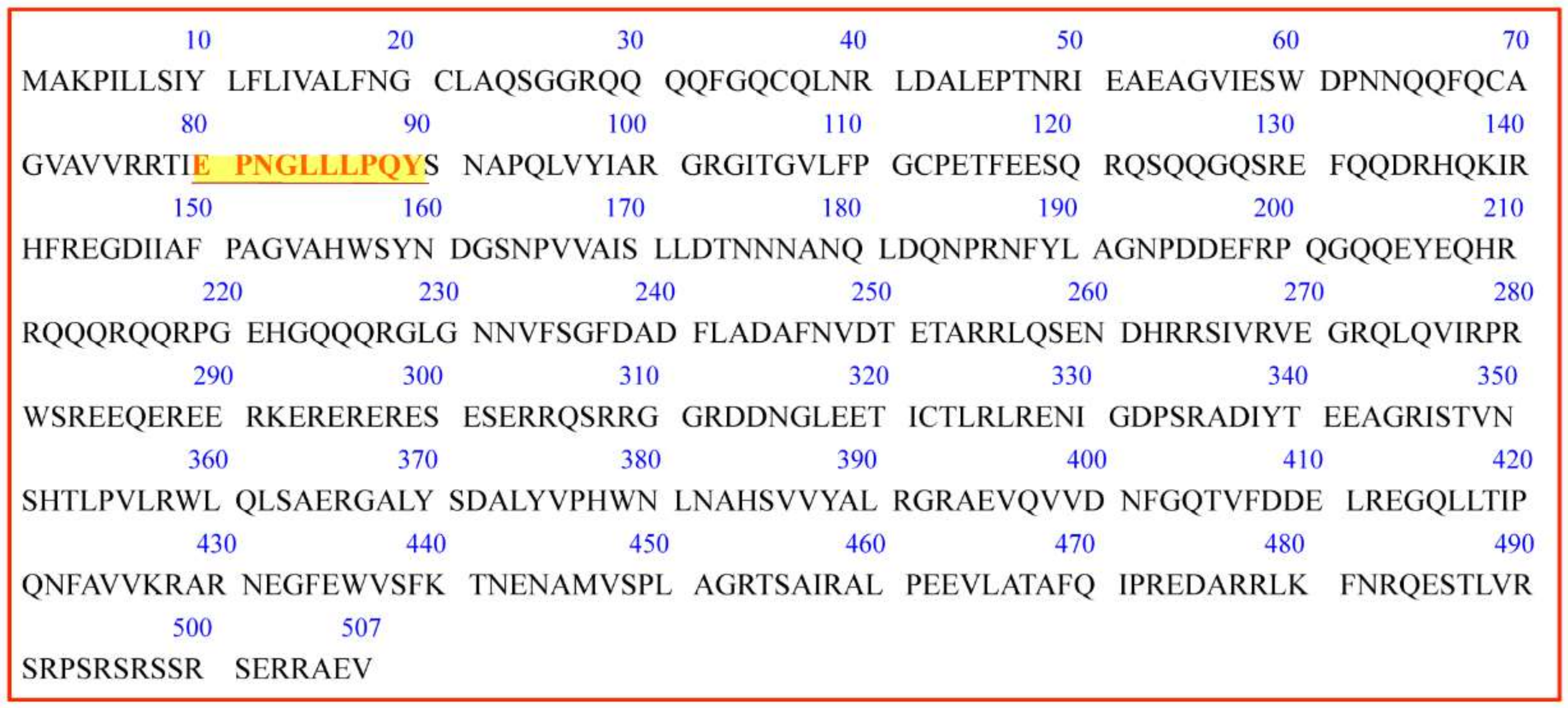
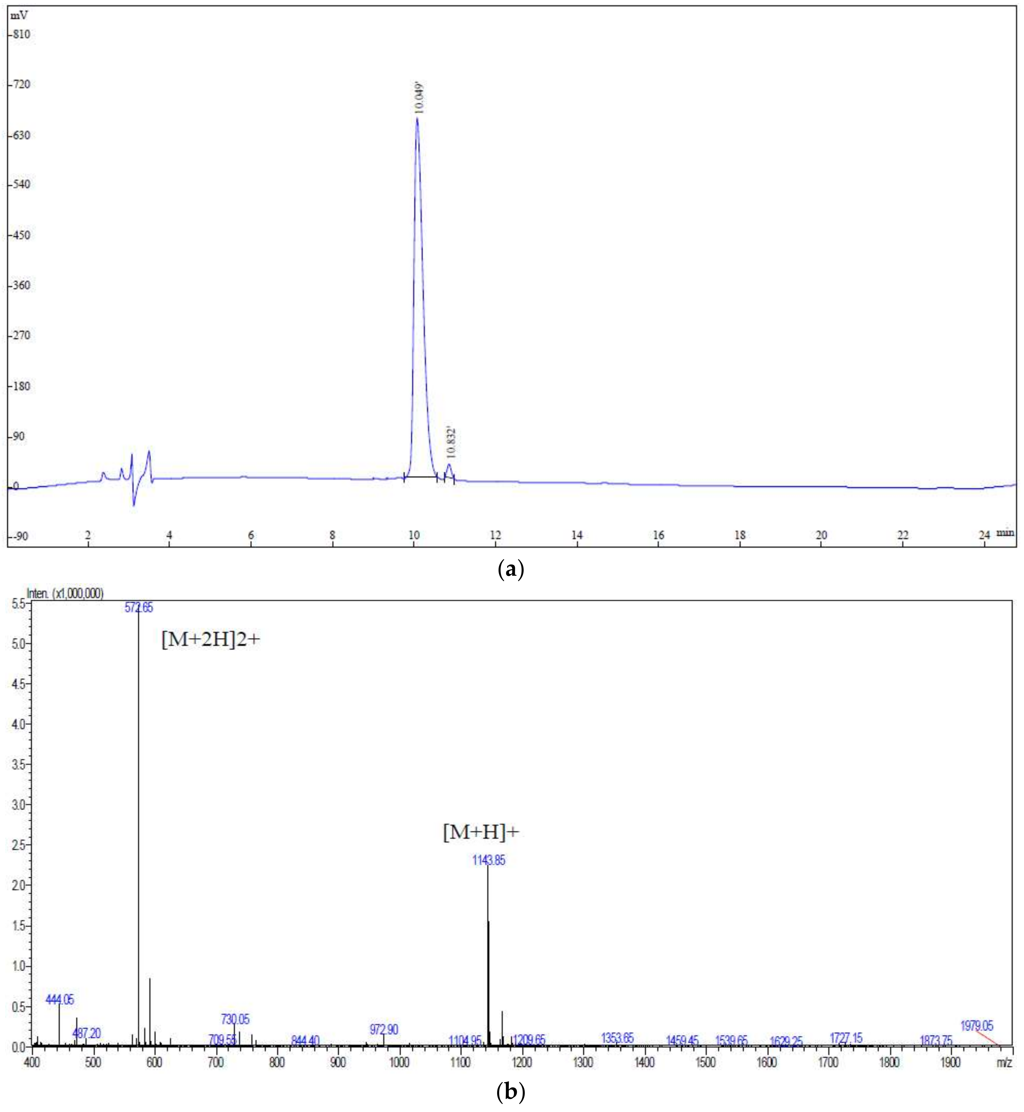
2.1. Identification of Peptides from Walnut Protein Hydrolysates

In the present study, in order to identify the peptides in the walnut hydrolysates, UPLC-Q-TOF-MS/MS combined with MASCOT searching was used to analyze the fragment spectra [24]. As shown in Table 1, in total, 6 kinds of proteins containing 33 unique peptides were identified from walnut protein hydrolysates. Many kinds of peptides were identified from walnut seed storage protein (Q2TPW5). Walnut seed storage protein consists of 507 amino acids, and it can probably release bioactive peptides by enzymatic digestion of pepsin and trypsin, and so on. Results of peptides along with the peptide sequences and molecular information are shown in Table 1. The Mascot searching program can only provide the information of the peptides with the number of amino acids more than six, otherwise the precision of the identification work may be impossible. Among the identified peptides derived from the hydrolysates of walnut seed storage proteins by trypsin, one peptide, EPNGLLLPQY (fragment residues 80–89), was predicted to be an ACE-inhibitory peptide according to the affinity analysis by molecular docking. Therefore, the EPNGLLLPQY was synthesized in order to clarify the mechanism of ACE-inhibitory activity. The HPLC and MS of the synthesized EPNGLLLPQY are shown as Figure 1. The peptide showed a retention time of 10.049 min in HPLC in Figure 1a with a high purity of 98.51%, which showed that it can be used for the following analysis. The molecular mass of the peptide was 1143.43 Da, which was determined by MS/MS as shown in Figure 1b.
In the present study, the peptide EPNGLLLPQY was identified from the hydrolysate of walnut protein digested by pepsin at 37 °C and pH 1.2 for 3 h. Moreover, there were several reports related to the peptides from walnut protein hydrolysates with ACE-inhibitory activity. Several ACE-inhibitory peptides prepared from walnut proteins, LPGRPPIKPWPL and WPERPPQIP, were identified and purified by column chromatography methods from the hydrolysate of walnut protein digested by neutral proteinase As1.398 at 45 °C for 5 h (pH 7.0) [12,20].
To screen the ACEI peptide, several in silico methods (Peptide Ranker and molecular docking) were used in the present study. Firstly, as the molecular weights of the identified protein ranged from 702.366–2590.46 Da with the number of amino acids from 7–23 and the study paid more attention to the peptides with a lower molecular weight, the identified peptides with amino acids no more than 10 were selected for further study. Secondly, Peptide Ranker was adopted to analyze the possibility of the peptide to be bioactive. As listed in Table 2, the peptide EPNGLLLPQY acquired the highest scores of Peptide Ranker, so it has the highest likelihood to inhibit ACE activity. Thirdly, molecular docking was employed to verify the affinity of the peptides binding to ACE. It is not hard to see that the peptide EPNGLLLPQY also has a relatively high affinity with ACE, which was evaluated by comparing the scores of “-CDOCKER_Interaction_Energy”, an evaluation function of the affinity of the donor and ligand in molecular docking in Discovery Studio 2017. Therefore, the peptide EPNGLLLPQY was chose for further ACEI analysis. The molecular mass of the peptide was 1142.60 Da, which was determined by MS/MS. Moreover, the location of EPNGLLLPQY (marked in red letter) in walnut seed storage proteins (Q2TPW5) from the PDB database is shown in Figure 2.

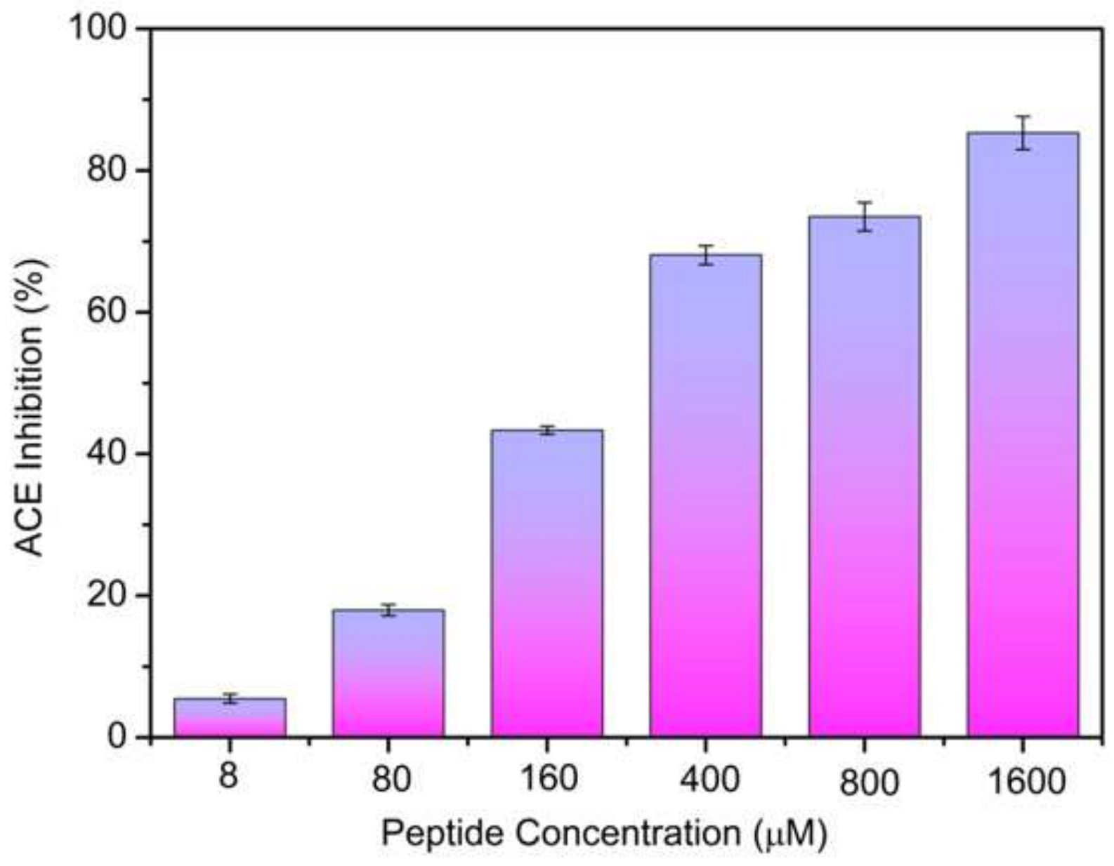
2.2. ACE-Inhibitory Activity Determination

In the present study, the ACE-inhibitory activity of the peptide, EPNGLLLPQY, released by pepsin digestion was evaluated, as shown in Figure 3. The ACE-inhibitory activity of the peptide at different concentrations (8–1600 µM) was determined, the inhibitory activity increased with the increase of the concentration in a dose-dependent pattern. The IC50 value was determined by the regression equation: Y = −(1.93948 × 10−4)X2 + 0.24469X + 3.48868 (R2 = 0.97179). The IC50 value of the peptide was 233.178 µM. There have been about 700 kinds of ACE-inhibitory peptides from many kinds of proteins reported till now, all of which were collected in the BIOPEP database http://www.uwm.edu.pl/biochemia/index.php/en/biopep [25], and the IC50 values of the peptides ranged from 0.01–10,000 µM. The peptide EPNGLLLPQY can be considered as a potential ACE inhibitor due to its high activity.

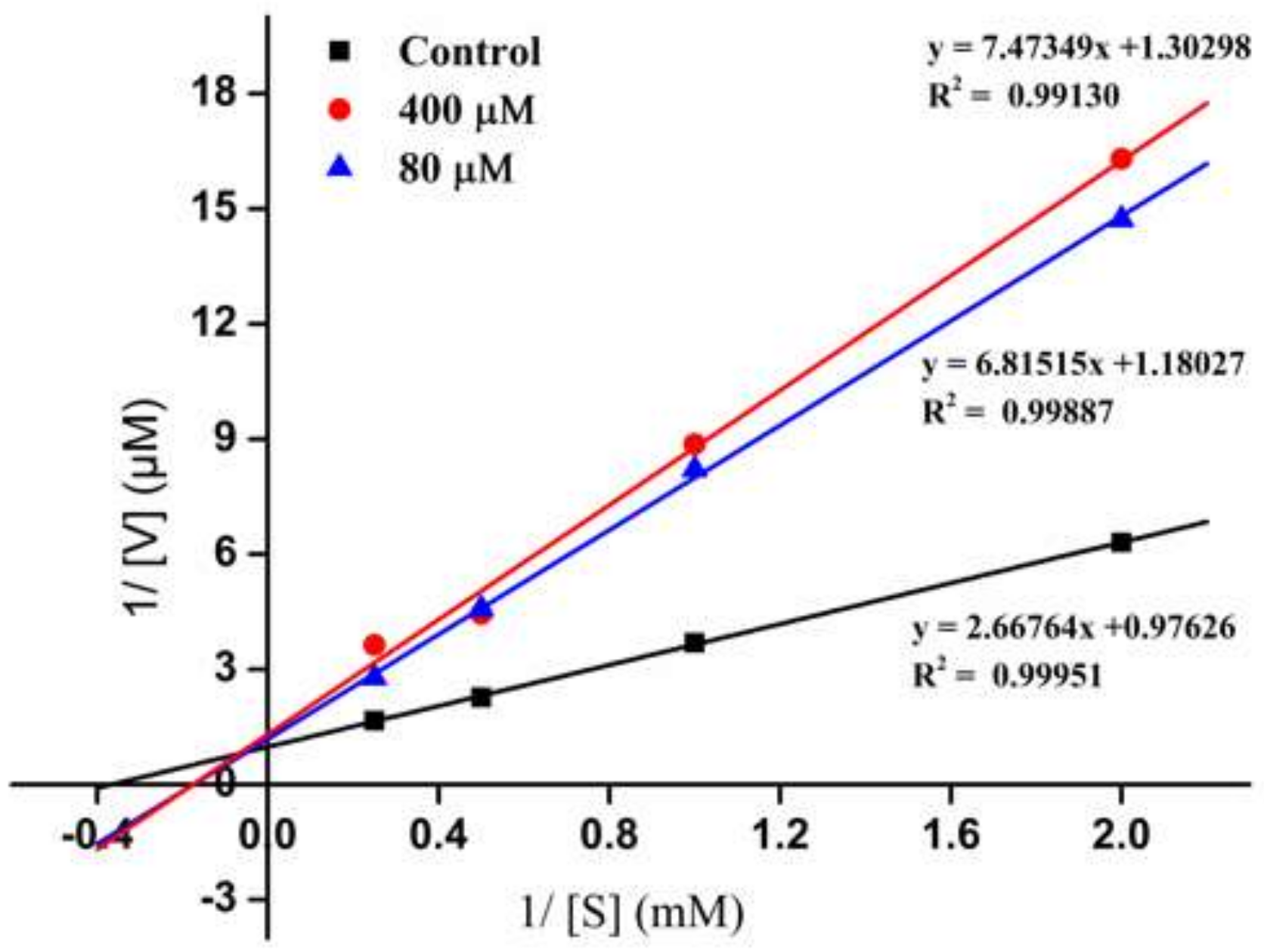
2.3. Inhibition Pattern of the ACE-Inhibitory Peptide

To further evaluate the active mechanism of the novel ACEI peptide, the inhibition pattern of the peptide, EPNGLLLPQY, against ACE was investigated by the Line weaver-Burk plot analysis method. As shown in Figure 4, three experimental groups (■ control; 80 μM; 400 μM) were tested, and 1/(S) and 1/(V) represent the reciprocal substrate concentration and velocity, respectively. The Vm values decreased as the peptide concentration increased, which confirmed that the existence of the peptide probably blocked the substrate from binding to ACE active sites. When the EPNGLLLPQY was added to the reaction system, the Km values become higher than the control, which indicated that a relative higher content of substrate was essential for the ACE catalytic reaction. Figure 4 presents an ACEI peptide exhibiting a mixed-type of inhibition of ACE. Similar food-derived peptides EVSQGRP, VSRHFASYAN and SAAVGSP also have been identified from Stichopus horrens [26]. The mixed-type inhibition indicated that the peptide binds to the ACE at positions including both the active and non-active sites, which consequently reduces the catalytic activity of ACE. Since ACE converts the hormone angiotensin I to the active vasoconstrictor angiotensin II, binding on the active site will directly prevent the cleavage ability of ACE. Binding on the non-active sites probably prevents the interaction of the ACE and the substrate by the steric hindrance effect.

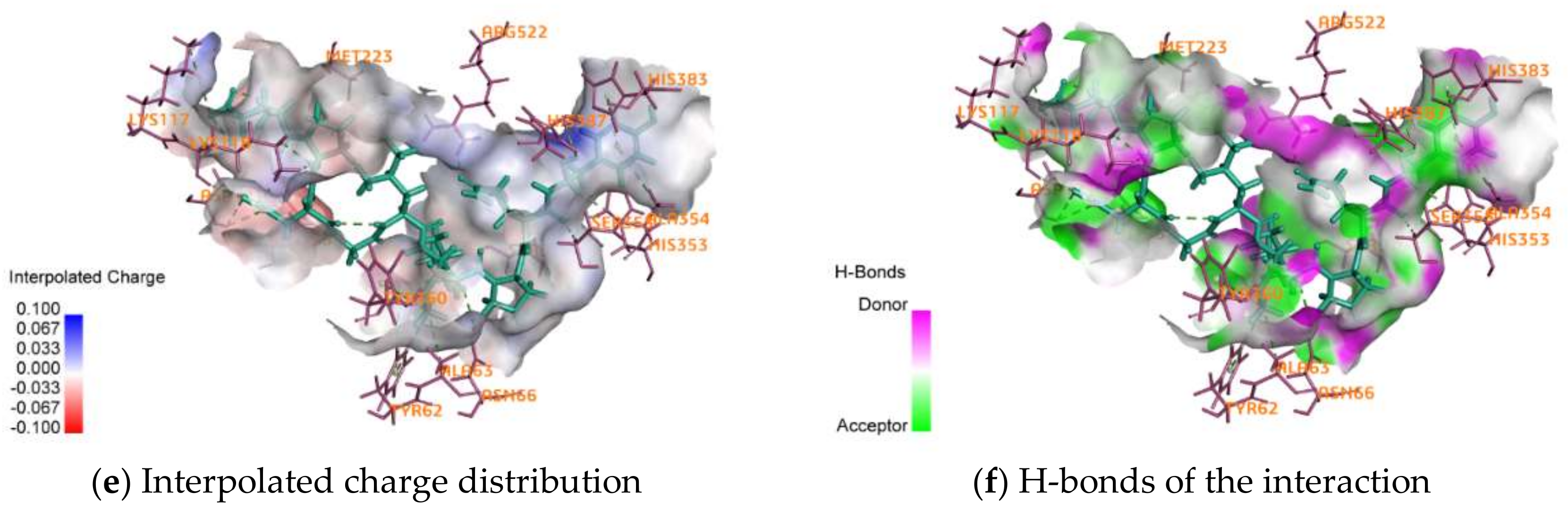
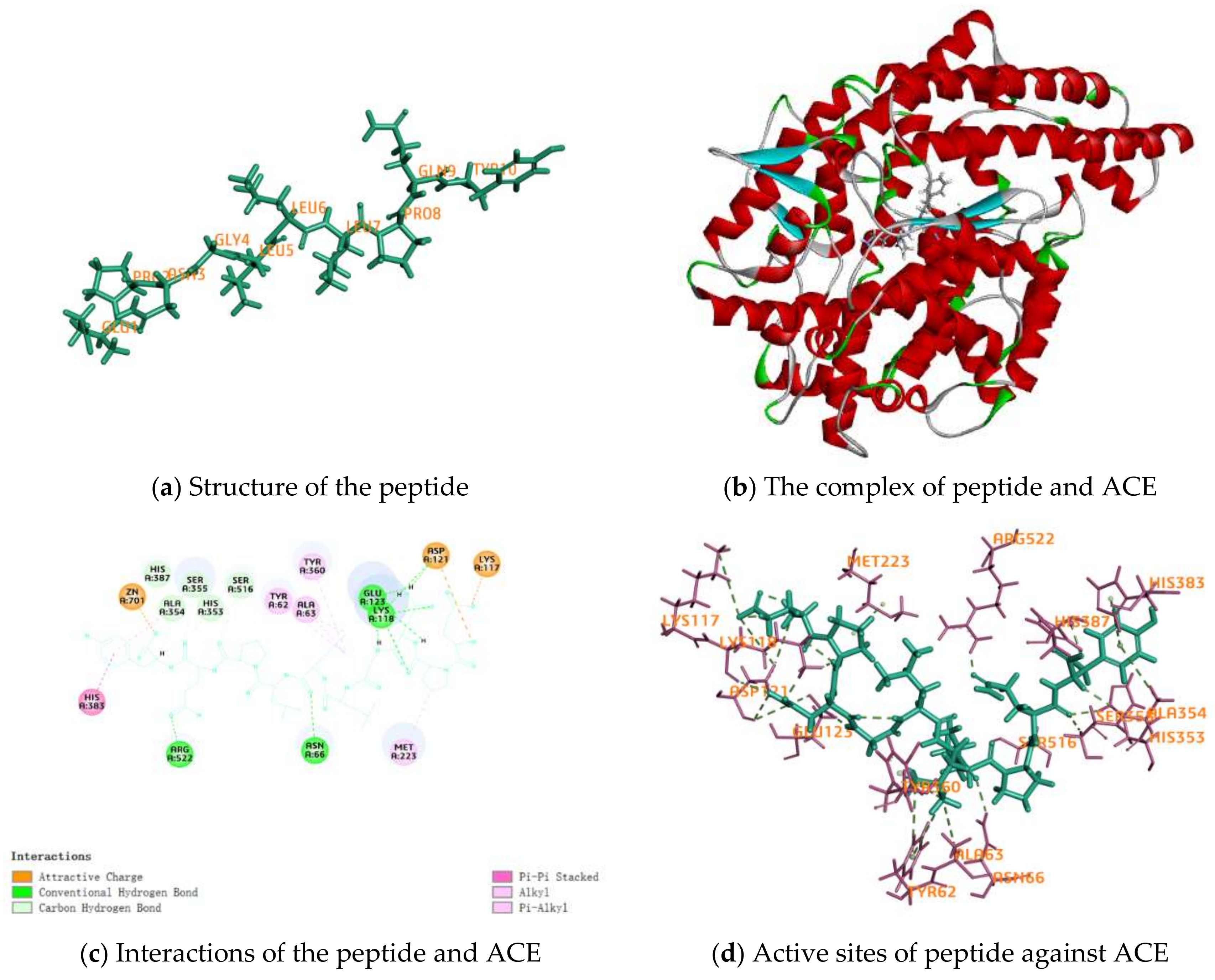
2.4. Activity Mechanism of EPNGLLLPQY

Molecular docking is a generally used method for the prediction of the interactions of the inhibitor as a donor and the enzyme as a receptor by calculating the affinity energies, indicating the binding sites and interactive bonds of the donor and receptor. In order to clarify the ACE inhibitive mechanism of EPNGLLLPQY, its molecular docking against ACE was performed by Discovery Studio 2017 R2 software. The values of “-CDOCKER_Interaction_Energy” (134.600 kcal mol−1) indicated the affinity between the EPNGLLLPQY and ACE, as shown in Table 2. Fu et al. [27] reported that the interactions of multiple hydrogen bonds between the inhibitor and ACE would promote the peptide-induced inhibition of ACE activity by the stabilization of the structure of the non-catalytic enzyme-peptide complex.
The structure of the peptide and the complex of it and ACE are shown in Figure 5a,b, which indicated that the peptide was a linear peptide, and its size was important for the inhibitory activity against ACE. There are three categories of interactions between EPNGLLLPQY as the ligand and ACE as the donor, electrostatic, hydrogen bond and hydrophobic interactions, and the majority of interactions were hydrogen bonds (Table 3). A total of 16 H-bonds has been found in the interactive site between the peptide EPNGLLLPQY and ACE, which is also shown in Figure 5c,d, including 10 amino acid residues in ACE, i.e., ASN66, LYS118, ASP121, GLU123, HIS353, ALA354, SER355, HIS387, SER516 and ARG522, as shown in Figure 5e. Among these residues, GLU123 and ARG522 also formed H-bonds with the ACE-inhibitory peptides (WG and PRY), as reported by Fu et al. [27]. SER516 was also identified in the C-domain complex of ACE-BPPb [28]. The large amount of ACE residues formed in ACE peptides by H-bonds could explain the greater interaction between ACE and peptides. Seven hydrophobic interactions have been found in the interactive site between the peptide and ACE, including six amino acid residues in ACE, i.e., TYR62, ALA63, MET223, ALA354, TYR360 and HIS383. Moreover, electrostatic interactions at the site of LYS117, ASP121 and ZN701 in ACE-EPNGLLLPQY also contributed to stabilization, as shown in Figure 5f. These results suggested that the intensive interactions of EPNGLLLPQY with ACE may contribute to its strong inhibitory activity.

3. Materials and Methods

3.1. Materials and Chemicals

Defatted walnut meals were purchased from Pretty Woman Changbai (Dalian, Liaoning, China). Pepsin (EC3.4.23.1, 2500 units/mg protein) was purchased from Novozymes (Beijing, China). ACE, hippuryl-l-histidyl-l-leucine (HHL), acetonitrile and trifluoroacetic acid (HPLC) were supplied by Sigma Aldrich Co. (St. Louis, MO, USA). Formic acid (FA) (HPLC) was supplied by Fluka (Buchs, Germany).

3.2. Enzymatic Hydrolysis of Walnut Proteins

Defatted walnut meals were soaked in water and smashed, the mixture was stirred in a water bath of 55 °C for 1 h after adjusting the pH to 8.5 with 1 mol/L NaOH. Then, it was centrifuged at 3000× g for 10 min, and the supernatant was centrifuged at 4000× g for 20 min after adjusting the pH to 4.5 with 1 mol/L HCl. The sediment was washed by 95% ethanol to remove the fat, and the crude proteins were prepared by freeze drying. Crude proteins were digested by pepsin at 37 °C for 3 h (pH 1.2) with an enzyme/substrate (E/S) ratio of 1/50. The concentration of walnut protein was 50 mg/mL, which was determined by the Lowry method with minor modifications with bovine serum albumin as the standard [20]. The hydrolysis course was finished by heating the samples in boiling water (10 min). The supernatant from the hydrolysate was collected by centrifugation at 8000× g at 4 °C for 10 min.

3.3. UPLC-Q-TOF-MS/MS Analysis

The peptides in walnut hydrolysates were analyzed by an ultra-high-performance liquid chromatography (UPLC) quadrupole time of flight (Q-TOF) system using a Microtof-II mass spectrometer (Bruker Daltonik GmbH, Bremen, Germany). Isolation of peptides was performed on a UPLC instrument (Dionex UltiMate 3000 RSLC System, Thermo Scientific, Waltham, MA, USA) equipped with a 150 mm × 3.0 mm C 18 column (Phenomenex Luna, Torrance, CA, USA) with a particle size of 3.0 μm. Acidified water (0.1% formic acid, v/v) and ACN (with 0.1% formic acid, v/v) were used as Mobile Phases A and B. Elution was performed with a gradient of 5–35% B at a flow rate of 0.25 mL min−1.
The MS spectra were obtained in the positive electrostatic ionization manner. The top 20 most intense ions were acquired based on auto-MS/MS after the fragmentation of protonated molecular ions [29]. The raw data of the peptide sequence was processed by Data Analysis 4.0 software (Bruker Daltonics, Bermen, Germany) and searched by MASCOT (http://www.expasy.ch/sprot/, Version 2.5.1.0, Matrix Science, London, UK).

3.4. Screening of the Potential ACEI Peptides

To screen peptides, in silico methods were adopted. Molecular docking was used to evaluate the affinity between the peptides and ACE. Peptide Ranker was used to analyze the possibility of the peptides to be bioactive by giving them scores from 0–1, and the higher the score, the higher the possibility.

3.5. Synthesis of Peptides

Peptide with potential ACE-inhibitory activity screened by molecular docking was chemically synthesized by the solid-phase method (Cellmano Biotechnology Co., LTD, Hefei, China). The purity of the peptide was determined by HPLC, and the sequence of the synthesized peptide was determined by HPLC-MS/MS.

3.6. Molecular Docking

Molecular docking between walnut peptides and ACE was performed using Discovery Studio 2017 software (NeoTrident Technology Ltd., Beijing, China). The crystal structure of ACE (PDB ID: 1O8A) was downloaded from the PDB database. Based on their amino acid sequence, the structure and the minimum energy forms of target peptides were automatically figured out before molecular docking [30]. The types of interactions between the docked receptor and the ligand were analyzed.

3.7. ACE-Inhibitory Activity Assay

ACE-inhibitory activity was determined by an HPLC method (e2695, Waters, Framingham, MA, USA). The reaction system contained 30 μL of HHL (2.5 mmol L−1), 20 μL of ACE (100 μM) in 100 mmol L−1 of sodium borate buffer at pH 8.3 and 10 μL of the sample. The reaction was performed at 37 °C for 60 min and terminated by adding 60 μL of HCl (1 mol L−1). The concentrations of HHL and HA were determined by HPLC equipped with a Waters C-18 column (150 × 4.6 mm, 5 μm). To analyze the components, isocratic elution was conducted for 25 min in 78% Mobile Phase A (0.05% TFA in H2O) and 22% Mobile Phase B (ACN) with a flow rate of 0.5 mL min−1. The ACE inhibition (%) was calculated using the following equation:
ACE Inhibitory Activity ( % ) = Δ A blank Δ A inhibitor Δ A blank × 100
where ΔAblank and ΔAinhibitor represent the peak areas of HA in the blank (sodium borate buffer) and sample, respectively. IC50 was defined as the concentration of the peptide that inhibited 50 percent of the ACE activity.

3.8. Statistical Analysis

All data were gathered in independent triplicates and described as the mean values and standard deviations. Statistics was performed using the SPSS 17.0 software.

4. Conclusions

A novel ACE-inhibitory peptide, EPNGLLLPQY, is a peptide (fragment of residues 80–89) located in walnut seed storage proteins, which exhibited inhibitory activity with an IC50 value of 233.178 µM in a mixed-type inhibition manner. The potential ACE-inhibitory mechanism was mainly attributed to the hydrogen bonds, hydrophobic and electrostatic interactions. Moreover, the peptides from walnut protein may be applied as a functional component in the research and development of functional products.

Acknowledgments

This study was financially supported by the National Natural Science Foundation of China (31371805) and the Basic Research Program of Liaoning Education Department (2017J080).

Author Contributions

Cong Wang conceived and designed the experiments, performed the experiments, analyzed the data, contributed reagents/materials/analysis tools, wrote the paper, prepared figures and tables, reviewed drafts of the paper. Prof. Lianzhou Jiang conceived and designed the experiments, analyze the data, contributed/reagent/materials/analysis tools, reviewed drafts of the paper. Maolin Tu, Di Wu, Hui Chen, Cheng Chen and Zhenyu Wang have reviewed and revised the drafts.

Conflicts of Interest

The authors declare no conflict of interest.

References

  1. World Health Organization. Cardiovascular Diseases (CVDs) Fact Sheet N 317; Updated January 2015; WHO: Geneva, Switzerland, 2016. [Google Scholar]
  2. Celermajer, D.S.; Chow, C.K.; Marijon, E.; Anstey, N.M.; Woo, K.S. Cardiovascular disease in the developing world. J. Am. Coll. Cardiol. 2012, 60, 1207–1216. [Google Scholar] [CrossRef] [PubMed]
  3. Staessen, J.A.; Wang, J.; Bianchi, G.; Birkenhäger, W.H. Essential hypertension. Lancet 2003, 361, 1629–1641. [Google Scholar] [CrossRef]
  4. Cannon, C.P. Cardiovascular disease and modifiable cardiometabolic risk factors. Clin. Cornerstone 2008, 9, 24–41. [Google Scholar] [CrossRef]
  5. Gu, Q.; Dillon, C.F.; Burt, V.L.; Gillum, R.F. Association of hypertension treatment and control with all-cause and cardiovascular disease mortality among US adults with hypertension. Am. J. Hypertens. 2010, 23, 38–45. [Google Scholar] [CrossRef] [PubMed]
  6. Skeggs, L.T.; Kahn, J.R.; Shumway, N.P. The preparation and function of the hypertensin-converting enzyme. J. Exp. Med. 1956, 103, 295–299. [Google Scholar] [CrossRef] [PubMed]
  7. Yang, H.; Erdös, E.; Levin, Y. A dipeptidyl carboxypeptidase that converts angiotensin I and inactivates bradykinin. Biochim. Biophys. Acta Protein Struct. 1970, 214, 374–376. [Google Scholar] [CrossRef]
  8. Hai-Lun, H.; Xiu-Lan, C.; Cai-Yun, S.; Yu-Zhong, Z.; Bai-Cheng, Z. Analysis of novel angiotensin-I-converting enzyme inhibitory peptides from protease-hydrolyzed marine shrimp Acetes chinensis. J. Pept. Sci. 2006, 12, 726–733. [Google Scholar] [CrossRef] [PubMed]
  9. López-Fandiño, R.; Otte, J.; Van Camp, J. Physiological, chemical and technological aspects of milk-protein-derived peptides with antihypertensive and ACE-inhibitory activity. Int. Dairy J. 2006, 16, 1277–1293. [Google Scholar] [CrossRef]
  10. Kim, S.-K.; Wijesekara, I. Development and biological activities of marine-derived bioactive peptides: A review. J. Funct. Foods 2010, 2, 1–9. [Google Scholar] [CrossRef]
  11. Atkinson, A.; Robertson, J. Captopril in the treatment of clinical hypertension and cardiac failure. Lancet 1979, 314, 836–839. [Google Scholar] [CrossRef]
  12. Liu, M.; Du, M.; Zhang, Y.; Xu, W.; Wang, C.; Wang, K.; Zhang, L. Purification and identification of an ACE inhibitory peptide from walnut protein. J. Agric. Food Chem. 2013, 61, 4097–4100. [Google Scholar] [CrossRef] [PubMed]
  13. Pihlanto-Leppälä, A. Bioactive peptides derived from bovine whey proteins: Opioid and ACE-inhibitory peptides. Trends Food Sci. Technol. 2000, 11, 347–356. [Google Scholar] [CrossRef]
  14. Vercruysse, L.; Van Camp, J.; Smagghe, G. ACE inhibitory peptides derived from enzymatic hydrolysates of animal muscle protein: A review. J. Agric. Food Chem. 2005, 53, 8106–8115. [Google Scholar] [CrossRef] [PubMed]
  15. Jia, J.; Ma, H.; Zhao, W.; Wang, Z.; Tian, W.; Luo, L.; He, R. The use of ultrasound for enzymatic preparation of ACE-inhibitory peptides from wheat germ protein. Food Chem. 2010, 119, 336–342. [Google Scholar] [CrossRef]
  16. Orio, L.P.; Boschin, G.; Recca, T.; Morelli, C.F.; Ragona, L.; Francescato, P.; Arnoldi, A.; Speranza, G. New ACE inhibitory peptides from hemp seed (Cannabis sativa L.) proteins. J. Agric. Food Chem. 2017, 65, 10482–10488. [Google Scholar] [CrossRef] [PubMed]
  17. Jamdar, S.N.; Rajalakshmi, V.; Pednekar, M.D.; Juan, F.; Yardi, V.; Arun, S. Influence of degree of hydrolysis on functional properties, antioxidant activity and ACE inhibitory activity of peanut protein hydrolysate. Food Chem. 2010, 121, 178–184. [Google Scholar] [CrossRef]
  18. Wang, C.; Song, W.; Jiang, L.; Du, M. Purification and identification of an ACE-inhibitory peptide from walnut protein hydrolysate. Eur. Food Res. Technol. 2014, 239, 333–338. [Google Scholar]
  19. Mukuddem-Petersen, J.; Stonehouse, O.W.; Jerling, J.C.; Hanekom, S.M.; White, Z. Effects of a high walnut and high cashew nut diet on selected markers of the metabolic syndrome: A controlled feeding trial. Br. J. Nutr. 2007, 97, 1144–1153. [Google Scholar] [CrossRef] [PubMed]
  20. Wang, C.; Du, M.; Zheng, D.; Kong, F.; Zu, G.; Feng, Y. Purification and characterization of nattokinase from Bacillus subtilis natto B-12. J. Agric. Food Chem. 2009, 57, 9722–9729. [Google Scholar] [CrossRef] [PubMed]
  21. Rawendra, R.D.; Chang, C.-I.; Chen, H.-H.; Huang, T.-C.; Hsu, J.-L. A novel angiotensin converting enzyme inhibitory peptide derived from proteolytic digest of Chinese soft-shelled turtle egg white proteins. J. Proteom. 2013, 94, 359–369. [Google Scholar] [CrossRef] [PubMed]
  22. Thomsen, R.; Christensen, M.H. MolDock: A new technique for high-accuracy molecular docking. J. Med. Chem. 2006, 49, 3315–3321. [Google Scholar] [CrossRef] [PubMed]
  23. Pooja, K.; Rani, S.; Prakash, B. In silico approaches towards the exploration of rice bran proteins-derived angiotensin-I-converting enzyme inhibitory peptides. Int. J. Food Prop. 2017, 20, 2178–2191. [Google Scholar] [CrossRef]
  24. Iqbal, J.; Li, W.; Ullah, K.; Hasan, M.; Linna, G.; Awan, U.; Zhang, Y.; Batool, S.; Qing, H.; Deng, Y. Study of rat hypothalamic proteome by HPLC/ESI ion trap and HPLC/ESI-Q-TOF MS. Proteomics 2013, 13, 2455–2468. [Google Scholar] [CrossRef] [PubMed]
  25. Dziuba, J.; Iwaniak, A.; Mine, Y.; Shahidi, F. Database of Protein and Bioactive Peptide Sequences. Nutraceutical Proteins & Peptides in Health & Disease; CRC/Taylor & Francis: Boca Raton, FL, USA, 2006; pp. 543–545. [Google Scholar]
  26. Forghani, B.; Zarei, M.; Ebrahimpour, A.; Philip, R.; Bakar, J.; Hamid, A.A.; Saari, N. Purification and characterization of angiotensin converting enzyme-inhibitory peptides derived from Stichopus horrens: Stability study against the ACE and inhibition kinetics. J. Funct. Foods 2016, 20, 276–290. [Google Scholar] [CrossRef]
  27. Fu, Y.; Alashi, A.M.; Young, J.F.; Therkildsen, M.; Aluko, R.E. Enzyme inhibition kinetics and molecular interactions of patatin peptides with angiotensin I-converting enzyme and renin. Int. J. Biol. Macromol. 2017, 101, 207–213. [Google Scholar] [CrossRef] [PubMed]
  28. Masuyer, G.; Schwager, S.L.; Sturrock, E.D.; Isaac, R.E.; Acharya, K.R. Molecular recognition and regulation of human angiotensin-I converting enzyme (ACE) activity by natural inhibitory peptides. Sci. Rep. 2012, 2, 717. [Google Scholar] [CrossRef] [PubMed]
  29. Zhao, Y.-Y.; Lei, P.; Chen, D.-Q.; Feng, Y.-L.; Bai, X. Renal metabolic profiling of early renal injury and renoprotective effects of Poria cocos epidermis using UPLC Q-TOF/HSMS/MS E. J. Pharm. Biomed. Anal. 2013, 81, 202–209. [Google Scholar] [PubMed]
  30. Tu, M.; Feng, L.; Wang, Z.; Qiao, M.; Shahidi, F.; Lu, W.; Du, M. Sequence analysis and molecular docking of antithrombotic peptides from casein hydrolysate by trypsin digestion. J. Funct. Foods 2017, 32, 313–323. [Google Scholar] [CrossRef]
Figure 1. Identification of EPNGLLLPQY by (a) HPLC and (b) MS.
Ijms 19 01156 g001
Figure 2. Location of EPNGLLLPQY in walnut seed storage protein (Q2TPW5).
Ijms 19 01156 g002
Figure 3. ACE-inhibitory activity of EPNGLLLPQY.
Ijms 19 01156 g003
Figure 4. Line weaver-Burk plot of ACE inhibition by EPNGLLLPQY.
Ijms 19 01156 g004
Figure 5. Molecular docking of EPNGLLLPQY against ACE (PDB: 1O8A).
Ijms 19 01156 g005aIjms 19 01156 g005b
Table 1. Walnut hydrolysate peptides identified by UPLC-Q-TOF-MS/MS.
No.Proteinm/z Meas.zMr. Calc.ScoreStartAA SequenceEndLength
1Histidine kinase OS529.307721056.59338.4LLQLSALADRAA10
2Legumin OS832.46331831.449121.37LLTIPQNFA7
3Legumin OS570.273621138.52527.31LSAERGALYSDAL11
4Legumin OS648.37421294.72533.74AIRALPEEVLANAL12
5Legumin OS710.677332127.99724.14AVALMDTTNNANQLDQNPRNF19
6Legumin OS759.70232275.06528.2AVALMDTTNNANQLDQNPRNFY20
7Legumin OS864.834632590.4629.51AVVRRTIEPNGLLLPQYSNAPQLLY23
8Legumin OS670.302732007.87124.94LMDTTNNANQLDQNPRNFY17
9Multi-sensor signal transduction histidine kinase529.307721056.59338.4PIQLSAERLQF9
10Receptor agonist peptide520.929931558.7721.66AKTPGLPPMPQSDYLK14
11Seed storage protein452.73252903.44528.1AFQIPREDA7
12Seed storage protein352.1932702.36629.19LSAERGALY7
13Seed storage protein520.314821038.60733.34AIRALPEEVLA9
14Seed storage protein574.293921146.56741.27LATAFQIPREDA10
15Seed storage protein555.833821109.64422.87AIRALPEEVLAT10
16Seed storage protein529.306321056.59338.51WLQLSAERGALY10
17Seed storage protein614.294831839.84621.95LLDTNNNANQLDQNPRNF16
18Seed storage protein582.968631745.87336.22FKTNENAMVSPLAGRTSAI17
19Seed storage protein994.9721986.91426.31LLDTNNNANQLDQNPRNFY17
20Seed storage protein644.645831930.90320.57PGEHGQQQRGLGNNVFSGFD18
21Seed storage protein921.442221839.85462.21FPAGVAHWSYNDGSNPVVAI18
22Seed storage protein719.028732153.04627.39AISLLDTNNNANQLDQNPRNF19
23Seed storage protein827.13932477.37529.43AVVRRTIEPNGLLLPQYSNAPQLV22
24Seed storage protein526.937131577.7826.95FLADAFNVDTETARRL14
25Seed storage protein863.913721725.80380.77LLDTNNNANQLDQNPRN15
26Seed storage protein572.3321142.6027-EPNGLLLPQYS10
27Seed storage protein681.013132039.00347.55AISLLDTNNNANQLDQNPRN18
28Seed storage protein711.850521421.67926.29FLADAFNVDTETARR13
29Vicilin-like protein precursor432.73562863.450125.24LLRGIENYR7
30Vicilin-like protein precursor425.72732849.434537.36YLRVFSNDI7
31Vicilin-like protein precursor482.27022962.518528.32LRVFSNDILV8
32Vicilin-like protein precursor538.813121075.60335.78YLRVFSNDILV9
33Vicilin-like protein precursor500.251131497.72123.9MESYFVPTERQSRR12
Mr. Calc.: Relative molecular mass calculated in MS analysis.
Table 2. Peptides analysis by Peptide Ranker and molecular docking.
No.PeptidesLength-CDOCKER_Energy Interaction (kcal mol−1)Peptide Ranker
1IRALPEEVL9155.9370.14
2LQLSALADRA10144.0810.23
3ATAFQIPRED10136.4010.18
4EPNGLLLPQY10134.6000.37
5LQLSAERGAL10130.5320.30
6LRVFSND7127.5560.16
7LRVFSNDIL9126.7760.30
8IQLSAERLQ9123.0610.12
9IRALPEEVLA10121.9180.14
10LTIPQNF7108.5180.34
11FQIPRED797.60960.27
12LRGIENY794.03960.12
13RVFSNDIL885.88070.31
14SAERGAL768.0270.29
Table 3. Interaction bonds of walnut peptide against ACE.
InteractionsDistanceCategoryTypes
A:LYS117:NZ–EPNGLLLPQY:O154.63933ElectrostaticAttractive Charge
A:ZN701:ZN–EPNGLLLPQY:O1622.22675ElectrostaticAttractive Charge
EPNGLLLPQY:N1–A:ASP121:OD24.00046ElectrostaticAttractive Charge
A:ASN66:HD21–EPNGLLLPQY:O902.53299Hydrogen BondConventional Hydrogen Bond
A:LYS118:HZ1–EPNGLLLPQY:O401.79716Hydrogen BondConventional Hydrogen Bond
A:LYS118:HZ3–EPNGLLLPQY:O312.13479Hydrogen BondConventional Hydrogen Bond
A:ARG522:HH12–EPNGLLLPQY:O1351.92374Hydrogen BondConventional Hydrogen Bond
EPNGLLLPQY:H33–A:GLU123:OE11.97968Hydrogen BondConventional Hydrogen Bond
EPNGLLLPQY:H42–A:LYS118:O2.29783Hydrogen BondConventional Hydrogen Bond
EPNGLLLPQY:H42–A:ASP121:O2.83625Hydrogen BondConventional Hydrogen Bond
EPNGLLLPQY:H43–A:ASP121:O2.80029Hydrogen BondConventional Hydrogen Bond
EPNGLLLPQY:H43–A:GLU123:OE22.2265Hydrogen BondConventional Hydrogen Bond
EPNGLLLPQY:H47–A:GLU123:OE12.01368Hydrogen BondConventional Hydrogen Bond
A:LYS118:HE2–EPNGLLLPQY:O312.33128Hydrogen BondCarbon Hydrogen Bond
A:HIS353:HD2–EPNGLLLPQY:O1402.40483Hydrogen BondCarbon Hydrogen Bond
A:SER355:HB1–EPNGLLLPQY:O1402.37051Hydrogen BondCarbon Hydrogen Bond
A:HIS387:HD2–EPNGLLLPQY:O1622.74483Hydrogen BondCarbon Hydrogen Bond
A:SER516:HB1–EPNGLLLPQY:O1232.80361Hydrogen BondCarbon Hydrogen Bond
EPNGLLLPQY:H144–A:ALA354:O2.26174Hydrogen BondCarbon Hydrogen Bond
A:HIS383–EPNGLLLPQY4.51235HydrophobicPi-Pi Stacked
A:ALA63–EPNGLLLPQY:C814.15055HydrophobicAlkyl
EPNGLLLPQY–A:MET2234.05541HydrophobicAlkyl
A:TYR62–EPNGLLLPQY:C815.26359HydrophobicPi-Alkyl
A:TYR62–EPNGLLLPQY:C855.3078HydrophobicPi-Alkyl
A:TYR360–EPNGLLLPQY:C815.23035HydrophobicPi-Alkyl
EPNGLLLPQY–A:ALA3544.78628HydrophobicPi-Alkyl

