Review

Pathogenesis of Lafora Disease: Transition of Soluble Glycogen to Insoluble Polyglucosan

by
Mitchell A. Sullivan
1,2,
Silvia Nitschke
1,
Martin Steup
1,3,4,
Berge A. Minassian
1,5 and
Felix Nitschke
1,*
1
Program in Genetics and Genome Biology, The Hospital for Sick Children Research Institute, 686 Bay Street, Toronto, ON M5G 0A4, Canada
2
Glycation and Diabetes, Mater Research Institute, Translational Research Institute, The University of Queensland, 37 Kent St., Woolloongabba, Brisbane 4102, Australia
3
The Impact Centre of the University of Toronto, 60 St. George Street, Toronto, ON M5S 1A7, Canada
4
Faculty of Mathematics and Science, University of Potsdam, 14476 Potsdam-Golm, Germany
5
Division of Neurology, Department of Pediatrics, University of Texas Southwestern, 5323 Harry Hines Blvd., Dallas, TX 75390-9063, USA
*
Author to whom correspondence should be addressed.
Int. J. Mol. Sci. 2017, 18(8), 1743; https://doi.org/10.3390/ijms18081743
Submission received: 17 July 2017 / Revised: 4 August 2017 / Accepted: 6 August 2017 / Published: 11 August 2017

Abstract

:
Lafora disease (LD, OMIM #254780) is a rare, recessively inherited neurodegenerative disease with adolescent onset, resulting in progressive myoclonus epilepsy which is fatal usually within ten years of symptom onset. The disease is caused by loss-of-function mutations in either of the two genes EPM2A (laforin) or EPM2B (malin). It characteristically involves the accumulation of insoluble glycogen-derived particles, named Lafora bodies (LBs), which are considered neurotoxic and causative of the disease. The pathogenesis of LD is therefore centred on the question of how insoluble LBs emerge from soluble glycogen. Recent data clearly show that an abnormal glycogen chain length distribution, but neither hyperphosphorylation nor impairment of general autophagy, strictly correlates with glycogen accumulation and the presence of LBs. This review summarizes results obtained with patients, mouse models, and cell lines and consolidates apparent paradoxes in the LD literature. Based on the growing body of evidence, it proposes that LD is predominantly caused by an impairment in chain-length regulation affecting only a small proportion of the cellular glycogen. A better grasp of LD pathogenesis will further develop our understanding of glycogen metabolism and structure. It will also facilitate the development of clinical interventions that appropriately target the underlying cause of LD.

Graphical Abstract

1. Introduction

Lafora disease (LD) is an autosomal recessively inherited disease that results from mutations in either the gene encoding for laforin (EPM2A) [1,2] or malin (EPM2B, also called NHLRC1) [3]. By 2009, over 200 independent families were found to suffer from LD with almost 100 distinct mutations in the two genes [4]. Since then, more mutations have been discovered [5,6]. While these two genes account for the majority of LD cases, there is evidence for at least one additional locus that may account for the remaining cases where neither EPM2A nor EPM2B are mutated [7].
The clinical outcomes of loss-of-function mutations in either EPM2A or EPM2B are generally similar, suggesting a closely related role of the two proteins laforin and malin in preventing LD. First epileptic episodes usually occur in adolescence. Subsequently, the disease progresses with ever worsening, increasingly intractable myoclonic seizures and cognitive decline through neurodegeneration. Eventually patients are bedridden, enduring in a vegetative state and finally succumbing to the disease, usually within ten years of symptom onset. Some studies have reported deviations from this general disease progression in patients with mutations in EPM2A or EPM2B, describing patients with a later onset or different rate of progression, e.g., a slower progression in particular patients with EPM2B mutations [8,9,10,11].
A hallmark of LD is the accumulation of Lafora bodies (LBs) in various tissues, including skin, skeletal muscle, heart, liver, and brain. LBs are stained on tissue sections with Schiff reagent following enzymatic degradation of soluble glycogen and periodate oxidation of vicinal hydroxyl groups present in the insoluble form of glycogen (polyglucosan). Their presence in skin tissue enables LD diagnosis based on the minimally invasive skin biopsy [12,13]. Those in the brain are considered to trigger neurodegeneration and epileptic symptoms [14].
Unfortunately to date there is no cure available for LD, with treatment being limited to alleviating the myoclonus epilepsy via antiepileptic drugs [15]. Understanding of the mechanisms underlying the pathogenesis of LD is paramount for the development of appropriate treatment strategies (Figure 1).

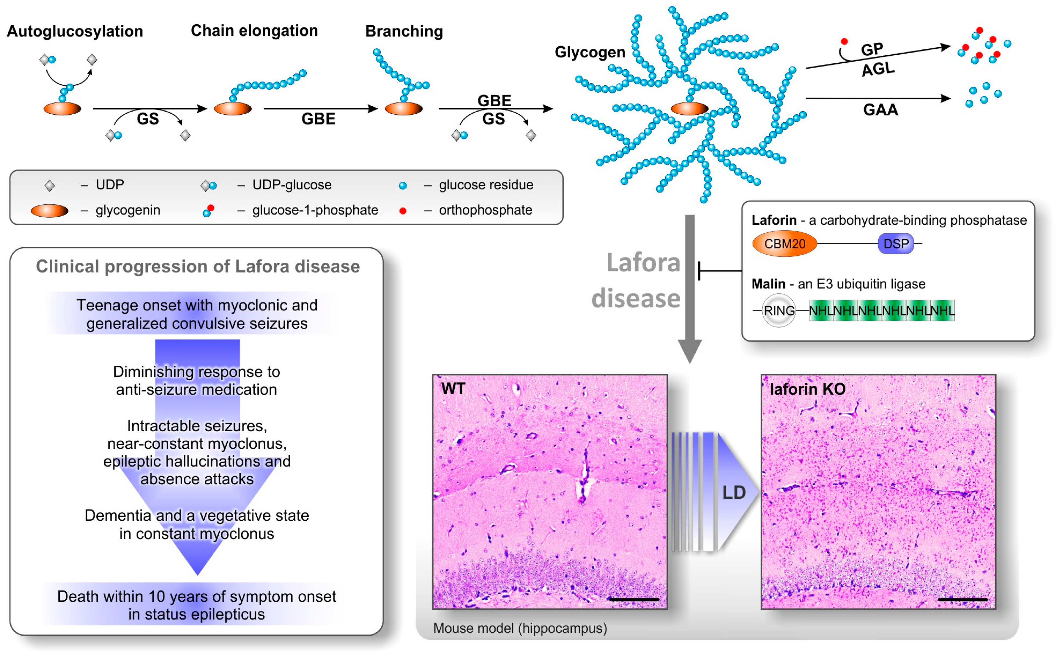
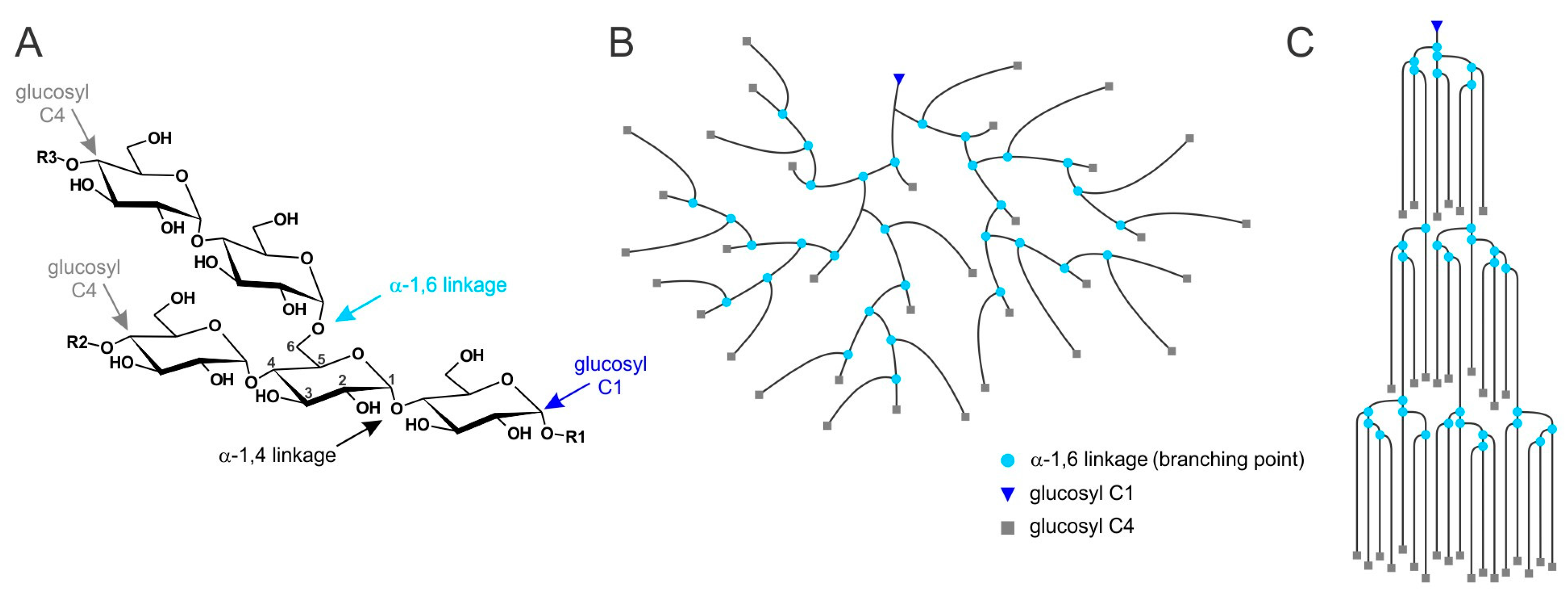
2. Lafora Bodies Put Lafora Disease in the Context of Polyglucan Metabolism

Consisting mostly of polyglucan chains, and to a minor extent proteins [16], the water-insoluble LBs are historically classified as polyglucosan bodies. Sensitivity of LBs to in vitro amylolysis implies the presence of α-(1,4) and α-(1,6) glucosidic linkages [17]. Both interglucose bonds are the essential linkages in water-soluble glycogen as well as in water-insoluble plant starches. The chemical similarity of the main constituent of LBs (polyglucosan) and soluble glycogen already indicates a close relation between glycogen metabolism and the formation of LBs (Figure 1). It is obvious that, despite similarities in the chemical composition of all these polyglucans, other features determine their (in)solubility in water.
Glycogen, serving as a cellular storage for reduced carbon and energy, is a regularly branched polymer of up to 55,000 glucosyl residues per molecule (β-particle). By a defined association of several β-particles even bigger assemblies (α-particles) can be formed, which have so far been described in liver [18] and heart [19] tissue. Glycogen is a disperse mixture of molecules with a wide range of molecule sizes [18,20,21]. Within glycogen molecules, chains have a distribution of lengths ranging from a degree of polymerisation of three to more than thirty-five [20,22], with an average length of approximately thirteen glucosyl residues [23].
De novo synthesis of glycogen molecules mainly involves autoglucosylation by glycogenin utilizing uridine 5’-diphosphoglucose (UDP-glucose) [24]. Recent results, however, suggest that glycogenin is not strictly necessary for glycogen synthesis [25]. Chains are elongated at the non-reducing end by glycogen synthase (GS) using UDP-glucose. Branching points are introduced by glycogen branching enzyme (GBE). Degradation is mediated by glycogen phosphorylase (GP) and glycogen debranching enzyme (AGL, see Figure 1) [24]. The enzymatic apparatus for glycogen turnover in mammals appears to be simple, especially when compared to the complexity of higher plant polyglucan (starch) metabolism, where several isoforms of synthesizing and degrading enzymes are expressed in a single cell [26]. However, in glycogen metabolism, additional enzymatic complexity is achieved, predominantly by regulatory features of the enzymes involved. For instance, while comparatively little is known about regulatory features of GBE, GS is strongly regulated by both covalent modifications and allosteric effects. Covalent modifications are essentially phosphorylations at multiple sites that partly occur in a hierarchical order. Increasing GS phosphorylation, as mediated by protein kinases such as glycogen synthase kinase 3 (GSK3), leads to an inactivation of GS. In turn, dephosphorylation by protein phosphatase 1 (PP1) activates GS [24]. Allosteric binding of glucose 6-phosphate (G6P) overrides the inhibition as imposed by GS phosphorylation and can restore full GS activity [27]. The complexity of GS regulation is increased by additional factors. First, main regulators of GS (GSK3 and PP1) are regulated themselves on a higher level by processes that include protein-(de)phosphorylation [28], binding to targeting subunits [29], and ubiquitination [30]. Secondly, concentrations of cellular constituents, such as UDP-glucose and/or of the allosteric activator G6P (and GS itself) may not be constant at all times and equal at all loci in the cell but underlie spatial and/or temporal variations. Interestingly, subcellular variations have been demonstrated for a variety of low molecular compounds such as O2, adenosine triphosphate (ATP), reduced nicotinamide adenine dinucleotide (NADH), glucose, Ca2+, and H+ [31,32,33]. Thus, during glycogen synthesis a certain degree of heterogeneity regarding the kinetic properties of individual GS molecules within the GS population of a single cell is likely to exist. This may contribute to the heterogeneity of structural properties observed in glycogen preparations, such as different chain lengths and molecule sizes.
In LD mouse models (laforin or malin knockout (KO), glycogen preparations have been shown to differ from wild-type (WT) by having, on average, longer glucan chains as well as by containing higher levels of covalently bound phosphate [20,34,35]. Important questions are (1) how the absence of laforin or malin inflicts these changes in glycogen structure, and (2) whether these changes contribute to glycogen insolubility, LBs accumulation, and LD.

3. Lafora Bodies—Cause or Effect of Lafora Disease?

The relatively slow progression of LD is consistent with a slow accumulation of LBs. However, the first question to be answered is whether LBs are actually pathological to, or merely symptomatic of LD. While there has been some disagreement in the field, the evidence is building that LBs are causative of LD. First, diminishing glycogen synthesis in malin KO mice by partial or full GS knockout revealed that decreasing glycogen and LB accumulation prevents the neurodegeneration and impaired autophagy seen in malin KO mice [14]. Furthermore, the knockout of PTG (protein targeting to glycogen, Ppp1r3c), a protein involved in the activation of GS, in laforin and malin-deficient mice prevented LB formation, seizure susceptibility, and neurodegeneration [36,37]. By contrast, an increase of total GS activity by overexpression of PTG in WT animals leads to accumulation of polyglucosan bodies and impaired autophagy [14], similar to what has been observed in mouse models of LD [14,38]. PTG overexpression in malin-deficient mice enhances both the glycogen accumulation and the observed autophagy phenotype [14]. Likewise, overexpression of constitutively active GS in WT mouse neurons results in accumulation of insoluble glycogen and neurodegeneration [39]. This strongly suggests that glycogen accumulation into LBs is a crucial process in the development of LD rather than a secondary effect. In turn it is possible that impaired autophagy is secondary to LB accumulation. As there is evidence that impaired autophagy can cause neurodegeneration [40,41,42], it appears to be possible that autophagy impairment is the link between LBs and the severe neurodegeneration of LD patients and mouse models. As the accumulation of LBs appears fundamental to LD, it is essential to understand how laforin-or malin-deficiency cause the accumulation of an insoluble form of glycogen (polyglucosan).

4. Laforin—A Carbohydrate-Binding Dual Specificity Phosphatase

Human laforin is encoded by EPM2A, a four-exon gene located on chromosome 6q24, resulting in a cytosolic protein of 331 amino acids [43]. Due to alternative splicing, additional isoforms may occur [44]. Within the laforin sequence, a putative dual specificity phosphatase domain (DSP) has been identified [1]. DSP domains are common in the large family of protein phosphatases capable of dephosphorylating phosphoproteins at the amino acid residues threonine/serine and tyrosine. Some DSP-containing phosphatases act also on various non-proteinaceous substrates [45]. In addition to the C-terminal DSP, laforin contains an N-terminal carbohydrate-binding module (CBM, family 20), which makes laforin to date the only protein in the animal kingdom to contain both a CBM and a DSP domain (Figure 1). The CBM promotes targeting of laforin to glycogen molecules, also locating the enzyme within the vicinity of glycogen metabolizing enzymes [46,47]. Given that LD is characterized by the accumulation of insoluble glycogen-derived LBs, it appears logical that CBM-mediated targeting of laforin to glycogen is crucial in preventing LB formation.
After identification of the laforin gene and its putative protein domains, the search was on for what key enzymes laforin dephosphorylates and how the lack of this leads to LD [1]. One proposed mechanism includes the GS regulator, glycogen synthase kinase 3 (GSK3) [48]. When overexpressing both laforin and GSK3β (isoform predominantly expressed in muscle) in Chinese hamster ovary (CHO) cells, two interesting results were obtained: Firstly, the two proteins interacted. Secondly, laforin was able to greatly reduce the amount of phosphate esters at residue Ser 9 of GSK3β [48]. This is expected to activate GSK3β and thereby to deactivate GS through increased phosphorylation [24].
Another interesting discovery was that laforin can function as a glucan phosphatase [49], removing the small amounts of phosphate groups attached to glycogen [34]. This finding, along with the hyperphosphorylation of glycogen from laforin KO mice [34], led to the hypothesis that elevated levels of phosphate esters cause the abnormal glycogen structure and trigger LB formation [35,50]. The phosphatase activity of laforin, however, appears not to be required to prevent the disease. This was concluded from the absence of the LD phenotype in mice lacking endogeneous laforin but overexpressing a mutated form of the protein (C265S in murine, C266S in human laforin) that lacks the phosphatase activity [38]. A more recent study clearly showed that muscle as well as brain glycogen from those mice is still hyperphosphorylated but the chain length distribution of glycogen is completely normalized in both tissues [51]. These results indicate that glycogen hyperphosphorylation does not cause the formation of LBs (Figure 2), making the search for a different cause paramount to the understanding of this disease.
The list of potential laforin interaction partners is still increasing [43]. Besides GSK3β [48], this list includes malin [52], three regulatory subunits of protein phosphatase 1 (PP1, the subunits are PTG [52], GL [49], R6 [30]), GS [49], the α and β unit of adenosine monophosphate-activated protein kinase (AMPK) [53], the proteins tau [54], Hira-interacting protein 5 (HIRIP5) [55], and EPM2A-interacting protein 1 (EPM2AIP1) [56]. These proteins appear to be related to glycogen metabolism, but also to endoplasmic reticulum stress as well as protein clearance, iron homeostasis, and tumor suppression. While the significance of some of the reported interactions is still controversial, it is clear that the role of laforin is complex and most likely involves multiple functions.
The interaction of laforin with malin and their collaborative function is supported by a large number of studies [30,48,52,53,57,58]. In most of these studies, both proteins were fused to an epitope tag and overexpressed in cell culture, their interaction having been demonstrated by co-immunoprecipitation. The biological relevance of protein-protein interactions using these experimental settings can be questioned due to the unrealistically high protein concentrations and the difficulties to replicate biologically representative environments. However, the existence of LD patient mutations (e.g., D146N in malin) that cause a loss of malin-laforin interaction [58] strongly suggests an interaction of both proteins in vivo. Furthermore, a functional laforin-malin complex being essential to prevent LD is in line with the generally indistinguishable clinical outcomes when one of the two interacting partners is non-functional. Many details about the laforin-malin complex such as the in vivo stoichiometry of the binding partners remain elusive but might be mechanistically relevant. To a large extent, the limited knowledge is due to the absence of a suitable antibody against malin.

5. Malin—An E3 Ubiquitin Ligase

Human malin, encoded by EPM2B (also called NHLRC1), a single-exon gene located on chromosome 6p22.3, is a 395 amino acid protein that contains a RING and six NHL-repeat domains, the former strongly suggesting an E3 ubiquitin ligase activity [59,60]. As in the case of laforin-mediated protein dephosphorylation, putative target proteins were sought that are ubiquitinated by malin. It was empirically confirmed that malin indeed functions as E3 ubiquitin ligase, with the capability of self-ubiquitination under in vitro conditions, a feature shared with other E3 ligases [48].
There is evidence that the laforin-malin complex predominantly incorporates K63-linked polyubiquitin chains [61,62,63]. This type of modification of target proteins appears to promote their autophagic inclusion and degradation [64,65,66]. However, it was also shown that malin-dependent target degradation can be counteracted by inhibitors of the proteasome [30,58]. One publication also demonstrates malin-dependent incorporation of K48-linked polyubiquitin [67] which suggests proteasomal targeting. Thus, it is still unclear whether targets of the laforin-malin complex are subjected to the cytosolic proteasome or to autophagy (or both). However, it seems very likely that malin targets are subjected to degradation.
Malin has been shown to ubiquitinate laforin, thereby promoting its degradation [68]. Furthermore, malin appears to be capable of ubiquitinating both GS [58] and AGL [69], but also PTG [30], an important scaffold protein that targets several enzymes to glycogen. This is, for example, the case for PP1, which by dephosphorylation leads to activation of GS [29]. In addition, the laforin-malin complex has been reported to ubiquitinate AMPK, a sensor of cellular energy stores [61], as well as both pyruvate kinase 1 and 2 [70]. For most of the proposed targets of the laforin-malin complex, however, ubiquitination and its down-stream effects have not been conclusively demonstrated at physiological concentrations in vivo. Doubt has been raised whether PTG, AGL and GS are substrates for malin ubiquitination, with reports of no evidence of altered levels of these proteins observed in either laforin or malin KO mice [71]. Some uncertainties are, however, inherent to these studies. Firstly, the total amount of any protein of interest in a tissue lysate, as analyzed by Western blotting, may not reflect changes of a small proportion of that protein, for example occurring on a subcellular level. Secondly, biological variation may require a higher number of biological replicates for a slight difference between genotypes to reach statistical significance. For instance, it has been shown that PTG levels in the muscle, while not reaching significance, were slightly increased (approximately by 20%) in malin KO mice compared to WT mice in the insoluble fraction of tissue lysates, as obtained in the so-called high speed pellet [71]. This may indicate an association of PTG with insoluble LBs, implying the possibility of PTG being indeed a target of the laforin-malin complex in vivo.
In summary, the function of polyubiquitination by the laforin-malin complex thus seems to link autophagy-related processes to glycogen metabolism. It therefore remains to be discussed how both processes could lead to LB accumulation.

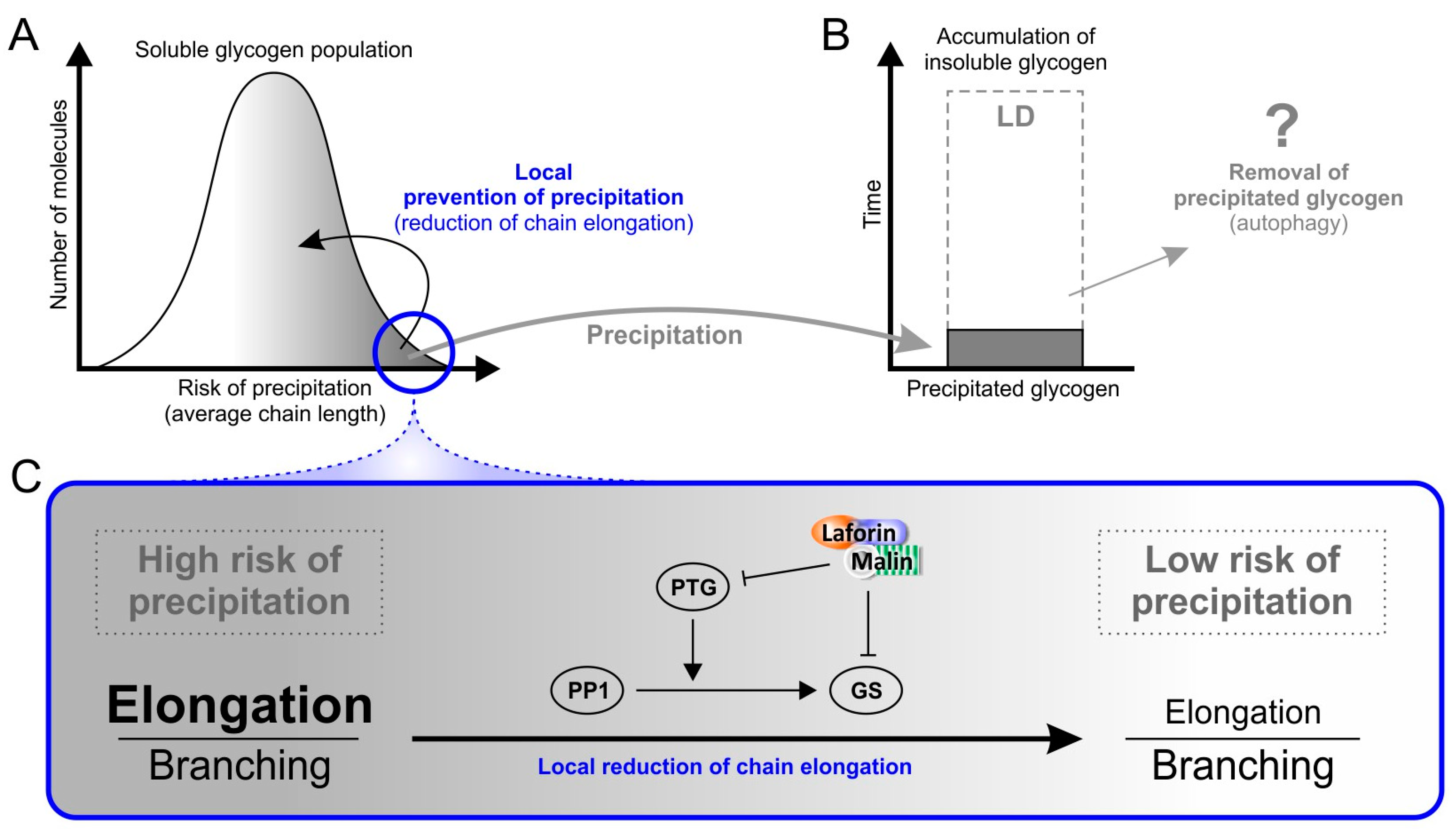
6. Factors That May Cause Glycogen Insolubility

A mutated laforin that possesses a catalytically inactive phosphatase site is able to prevent LD in mice deficient of endogenous laforin [38]. Moreover, hyperphosphorylation of glycogen is not strictly correlated with LB accumulation [51]. These results demonstrate that the increased glycogen phosphate levels per se cannot be the underlying cause of LD. The exact role of the glycogen phosphate remains to be elucidated.
It may, however, be informative to summarize the current knowledge of (de)phosphorylation of another polyglucan-storing biological system, i.e., plant starch metabolism. Typically, plants form and mobilize water-insoluble starch granules as a functional equivalent to glycogen in animals [72]. With the exception of some mutants, plant starch contains two types of polyglucans, amylopectin and amylose. Amylopectin is the dominant polyglucan that defines most of the properties of the entire starch particle. Furthermore, amylopectin possesses a relatively high content of branching points, a higher average molecular weight, and is, therefore, more similar to glycogen as compared to amylose (Figure 3) [73,74]. Phosphate esters are introduced into amylopectin by distinct dikinases, both during synthesis and degradation [75,76]. It is widely accepted that the covalent modification by phosphorylation mediates a reversible (and local) transition from a highly ordered but less hydrated form of the polyglucan into a less ordered but more hydrated form of starch [47,77,78]. During degradation one established role of phosphate esters is, due to the phosphate’s high charge and hydrophilia, to separate double-helices from each other, rendering them fully hydrated and, thus, facilitating access of starch degrading enzymes (Figure 4) [77,78]. As both glycogen and amylopectin have a common chemical basis, both being composed of branched α-glucan chains, local physicochemical effects of branching points or of similar covalent modifications (such as phosphorylation), are expected to have, in principle, similar consequences. Regarding what is known from starch metabolism, phosphorylation of glycogen should actually counteract glycogen insolubility. This implies that in the pathological LD state hyperphosphorylation would be beneficial rather than harmful.
Another factor that has been discussed as causative of LB accumulation is simply the cellular glycogen concentration. This hypothesis is supported by the fact that accumulation of insoluble glycogen (polyglucosan) is observed as a result of increased GS activity by overexpression of PTG or of GS itself [14,39]. While the cellular concentration of brain glycogen in these PTG or GS overexpressing mice is significantly higher than in the controls, these values are still 30–100 times lower than what is observed in healthy liver tissue [79]. This, together with the fact that in the brain LBs are mostly formed in neurons which have much lower glycogen contents than for instance astrocytes [80], makes it unlikely that glycogen becomes insoluble simply due to an increased concentration.
By contrast, the abnormal chain length distribution of glycogen strictly correlates with the occurrence of LBs in laforin- and malin-deficient mice [51]. The relevance of the chain length and branching pattern for the hydrosolubility of glucan polymers is obvious when comparing the solubility of the different polyglucans, starch, glycogen, and phytoglycogen. The latter is found in some plants or plant tissues with an altered expression of a few starch-related enzymes. These alterations are mostly biotechnologically induced (mutants) but can also occur naturally [81,82]. Non-pathological glycogen, amylopectin, and phytoglycogen all consist of chains of glucosyl residues connected via α-(1,4) linkages, which contain branch points via α-(1,6) linkages [83,84]. Glycogen and phytoglycogen are water-soluble while amylopectin is not. The average chain length of amylopectin varies depending on the plant species and tissue but is generally much higher than that of glycogen and phytoglycogen [85]. However, while in the soluble polyglucans the branching points are equally distributed throughout the molecule, in amylopectin they form clusters (Figure 3) [73,74]. The clustered arrangement gives rise to areas with low numbers of branching points. In these areas, chains have been shown to occur as double-helices including all but a few glucosyl units that are closest to the branch point and, therefore, are excluded from the double-helix due to steric constraints. Tight packing of the double-helices induces crystallization, hence water-insolubility of the highly dense starch particles [73,85,86]. Using purified malto-oligosaccharides as polyglucan models, the minimum length for glucan chain crystallization is reported to be 10 glucose units. In the presence of longer chains, however, malto-oligosaccharides of at least 6 units can co-crystallise [87].
Thus, it is obvious that frequent and regularly distributed branching points lead to shorter chains and less double-helix formation. Indeed, a strong correlation of chain length distribution and polyglucan crystallinity has been demonstrated using a range of mutant starches. In these studies, a high abundance of short glucan chains was consistently found to interfere with polyglucan crystallinity [88,89]. Furthermore, phytoglycogen-producing mutant plants accumulate insoluble polyglucans when additionally overexpressing a chain-elongating starch synthase, leading to (1) a higher abundance of longer glucan chains, and (2) to a substantial increase in polyglucan crystallinity as induced by double-helix formation [82].
A clear difference between water-soluble normal glycogen and the partially insoluble LD glycogen is the enrichment of longer α-glucan chains in the latter, as seen by its abnormal chain length distribution [20]. Similar to LD-related glycogen insolubility, a deficiency of GBE also leads to a dramatic accumulation of insoluble glycogen in the mouse model of GBE deficiency [90]. In turn, overexpression of GS or PTG, both leading to increased GS activity, results in polyglucosan body formation in mouse muscle and brain [39,91]. Thus, evidence is compelling that the abnormal chain length distribution of glycogen (i.e., the higher abundance of longer glucan chains) is the general reason for glycogen insolubility in LD.
It should be noted that the chain length distribution has to date been determined as an average of all glycogen molecules present in the respective tissue sample. At a subcellular/submolecular level, minor differences in the activities of GS and GBE may be part of normal glycogen metabolism. Such localized differences could lead to variations of chain length distributions in individual molecules and/or even in some regions of a given glycogen molecule, but remain undetected by conventional chain length distribution analyses. There is, however, evidence that even in wild-type mice some glycogen molecules possess longer chains than others, i.e., that not all molecules within one cell have an identical chain length distribution [20,92]. It is therefore possible that in the disperse mixture of glycogen molecules within a cell, not all molecules are affected to the same extent by the chain length abnormality seen in LD. Taking into account that the chain length greatly determines polyglucan solubility (see above), it implies that not all glycogen molecules may have the same risk (i.e., probability) to precipitate. Precipitation of glycogen likely affects a small proportion of glycogen molecules that have the most severe chain length abnormality. By contrast, less affected glycogen molecules (which probably represent the vast majority) can still be metabolized normally (Figure 5A). The hypothesized scenario is fully in line with the fact that insoluble polyglucosan, seen in LD, accumulates slowly.

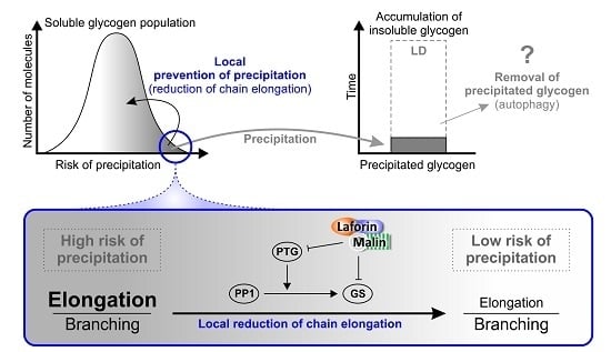
7. Why Does Insoluble Glycogen Accumulate in Lafora Disease?

With a loss-of-function mutation in either laforin or malin causing essentially the same pathological phenotype, and the strong evidence for in vivo laforin-malin interaction [48,53,68,93], it seems likely that the primary cause of LD is the lack of a functional laforin-malin complex. One function of this complex must therefore be associated with the avoidance of LB accumulation. From a mechanistic point of view, LBs are accumulating when the rate of production exceeds the rate of removal of the insoluble glycogen that aggregates in LBs. To accumulate bodies over time, the rate of removal can be substantial or infinitely small, but, in any case, must be smaller than the rate of production. In principle, deficiency in the laforin-malin complex may lead to an increased formation, or to an impaired removal of LBs, or may even affect both processes (Figure 5A,B). However, there is yet no evidence for a mechanism to remove LBs in vivo.
While plants possess the capacity to mobilize water-insoluble polyglucans (starch) under a wide range of external conditions, this is certainly not the case in animals, which generally have evolved to use water-soluble polyglucans (glycogen). Insolubility and aggregation of glycogen can have severe consequences in mammalian cells which becomes obvious in the neurons of LD patients. Thus, it is likely that hypothetical mechanisms to dispose of insoluble glucan material work at a low rate and may practically render precipitated glycogen metabolically inert.
Avoiding precipitation altogether would be biologically important. One possibility is that the laforin-malin complex is able to decrease GS activity selectively on molecules that are likely to precipitate, for example molecules that are still soluble but are, on average or in particular molecule regions, composed of longer glucan chains (Figure 5A). Indeed, it has been shown that laforin preferentially binds solubilized potato starch as compared to glycogen in vitro and polyglucosan bodies relative to glycogen in vivo [94], implying a preference of laforin for substrates composed of longer glucan chains and possessing less branching points. One role of laforin in the complex with malin would be to target malin via its CBM to a subset of glycogen molecules that possess abnormally long chains and are at a higher risk to precipitate. By polyubiquitination, malin promotes targeting of GS and PTG to degradation, whereby it could locally decrease chain elongation. Continued glycogen synthesis with locally reduced GS activity may lead to an increased frequency of branching points promoting water-solubility. Consequently, the risk of precipitation is diminished for distinct glycogen molecules that were otherwise prone to precipitate (Figure 5A,C). This model of locally focussed action of the laforin-malin complex consolidates the apparently conflicting results that (1) malin ubiquitinates PTG [30,58] and GS [58] leading to their degradation, and (2) total GS and PTG levels are not significantly altered in LD mice [71].
However, a mechanism for the removal of abnormal and insoluble glycogen cannot be ruled out and may also involve a functional laforin-malin complex. One possible mechanism of removal could proceed via autophagy which has been shown to be impaired in some LD mouse models but not in others [22,38,51]. There is increasing evidence that accumulation of insoluble glycogen underlies the inconsistently detected general autophagy impairment [14,95]. In cell culture studies, a laforin-malin complex has been associated with two autophagy-related proteins, designated LC3 and p62. The first is located in the autophagosome membrane, the second is an adapter protein that interacts with the laforin-malin complex and binds ubiquitinated proteins that are targeted to autophagy [57]. It has been shown that a general defect of autophagy cannot be strictly correlated with LB formation, and hence is not the primary cause of LD [22,51,96]. Nevertheless, it is possible to hypothesize that autophagy is involved in LB removal (Figure 5B). This process may be dependent on a functional laforin-malin complex and precisely act to target and dispose of abnormal and precipitated glycogen (Figure 5). Its impairment in LD mouse models may not consistently and detectably affect markers of general autophagy.
Thus, the function of the laforin-malin complex may be implicated in (1) a locally focussed alleviation of glycogen precipitation risks, (2) a selective removal of abnormally formed and precipitated glycogen, or (3) in both. All of these hypotheses are unified in the assumption of an underlying intrinsic property of glycogen metabolism to inadvertently produce disadvantageous structural variations of glycogen molecules including some that are at risk to precipitate. The hypotheses differ in how they assume the cell copes with the risk of glycogen precipitation. While according to the first hypothesis, glycogen precipitation is avoided as much as possible, the second hypothesis implies a removal of abnormal and precipitated glycogen by autophagy. It is not inconceivable that mammalian cells employ both of these strategies at the same time (hypothesis 3, Figure 5).

Acknowledgments

This work was supported by the Ontario Brain Institute and the National Institute of Neurological Disorders and Stroke of the National Institutes of Health (NIH) under award number P01 NS097197. Berge A. Minassian holds the University of Toronto Michael Bahen Chair in Epilepsy Research and the University of Texas Southwestern Jimmy Elizabeth Westcott Chair in Pediatric Neurology. Mitchell A. Sullivan was supported by an NHMRC CJ Martin Fellowship (GNT1092451). Martin Steup is visiting professor at the Impact Centre of the University of Toronto.

Conflicts of Interest

The authors declare no conflict of interest.

Abbreviations

AGLGlycogen debranching enzyme
AMPKAMP-activated protein kinase
CBMCarbohydrate-binding module
CLDChain length distribution
DSPDual specificity phosphatase
G6PGlucose-6-phosphate
GBEGlycogen branching enzyme
GPGlycogen phosphorylase
GSGlycogen synthase
GSK3Glycogen synthase kinase 3
KOKnockout
LBLafora body
LDLafora disease
PP1Protein phosphatase 1
PTGProtein targeting to glycogen – regulatory subunit of PP1
WTWild-type

References

  1. Minassian, B.A.; Lee, J.R.; Herbrick, J.A.; Huizenga, J.; Soder, S.; Mungall, A.J.; Dunham, I.; Gardner, R.; Fong, C.G.; Carpenter, S.; et al. Mutations in a gene encoding a novel protein tyrosine phosphatase cause progressive myoclonus epilepsy. Nat. Genet. 1998, 20, 171–174. [Google Scholar] [CrossRef] [PubMed]
  2. Serratosa, J.M.; Gomez-Garre, P.; Gallardo, M.E.; Anta, B.; de Bernabe, D.B.; Lindhout, D.; Augustijn, P.B.; Tassinari, C.A.; Malafosse, R.M.; Topcu, M.; et al. A novel protein tyrosine phosphatase gene is mutated in progressive myoclonus epilepsy of the lafora type (EPM2). Hum. Mol. Genet. 1999, 8, 345–352. [Google Scholar] [CrossRef] [PubMed]
  3. Chan, E.M.; Bulman, D.E.; Paterson, A.D.; Turnbull, J.; Andermann, E.; Andermann, F.; Rouleau, G.A.; Delgado-Escueta, A.V.; Scherer, S.W.; Minassian, B.A. Genetic mapping of a new lafora progressive myoclonus epilepsy locus (EPM2B) on 6p22. J. Med. Genet. 2003, 40, 671–675. [Google Scholar] [CrossRef] [PubMed]
  4. Singh, S.; Ganesh, S. Lafora progressive myoclonus epilepsy: A meta-analysis of reported mutations in the first decade following the discovery of the EPM2A and NHLRC1 genes. Hum. Mutat. 2009, 30, 715–723. [Google Scholar] [CrossRef] [PubMed]
  5. Lesca, G.; Boutry-Kryza, N.; de Toffol, B.; Milh, M.; Steschenko, D.; Lemesle-Martin, M.; Maillard, L.; Foletti, G.; Rudolf, G.; Nielsen, J.E.; et al. Novel mutations in EPM2A and NHLRC1 widen the spectrum of lafora disease. Epilepsia 2010, 51, 1691–1698. [Google Scholar] [CrossRef] [PubMed]
  6. Poyrazoglu, H.G.; Karaca, E.; Per, H.; Gumus, H.; Onay, H.; Canpolat, M.; Canoz, O.; Ozkinay, F.; Kumandas, S. Three patients with lafora disease: Different clinical presentations and a novel mutation. J. Child. Neurol. 2015, 30, 777–781. [Google Scholar] [CrossRef] [PubMed]
  7. Chan, E.M.; Omer, S.; Ahmed, M.; Bridges, L.R.; Bennett, C.; Scherer, S.W.; Minassian, B.A. Progressive myoclonus epilepsy with polyglucosans (lafora disease)—Evidence for a third locus. Neurology 2004, 63, 565–567. [Google Scholar] [CrossRef] [PubMed]
  8. Baykan, B.; Striano, P.; Gianotti, S.; Bebek, N.; Gennaro, E.; Gurses, C.; Zara, F. Late-onset and slow-progressing lafora disease in four siblings with EPM2B mutation. Epilepsia 2005, 46, 1695–1697. [Google Scholar] [CrossRef] [PubMed]
  9. Gomez-Abad, C.; Gomez-Garre, P.; Gutierrez-Delicado, E.; Saygi, S.; Michelucci, R.; Tassinari, C.A.; de Cordoba, S.R.; Serratosa, J.M. Lafora disease due to EPM2B mutations—A clinical and genetic study. Neurology 2005, 64, 982–986. [Google Scholar] [CrossRef] [PubMed]
  10. Franceschetti, S.; Gambardella, A.; Canafoglia, L.; Striano, P.; Lohi, H.; Gennaro, E.; Ianzano, L.; Veggiotti, P.; Sofia, V.; Biondi, R.; et al. Clinical and genetic findings in 26 Italian patients with lafora disease. Epilepsia 2006, 47, 640–643. [Google Scholar] [CrossRef] [PubMed]
  11. Jara-Prado, A.; Ochoa, A.; Alonso, M.E.; Lima Villeda, G.A.; Fernandez-Valverde, F.; Ruano-Calderon, L.; Vargas-Canas, S.; Duron, R.M.; Delgado-Escueta, A.V.; Martinez-Juarez, I.E. Late onset lafora disease and novel EPM2A mutations: Breaking paradigms. Epilepsy Res. 2014, 108, 1501–1510. [Google Scholar] [CrossRef] [PubMed]
  12. Minassian, B.A. Lafora’s disease: Towards a clinical, pathologic, and molecular synthesis. Pediatr. Neurol. 2001, 25, 21–29. [Google Scholar] [CrossRef]
  13. Carpenter, S.; Karpati, G. Sweat gland duct cells in lafora disease: Diagnosis by skin biopsy. Neurology 1981, 31, 1564–1568. [Google Scholar] [CrossRef] [PubMed]
  14. Duran, J.; Gruart, A.; Garcia-Rocha, M.; Delgado-Garcia, J.M.; Guinovart, J.J. Glycogen accumulation underlies neurodegeneration and autophagy impairment in lafora disease. Hum. Mol. Genet. 2014, 23, 3147–3156. [Google Scholar] [CrossRef] [PubMed]
  15. Striano, P.; Zara, F.; Turnbull, J.; Girard, J.M.; Ackerley, C.A.; Cervasio, M.; de Rosa, G.; del Basso-de Caro, M.L.; Striano, S.; Minassian, B.A. Typical progression of myoclonic epilepsy of the lafora type: A case report. Nat. Clin. Pract. Neurol. 2008, 4, 106–111. [Google Scholar] [CrossRef] [PubMed]
  16. Yokoi, S.; Austin, J.; Witmer, F.; Sakai, M. Studies in myoclonus epilepsy (lafora body form): I. Isolation and preliminary characterization of lafora bodies in two cases. Arch. Neurol. 1968, 19, 15–33. [Google Scholar] [CrossRef] [PubMed]
  17. Sakai, M.; Austin, J.; Witmer, F.; Trueb, L. Studies in myoclonus epilepsy (lafora body form). II. Polyglucosans in systemic deposits of myoclonus epilepsy and in corpora-amylacea. Neurology 1970, 20, 160. [Google Scholar] [CrossRef] [PubMed]
  18. Sullivan, M.A.; Vilaplana, F.; Cave, R.A.; Stapleton, D.; Gray-Weale, A.A.; Gilbert, R.G. Nature of α and β particles in glycogen using molecular size distributions. Biomacromolecules 2010, 11, 1094–1100. [Google Scholar] [CrossRef] [PubMed]
  19. Besford, Q.A.; Sullivan, M.A.; Zheng, L.; Gilbert, R.G.; Stapleton, D.; Gray-Weale, A. The structure of cardiac glycogen in healthy mice. Int. J. Biol. Macromol. 2012, 51, 887–891. [Google Scholar] [CrossRef] [PubMed]
  20. Nitschke, F.; Wang, P.; Schmieder, P.; Girard, J.-M.; Awrey, D.E.; Wang, T.; Israelian, J.; Zhao, X.; Turnbull, J.; Heydenreich, M.; et al. Hyperphosphorylation of glucosyl C6 carbons and altered structure of glycogen in the neurodegenerative epilepsy lafora disease. Cell Metab. 2013, 17, 756–767. [Google Scholar] [CrossRef] [PubMed]
  21. Wanson, J.C.; Drochmans, P. Rabbit skeletal muscle glycogen: A morphological and biochemical study of glycogen beta-particles isolated by the precipitation-centrifugation method. J. Cell Biol. 1968, 38, 130. [Google Scholar] [CrossRef] [PubMed]
  22. Irimia, J.M.; Tagliabracci, V.S.; Meyer, C.M.; Segvich, D.M.; DePaoli-Roach, A.A.; Roach, P.J. Muscle glycogen remodeling and glycogen phosphate metabolism following exhaustive exercise of wild type and laforin knockout mice. J. Biol. Chem. 2015, 290, 22686–22698. [Google Scholar] [CrossRef] [PubMed]
  23. Jayachandran, K.N.; Takacs-Cox, A.; Brooks, D.E. Synthesis and characterization of polymer brushes of poly(n,n-dimethylacrylamide) from polystyrene latex by aqueous atom transfer radical polymerization. Macromolecules 2002, 35, 4247–4257. [Google Scholar] [CrossRef]
  24. Roach, P.J.; Depaoli-Roach, A.A.; Hurley, T.D.; Tagliabracci, V.S. Glycogen and its metabolism: Some new developments and old themes. Biochem. J. 2012, 441, 763–787. [Google Scholar] [CrossRef] [PubMed]
  25. Testoni, G.; Duran, J.; Garcia-Rocha, M.; Vilaplana, F.; Serrano, A.L.; Sebastian, D.; Lopez-Soldado, I.; Sullivan, M.A.; Slebe, F.; Vilaseca, M.; et al. Lack of glycogenin causes glycogen accumulation and muscle function impairment. Cell. Metab. 2017, 26, 256–266. [Google Scholar] [CrossRef] [PubMed]
  26. Szydlowski, N.; Ragel, P.; Raynaud, S.; Lucas, M.M.; Roldán, I.; Montero, M.; Munoz, F.J.; Ovecka, M.; Bahaji, A.; Planchot, V.; et al. Starch granule initiation in arabidopsis requires the presence of either class IV or class III starch synthases. Plant Cell 2009, 21, 2443–2457. [Google Scholar] [CrossRef] [PubMed]
  27. Hunter, R.W.; Zeqiraj, E.; Morrice, N.; Sicheri, F.; Sakamoto, K. Expression and purification of functional human glycogen synthase-1:Glycogenin-1 complex in insect cells. Protein Expr. Purif. 2015, 108, 23–29. [Google Scholar] [CrossRef] [PubMed]
  28. Patel, S.; Doble, B.W.; MacAulay, K.; Sinclair, E.M.; Drucker, D.J.; Woodgett, J.R. Tissue-specific role of glycogen synthase kinase 3β in glucose homeostasis and insulin action. Mol. Cell Biol. 2008, 28, 6314–6328. [Google Scholar] [CrossRef] [PubMed]
  29. Fong, N.M.; Jensen, T.C.; Shah, A.S.; Parekh, N.N.; Saltiel, A.R.; Brady, M.J. Identification of binding sites on protein targeting to glycogen for enzymes of glycogen metabolism. J. Biol. Chem. 2000, 275, 35034–35039. [Google Scholar] [CrossRef] [PubMed]
  30. Worby, C.A.; Gentry, M.S.; Dixon, J.E. Malin decreases glycogen accumulation by promoting the degradation of protein targeting to glycogen (PTG). J. Biol. Chem. 2008, 283, 4069–4076. [Google Scholar] [CrossRef] [PubMed]
  31. Petty, H.R.; Worth, R.G.; Kindzelskii, A.L. Imaging sustained dissipative patterns in the metabolism of individual living cells. Phys. Rev. Lett. 2000, 84, 2754–2757. [Google Scholar] [CrossRef] [PubMed]
  32. Rickey Welch, G.; Easterby, J.S. Metabolic channeling versus free diffusion: Transition-time analysis. Trends Biochem. Sci. 1994, 19, 193–197. [Google Scholar] [CrossRef]
  33. Aw, T.Y. Intracellular compartmentation of organelles and gradients of low molecular weight species. Int. Rev. Cytol. 1999, 192, 223–253. [Google Scholar]
  34. Tagliabracci, V.S.; Turnbull, J.; Wang, W.; Girard, J.-M.; Zhao, X.; Skurat, A.V.; Delgado-Escueta, A.V.; Minassian, B.A.; DePaoli-Roach, A.A.; Roach, P.J. Laforin is a glycogen phosphatase, deficiency of which leads to elevated phosphorylation of glycogen in vivo. Proc. Natl. Acad. Sci. USA 2007, 104, 19262–19266. [Google Scholar] [CrossRef] [PubMed]
  35. Tagliabracci, V.S.; Girard, J.M.; Segvich, D.; Meyer, C.; Turnbull, J.; Zhao, X.C.; Minassian, B.A.; DePaoli-Roach, A.A.; Roach, P.J. Abnormal metabolism of glycogen phosphate as a cause for lafora disease. J. Biol. Chem. 2008, 283, 33816–33825. [Google Scholar] [CrossRef] [PubMed]
  36. Turnbull, J.; DePaoli-Roach, A.A.; Zhao, X.C.; Cortez, M.A.; Pencea, N.; Tiberia, E.; Piliguian, M.; Roach, P.J.; Wang, P.X.; Ackerley, C.A.; et al. PTG depletion removes lafora bodies and rescues the fatal epilepsy of lafora disease. PLoS Genet. 2011, 7, e1002037. [Google Scholar] [CrossRef] [PubMed]
  37. Turnbull, J.; Epp, J.R.; Goldsmith, D.; Zhao, X.C.; Pencea, N.; Wang, P.X.; Frankland, P.W.; Ackerley, C.A.; Minassian, B.A. PTG protein depletion rescues malin-deficient lafora disease in mouse. Ann. Neurol. 2014, 75, 442–446. [Google Scholar] [CrossRef] [PubMed]
  38. Gayarre, J.; Duran-Trio, L.; Garcia, O.C.; Aguado, C.; Juana-Lopez, L.; Crespo, I.; Knecht, E.; Bovolenta, P.; de Cordoba, S.R. The phosphatase activity of laforin is dispensable to rescue Epm2a−/− mice from lafora disease. Brain 2014, 137, 806–818. [Google Scholar] [CrossRef] [PubMed]
  39. Duran, J.; Tevy, M.F.; Garcia-Rocha, M.; Calbo, J.; Milan, M.; Guinovart, J.J. Deleterious effects of neuronal accumulation of glycogen in flies and mice. EMBO Mol. Med. 2012, 4, 1–11. [Google Scholar] [CrossRef] [PubMed]
  40. Komatsu, M.; Waguri, S.; Chiba, T.; Murata, S.; Iwata, J.; Tanida, I.; Ueno, T.; Koike, M.; Uchiyama, Y.; Kominami, E.; et al. Loss of autophagy in the central nervous system causes neurodegeneration in mice. Nature 2006, 441, 880–884. [Google Scholar] [CrossRef] [PubMed]
  41. Hara, T.; Nakamura, K.; Matsui, M.; Yamamoto, A.; Nakahara, Y.; Suzuki-Migishima, R.; Yokoyama, M.; Mishima, K.; Saito, I.; Okano, H.; et al. Suppression of basal autophagy in neural cells causes neurodegenerative disease in mice. Nature 2006, 441, 885–889. [Google Scholar] [CrossRef] [PubMed]
  42. McMahon, J.; Huang, X.X.; Yang, J.; Komatsu, M.; Yue, Z.Y.; Qian, J.; Zhu, X.J.; Huang, Y.F. Impaired autophagy in neurons after disinhibition of mammalian target of rapamycin and its contribution to epileptogenesis. J. Neurosci. 2012, 32, 15704–15714. [Google Scholar] [CrossRef] [PubMed]
  43. Gentry, M.S.; Roma-Mateo, C.; Sanz, P. Laforin, a protein with many faces: Glucan phosphatase, adapter protein, et alii. FEBS J. 2013, 280, 525–537. [Google Scholar] [CrossRef] [PubMed] [Green Version]
  44. Dubey, D.; Parihar, R.; Ganesh, S. Identification and characterization of novel splice variants of the human EPM2A gene mutated in lafora progressive myoclonus epilepsy. Genomics 2012, 99, 36–43. [Google Scholar] [CrossRef] [PubMed]
  45. Pulido, R.; Hooft van Huijsduijnen, R. Protein tyrosine phosphatases: Dual-specificity phosphatases in health and disease. FEBS J. 2008, 275, 848–866. [Google Scholar] [CrossRef] [PubMed]
  46. Wang, J.Y.; Stuckey, J.A.; Wishart, M.J.; Dixon, J.E. A unique carbohydrate binding domain targets the lafora disease phosphatase to glycogen. J. Biol. Chem. 2002, 277, 2377–2380. [Google Scholar] [CrossRef] [PubMed]
  47. Emanuelle, S.; Brewer, M.K.; Meekins, D.A.; Gentry, M.S. Unique carbohydrate binding platforms employed by the glucan phosphatases. Cell. Mol. Life Sci. 2016, 73, 2765–2778. [Google Scholar] [CrossRef] [PubMed]
  48. Lohi, H.; Ianzano, L.; Zhao, X.C.; Chan, E.M.; Turnbull, J.; Scherer, S.W.; Ackerley, C.A.; Minassian, B.A. Novel glycogen synthase kinase 3 and ubiquitination pathways in progressive myoclonus epilepsy. Hum. Mol. Genet. 2005, 14, 2727–2736. [Google Scholar] [CrossRef] [PubMed]
  49. Worby, C.A.; Gentry, M.S.; Dixon, J.E. Laforin, a dual specificity phosphatase that dephosphorylates complex carbohydrates. J. Biol. Chem. 2006, 281, 30412–30418. [Google Scholar] [CrossRef] [PubMed]
  50. Roach, P.J. Glycogen phosphorylation and lafora disease. Mol. Asp. Med. 2015, 46, 78–84. [Google Scholar] [CrossRef] [PubMed]
  51. Nitschke, F.; Sullivan, M.A.; Wang, P.; Zhao, X.; Chown, E.E.; Perri, A.M.; Israelian, L.; Juana-Lopez, L.; Bovolenta, P.; Rodriguez de Cordoba, S.; et al. Abnormal glycogen chain length pattern, not hyperphosphorylation, is critical in lafora disease. EMBO Mol. Med. 2017, 9, 906–917. [Google Scholar] [CrossRef] [PubMed]
  52. Fernandez-Sanchez, M.E.; Criado-Garcia, O.; Heath, K.E.; Garcia-Fojeda, B.; Medrano-Fernandez, I.; Gomez-Garre, P.; Sanz, P.; Serratosa, J.M.; de Cordoba, S.R. Laforin, the dual-phosphatase responsible for lafora disease, interacts with R5 (PTG), a regulatory subunit of protein phosphatase-1 that enhances glycogen accumulation. Hum. Mol. Genet. 2003, 12, 3161–3171. [Google Scholar] [CrossRef] [PubMed]
  53. Solaz-Fuster, M.C.; Gimeno-Alcaniz, J.V.; Ros, S.; Fernandez-Sanchez, M.E.; Garcia-Fojeda, B.; Garcia, O.C.; Vilchez, D.; Dominguez, J.; Garcia-Rocha, M.; Sanchez-Piris, M.; et al. Regulation of glycogen synthesis by the laforin-malin complex is modulated by the amp-activated protein kinase pathway. Hum. Mol. Genet. 2008, 17, 667–678. [Google Scholar] [CrossRef] [PubMed]
  54. Puri, R.; Suzuki, T.; Yamakawa, K.; Ganesh, S. Hyperphosphorylation and aggregation of tau in laforin-deficient mice, an animal model for lafora disease. J. Biol. Chem. 2009, 284, 22657–22663. [Google Scholar] [CrossRef] [PubMed]
  55. Ganesh, S.; Tsurutani, N.; Suzuki, T.; Ueda, K.; Agarwala, K.L.; Osada, H.; Delgado-Escueta, A.V.; Yamakawa, K. The lafora disease gene product laforin interacts with HIRIP5, a phylogenetically conserved protein containing a NifU-like domain. Hum. Mol. Genet. 2003, 12, 2359–2368. [Google Scholar] [CrossRef] [PubMed]
  56. Ianzano, L.; Zhao, X.C.; Minassian, B.A.; Scherer, S.W. Identification of a novel protein interacting with laforin, the EPM2A progressive myoclonus epilepsy gene product. Genomics 2003, 81, 579–587. [Google Scholar] [CrossRef]
  57. Sanchez-Martin, P.; Roma-Mateo, C.; Viana, R.; Sanz, P. Ubiquitin conjugating enzyme E2-N and sequestosome-1 (p62) are components of the ubiquitination process mediated by the malin-laforin E3-ubiquitin ligase complex. Int. J. Biochem. Cell Biol. 2015, 69, 204–214. [Google Scholar] [CrossRef] [PubMed]
  58. Vilchez, D.; Ros, S.; Cifuentes, D.; Pujadas, L.; Valles, J.; Garcia-Fojeda, B.; Criado-Garcia, O.; Fernandez-Sanchez, E.; Medrano-Fernandez, I.; Dominguez, J.; et al. Mechanism suppressing glycogen synthesis in neurons and its demise in progressive myoclonus epilepsy. Nat. Neurosci. 2007, 10, 1407–1413. [Google Scholar] [CrossRef] [PubMed]
  59. Freemont, P.S. Ubiquitination: Ring for destruction? Curr. Biol. 2000, 10, R84–R87. [Google Scholar] [CrossRef]
  60. Chan, E.M.; Young, E.J.; Ianzano, L.; Munteanu, I.; Zhao, X.C.; Christopoulos, C.C.; Avanzini, G.; Elia, M.; Ackerley, C.A.; Jovic, N.J.; et al. Mutations in NHLRC1 cause progressive myoclonus epilepsy. Nat. Genet. 2003, 35, 125–127. [Google Scholar] [CrossRef] [PubMed]
  61. Moreno, D.; Towler, M.C.; Hardie, D.G.; Knecht, E.; Sanz, P. The laforin-malin complex, involved in lafora disease, promotes the incorporation of K63-linked ubiquitin chains into amp-activated protein kinase beta subunits. Mol. Biol. Cell 2010, 21, 2578–2588. [Google Scholar] [CrossRef] [PubMed]
  62. Roma-Mateo, C.; Moreno, D.; Vernia, S.; Rubio, T.; Bridges, T.M.; Gentry, M.S.; Sanz, P. Lafora disease E3-ubiquitin ligase malin is related to TRIM32 at both the phylogenetic and functional level. BMC Evol. Biol. 2011, 11. [Google Scholar] [CrossRef] [PubMed] [Green Version]
  63. Rubio-Villena, C.; Garcia-Gimeno, M.A.; Sanz, P. Glycogenic activity of R6, a protein phosphatase 1 regulatory subunit, is modulated by the laforin-malin complex. Int. J. Biochem. Cell Biol. 2013, 45, 1479–1488. [Google Scholar] [CrossRef] [PubMed]
  64. Deshaies, R.J.; Joazeiro, C.A.P. Ring domain E3 ubiquitin ligases. Ann. Rev. Biochem. 2009, 78, 399–434. [Google Scholar] [CrossRef] [PubMed]
  65. Tan, J.M.M.; Wong, E.S.P.; Kirkpatrick, D.S.; Pletnikova, O.; Ko, H.S.; Tay, S.P.; Ho, M.W.L.; Troncoso, J.; Gygi, S.P.; Lee, M.K.; et al. Lysine 63-linked ubiquitination promotes the formation and autophagic clearance of protein inclusions associated with neurodegenerative diseases. Hum. Mol. Genet. 2008, 17, 431–439. [Google Scholar] [CrossRef] [PubMed]
  66. Nathan, J.A.; Kim, H.T.; Ting, L.; Gygi, S.P.; Goldberg, A.L. Why do cell proteins linked to K63-polyubiquitin chains not associate with proteasomes? EMBO J. 2013, 32, 552–565. [Google Scholar] [CrossRef] [PubMed]
  67. Sharma, J.; Mulherkar, S.; Mukherjee, D.; Jana, N.R. Malin regulates wnt signaling pathway through degradation of dishevelled2. J. Biol. Chem. 2012, 287, 6830–6839. [Google Scholar] [CrossRef] [PubMed]
  68. Gentry, M.S.; Worby, C.A.; Dixon, J.E. Insights into lafora disease: Malin is an E3 ubiquitin ligase that ubiquitinates and promotes the degradation of laforin. Proc. Natl. Acad. Sci. USA 2005, 102, 8501–8506. [Google Scholar] [CrossRef] [PubMed]
  69. Cheng, A.; Zhang, M.; Gentry, M.S.; Worby, C.A.; Dixon, J.E.; Saltiel, A.R. A role for AGL ubiquitination in the glycogen storage disorders of lafora and Cori’s disease. Genes Dev. 2007, 21, 2399–2409. [Google Scholar] [CrossRef] [PubMed]
  70. Viana, R.; Lujan, P.; Sanz, P. The laforin/malin E3-ubiquitin ligase complex ubiquitinates pyruvate kinase M1/M2. BMC Biochem. 2015, 16. [Google Scholar] [CrossRef] [PubMed]
  71. DePaoli-Roach, A.A.; Tagliabracci, V.S.; Segvich, D.M.; Meyer, C.M.; Irimia, J.M.; Roach, P.J. Genetic depletion of the malin E3 ubiquitin ligase in mice leads to lafora bodies and the accumulation of insoluble laforin. J. Biol. Chem. 2010, 285, 25372–25381. [Google Scholar] [CrossRef] [PubMed]
  72. Cenci, U.; Nitschke, F.; Steup, M.; Minassian, B.A.; Colleoni, C.; Ball, S.G. Transition from glycogen to starch metabolism in archaeplastida. Trends Plant Sci. 2014, 19, 18–28. [Google Scholar] [CrossRef] [PubMed]
  73. Buleon, A.; Colonna, P.; Planchot, V.; Ball, S. Starch granules: Structure and biosynthesis. Int. J. Biol. Macromolecules 1998, 23, 85–112. [Google Scholar] [CrossRef]
  74. Manners, D.J. Recent developments in our understanding of glycogen structure. Carbohydr. Polym. 1991, 16, 37–82. [Google Scholar] [CrossRef]
  75. Ritte, G.; Scharf, A.; Eckermann, N.; Haebel, S.; Steup, M. Phosphorylation of transitory starch is increased during degradation. Plant. Physiol. 2004, 135, 2068–2077. [Google Scholar] [CrossRef] [PubMed]
  76. Pfister, B.; Zeeman, S.C. Formation of starch in plant cells. Cell. Mol. Life Sci. 2016, 73, 2781–2807. [Google Scholar] [CrossRef] [PubMed]
  77. Hejazi, M.; Fettke, J.; Haebel, S.; Edner, C.; Paris, O.; Frohberg, C.; Steup, M.; Ritte, G. Glucan, water dikinase phosphorylates crystalline maltodextrins and thereby initiates solubilization. Plant J. 2008, 55, 323–334. [Google Scholar] [CrossRef] [PubMed]
  78. Zeeman, S.C.; Kossmann, J.; Smith, A.M. Starch: Its metabolism, evolution, and biotechnological modification in plants. Annu. Rev. Plant Biol. 2010, 61, 209–234. [Google Scholar] [CrossRef] [PubMed]
  79. Chen, C.B.; Williams, P.F.; Cooney, G.J.; Caterson, I.D. Diurnal rhythms of glycogen-metabolism in the liver and skeletal-muscle in gold thioglucose induced-obese mice with developing insulin resistance. Int. J. Obes. 1992, 16, 913–921. [Google Scholar]
  80. Saez, I.; Duran, J.; Sinadinos, C.; Beltran, A.; Yanes, O.; Tevy, M.F.; Martinez-Pons, C.; Milan, M.; Guinovart, J.J. Neurons have an active glycogen metabolism that contributes to tolerance to hypoxia. J. Cereb. Blood Flow Metab. 2014, 34, 945–955. [Google Scholar] [CrossRef] [PubMed]
  81. Bischof, S.; Umhang, M.; Eicke, S.; Streb, S.; Qi, W.; Zeeman, S.C. Cecropia peltata accumulates starch or soluble glycogen by differentially regulating starch biosynthetic genes. Plant Cell 2013, 25, 1400–1415. [Google Scholar] [CrossRef] [PubMed] [Green Version]
  82. Fujita, N.; Hanashiro, I.; Suzuki, S.; Higuchi, T.; Toyosawa, Y.; Utsumi, Y.; Itoh, R.; Aihara, S.; Nakamura, Y. Elongated phytoglycogen chain length in transgenic rice endosperm expressing active starch synthase IIa affects the altered solubility and crystallinity of the storage α-glucan. J. Exp. Bot. 2012, 63, 5859–5872. [Google Scholar] [CrossRef] [PubMed]
  83. Gilbert, R.G.; Wu, A.C.; Sullivan, M.A.; Sumarriva, G.E.; Ersch, N.; Hasjim, J. Improving human health through understanding the complex structure of glucose polymers. Anal. Bioanal. Chem. 2013, 405, 8969–8980. [Google Scholar] [CrossRef] [PubMed]
  84. Powell, P.O.; Sullivan, M.A.; Sweedman, M.C.; Stapleton, D.I.; Hasjim, J.; Gilbert, R.G. Extraction, isolation and characterisation of phytoglycogen from su-1 maize leaves and grain. Carbohyd. Polym. 2014, 101, 423–431. [Google Scholar] [CrossRef] [PubMed]
  85. Wu, A.C.; Gilbert, R.G. Molecular weight distributions of starch branches reveal genetic constraints on biosynthesis. Biomacromolecules 2010, 11, 3539–3547. [Google Scholar] [CrossRef] [PubMed]
  86. Umeki, K.; Kainuma, K. Fine structure of naegeli amylodextrin obtained by acid treatment of defatted waxy-maize starch—Structural evidence to support the double-helix hypothesis. Carbohydr. Res. 1981, 96, 143–159. [Google Scholar] [CrossRef]
  87. Gidley, M.J.; Bulpin, P.V. Chrystallization of maltoologosaccharides as models of the crystalline forms of starch-minimum chain-length requirement for the formation of double helices. Carbohydr. Res. 1987, 161, 291–300. [Google Scholar] [CrossRef]
  88. Wong, K.-S.; Kubo, A.; Jane, J.-L.; Harada, K.; Satoh, H.; Nakamura, Y. Structures and properties of amylopectin and phytoglycogen in the endosperm of sugary-1 mutants of rice. J. Cereal Sci. 2003, 37, 139–149. [Google Scholar] [CrossRef]
  89. Fujita, N.; Kubo, A.; Suh, D.S.; Wong, K.S.; Jane, J.L.; Ozawa, K.; Takaiwa, F.; Inaba, Y.; Nakamura, Y. Antisense inhibition of isoamylase alters the structure of amylopectin and the physicochemical properties of starch in rice endosperm. Plant Cell Physiol. 2003, 44, 607–618. [Google Scholar] [CrossRef] [PubMed]
  90. Akman, H.O.; Sheiko, T.; Tay, S.K.H.; Finegold, M.J.; DiMauro, S.; Craigen, W.J. Generation of a novel mouse model that recapitulates early and adult onset glycogenosis type IV. Hum. Mol. Genet. 2011, 20, 4430–4439. [Google Scholar] [CrossRef] [PubMed]
  91. Raben, N.; Danon, M.; Lu, N.; Lee, E.; Shliselfeld, L.; Skurat, A.V.; Roach, P.J.; Lawrence, J.C., Jr.; Musumeci, O.; Shanske, S.; et al. Surprises of genetic engineering: A possible model of polyglucosan body disease. Neurology 2001, 56, 1739–1745. [Google Scholar] [CrossRef] [PubMed]
  92. Palmer, T.N.; Macaskie, L.E.; Grewal, K.K. Spatial-distribution of unit chains in glycogen. Carbohydr. Res. 1983, 115, 139–150. [Google Scholar] [CrossRef]
  93. Liu, Y.; Zeng, L.; Ma, K.L.; Baba, O.; Zheng, P.; Liu, Y.; Wang, Y. Laforin-malin complex degrades polyglucosan bodies in concert with glycogen debranching enzyme and brain isoform glycogen phosphorylase. Mol. Neurobiol. 2014, 49, 645–657. [Google Scholar] [CrossRef] [PubMed]
  94. Chan, E.M.; Ackerley, C.A.; Lohi, H.; Ianzano, L.; Cortez, M.A.; Shannon, P.; Scherer, S.W.; Minassian, B.A. Laforin preferentially binds the neurotoxic starch-like polyglucosans, which form in its absence in progressive myoclonus epilepsy. Hum. Mol. Genet. 2004, 13, 1117–1129. [Google Scholar] [CrossRef] [PubMed]
  95. Duran, J.; Guinovart, J.J. Brain glycogen in health and disease. Mol. Aspects Med. 2015, 46, 70–77. [Google Scholar] [CrossRef] [PubMed]
  96. Wang, P.; Israelian, L.; Xue, Y.; Song, S.; Attisano, L.; Minassian, B.A. SGK1 (glucose transport), dishevelled2 (wnt signaling), LC3/p62(autophagy) and p53 (apoptosis) proteins are unaltered in lafora disease. All Results J. Biol. 2016, 7, 28–33. [Google Scholar]
Figure 1. Lafora disease (LD) in the context of glycogen metabolism. Synthesis of glycogen molecules in the cytosol (top row) essentially includes autoglucosylation of glycogenin, elongation of chains by glycogen synthase (GS), and introduction of branching points by glycogen branching enzyme (GBE). The regularly branched and water-soluble glycogen molecules are degraded by glycogen phosphorylase (GP) and glycogen debranching enzyme (AGL, indirect debranching) in the cytosol or by lysosomal enzymes such as acid α-glucosidase (GAA). Functional laforin and malin essentially inhibit the formation of Lafora bodies and prevent LD as indicated by the inhibitory arrow. Mutations leading to non-functional laforin or malin (protein domains explained in the text) result in formation and accumulation of Lafora bodies as exemplarily shown in brain sections of the laforin-deficient mouse model (bottom right, scale bar equals 100 µm). In humans, LD manifests in an eventually fatal progression of clinical symptoms (bottom left).
Ijms 18 01743 g001
Figure 2. Prevention of LBs depends on the presence of malin but not on laforin’s phosphatase activity. The phenotypic features (C6 phosphate and chain length distribution, CLD) of brain glycogen as well as LB occurrence in the hippocampus are summarized for different LD-related mouse lines. Overexpression of phosphatase inactive laforin (C266S mutation in the DSP domain) leads to glycogen hyperphosphorylation but prevents both CLD abnormality and LB accumulation provided malin is functional. Hyperphosphorylation per se does not cause LB formation and accumulation.
Ijms 18 01743 g002
Figure 3. Branching structure of amylopectin and glycogen. (A) Chemical basis of a branched α-glucan. R1, glucan chain at the reducing end; R2 and R3, chains at the nonreducing ends; (B,C) Segments of glycogen and amylopectin, respectively. In glycogen, branching points are essentially distributed evenly; in amylopectin, they are clustered. In areas with a low abundance of branching points, double-helices form. Modified version from [20].
Ijms 18 01743 g003
Figure 4. Role of phosphate esters during degradation of plant amylopectin. Degradation of starch granules in higher plants is facilitated by cycles of phosphorylation and dephosphorylation. Tightly packed, semi-crystalline exterior glucan chains of amylopectin undergo phase transition (specified in the text) through incorporation of phosphate esters by glucan water dikinase (GWD) and phosphoglucan water dikinase (PWD), both utilizing ATP. Exo-acting β-amylases (BAM) remove maltose units from the non-reducing ends, their action being obstructed by phosphate esters and branching points. The glucan phosphatases SEX4 (starch-excess 4) and LSF2 (Like-SEX4 2) remove phosphate esters and enable further BAM-mediated degradation. Isoamylase (ISA) is required for direct debranching in the amorphous layer of amylopectin before GWD-mediated phosphorylation initiates degradation of the subsequent semi-crystalline layer. Figure modified from [47,78].
Ijms 18 01743 g004
Figure 5. Mechanistic model for Lafora body formation and accumulation. (A) Glycogen as a disperse mixture of molecules that possess different contents of long glucan chains and according risks of precipitation. The minor proportion of molecules (blue circle) actually subject to precipitation (grey arrow) may be modified by local reduction of chain elongation that is achieved by a well-supported mechanism explained in (C). This modification increases the relative branching frequency and decreases the risk to precipitate; (B) If glycogen precipitation is not (entirely) avoided, precipitated glycogen is accumulating over time (y-axis) as seen in Lafora disease, unless cells are able to mobilize and remove precipitated glycogen (e.g., hypothetically (question mark) through autophagy) at a rate that is higher than that of precipitation; (C) The mechanism by which a functional laforin-malin complex can reduce GS activity as supported by experimental data obtained in cell-culture [30,58]. Negative effects are indicated by inhibitory arrows while normal arrows indicate a positive effect. By targeting GS and PTG to degradation, the laforin-malin complex reduces GS activity and relatively increases branching frequency. It is proposed that this process may not significantly affect the entire GS population but is occurring locally, i.e., predominantly at a minor proportion of glycogen molecules with a high risk of precipitation (blue circle in (A)).
Ijms 18 01743 g005

