Article

Divergent Expression Patterns and Function Implications of Four nanos Genes in a Hermaphroditic Fish, Epinephelus coioides

1
State Key Laboratory of Freshwater Ecology and Biotechnology, Institute of Hydrobiology, Chinese Academy of Sciences, Graduate University of the Chinese Academy of Sciences, Wuhan 430072, China
2
State Key Laboratory of Biocontrol, Guangdong Province Key Laboratory for Aquatic Economic Animals, The Institute of Aquatic Economic Animals, School of Life Sciences, Sun Yat-Sen University, Guangzhou 510275, China
*
Authors to whom correspondence should be addressed.
Int. J. Mol. Sci. 2017, 18(4), 685; https://doi.org/10.3390/ijms18040685
Submission received: 13 February 2017 / Revised: 12 March 2017 / Accepted: 17 March 2017 / Published: 23 March 2017
(This article belongs to the Section Biochemistry)

Abstract

:
Multiple nanos genes have been characterized in several fishes, but the functional implications of their various expression patterns remain unclear. In this study, we identified and characterized four nanos genes from a hermaphroditic fish orange-spotted grouper, Epinephelus coioides. Ecnanos1a and Ecnanos1b show divergent expression patterns, and the dynamic expression change of Ecnanos1a in pituitaries during sex change is associated with testis differentiation and spermatogenesis. Ecnanos2 and Ecnanos3 might be germline stem cells (GSCs) and primordial germ cells (PGCs)-specific markers, respectively. Significantly, Ecnanos3 3′-untranslated region (UTR) is necessary for PGC specific expression, where a non-canonical “GCACGTTT” sequence is required for miR-430-mediated repression of Ecnanos3 RNA. Furthermore, grouper Dead end (Dnd) can relieve miR-430 repression in PGCs by associating with a 23 bp U-rich region (URR) in Ecnanos3 3′-UTR. The current study revealed the functional association of multiple nanos genes with PGC formation and germ cell development in orange-spotted grouper, and opened up new possibilities for developing biotechnologies through utilizing the associations between Ecnanos3 and PGCs or between Ecnanos2 and GSCs in the hermaphroditic fish.

1. Introduction

Germ-line cells, including primordial germ cells (PGCs), germline stem cells (GSCs) and gametes, have been attracting more and more attention. The basic knowledge of germ-line cell specialization and development is not only crucial for understanding successful reproduction strategies of aquaculture species [1], but also has opened up new possibilities for developing biotechnology of PGCs, such as interspecific surrogate [2,3,4]. Several conserved germline genes, such as vasa, dazl and dead end (dnd), have been demonstrated to be required for specifying and/or maintaining the germline development [5,6,7]. Nanos, an RNA binding protein, was first identified as a determinant of abdomen formation in Drosophila [8], and later its evolutionarily conserved functions in germ cell development were revealed in both invertebrates [9] and vertebrates [10]. It was also confirmed to be essential for the specification and migration of PGCs [9,11,12] and for the maintenance of GSCs self-renewal [13].
Although only one nanos gene was found in Drosophila, multiple nanos genes, including nanos1, nanos2, and nanos3, have been cloned in diverse organisms. They show varied expression patterns, functions and regulatory mechanisms among different species. In human, NANOS1 is expressed ubiquitously, while NANOS2 and NANOS3 are enriched in ovary, testis and fetal brain [14]. In mouse, Nanos1 was observed to be expressed in the central nervous system [15], NANOS3 was detected in the PGCs as early as Embryonic Day 9.5 (E9.5) and expressed in ovary and testis at a later stage of development, whereas NANOS2 was found to be restricted in the developing male PGCs at E13.5 and then to be expressed predominantly in male germ cells [10]. In fish, multiple nanos genes and their diverse expression patterns have been identified and characterized in several species. In medaka, nanos1a and nanos1b are expressed in brain during early embryogenesis, nanos3 was detected in both ovary and testis, and nanos2 was expressed in oogonia and spermatogonia [16]. However, fewer nanos genes were identified in other fish, which exhibit some different expression patterns compared to that in medaka. In model vertebrate organism zebrafish [17,18], three nanos genes were predicted from bioinformatics analysis, and only nanos2 and nanos3 were characterized [19,20,21,22,23]. In rainbow trout, nanos3 was observed to be expressed in ovary and testis, while nanos2 expression was found to be restricted to a subpopulation (less than 20%) of undifferentiated spermatogonia [24]. In Chinese sturgeon and Japanese eel, only nanos1 was identified, and their expression patterns were not detailed [25,26]. In olive flounder, Atlantic cod and common carp, only nanos3 was identified as PGCs marker [27,28,29]. Therefore, the various expression patterns and function implications of nanos genes should be further investigated in diverse fishes.
The expression of nanos is usually mediated by its 3′-untranslated region (UTR). Nanos3 protein is maternal and expressed specifically in PGCs during early embryogenesis. However, nanos3 mRNA exists in both PGCs and somatic cells [14,21,26]. The PGC-specific expression of Nanos3 protein is mainly due to the posttranscriptional suppression in somatic cells. Several elements in nanos3 3′-UTR mediate the mRNA stability and translation in germ cells [30,31,32,33]. MicroRNAs (miRNAs) are a class of 22~24-nucleotide (nt) RNAs, which base pair with the 3′-UTR of mRNAs and lead to translational repression and/or mRNA degradation [34,35]. miR-430 is abundantly produced during maternal-zygotic transition (MZT), and plays essential roles in inducing the deadenylation, degradation, and translational repression of germplasm mRNAs in somatic cells through binding to their 3′-UTRs. Moreover, Dnd protein combines the U-rich regions of nanos3 3′-UTR to block miRNA accessibility [34,36]. The miR-430 family uses the same seed sequence (GCACUU) to target the mRNAs and cause their degradation [33,37]. However, this core binding site is absent in nanos3 3′-UTRs of some teleost fish, including cod, salmon, and olive flounder [32]. Therefore, the interaction mechanism of nanos3 3′-UTRs, miRNA-430 and Dnd protein should be further investigated in various vertebrates.
Orange-spotted grouper is a protogynous hermaphrodite teleost [38,39]. It is an important mariculture species in Southeast Asia because of its superior taste and high commercial value [40,41]. Recently, its sex differentiation and regulatory mechanisms have attracted considerable attentions [42,43,44]. Several genes related to sex differentiation and sex change, such as dmrt1 [45], sox3 [40], tshβ [46], cyp19a [47], and β-defensin [48], have been identified and a hypothetical molecular mechanism underlying sex change has also been proposed [43]. However, no detailed studies about PGC formation and germ cell differentiation were performed in orange-spotted grouper. In the present study, we attempted to identify diverse nanos genes, to characterize the expression patterns, to reveal the key sequence interaction with miR-430 and Dnd protein, and thereby to investigate the function implications in this hermaphroditic fish.

2. Results

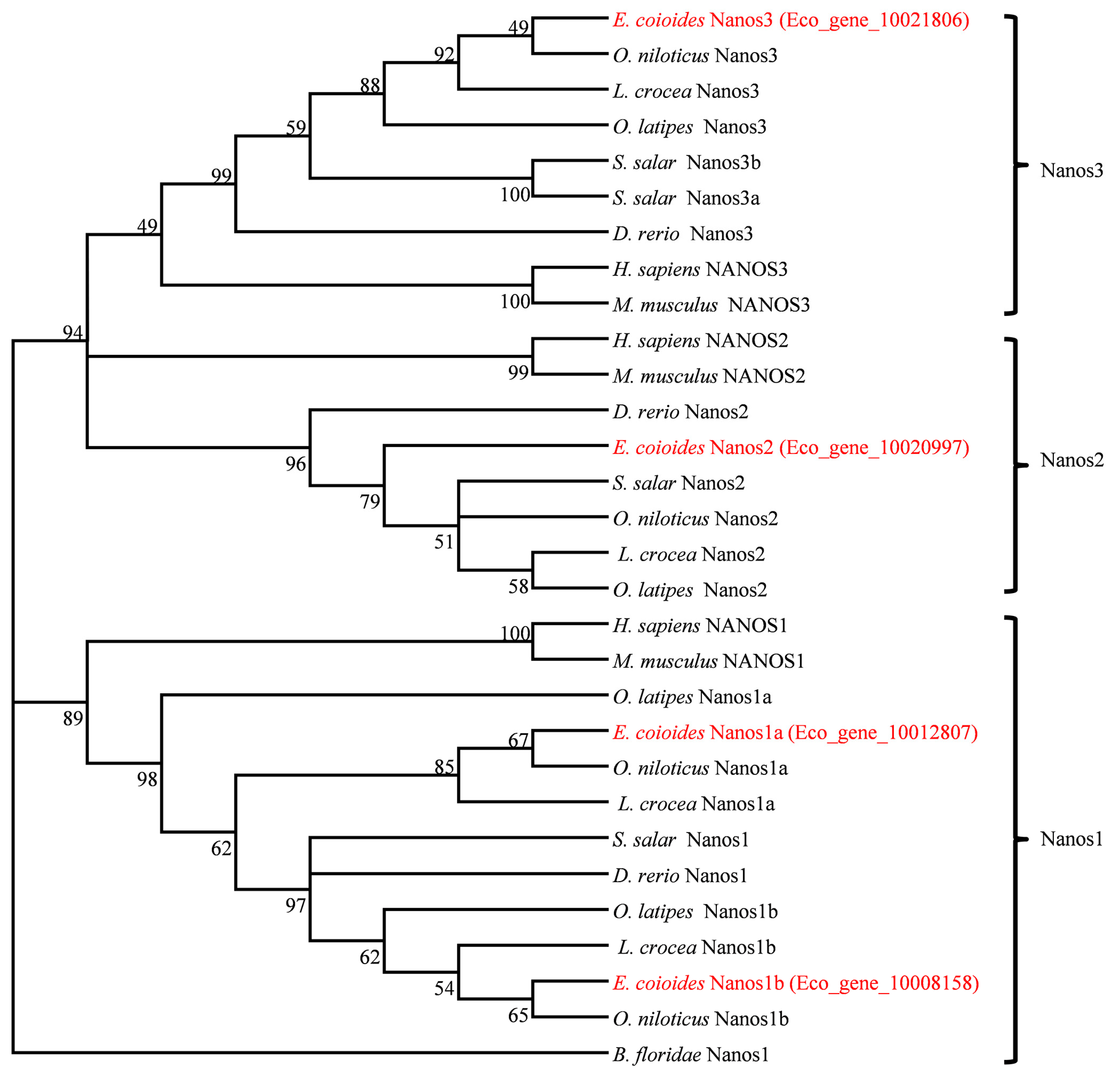
2.1. Molecular Characterization and Phylogenetic Relationship of Four Nanos Genes

A draft genome of orange-spotted grouper has been assembled by whole-genome shotgun strategy in Sun Yat-Sen University (unreported data). In order to identify nanos genes in orange-spotted grouper, BLAST search was first performed from the draft genome by using the common conserved RNA-binding zinc finger domain (zf-domain) sequence of human NANOS1 (NP_955631) as the query, and four predicted nanos genes (Eco_gene_10012807, Eco_gene_10008158, Eco_gene_10020997 and Eco_gene_10021806) were identified. Subsequently, four full-length nanos cDNA sequences were obtained by 5′- and 3′-rapid-amplification of cDNA ends (RACE). Eco_gene_10012807 and Eco_gene_10008158 are 1753 and 1263 bp, and the open reading frames (ORFs) are 672 and 711 bp, respectively. The Eco_gene_10020997 cDNA is 1169 bp in length, and encodes a protein of 250 amino acids. Eco_gene_10021806 cDNA is 1451 bp in total length, consisting of a coding sequence of 666 bp.
To define these four nanos genes, we downloaded 39 nanos genes from 12 species based on the zf-domain of human NANOS from Ensembl and NCBI database and performed comparative analysis. Multiple protein sequence alignments showed that the deduced amino acid sequences of Eco_gene_10012807 and Eco_gene_0008158 exhibited high identities to other teleost Nanos1, ranging from 58.5% to 88.6% and 77.5% to 93.2% respectively (Figures S1 and S2), and named as EcNanos1a (KX262959) and EcNanos1b (KX262960). The identity between EcNanos1a and EcNanos1b is about 60.7%, and the identity of zf-domain is about 92.6%. Although Nanos2 protein possesses species specificity, the zf-domain of Eco_gene_10020997 showed 58.1% to 76.8% identities to other vertebrate Nanos2 (Figure S3). Thus, we named it EcNanos2 (KX262962). The deduced amino acid sequences of Eco_gene_10021806 shows 30.2% to 62.6% identities to other teleost Nanos3 proteins (Figure S4), and named as EcNanos3 (KX262961). Similar to those in other species, all four Ecnanos genes contain just a single exon and are characterized by a highly conserved RNA-binding zf-domain with eight invariant cysteine and histidine residues in a “CCHC CCHC configuration” and a conserved CCR4-NOT deadenylase interaction domain in the NH2 terminus (Figures S1–S4).
Based on these Nanos amino acid sequences, a phylogenetic relationship tree was constructed. As shown in Figure 1, these Nanos proteins are divided into three branches (Nanos1, -2, and -3). EcNanos1a and EcNanos1b are clustered into Nanos1 branch. EcNanos2 and EcNanos3 are grouped with Nanos2 and Nanos3 in other species, respectively. The topology of clades of Nanos1, Nanos2 and Nanos3 are basically consistent with the known taxonomic relationship among these species.

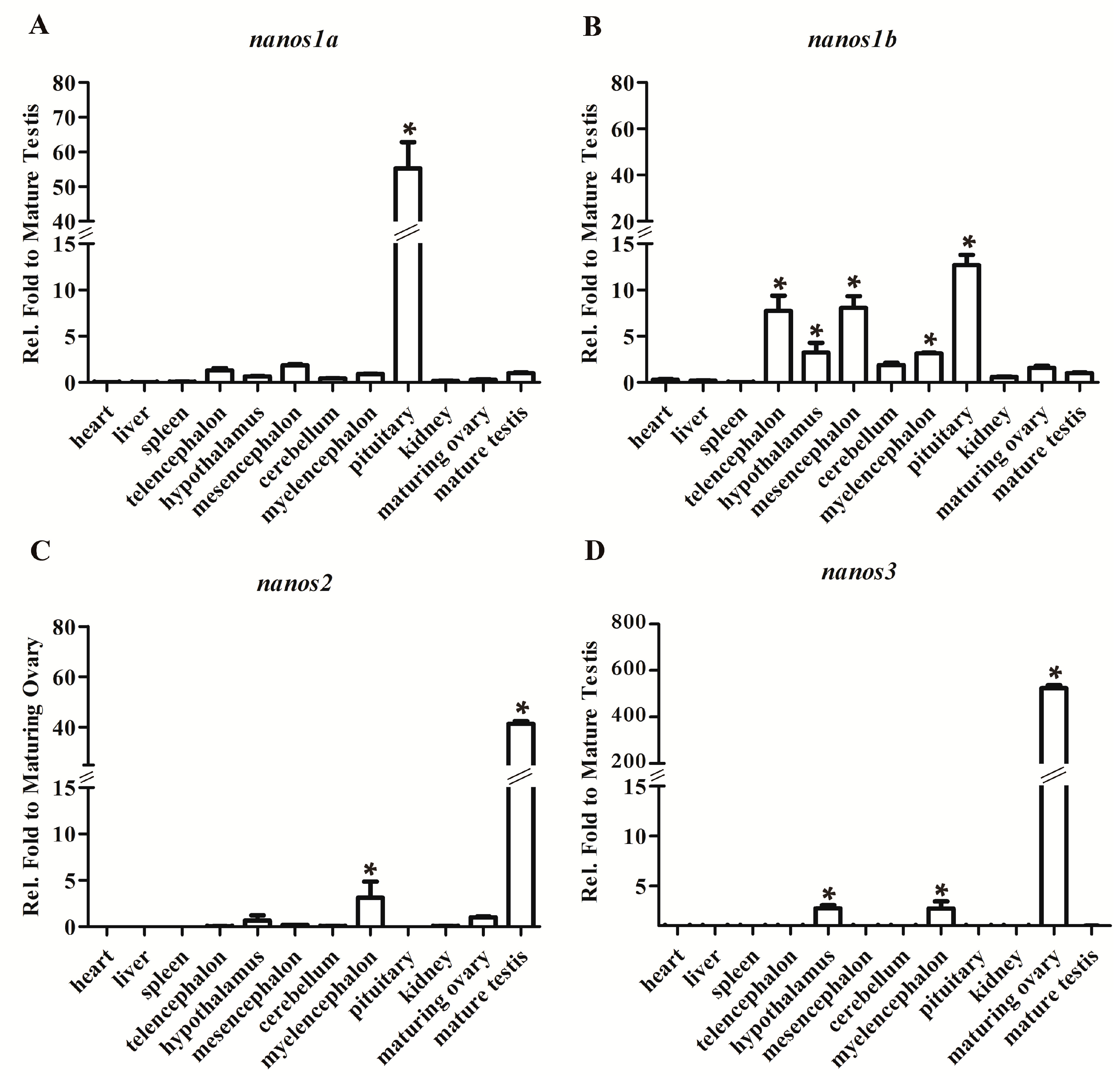
2.2. Differential Expression Patterns of Four Ecnanos Genes in Adult Tissues

To reveal the expression difference, we detected the transcripts of all four Ecnanos genes by RT-qPCR in adult tissues, including heart, liver, spleen, telencephalon, hypothalamus, mesencephalon, cerebellum, myelencephalon, pituitary, kidney, ovary and testis. As shown in Figure 2, Ecnanos1a is expressed predominantly in pituitary (Figure 2A). Compared to Ecnanos1a, Ecnanos1b is mainly transcribed in pituitary and diverse brain tissues (Figure 2B). Unlike Ecnanos1, Ecnanos2 and Ecnanos3 are predominantly in mature testis (Figure 2C) and maturing ovary (Figure 2D), respectively.

2.3. Ecnanos1a Is Up-Regulated in Pituitaries during Sex Change

Considering Ecnanos1a is expressed predominantly in pituitary (Figure 2A), we sampled pituitaries at different stages during gonad development and sex change to examine relative expression association of Ecnanos1s with the gonad differentiation and sex change of orange-spotted grouper.
Firstly, gonadal histology was performed to determine the developmental phase of gonad. As previously described [46,49], the gonads of orange-spotted grouper are classified into six stages, including undifferentiated-stage gonad, resting ovary, maturing ovary, early transitional gonad, late transitional gonad and mature testis. In the undifferentiated-stage, the paired gonadal primordia (GP) with a few scattered PGCs adhere to the swim-bladder wall (Figure 3A). The resting ovary gonad is small, compact and translucent with a large number of proliferating primary-growth oocytes (PO) and previtellogenic oocytes (PVO) (Figure 3B). The maturing ovary contains a lot of vitellogenic oocytes (VO), PVO and PO (Figure 3C). The early transitional gonad shows obvious lobules, where degenerating primary oocytes (DPO), cavities (CV) and PO are mixed with spermatocytes (SPC) and spermatids (SPD) (Figure 3D). In late transitional gonad, there are many spermatogonia (SPG), SPC and SPD, and only a few of PO scatters in the lobules (Figure 3E). In the mature testis, a great variety of male germ cells at all spermatogenic stages fill the gonads and numerous free spermatozoa (SPZ) congregate into the lobule lumen (Figure 3F).
Then, the total RNAs of pituitaries at the six stages were isolated for RT-qPCR detection. As shown in Figure 3G, Ecnanos1a shows similar expression level in the pituitaries at undifferentiated-stage, resting ovary stage and maturing ovary stage. As sex change to early transitional gonad and late transitional gonad stages, the Ecnanos1a transcripts start to rise significantly, and reach the highest level in the pituitaries of mature male individuals, indicating a close correlation between the Ecnanos1a expression and the male sex differentiation. In contrast to Ecnanos1a, Ecnanos1b expression in pituitaries at the corresponding stages is much lower, and there is no significant difference among the six gonadal developmental stages. Furthermore, the expression level of Ecnanos1a is up to about 60 folds against that of Ecnanos1b in the pituitaries of mature male individuals (Figure 3G). The data suggest that functional divergence might have occurred between Ecnanos1a and Ecnanos1b, and Ecnanos1a might play an important role in regulating orange-spotted grouper sex change.

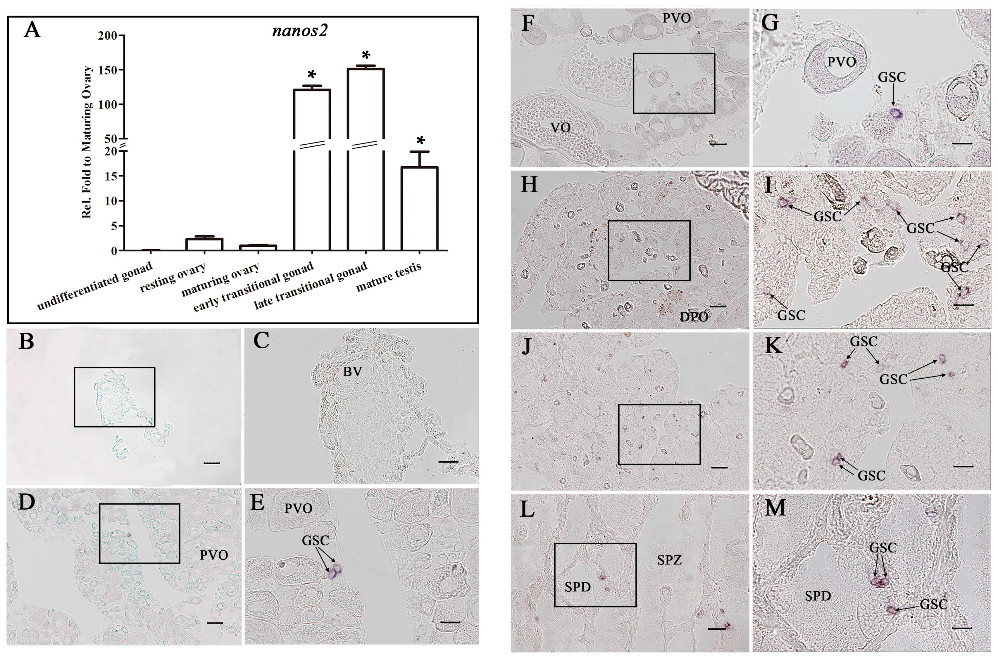
2.4. Ecnanos2 Is Expressed Abundantly in Transitional Gonads and Specifically in Germline Stem Cells

Owing to the differential expression pattern of Ecnanos2 in ovary and testis (Figure 2C), we evaluated the expression of Ecnanos2 in the gonads at the above six developmental stages by RT-qPCR and in-situ hybridization (ISH). Consistent with the former RT-qPCR results of adult tissues (Figure 2C), a very low level of Ecnanos2 transcript exists in the gonads at resting ovary and maturing ovary stages (Figure 4A). Along with sex change occurrence, the expression level of Ecnanos2 sharply increases up to 60–120 folds at early transitional gonad, and reaches its peak at late transitional gonad. After testis maturation, the expression level of Ecnanos2 significantly decreases, which are down to 7–9 folds against those of early and late transitional gonads (Figure 4A).
ISH detection not only observed similar expression dynamics but also revealed specifically expressed cells of Ecnanos2. Similar to the RT-qPCR data (Figure 4A), no Ecnanos2 positive cells were detected in gonadal primordia (Figure 4B,C). In resting ovary and maturing ovary, Ecnanos2 was found to be expressed only in a subset of small germ cells with a diameter of <20 μm (Figure 4D–G). Moreover, more abundant positive Ecnanos2 cells were observed in early and late transitional gonads (Figure 4H–K), and some of them were still seen to distribute in the testicular lobules of mature testis (Figure 4L,M). According to previous report in medaka and zebrafish [17], the positive cells might be GSCs, where Ecnanos2 is expressed specifically.

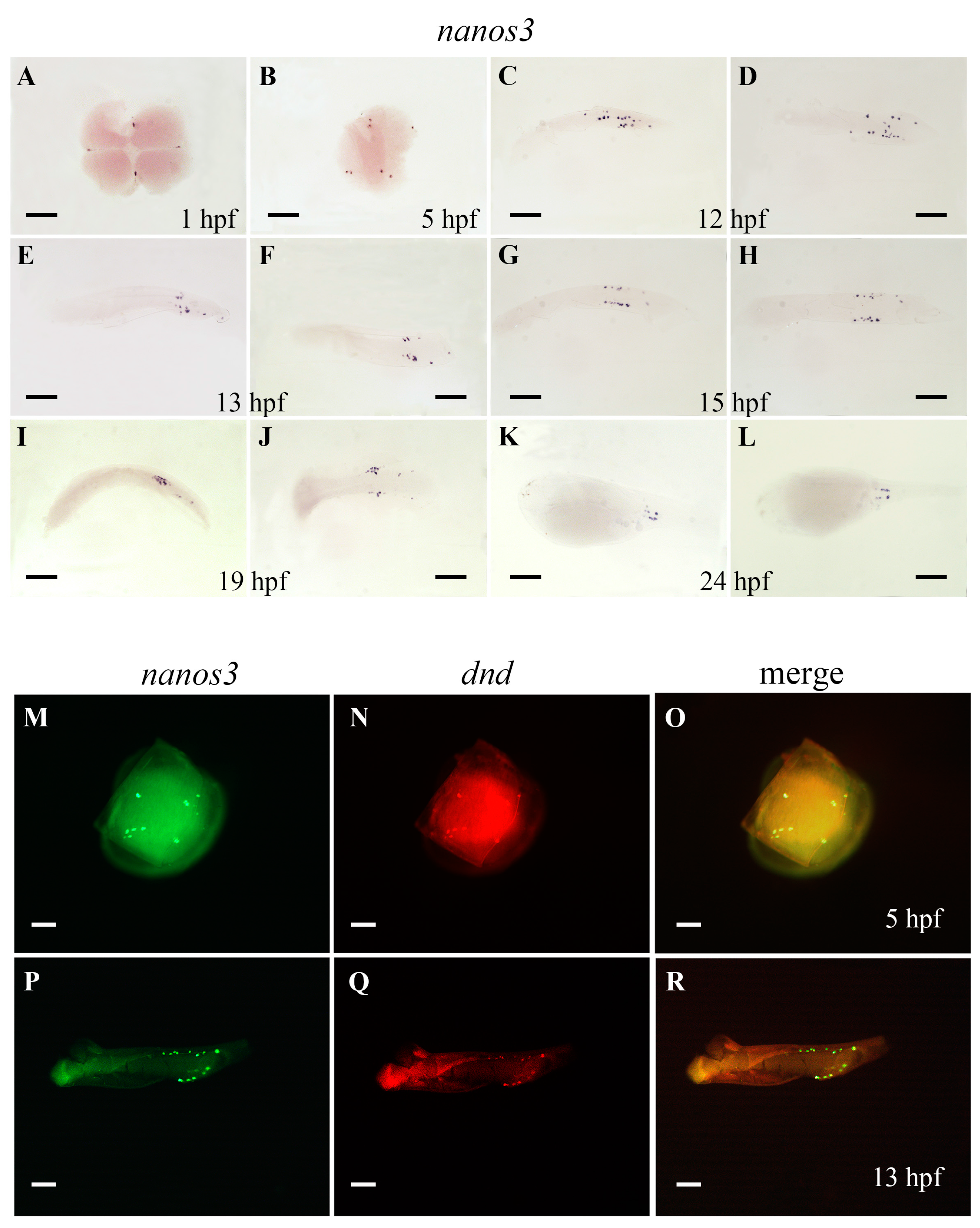
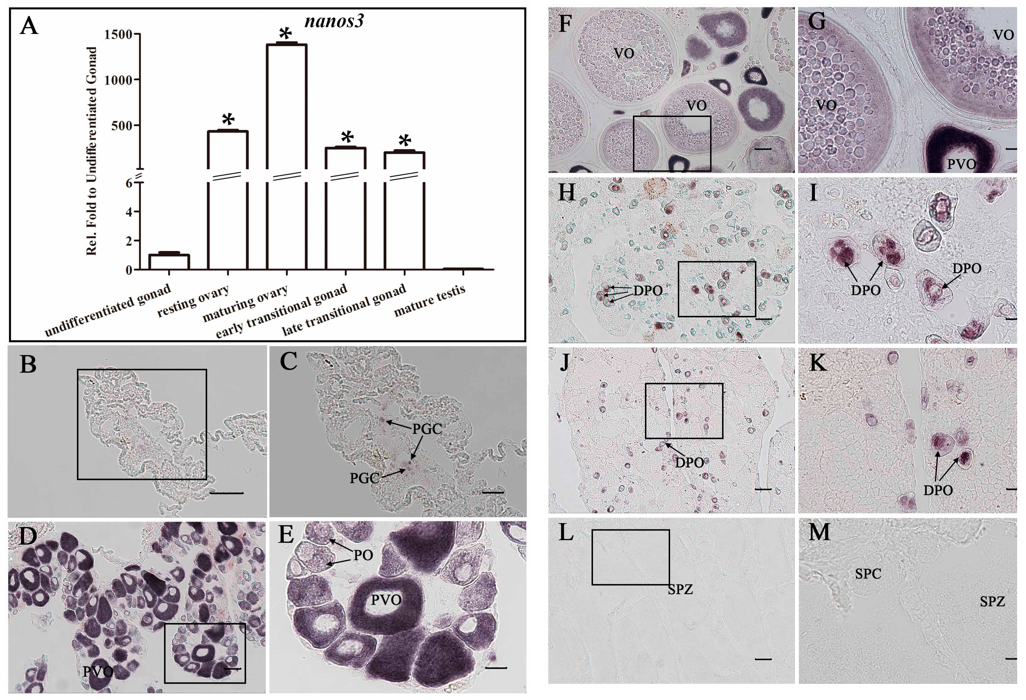
2.5. Ecnanos3 Is Expressed in PGCs and Different Stage Oocytes of Oogenesis

In contrast to Ecnanos1s and Ecnanos2, Ecnanos3 was revealed to be maternal and to be restricted in PGCs during embryogenesis by WISH detection. Ecnanos3 transcript is localized as four clumps in the cleavage furrows of four-cell stage (Figure 5A). At blastula embryo (five hours post fertilization, 5 hpf), about eight positive cells are separated into four groups (Figure 5B). It appears that the four cells inherit maternal nanos3 transcript and have undergone one cell division to generate these positive cells. At 12 hpf, the positive Ecnanos3 cells have been greatly proliferated and are located bilaterally in the posterior trunk (Figure 5C,D). From 13 to 24 hpf, these positive Ecnanos3 cells migrate and align more tightly on both sides of the posterior embryo and reach the position of presumptive gonad after 19 hpf (Figure 5E–L). The migration of Ecnanos3-expressing cells is similar to the PGC migration patterns in other teleosts. To confirm the PGC-specific expression of Ecnanos3, a double fluorescent ISH of Ecnanos3 and Ecdnd, a highly conserved PGC-specific gene during embryogenesis [50,51,52,53], was performed. At 5 and 13 hpf, red fluorescence co-localized with green fluorescence within a cluster of cells which both express Ecnanos3 and Ecdnd (Figure 5M–R). The data indicate that Ecnanos3 is specifically localized and expressed in PGCs during embryogenesis of orange-spotted grouper.
Moreover, we detected dynamic change of Ecnanos3 expression in gonads during gonad development and reversal by RT-qPCR and ISH. In undifferentiated gonad, only a minute amount of Ecnanos3 transcript was detected, but relative expression levels increased several hundreds or even more than one thousand times in resting ovary and maturing ovary (Figure 6A). Along with gonad reversal from ovary to testis, its expression level significantly decreased in early and late transitional gonads, and no Ecnanos3 transcript were detected in mature testis (Figure 6A). ISH detection further revealed its expression localization and specificity in PGCs and different stage oocytes of oogenesis. In undifferentiated gonad, Ecnanos3 transcript exists only in PGCs (Figure 6A,B). Along with oogenesis progress, Ecnanos3 is largely expressed in primary oocytes, previtellogenic oocytes and vitellogenic oocytes of the resting and maturing ovaries (Figure 6D–G). In early and late transitional gonads, the Ecnanos3 transcript is still in some residual primary oocytes, but most of them are in degenerating primary oocytes (DPO) (Figure 6H–K). Consistent with RT-qPCR detection (Figure 6A), no positive Ecnanos3 cells were observed in mature testis (Figure 6L,M). The above data indicate that Ecnanos3 is specifically expressed in PGCs and different stage oocytes of orange-spotted grouper.

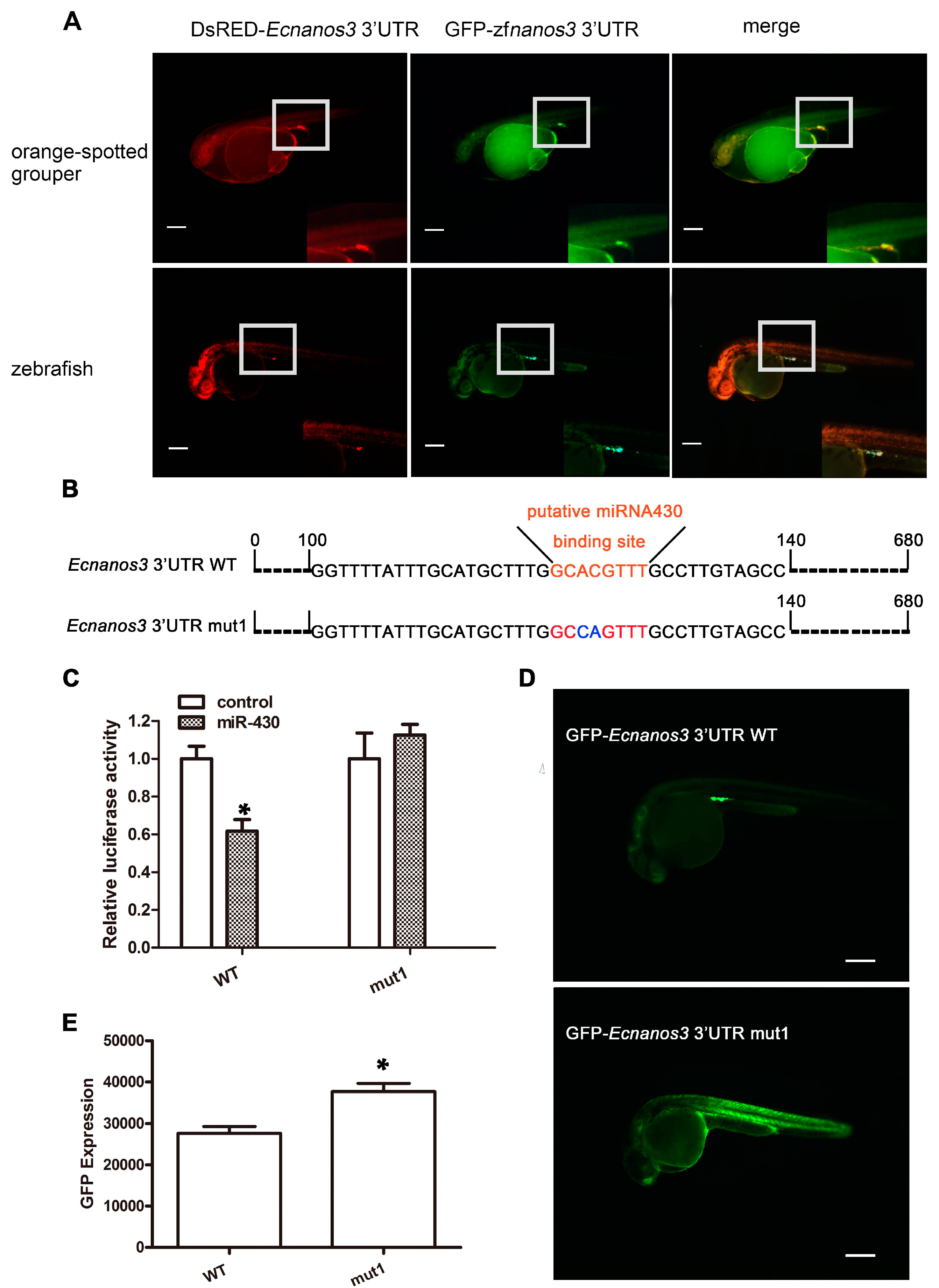
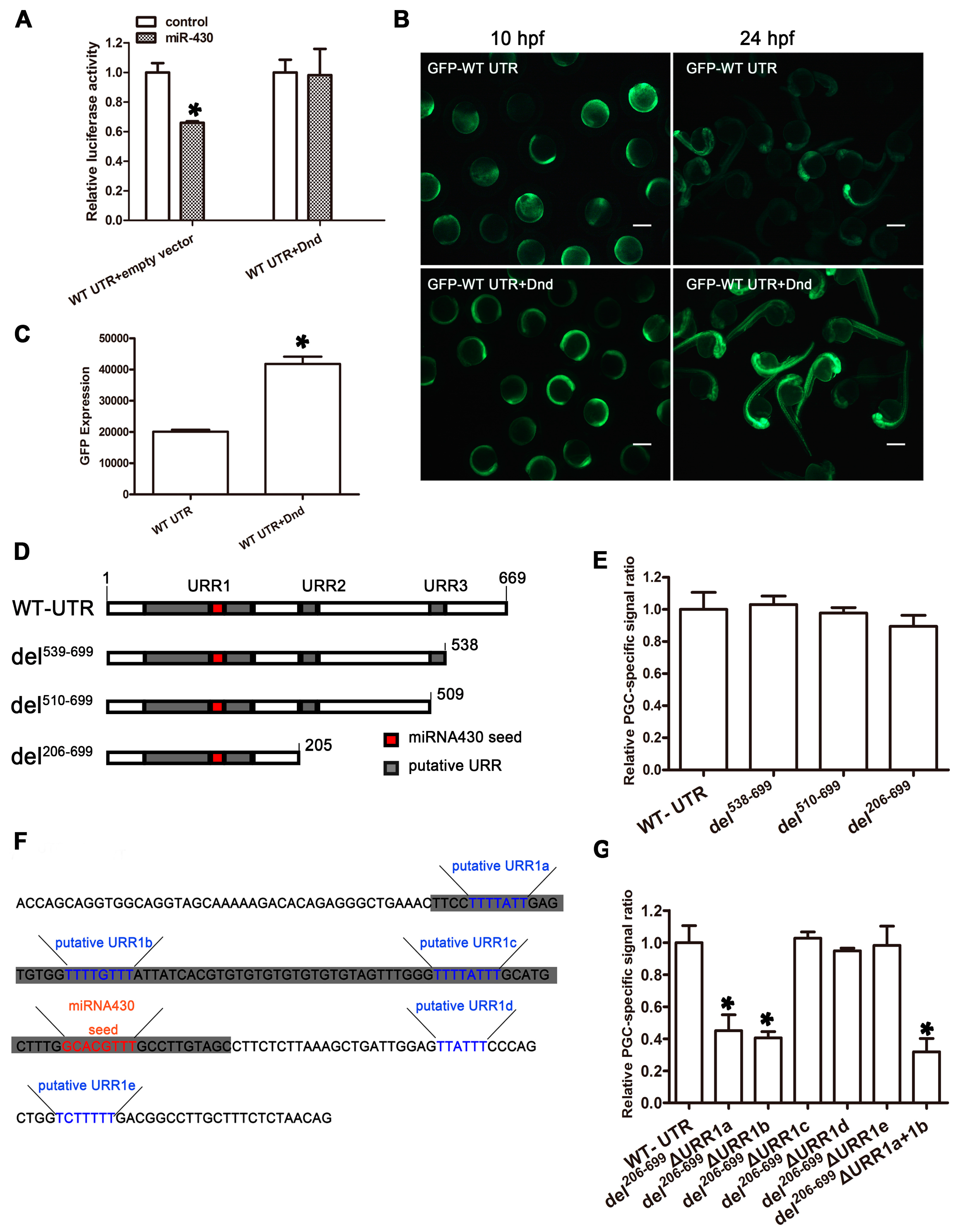
2.6. A Non-Canonical Seed Fragment in Ecnanos3 3′-UTR Is Required for miR-430-Mediated Repression

U-rich motif and miR-430 binding site (GCACTTT) are two crucial elements for nanos3 3′-UTR mediating PGC-specific mRNA stabilization in teleost [32]. Although none of identical miR-430 binding site found in Ecnanos3 3′-UTR, a similar fragment (GCACGTTT) exists (Figure S5). Three URRs in Ecnanos3 3′-UTR were revealed (Figure S5) using MEME Suite online. The first URR (URR1) is rather long with 90 base pairs, in which the seed sequence of miR-430 is included. To determinate whether Ecnanos3 3′-UTR has conserved function as its homologous genes in other teleost, we co-injected DsRED-Ecnanos3 3′-UTR RNA and GFP-zfnanos3 3′-UTR RNA into orange-spotted grouper or zebrafish eggs at 1-cell stage, respectively. At the 26 hpf, red fluorescence co-localized with green fluorescence within a cluster of presumptive PGCs (GFP-positive cells) above the anterior region of the yolk extension in both teleost (Figure 7A).
To examine whether Ecnanos3 is a target of miR-430, we mutated the putative miR-430 binding site “GCACGTTT” to “GGCAGTTT” (Ecnanos3 3′-UTR mut1) (Figure 7B), then linked the Ecnanos3 3′-UTR mut1 and Ecnanos3 3′-UTR WT to the C-terminus of firefly luciferase presenting in pmirGLO vector (Figure S6A) to perform luciferase reporter assay. pmirGLO/WT or pmirGLO/mut1 were co-transfected with miRNA-430 mimics or control miRNA mimics into HEK293T cells, respectively. Luciferase activity of pmirGLO/WT co-transfected with miR-430 mimic was significantly decreased (39%) against that of co-transfected with control miRNA-mimic (Figure 7C). In contrast, there is no significant difference in luciferase activity of pmirGLO/mut1 between miR-430 mimic and control miRNA mimic (Figure 7C). Thus, miR-430 specifically suppresses luciferase expression through a non-canonical seed sequence in Ecnanos3 3′-UTR in vitro. Furthermore, we examined this suppression effect in zebrafish embryos by using GFP reporter (Figure S6B). As shown in Figure 7D, the green fluorescence in the embryos injected with GFP-Ecnanos3 3′-UTR mut1 mRNA was ubiquitously distributed, while the PGCs were specifically labeled by GFP with WT Ecnanos3 3′-UTR. The total GFP expression level in mut1 embryos was about 1.3 folds against that of WT embryos (Figure 7E). Therefore, a non-canonical miR-430 binding site is necessary for Ecnanos3 3′-UTR-mediated mRNA suppression in somatic cells, but not in PGCs.

2.7. EcDnd Protects Ecnanos3 from miR-430 Repression in PGCs through a 23 bp U-Rich Element

To explore why miR-430-mediated suppression on nanos3 invalids in PGCs in orange-spotted grouper, Ecdnd effects on reporter activities driven by Ecnanos3 3′-UTR were assessed both in vitro and in vivo. Luciferase reporter assay showed that there was no significant difference between miR-430 and control miRNA mimics when EcDnd presented (Figure 8A), suggesting that the suppression of miR-430 on nanos3 3′-UTR is inhibited by EcDnd. Furthermore, GFP-Ecnanos3 3′-UTR RNA and Ecdnd RNA were co-injected into zebrafish embryos to investigate GFP expression. As shown in Figure 7B, GFP was ubiquitously expressed in zebrafish embryos at bud stage (10 hpf) and significantly decreased in the somatic cells at 24 hpf in the embryos injected with GFP-Ecnanos3 3′-UTR mRNA, while GFP expression in somite cells did not decrease in the presence of EcDnd (Figure 8B). The total GFP expression level rose up to two folds in the embryos co-injected with GFP-Ecnanos3 3′-UTR and Ecdnd mRNA compared to those only injected with GFP-Ecnanos3 3′-UTR mRNA (Figure 8C), suggesting that the exogenous EcDnd protein protects GFP-Ecnanos3 3′-UTR RNA in the somatic cells.
As a RNA-binding protein, EcDnd has two single-strand RNA recognition motifs that bind URRs within mRNAs [34,54]. Considering three URRs in Ecnanos3 3′-UTR (Figure S5), we first generated a series of 3′-truncated mutants to address the role of the URRs (Figure 8D). GFP was fused to the mutant del538−699 that lacked polyA, the mutant del510−699 where URR3 and the following fragment were deleted or the mutant del206−699 where URR2 and the following fragment were removed. At 26 hpf, each injected embryo was observed for calculating PGC-specific labeling efficiency. As shown in Figure 8E, PGC-specific GFP signal was no significantly different between the full length and the truncated variants of Ecnanos3 3′-UTR. This suggests that URR2, URR3 and polyA in Ecnanos3 3′-UTR is not necessary for the stable expression of GFP in PGCs. Since the predicted URR1 is 90 bp in length and the miR-430 binding site is included, five more subtle U-rich regions in URR1 were deleted by using mutant del206−699 as backbone, respectively (Figure 8F). Interestingly, the PGC-specific signal significantly decreased about 60% when the URR1a or URR1b was deleted, while the deletions of the other three small URRs did not (Figure 8G). The double-deletion mutant, where a 23 bp fragment containing URR1a and URR1b was removed, resulted in lower ratio (decrease about 70%) of specific-PGC labeling signal compared with the single mutants. In addition, the mutant del44−139 where URR1 alone was deleted (Figure S7A) was linked to the GFP reporter and GFP-Ecnanos3 3′-UTR del44−139 mRNA was injected into one-cell embryos. Owing to the deletion of miR-430 binding site, the green fluorescence in the embryos injected with GFP-Ecnanos3 3′-UTR del44−139 mRNA was ubiquitously distributed (Figure S7B), similar to the phenotype of embryos injected with GFP-Ecnanos3 3′-UTR mut1 mRNA (Figure 7D).

3. Discussion

In this study, we first identified four nanos genes in protogynous hermaphroditic orange-spotted grouper and revealed their molecular characterization, phylogenetic relationship, and divergence expression patterns. Importantly, we identified Ecnanos2 as a GSC-specific marker to mark GSCs distribution in different stage gonads during sex differentiation and reversal, and confirmed Ecnanos3 as PGC and oocyte marker to trace PGC migration and oocyte development processes during embryogenesis and oogenesis in the hermaphroditic vertebrate. Moreover, we revealed that a non-canonical “GCACGTTT” sequence and a 23 bp U-rich element with Ecnanos3 3′-UTR are required for Ecnanos3 PGC specific expression through interacting with miR-430 and Dnd protein.
Gene duplication has been believed to be very important and frequent events in fish evolution [55]. Besides the two rounds of whole-genome duplication (WGD) events occurred at the root of vertebrate lineage [56], a third round of WGD, known as teleost genome duplication (TGD), was estimated to occur in an ancestor of teleosts 320–350 million years ago (Mya) after the divergence of tetrapods and teleosts [57,58,59,60]. Indicated by phylogenetic relationship analysis (Figure 1), all analyzed vertebrates have at least three nanos genes, while lancelet (Branchiostoma floridae), the ancient chordate, has only one. As suggested previously, lancelet is an ancient chordate lineage divided from other phylum Chordata species before two rounds of WGD that occurred in vertebrate stem [61]. Thus, it seems likely that the diverse nanos genes in vertebrates are the products resulted from WGDs and TGD. Furthermore, clade Nanos1 is more distantly related to clades Nanos2 and Nanos3, suggesting that Nanos1 would have more differentiated functions from Nanos2 and Nanos3. In comparison with mammals with only one Nanos1, interestingly, most teleost fishes have two nanos1 genes, which suggests a divergence between nanos1a and nanos1b might occur specifically in teleost, consistent with the previous point in medaka [16]. Additionally, zebrafish was found to have only one nanos1 that was clustered into Nanos1b branch, suggesting that nanos1a gene might be lost during evolution, because a rapid gene loss was demonstrated to occur just following the gene duplication in vertebrates [62,63,64]. As a special linkage that was confirmed to undergo additional genome duplication and large genomic reorganization during the following rediploidization process [65], salmon was revealed to have two nanos3 genes, one nanos1 and one nanos2 gene. Although phylogenetic analysis based on one gene would be insufficient or yield contrasting species tree [66], our phylogenetic tree has provided a special case about the phylogeny of nanos gene family to understand teleost evolution.
Significantly, our current results not only revealed differential expression patterns and possible divergent functions of four nanos genes from the hermaphroditic orange-spotted grouper, but also confirmed the conserved roles of nanos genes in germ-line cells as reported previously in both vertebrates and invertebrates [67,68]. In Drosophila, the single nanos gene is essential for germ cell migration, maintenance and differentiation [67]. In most of vertebrates, three nanos genes, nanos1, nanos2 and nanos3, were found, and functional divergence was verified to occur during evolution. Nanos1 was not considered as a key fertility factor [15], but nanos2 and nanos3 were demonstrated to play important roles in germ cells. Ecnanos3 is specifically expressed in PGCs and different stage oocytes of the orange-spotted grouper (Figure 5 and Figure 6). Zebrafish nanos3, identified firstly as nanos1 [20], was also found to be expressed in the germ plasm, PGCs and oocytes [20,21]. Medaka nanos3 was revealed to be expressed in migrating PGCs, then to diminish as PGCs come to contact with gonadal somatic cells [69], and finally to resume only in oocytes [16,70]. By utilizing specific localization of the microinjected GFP-nanos3 3′-UTR mRNA in PGCs, different migration patterns or routes have been observed in several teleost fishes [27]. The PGC migration route in orange-spotted grouper (Figure 5) was similar to that of zebrafish. In adult gonads, Ecnanos3 transcript was only detected in oogenic germ cells, whereas not in spermatogenic germ cells (Figure 6D–M). The specific expression of Ecnanos3 in oogenic germ cells suggests that it might regulate oocyte production, as it does in zebrafish [19,20].
Conserved 3′-UTR-mediated mechanism is crucial for PGC-specific mRNA stabilization in teleost [71]. Injecting fish embryos with chimeric RNA where the GFP ORF was fused to the zebrafish nanos 3′-UTR labeled the PGCs of medaka, icegoby, herring and other four cyprinid species [70]. In this study, the co-injection of Ecnanos3 3′-UTR with DsRED and zfnanos3 3′-UTR with GFP can co-label both orange-spotted grouper and zebrafish PGCs. This indicates that both zebrafish and grouper nanos3 3′-UTR protect the fluorescence mRNAs in PGCs, while the mRNAs in somatic cells inactivate quickly. Therefore, Ecnanos3 3′-UTR plays an evolutionarily conserved role in PGC specific expression of nanos3. nanos3 transcripts exist both in PGCs and somatic cells, but their translation are suppressed in somatic cells [20,33,72]. Several mechanisms would contribute to the selective enrichment of Nanos protein in PGCs [73]. In sea urchin, the GNARLE (Global Nanos Associated RNA Lability Element) within nanos2 3′-UTR interacts with some proteins involved in RNA stability and/or protein translation, such as nucleases or deadenylases to destabilize the RNA in somite cells [74]. Moreover, nanos3 is also regulated by binding to miRNAs and piRNAs through its 3′-UTR. In zebrafish and human cells, miRNA-430 uses a seed sequence “GCACUUU” to associate with the nanos3 and tdrd7 3′-UTR, leading to the degradation of mRNAs [33,34]. In Carassius auratus, miRNA-430 uses the same seed sequence to bind to the 3′-UTR of C1q-like and guides its expression level in PGCs during early embryogenesis [75]. In olive flounder, two “GCACs” were identified as miRNA-430 seed-matched sites and responsible for the degradation of nanos3 mRNA [54]. In our case, miR-430 represses Ecnanos3 through the non-canonical binding site “GCACGTTT” in 3′-UTR of Ecnanos3. Since miRNAs recognize their targets via a “loose” base-pairing with several mismatches, miR-430 suppression on nanos3 3′-UTR is valid among species.
Dnd is required for PGC survival in vertebrates [50]. Our results indicate that EcDnd prevents miR-430-mediated repression on Ecnanos3 3′-UTR to maintain Ecnanos3 PGC specific expression. Dnd protein associates with URRs of nanos3 3′-UTR to suppress miR-430 function in zebrafish and human cells [34,36]. Although teleost nanos3 3′-UTRs vary in length (from 124 to 680 nt) and show low sequence similarity among each other, conserved URRs exist in all analyzed teleost species, including zebrafish, cod, salmon, tetraodon, medaka, and so on [32]. Although URRs are extensively existed in many teleost fish, their locations in 3′-UTR display species-specific. Two minimal URRs in zebrafish nanos3 3′-UTR locate on 11 bp before and 41 bp after miR-430 binding site, respectively [34,36]. In olive flounder, a 31 bp U-rich region located 11 bp behind the first GCAC site is the binding site of Dnd [54]. Similar as other teleost nanos3 3′-UTR, three URRs were identified in Ecnanos3 3′-UTR (Figure S5), where two functional minimal URRs (URR1a and URR1b) located on 50 bp before miR-430 seed are required for Dnd function (Figure 8F,G). It seems that the binding of Dnd to the region containing URR1a and URR1b prevents the access of miR-430 to its target sequence. Therefore, a 23 bp U-rich region in Ecnanos3 3′-UTR is required for Dnd-mediated mRNA stability in PGCs. Additionally, the PGC labeling efficiencies of del206–699ΔURR1a and del206–699ΔURR1b remain 38.9% and 35.0% compared to the full-length 3′-UTR respectively, implying other mechanisms may regulate nanos3 PGC-specific expression. Thus, the molecular mechanism and the interactions among PGC-specific RNAs, miRNAs and RNA-binding proteins need further study.
In contrast to Ecnanos3, orange-spotted grouper nanos2 was observed to be expressed continuously and specifically in a subset of small germ cells with a diameter of <20 μm in different stage gonads from the undifferentiated, differentiation and maturing ovary to sex reversal and mature testis (Figure 4). In medaka and zebrafish, a small subset of <20 μm nanos2 positive cells were identified as GSCs [17,48,49]. Considering the similar diameter and distribution in gonads of orange-spotted grouper, Ecnanos2 might be a marker of GSCs that continuously give rise to oocytes or sperms, as other teleost nanos2 reported previously [76,77]. As orange-spotted grouper is a protogynous hermaphrodite, we are able to continuously trace GSCs in the different stage gonads. Interestingly, expression level of Ecnanos2 in gonads at the early and late transitional stages raise highly up to 60–120 and 65–150 folds relative to that of resting ovary and maturing ovary (Figure 4A). The expression level elevation might be related to spermatogenesis in the transitional gonad because a large number of spermatogenic cells will be raised from these GSCs with Ecnanos2 expression during sex change process and spermatogenesis. Based on these results, nanos2 is very likely a GSC-specific gene and play a conserved role in maintaining GSCs self-renew [13] in vertebrate. Same as other teleost nanos2, orange-spotted grouper nanos2 was not found in embryos (not shown), which is different from mouse nanos2 that is expressed in male PGCs [10]. Recently, germ cell transplantation was applied successfully in assisted reproductive technologies and the studies of germ cell development. As described by Okutsu et al., triploid sterile masu salmon (Oncorhynchus masou) only produced rainbow trout (O. mykiss) sperm and eggs at two years after transplanting pvasa-GFP labelled spermatogonia of rainbow trout [4]. The single GFP-nanos1 3′-UTR tagged PGC of goldfish or loach was successfully transplanted to zebrafish embryo which blocked PGCs development by injecting a dnd antisense morpholino oligonucleotide (MO) [78]. The first step in establishing a viable PGC or GSC transplantation method is to visualize PGCs or GSCs. The identification of Ecnanos2 and Ecnanos3 as GSCs or PGCs-specific marker respectively (Figure 4, Figure 5 and Figure 6) and the visualization of PGCs by Ecnanos3 3′-UTR RNA (Figure 7A) will open up new possibilities for developing germ cell transplantation in the hermaphroditic fish.
In vertebrates, nanos1 has been identified in human [14], mouse [15], cynomolgus monkey [79], Xenopus [80,81,82], zebrafish [21], medaka [16], Chinese sturgeon [26], Japanese eel [25,26], Atlantic cod, and Atlantic salmon [32]. The expression pattern of nanos1 was revealed to be various among different vertebrate species. Human NANOS1 and Chinese sturgeon nanos1 were found to be expressed in multiple tissues ubiquitously, while mouse Nanos1 predominantly in brain, oocytes and seminiferous tubules of testis [15]. Cynomolgus monkey nanos1 was expressed abundantly in lung and liver, and slightly in brain, heart, kidney, muscle, and testis [79]. Xenopus nanos1 was also detected in adult ovary, testis and brain [81]. In this study, Ecnanos1a was observed to be expressed predominantly in pituitary, while Ecnanos1b mainly in pituitary and other brain tissues (Figure 2A,B). Therefore, Ecnanos1a and Ecnanos1b might process neofunctionalization after their divergence. Significantly, we found that Ecnanos1a was up-regulated in pituitaries during sex change and detected the highest Ecnanos1a expression level in the pituitaries of mature male individuals, while Ecnanos1b seemed to maintain same low expression level in pituitaries during the transitional process (Figure 3G). Therefore, the association of Ecnanos1a expression level with testis differentiation and spermatogenesis suggests Ecnanos1a might play a role in grouper male sex differentiation. Of course, the underlying mechanism awaits further investigation.
In conclusion, the current study identified and characterized four nanos genes in protogynous hermaphroditic orange-spotted grouper, and revealed their divergence expression patterns in different stage gonads during sex differentiation and reversal. Based on the present investigations, we confirmed the conserved function roles of nanos genes in germ-line cells, in which nanos2 might be a GSC-specific marker, and nanos3 might be a PGC and oocyte marker. In addition, we revealed that PGC-specific expression of nanos3 is mediated by interacting with miR-430 and Dnd protein in orange-spotted grouper.

4. Materials and Methods

4.1. Experimental Fish

Adult orange-spotted groupers were purchased from markets in Wuhan, China. The samples of early embryogenesis were obtained from Marine Fisheries Development Center of Guandong Province, China. The animal treatments and experimental protocols were agreed by the Institute of Hydrobiology Institutional Animal Care and Use Committee (Approval ID: keshuizhuan 0829).

4.2. RNA Extraction and Real-Time Quantitative PCR (RT-qPCR)

Total RNAs were extracted from different tissues, including heart, liver, spleen, telencephalon, mesencephalon, cerebellum, hypothalamus, myelencephalon, kidney, pituitary and gonad by using Spin/Vacuum (SV) Tolal RNA Isolation System (Promaga Z3100, Madison, WI, USA) according to the recommendations manufacture’s protocols. One microgram of total RNAs were used to establish cDNA library through SMARTer™ RACE cDNA Amplification Kit (Clontech 634923, Mountain View, CA, USA) based on the protocols. Then, first-strand cDNAs were synthesized following the protocols of GoScriptTM Reverse Transcription System (Promega A5000, Madison, WI, USA).
RT-qPCR experiments were performed in a final volume of 20 μL containing 1 μL of cDNA, 0.5 μL of each 10 mM primer, and 10 μL of SYBRH Premix Ex TaqTM II (Perfect Real Time, Takara, Dalian, China). The protocol was 94 °C (2 min) for heat denaturing, then, 40 cycles of 94 °C (15 s), 57 °C (15 s), 72 °C (20 s), and additional 72 °C (2 min). The amplification efficiency of each pair of primers was determined by gradient dilution of template. β-Actin was used as internal control. In addition, total RNAs were used as negative control to exclude the contaminant of genomic DNA. All primers used were designed by http://biotools.nubic.northwestern.edu/OligoCalc.html [83] (Table S1). The samples were analyzed in triplicates, and relative expression levels of target genes were calculated with the 2−ΔΔCt method. For statistical analysis, Tukey’s test was calculated with SPSS software (SPSS Inc., Chicago, IL, USA). A probability (p) of <0.05 was considered statistically significant.

4.3. Sequence and Phylogenetic Analyses

The full-length cDNAs of Ecnanos1a, Ecnanos1b, Ecnanos2 and Ecnanos3 were achieved by 5’ and 3′ rapid amplification of cDNA ends (RACE), and the amino-acid sequence was predicted by using the DNAMAN software. The protein domain was predicted by SMART online http://smart.embl-heidelberg.de/ [84].
Multiple protein sequence alignment was performed by ClustalW, and then manually adjusted for phylogenetic construction by bootstrap analysis (1000 replicates) using the neighbor-joining and maximum likelihood method in MEGA version 5.0 (The Biodesign Institute, Tempe, AZ, USA), both of which produced similar trees. 3′-UTR of Ecnanos3 was analyzed by using Motif-based sequence analysis tools with default parameters (MEME Suite, http://meme-suite.org/) [85,86].

4.4. Histological Examination of Different Gonadal Developmental Stages

Gonadal tissues were fixed in 4% paraformaldehyde (PFA) overnight at 4 °C, and embedded in Opti-mum Cutting Temperature (O.C.T.) Compoundfor section as previously described [87]. The sections were stained with hematoxylin/eosin (HE; Beyotime Institute of Biology, Suzhou, China) and observed in Zeiss microscopy (Jena, Germany).

4.5. In Situ Hybridization

Riboprobes were made by using digoxigenin (DIG) RNA labeling kit (Roche, Mannheim, Germany) or Fluorescein labeling kit (Roche) according to the protocols. Section tissue in-situ hybridization was performed as described previously [46]. Whole-mount in situ hybridization was carried out as previously described [88,89]. Double fluorescent ISH was performed following the recommendations manufacture’s protocols of TSATM Plus Fluorescence Systems (PerkinElmer, Boston, MA, USA). In briefly, 500 ng DIG-labeled Ecnanos3 probe and 500 ng fluorescein-labeled Ecdnd probe were added in the permeabilizated embryos for hybridization, then incubated in 59 °C for 16 h. After washing away excess probes, hybrids are detected by anti-DIG-peroxidase (Roche, Mannheim, Germany) and anti-fluorescein-peroxidase (Roche, Germany) with tyramide signal amplification.

4.6. RNAs and Microinjection

EcDnd (GenBank: KX881943) ORF, GFP ORF/DsRed ORF linked to Ecnanos3 3′-UTR and GFP ORF fused with zfnanos3 3′-UTR were inserted into pCS2+ vector digested with BamH1 and EcoR1, respectively. The series of 3′-truncate mutations (del538–699, del510–699, del206–699) fused with GFP ORF similarly cloned into pCS2+ vector. The miR-430 binding site “GCACGTTT” in the constructed GFP-Ecnanos3 3′-UTR was replaced with “GCCAGTTT” by overlap PCR. The mutations of deleted URR1a, URR1b, URR1c, URR1d and URR1e were cloned by overlap PCR from the del206−699 plasmid. The mutant del44–139 where URR1 alone was deleted was cloned by overlap PCR from the WT-UTR plasmid.
For RNA synthesis, the plasmids linearized by NotI were transcribed in vitro by using Message Machine-Kit (Ambion, Austin, TX, USA). Microinjections were performed as described previously [90]. Subsequently, every embryo was observed in Leica M205FA stereomicroscope to analyze GFP expression pattern (n > 150). For measuring GFP expression in vivo, 40 embryos were collected to perform fluorophotometry detection by fluorophotometric scan (TECAN, Grödig, Austria) [84].

4.7. Luciferase Reporter Assay

Full lengths sequence of Ecnanos3 3′-UTR, miR-430 binding site mutation was amplified and subcloned into Nhe1/Sal1 sites of pmir-GLO plasmid (Promega, USA). In this study, HEK293T cells were selected to study the function of Ecnanos3 3′-UTR, which has been widely utilized to study miRNA-mediated posttranscriptional suppression of teleost genes in vitro due to its highly efficient transfection [34,91,92]. HEK293T cells were cultured as described previously [93]. MicroRNA mimics were synthesized by Genepharma (Shanghai, China). For luciferase analysis, HEK293T cells seeded in 24-well plates overnight were transiently transfected with 150 ng plasmid, and 30 pM miRNA mimics or negative control by DharmaFECT transfection reagent (Dharmacon, Suzhou, China) per well. At 36 h post transfection, luciferase activity was measured by a Junior LB9509 luminometer (Berthod, Pforzhiem, Germany) and normalized to Renilla luciferase activity. Each independent experiment was performed in triplicate. For Statistical analysis, Tukey’s test was calculated with SPSS software (SPSS Inc., Chicago, IL, USA). A probability (p) of <0.05 was considered statistically significant.

Supplementary Materials

Supplementary materials can be found at www.mdpi.com/1422-0067/18/4/685/s1.

Acknowledgments

We thank Hai-Fa Zhang and the members of the Marine Fisheries Development Center of Guandong Province for help with artificial propagation of orange-spotted grouper. This study was supported by the National Natural Science Foundation of China (Grant Nos. U1401213) and the Autonomous Project of State Key Laboratory of Biocontrol, Guangdong Province Key Laboratory for Improved Variety Reproduction of Aquatic Economic Animals.

Author Contributions

Yang Wang, Li Zhou and Zhi-Hui Sun conceived and designed the experiments; Zhi-Hui Sun and Zhi Li performed the experiments; Zhi-Hui Sun and Wei-Jia Lu analyzed the data; Xiao-Chun Liu and Shui-Sheng Li contributed materials; and Zhi-Hui Sun, Li Zhou and Jian-Fang Gui wrote the paper.

Conflicts of Interest

The authors declare no conflict of interest. The founding sponsors had no role in the design of the study; in the collection, analyses, or interpretation of data; in the writing of the manuscript, and in the decision to publish the results.

References

  1. Xu, H.Y.; Li, M.Y.; Gui, J.F.; Hong, Y.H. Fish germ cells. Sci. China Life Sci. 2010, 53, 435–446. [Google Scholar] [CrossRef] [PubMed]
  2. Takeuchi, Y.; Yoshizaki, G.; Takeuchi, T. Generation of live fry from intraperitoneally transplanted primordial germ cells in rainbow trout. Biol. Rep. 2003, 69, 1142–1149. [Google Scholar]
  3. Takeuchi, Y.; Yoshizaki, G.; Takeuchi, T. Biotechnology: Surrogate broodstock produces salmonids. Nature 2004, 430, 629–630. [Google Scholar] [CrossRef] [PubMed]
  4. Okutsu, T.; Shikina, S.; Kanno, M.; Takeuchi, Y.; Yoshizaki, G. Production of trout offspring from triploid salmon parents. Science 2007, 317, 1517–1517. [Google Scholar] [CrossRef] [PubMed]
  5. Xu, H.; Gui, J.; Hong, Y. Differential expression of vasa RNA and protein during spermatogenesis and oogenesis in the gibel carp (Carassius auratus gibelio), a bisexually and gynogenetically reproducing vertebrate. Dev. Dyn. 2005, 233, 872–882. [Google Scholar] [CrossRef] [PubMed]
  6. Peng, J.X.; Xie, J.L.; Zhou, L.; Hong, Y.H.; Gui, J.F. Evolutionary conservation of dazl genomic organization and its continuous and dynamic distribution throughout germline development in gynogenetic gibel carp. J. Exp. Zool. Part B 2009, 312, 855–871. [Google Scholar] [CrossRef] [PubMed]
  7. Liu, W.; Li, S.Z.; Li, Z.; Wang, Y.; Li, X.Y.; Zhong, J.X.; Zhang, X.J.; Zhang, J.; Zhou, L.; Gui, J.F. Complete depletion of primordial germ cells in an All-female fish leads to sex-biased gene expression alteration and sterile all-male occurrence. BMC Genom. 2015, 16, 971. [Google Scholar] [CrossRef] [PubMed]
  8. Wang, C.; Lehmann, R. Nanos is the localized posterior determinant in Drosophila. Cell 1991, 66, 637–647. [Google Scholar] [CrossRef]
  9. Asaoka-Taguchi, M.; Yamada, M.; Nakamura, A.; Hanyu, K.; Kobayashi, S. Maternal pumilio acts together with nanos in germline development in Drosophila embryos. Nat. Cell Biol. 1999, 1, 431–437. [Google Scholar] [PubMed]
  10. Tsuda, M.; Sasaoka, Y.; Kiso, M.; Abe, K.; Haraguchi, S.; Kobayashi, S.; Saga, Y. Conserved role of NANOS proteins in germ cell development. Science 2003, 301, 1239. [Google Scholar] [CrossRef] [PubMed]
  11. Hayashi, Y.; Hayashi, M.; Kobayashi, S. Nanos suppresses somatic cell fate in Drosophila germ line. Proc. Natl. Acad. Sci. USA 2004, 101, 10338–10342. [Google Scholar] [CrossRef] [PubMed]
  12. Suzuki, A.; Tsuda, M.; Saga, Y. Functional redundancy among Nanos proteins and a distinct role of nanos2 during male germ cell development. Development 2007, 134, 77–83. [Google Scholar] [PubMed]
  13. Wang, Z.; Lin, H. Nanos maintains germline stem cell self-renewal by preventing differentiation. Science 2004, 303, 2016–2019. [Google Scholar] [CrossRef] [PubMed]
  14. Julaton, V.T.A.; Pera, R.A.R. Nanos3 function in human germ cell development. Hum. Mol. Genet. 2011, 20, 2238–2250. [Google Scholar] [CrossRef] [PubMed]
  15. Haraguchi, S.; Tsuda, M.; Kitajima, S.; Sasaoka, Y.; Nomura-Kitabayashid, A.; Kurokawa, K.; Saga, Y. Nanos1: A mouse nanos gene expressed in the central nervous system is dispensable for normal development. Mech. Dev. 2003, 120, 721–731. [Google Scholar] [CrossRef]
  16. Aoki, Y.; Nakamura, S.; Ishikawa, Y.; Tanaka, M. Expression and syntenic analyses of four nanos genes in medaka. Zool. Sci. 2009, 26, 112–118. [Google Scholar] [PubMed]
  17. Lele Z, K.P.H. The zebrafish as a model system in developmental, toxicological and transgenic research. Biotechnol. Adv. 1996, 14, 57–72. [Google Scholar] [PubMed]
  18. Grunwald, D.J.; Eisen, J.S. Headwaters of the zebrafish—Emergence of a new model vertebrate. Nat. Rev. Genet. 2002, 3, 711–724. [Google Scholar]
  19. Beer, R.L.; Draper, B.W. Nanos3 maintains germline stem cells and expression of the conserved germline stem cell gene nanos2 in the zebrafish ovary. Dev. Biol. 2013, 374, 308–318. [Google Scholar] [CrossRef] [PubMed]
  20. Draper, B.W.; McCallum, C.M.; Moens, C.B. Nanos1 is required to maintain oocyte production in adult zebrafish. Dev. Biol. 2007, 305, 589–598. [Google Scholar] [CrossRef] [PubMed]
  21. Köprunner, M.; Thisse, C.; Bernard, T.; Erez, R. A zebrafish nanos-related gene is essential for the development of primordial germ cells. Genes Dev. 2001, 15, 2877–2885. [Google Scholar] [PubMed]
  22. Hashimoto, Y.; Maegawa, S.; Nagai, T.; Yamaha, E.; Suzuki, H.; Yasuda, K.; Inoue, K. Localized maternal factors are required for zebrafish germ cell formation. Dev. Biol. 2004, 268, 152–161. [Google Scholar] [PubMed]
  23. Raz, E. Primordial germ-cell development: The zebrafish perspective. Nat. Rev. Genet. 2003, 4, 690–700. [Google Scholar] [PubMed]
  24. Bellaiche, J.; Lareyre, J.J.; Cauty, C.; Yano, A.; Allemand, I.; Le Gac, F. Spermatogonial stem cell quest: Nanos2, marker of a subpopulation of undifferentiated a spermatogonia in trout testis. Biol. Rep. 2014, 90, 79. [Google Scholar]
  25. Kawakami, Y.; Ishihara, M.; Saito, T.; Fujimoto, T.; Adachi, S.; Arai, K.; Yamaha, E. Cryopreservation of green fluorescent protein (GFP)-labeled primordial germ cells with GFP fused to the 3′-untranslated region of the nanos gene by vitrification of japanese eel (Anguilla japonica) somite stage embryos. J. Anim. Sci. 2012, 90, 4256–4265. [Google Scholar] [CrossRef] [PubMed]
  26. Ye, H.; Chen, X.; Wei, Q.; Zhou, L.; Liu, T.; Gui, J.; Li, C.; Cao, H. Molecular and expression characterization of a nanos1 homologue in chinese sturgeon, acipenser sinensis. Gene 2012, 511, 285–292. [Google Scholar] [PubMed]
  27. Li, M.; Tan, X.; Jiao, S.; Wang, Q.; Wu, Z.; You, F.; Zou, Y. A new pattern of primordial germ cell migration in olive flounder (Paralichthys olivaceus) identified using nanos3. Dev. Genes Evol. 2015, 225, 195–206. [Google Scholar] [PubMed]
  28. Presslauer, C.; Nagasawa, K.; Fernandes, J.M.; Babiak, I. Expression of vasa and nanos3 during primordial germ cell formation and migration in atlantic cod (Gadus morhual). Theriogenology 2012, 78, 1262–1277. [Google Scholar] [CrossRef] [PubMed]
  29. Kawakami, Y.; Saito, T.; Fujimoto, T.; Goto-Kazeto, R.; Takahashi, E.; Adachi, S.; Arai, K.; Yamaha, E. Visualization and motility of primordial germ cells using green fluorescent protein fused to 3′-UTR of common carp nanos-related gene. Aquaculture 2011, 317, 245–250. [Google Scholar] [CrossRef]
  30. D’Agostino, I.; Merritt, C.; Chen, P.L.; Seydoux, G.; Subramaniam, K. Translational repression restricts expression of the C. elegans nanos homolog NOS-2 to the embryonic germline. Dev. Biol. 2006, 292, 244–252. [Google Scholar] [PubMed]
  31. Gavis, E.R.; Lehmann, R. Translation regulation of nanos by RNA localization. Lett. Nat. 1994, 369, 315–318. [Google Scholar]
  32. Škugor, A.; Slanchev, K.; Torgersen, J.S.; Tveiten, H.; Andersen, O. Conserved mechanisms for germ cell-specific localization of nanos3 transcripts in teleost species with aquaculture significance. Mar. Biotechnol. 2014, 16, 256–264. [Google Scholar] [PubMed]
  33. Mishima, Y.; Giraldez, A.J.; Takeda, Y.; Fujiwara, T.; Sakamoto, H.; Schier, A.F.; Inoue, K. Differential regulation of germline mRNAs in soma and germ cells by zebrafish miR-430. Curr. Biol. 2006, 16, 2135–2142. [Google Scholar] [CrossRef] [PubMed]
  34. Kedde, M.; Strasser, M.J.; Boldajipour, B.; Oude Vrielink, J.A.; Slanchev, K.; le Sage, C.; Nagel, R.; Voorhoeve, P.M.; van Duijse, J.; Orom, U.A.; et al. RNA-binding protein DND1 inhibits microRNA access to target mRNA. Cell 2007, 131, 1273–1286. [Google Scholar] [PubMed]
  35. Giraldez, A.J.; Mishima, Y.; Rihel, J.; Grocock, R.J.; van Dongen, S.; Inoue, K.; Schier, A.F. Zebrafish miR-430 promotes deadenylation and clearance of maternal mRNAs. Science 2006, 312, 75–79. [Google Scholar] [CrossRef] [PubMed]
  36. Slanchev, K.; Stebler, J.; Goudarzi, M.; Cojocaru, V.; Weidinger, G.; Raz, E. Control of dead end localization and activity—Implications for the function of the protein in antagonizing miRNA function. Mech. Dev. 2009, 126, 270–277. [Google Scholar] [CrossRef] [PubMed]
  37. Rosa, A.; Spagnoli, F.M.; Brivanlou, A.H. The mir-430/427/302 family controls mesendodermal fate specification via species-specific target selection. Dev. Cell 2009, 16, 517–527. [Google Scholar] [PubMed]
  38. Yeh, S.L.; Dai, Q.C.; Chu, Y.T.; Kuo, C.M.; Ting, Y.Y.; Chang, C.F. Induced sex change, spawning and larviculture of potato grouper, Epinephelus tukula. Aquaculture 2003, 228, 371–381. [Google Scholar]
  39. Chao, T.M.; Lim, L.C. Recent developments in the breeding of grouper (Epinephelus spp.) in singapore. Singap. J. Prim. Ind. 1991, 19, 78–93. [Google Scholar]
  40. Yao, B.; Zhou, l.; Wang, Y.; Xia, W.; Gui, J. Differential expression and dynamic changes of SOX3 during gametogenesis and sex reversal in protogynous hermaphroditic fish. J. Exp. Zool. 2007, 307A, 207–219. [Google Scholar]
  41. Ko, C.F.; Chiou, T.T.; Chen, T.T.; Wu, J.L.; Chen, J.C.; Lu, J.K. Molecular cloning of myostatin gene and characterization of tissue-specific and developmental stage-specific expression of the gene in orange spotted grouper, Epinephelus coioides. Mar. Biotechnol. 2007, 9, 20–32. [Google Scholar] [PubMed]
  42. Li, W.S.; Chen, D.; Wong, A.O.; Lin, H.R. Molecular cloning, tissue distribution, and ontogeny of mRNA expression of growth hormone in orange-spotted grouper (Epinephelus coioides). Gen. Comp. Endocr. 2005, 144, 78–89. [Google Scholar] [CrossRef]
  43. Zhou, L.; Gui, J.F. Molecular mechanisms underlying sex change in hermaphroditic groupers. Fish Physiol. Biochem. 2010, 36, 181–193. [Google Scholar] [CrossRef] [PubMed]
  44. Mei, J.; Gui, J.F. Genetic basis and biotechnological manipulation of sexual dimorphism and sex determination in fish. Sci. China Life Sci. 2015, 58, 124–136. [Google Scholar] [CrossRef] [PubMed]
  45. Xia, W.; Zhou, L.; Yao, B.; Li, C.J.; Gui, J.F. Differential and spermatogenic cell-specific expression of dmrt1 during sex reversal in protogynous hermaphroditic groupers. Mol. Cell. Endocr. 2007, 263, 156–172. [Google Scholar]
  46. Wang, Y.; Zhou, L.; Yao, B.; Li, C.J.; Gui, J.F. Differential expression of thyroid-stimulating hormone β subunit in gonads during sex reversal of orange-spotted and red-spotted groupers. Mol. Cell. Endocr. 2004, 220, 77–88. [Google Scholar]
  47. Huang, W.; Zhou, L.; Li, Z.; Gui, J.F. Expression pattern, cellular localization and promoter activity analysis of ovarian aromatase (cyp19a1a) in protogynous hermaphrodite red-spotted grouper. Mol. Cell. Endocri. 2009, 307, 224–236. [Google Scholar] [CrossRef] [PubMed]
  48. Jin, J.Y.; Zhou, L.; Wang, Y.; Li, Z.; Zhao, J.J.; Zhang, Q.Y.; Gui, J.F. Antibacterial and antiviral roles of a fish β-defensin expressed both in pituitary and testis. PLoS ONE 2010, 5, e12883. [Google Scholar] [CrossRef] [PubMed]
  49. Marino, G.; Azzurro, E.; Massari, A.; Finoia, M.G.; Mandich, A. Reproduction in the dusky grouper from the southern Mediterranean. J. Fish Biol. 2001, 58, 909–927. [Google Scholar] [CrossRef]
  50. Weidinger, G.; Stebler, J.; Slanchev, K.; Dumstrei, K.; Wise, C.; Lovell-Badge, R.; Thisse, C.; Thisse, B.; Raz, E. Dead end, a novel vertebrate germ plasm component, is required for zebrafish primordial germ cell migration and survival. Curr. Biol. 2003, 13, 1429–1434. [Google Scholar] [CrossRef]
  51. Horvay, K.; Claussen, M.; Katzer, M.; Landgrebe, J.; Pieler, T. Xenopus dead end mRNA is a localized maternal determinant that serves a conserved function in germ cell development. Dev. Biol. 2006, 291, 1–11. [Google Scholar] [PubMed]
  52. Sakurai, T.; Iguchi, T.; Moriwaki, K.; Noguchi, M. The ter mutation first causes primordial germ cell deficiency in ter/ter mouse embryos at 8 days of gestation. Dev. Growth Differ. 1995, 37, 293–302. [Google Scholar] [CrossRef]
  53. Aramaki, S.; Sato, F.; Kato, T.; Soh, T.; Kato, Y.; Hattori, M.A. Molecular cloning and expression of dead end homologue in chicken primordial germ cells. Cell Tissue Res. 2007, 330, 45–52. [Google Scholar] [CrossRef] [PubMed]
  54. Li, M.; Tan, X.; Sui, Y.; Jiao, S.; Wu, Z.; You, F. Conserved elements in the nanos3 3′-UTR of olive flounder are responsible for the selective retention of RNA in germ cells. Comp. Biochem. Phys. B 2016, 198, 66–72. [Google Scholar]
  55. Volff, J.N.; Schartl, M. Evolution of signal transduction by gene and genome duplication in fish. J. Struct. Funct. Genom. 2003, 3, 139–150. [Google Scholar] [CrossRef]
  56. Dehal, P.; Boore, J.L. Two rounds of whole genome duplication in the ancestral vertebrate. PLoS Biol. 2005, 3, e314. [Google Scholar]
  57. Pasquier, J.; Cabau, C.; Nguyen, T.; Jouanno, E.; Severac, D.; Braasch, I.; Journot, L.; Pontarotti, P.; Klopp, C.; Postlethwait, J.H.; et al. Gene evolution and gene expression after whole genome duplication in fish: The phylofish database. BMC Genom. 2016, 17, 368. [Google Scholar] [CrossRef] [PubMed]
  58. Amores, A.; Catchen, J.; Fontenot, Q.; Postlethwait, J.H. Genome evolution and meiotic maps by massively parallel DNA sequencing: Spotted gar, an outgroup for the teleost genome duplication. Genetics 2011, 188, 799–808. [Google Scholar] [CrossRef] [PubMed]
  59. Meyer, A.; van de Peer, Y. From 2R to 3R: Evidence for a fish-specific genome duplication (FSGD). BioEssays 2005, 27, 937–945. [Google Scholar] [CrossRef] [PubMed]
  60. Braasch, I.; Gehrke, A.R.; Smith, J.J.; Kawasaki, K.; Manousaki, T.; Pasquier, J.; Amores, A.; Desvignes, T.; Batzel, P.; Catchen, J. The spotted gar genome illuminates vertebrate evolution and facilitates human-teleost comparisons. Nat. Genet. 2016, 48, 427–437. [Google Scholar] [CrossRef] [PubMed]
  61. Putnam, N.H.; Butts, T.; Ferrier, D.E.; Furlong, R.F.; Hellsten, U.; Kawashima, T.; Robinson-Rechavi, M.; Shoguchi, E.; Terry, A.; Yu, J.K.; et al. The amphioxus genome and the evolution of the chordate karyotype. Nature 2008, 453, 1064–1071. [Google Scholar] [CrossRef] [PubMed]
  62. Wapinski, I.; Pfeffer, A.; Friedman, N.; Regev, A. Natural history and evolutionary principles of gene duplication in Fungi. Nature 2007, 449, 54–61. [Google Scholar] [CrossRef]
  63. Scannell, D.R.; Byrne, K.P.; Gordon, J.L.; Wong, S.; Wolfe, K.H. Multiple rounds of speciation associated with reciprocal gene loss in polyploid yeasts. Nature 2006, 440, 341–345. [Google Scholar] [CrossRef] [PubMed]
  64. Otto, S.P. The evolutionary consequences of polyploidy. Cell 2007, 131, 452–462. [Google Scholar] [PubMed]
  65. Lien, S.; Koop, B.F.; Sandve, S.R.; Miller, J.R.; Kent, M.P.; Nome, T.; Hvidsten, T.R.; Leong, J.S.; Minkley, D.R.; Zimin, A.; et al. The atlantic salmon genome provides insights into rediploidization. Nature 2016, 533, 200–205. [Google Scholar] [CrossRef] [PubMed]
  66. Jarvis, E.D.; Mirarab, S.; Aberer, A.J.; Li, B.; Houde, P.; Li, C.; Ho, S.Y.; Faircloth, B.C.; Nabholz, B.; Howard, J.T. Whole-genome analyses resolve early branches in the tree of life of modern birds. Science 2014, 346, 1320–1331. [Google Scholar] [CrossRef] [PubMed]
  67. Forbes, A.; Lehmann, R. Nanos and pumilio have critical roles in the development and function of Drosophila germline stem cells. Development 1998, 125, 679–690. [Google Scholar]
  68. Sada, A.; Suzuki, A.; Suzuki, H.; Saga, Y. The RNA-binding protein nanos2 is required to maintain murine spermatogonial stem cells. Science 2009, 325. [Google Scholar] [CrossRef] [PubMed]
  69. Aoki, Y.; Nagao, I.; Saito, D.; Ebe, Y.; Kinjo, M.; Tanaka, M. Temporal and spatial localization of three germline-specific proteins in medaka. Dev. Dyn. 2008, 237, 800–807. [Google Scholar] [PubMed]
  70. Saito, T.; Fujimoto, T.; Maegawa, S.; Inoue, K.; Tanaka, M.; Arai, K.; Yamaha, E. Visualization of primordial germ cells in vivo using GFP-nos1 3′-UTR mRNA. Int. J. Dev. Biol. 2006, 50, 691. [Google Scholar] [CrossRef] [PubMed]
  71. Staton, A.A.; Knaut, H.; Giraldez, A.J. miRNA regulation of sdf1 chemokine signaling provides genetic robustness to germ cell migration. Nat. Genet. 2011, 43, 204–211. [Google Scholar] [CrossRef] [PubMed]
  72. Abrams, E.W.; Mullins, M.C. Early zebrafish development: It’s in the maternal genes. Curr.Opin. Genet. Dev. 2009, 19, 396–403. [Google Scholar] [CrossRef] [PubMed]
  73. Cinalli, R.M.; Rangan, P.; Lehmann, R. Germ cells are forever. Cell 2008, 132, 559–562. [Google Scholar] [CrossRef] [PubMed]
  74. Oulhen, N.; Yoshida, T.; Yajima, M.; Song, J.L.; Sakuma, T.; Sakamoto, N.; Yamamoto, T.; Wessel, G.M. The 3′-UTR of nanos2 directs enrichment in the germ cell lineage of the sea urchin. Dev. Biol. 2013, 377, 275–283. [Google Scholar] [PubMed]
  75. Mei, J.; Yue, H.M.; Li, Z.; Chen, B.; Zhong, J.X.; Dan, C.; Zhou, L.; Gui, J.F. C1q-like factor, a target of mir-430, regulates primordial germ cell development in early embryos of carassius auratus. Int. J. Biol. Sci. 2013, 10, 15–24. [Google Scholar]
  76. Lin, H. The tao of stem cells in the germline. Annu. Rev. Genet. 1997, 31, 455–491. [Google Scholar] [CrossRef] [PubMed]
  77. Nakamura, S.; Kobayashi, K.; Nishimura, T.; Higashijima, S.; Tanaka, M. Identification of germline stem cells in the ovary of the teleost medaka. Science 2010, 328, 1561–1563. [Google Scholar] [CrossRef] [PubMed]
  78. Saito, T.; Goto-Kazeto, R.; Arai, K.; Yamaha, E. Xenogenesis in teleost fish through generation of germ-line chimeras by single primordial germ cell transplantation. Biol. Rep. 2008, 78, 159–166. [Google Scholar] [CrossRef] [PubMed]
  79. Yamauchi, K.; Hasegawa, K.; Chuma, S.; Nakatsuji, N.; Suemori, H. In vitro germ cell differentiation from cynomolgus monkey embryonic stem cells. PLoS ONE 2009, 4, e5338. [Google Scholar]
  80. Lai, F.; Singh, A.; King, M.L. Xenopus nanos1 is required to prevent endoderm gene expression and apoptosis in primordial germ cells. Development 2012, 139, 1476–1486. [Google Scholar] [CrossRef]
  81. Lai, F.; Zhou, Y.; Luo, X.; Fox, J.; King, M.L. Nanos1 functions as a translational repressor in the Xenopus germline. Mech. Dev. 2011, 128, 153–163. [Google Scholar] [CrossRef] [PubMed]
  82. Mosquera, L.; Forristall, C.; Zhou, Y.; Lou King, M. A mRNA localized to the vegetal cortex of Xenopus oocytes encodes a protein with a nanos-like zinc finger domain. Development 1993, 117, 377–386. [Google Scholar] [PubMed]
  83. Mitrofanov, K.Y.; Zhelankin, A.V.; Shiganova, G.M.; Sazonova, M.A.; Bobryshev, Y.V.; Postnov, A.Y.; Orekhov, A.N. Analysis of mitochondrial DNA heteroplasmic mutations A1555G, C3256T, T3336C, C5178A, G12315A, G13513A, G14459A, G14846A and G15059A in CHD patients with the history of myocardial infarction. Exp. Mol. Pathol. 2016, 100, 87–91. [Google Scholar] [CrossRef] [PubMed]
  84. Zhuo, M.Q.; Pan, Y.X.; Wu, K.; Xu, Y.H.; Zhang, L.H.; Luo, Z. IRS1 and IRS2: Molecular characterization, tissue expression and transcriptional regulation by insulin in yellow catfish Pelteobagrus fulvidraco. Fish Physiol. Biochem. 2016. [Google Scholar] [CrossRef] [PubMed]
  85. Bailey, T.L.; Boden, M.; Buske, F.A.; Frith, M.; Grant, C.E.; Clementi, L.; Ren, J.; Li, W.W.; Noble, W.S. MEME Suite: Tools for motif discovery and searching. Nucleic Acids Res. 2009, gkp335. [Google Scholar] [CrossRef]
  86. Bailey, T.L.; Johnson, J.; Grant, C.E.; Noble, W.S. The MEME Suite. Nucleic. Acids. Res. 2015, 43, W39–W49. [Google Scholar] [CrossRef] [PubMed]
  87. Zhai, Y.H.; Zhou, L.; Wang, Y.; Wang, Z.W.; Li, Z.; Zhang, X.J.; Gui, J.F. Proliferation and resistance difference of a liver-parasitized myxosporean in two different gynogenetic clones of gibel carp. Parasitol. Res. 2014, 113, 1331–1341. [Google Scholar] [CrossRef] [PubMed]
  88. Wang, Y.; Sun, Z.H.; Zhou, L.; Li, Z.; Gui, J.F. Grouper tshβ promoter-driven transgenic zebrafish marks proximal kidney tubule development. PLoS ONE 2014, 9, e97806. [Google Scholar] [CrossRef] [PubMed]
  89. Wang, Y.; Zhou, L.; Li, Z.; Li, W.; Gui, J. Apolipoprotein C1 regulates epiboly during gastrulation in zebrafish. Sci. China Life Sci. 2013, 56, 975–984. [Google Scholar] [CrossRef] [PubMed]
  90. Yue, H.M.; Li, Z.; Wu, N.; Liu, Z.; Wang, Y.; Gui, J.F. Oocyte-specific H2A variant H2AF1O is required for cell synchrony before midblastula transition in early zebrafish embryos. Biol. Rep. 2013, 89, 82. [Google Scholar] [CrossRef] [PubMed]
  91. Lu, Z.; Liu, M.; Stribinskis, V.; Klinge, C.M.; Ramos, K.S.; Colburn, N.H.; Li, Y. MicroRNA-21 promotes cell transformation by targeting the programmed cell death 4 gene. Oncogene 2008, 27, 4373–4379. [Google Scholar] [CrossRef] [PubMed]
  92. Jing, J.; Xiong, S.; Li, Z.; Wu, J.; Zhou, L.; Gui, J.F.; Mei, J. A feedback regulatory loop involving p53/miR-200 and growth hormone endocrine axis controls embryo size of zebrafish. Sci. Rep. 2015, 5, 15906. [Google Scholar] [CrossRef]
  93. Sun, F.; Zhang, Y.-B.; Liu, T.-K.; Gan, L.; Yu, F.-F.; Liu, Y.; Gui, J.-F. Characterization of fish IRF3 as an IFN-inducible protein reveals evolving regulation of ifn response in vertebrates. J. Immunol. 2010, 185, 7573–7582. [Google Scholar] [CrossRef]
Figure 1. Phylogenetic tree of Nanos protein. The phylogenetic tree was constructed with MEGA version 5.0 program by bootstrap analysis using maximum likelihood method (1000 replicates). The red color fonts indicates the Nanos protein of orange spotted grouper.
Ijms 18 00685 g001
Figure 2. Ecnanos expression in adult tissues. β-Actin was used as control. (A) nanos1a; (B) nanos1b; (C) nanos2; and (D) nanos3. Each bar represents mean ± standard deviation (SD) (n = 3). Asterisks (*) indicate significant differences (p  ≤ 0.05) between other tissues and mature testis or maturing ovary. No bar indicates that the transcript was undetected. Data were performed from three independent experiments.
Ijms 18 00685 g002
Figure 3. Ecnanos1s expression in pituitaries at six different gonadal development stages: (AF) Histological structures of gonads at six different developmental stages. (A) Undifferentiated-stage gonad: The gonadal primordia (GP) with blood vessel (BV) and a few scattered PGCs; (B) Resting ovary: Appearance of mostly primary-growth stage oocytes (PO); (C) Maturing ovary: Ovary with numbers of vitellogenic stage oocytes (VO) and previtellogenic oocytes (PVO); (D) Early transitional gonad: Gonad with the degeneration primary oocytes (DPO), few spermatocytes (SPC) and spermatids (SPD); (E) Late transitional gonad: Gonad with abundant spermatocytes (SPC), spermatids (SPD) and the degeneration of primary-growth stage oocytes (DPO); (F) Mature testis: Gonad with the abundant spermatogonia (SPG), spermatocytes (SPC), spermatids (SPD) and spermatozoa (SPZ). OL: ovary lumen; GW: gonad wall; CV: cavities. Bar: (AF) 50 μm; and (G) Real-time-qPCR detection of Ecnanos1a and Ecnanos1b expression in pituitaries at six different developmental stages. P1–P6: the pituitaries from individuals at undifferentiated-stage gonadal, resting ovary, maturing ovary, bisexual gonadal, late transitional gonadal and mature testis stage, respectively. β-Actin was used as control. Each bar represents mean ± SD (n = 3). Asterisks (*) indicate significant differences (p  ≤  0.001) between P4–P6 and P1. Data were performed from three independent experiments.
Ijms 18 00685 g003
Figure 4. Ecnanos2 expression in gonads at six different gonadal development stages. (A) Real-time -qPCR analysis of Ecnanos2 expression in gonads at six different development stages. Each bar represents mean ± SD (n = 3). Asterisks (*) indicate significant differences (p  ≤  0.001) between other gonads and maturing ovary. Data were performed from three independent experiments; (BM) in-situ hybridization (ISH) analyses of Ecnanos2 transcripts at: undifferentiated-stage gonad (B,C); resting ovary (D,E); maturing ovary (F,G); bisexual gonad (H,I); late transitional gonad (J,K); and mature testis (L,M). The boxed areas on the left are shown on the right at higher magnification. BV: blood vessel, GSC: germline stem cell, PVO: previtellogenic oocytes, VO: vitellogenic oocyte, DPO: degenerating primary oocytes, SPD: spermatids, SPZ: spermatozoa. Bar: (B,D,F,H,J,L) 50 μm; and (C,E,G,I,K,M) 20 μm.
Ijms 18 00685 g004
Figure 5. Whole-mount in situ hybridization (WISH) analyses of Ecnanos3 transcripts (AL); and double fluorescent ISH analyses of Ecnanos3 and Ecdnd transcripts (MR) in orange-spotted grouper embryos. Development stages are marked in the bottom of panel. (C,E,G,I,K) lateral view; (A,B,D,F,H,J,L,MR) dorsal view. The animal pole is to the top for the early stages (A,B,MO); and anterior is to the left later (CL,PR). Bar: (AR) 250 μm.
Ijms 18 00685 g005
Figure 6. Ecnanos3 expression in gonads at six different gonadal development stages. (A) Real-time RT-qPCR analysis of Ecnanos3 expression in gonads at six different stages. β-Actin was used as control. Each bar represents mean ± SD (n = 3). Asterisks (*) indicate significant differences (p  ≤  0.001) between other gonads and undifferentiated-stage gonad; (BM) ISH analysis of Ecnanos3 transcripts at: undifferentiated-stage gonad (B,C); resting ovary (D,E); maturing ovary (F,G); bisexual gonad (H,I); late transitional gonad (J,K); and mature testis (L,M). The boxed areas on the left are shown on the right with higher magnification. PGC: primordial germ cell, PO: primary oocyte, PVO: previtellogenic oocytes, VO: vitellogenic oocyte, DPO: degenerating primary oocytes, SPZ: spermatozoa. SPC: spermatocytes. Bar: (B,D,F,H,J,L) 50 μm; and (C,E,G,I,K,M) 20 μm.
Ijms 18 00685 g006
Figure 7. miR-430 regulates 3′-UTR-mediated expression of Ecnanos3. (A) Visualization of PGCs in orange spotted grouper and zebrafish embryos at 26 hpf using DsRED-Ecnanos3 3′-UTR and GFP-zfnanos3 3′-UTR mRNA. The boxes are shown on the bottom-right with higher magnification. Lateral view; (B) Schematic representation of Ecnanos3 3′-UTR WT and Ecnanos3 3′-UTR mut1. Putative miR-430 binding site is shown by red color and mutated nucleotides in miR-430 binding site are shown with blue color; (C) Dual-luciferase activity assays for validation of miR-430 target site in HEK293T cells; (D) PGCs visualization at 26 hpf by using GFP fused Ecnanos3 3′-UTR WT (n = 92) and Ecnanos3 3′-UTR mut1 (n = 64), respectively; and (E) Fluorophotometric measurement of green fluorescent protein (GFP) in the wild type (WT) and mutant Ecnanos3-3′-UTR embryos. n = 40 from three independent experiments for each treatment. Each error bar represents mean ± SD * p < 0.05. Bar = 250 μm.
Ijms 18 00685 g007
Figure 8. EcDnd relieves miR-430 inhibition to Ecnanos3 in PGCs. (A) Detection of dual-luciferase activity in HEK293T cells cotransfected with PmirGLO/WT 3′-UTR and miRNA mimic/negative control in the present of EcDnd or empty vectors; (B) GFP expression pattern in the embryos injecting GFP-WT UTR RNA with or without EcDnd RNA. Development stages are marked in the bottom of panel. Bar = 250 μm; (C) Fluorophotometric measurement of GFP in the GFP-WT 3′-UTR embryos with or without EcDnd RNA. n = 40 from three independent experiments for each treatment; (D) Schematic representation of Ecnanos3-3′-UTR truncated mutants. miR-430 seed and URRs predicted by MEME are indicated by red and grey color, respectively; (F) Schematic representation of putative small URRs in URR1. Blue and red color letters represent putative small URRs and miR-430 seed respectively; and (E,G) Relative PGC-specific labeling efficiency. n > 150 from three independent experiments for each treatment. Error bars represent mean + SD, * p < 0.05.
Ijms 18 00685 g008

