Article

Genetic Polymorphism of miR-196a-2 is Associated with Bone Mineral Density (BMD)

by
Irma Karabegović
1,
Silvana Maas
1,
Carolina Medina-Gomez
1,2,
Maša Zrimšek
2,
Sjur Reppe
3,4,
Kaare M. Gautvik
4,5,
André G. Uitterlinden
1,2,
Fernando Rivadeneira
1,2 and
Mohsen Ghanbari
1,6,*
1
Department of Epidemiology, Erasmus University Medical Center, ’s-Gravendijkwal 230, 3015 CE Rotterdam, the Netherlands
2
Department of Internal Medicine, Erasmus University Medical Center, ’s-Gravendijkwal 230, 3015 CE Rotterdam, the Netherlands
3
Department of Medical Biochemistry, Oslo University Hospital, Ullevaal, 0450 Oslo, Norway
4
Unger-Vetlesen Institute, Oslo Diakonale Hospital, 0456 Oslo, Norway
5
Department of Molecular Medicine, University of Oslo, 0372 Oslo, Norway
6
Department of Genetics, School of Medicine, Mashhad University of Medical Sciences, 91388-13944 Mashhad, Iran
*
Author to whom correspondence should be addressed.
Int. J. Mol. Sci. 2017, 18(12), 2529; https://doi.org/10.3390/ijms18122529
Submission received: 1 November 2017 / Revised: 22 November 2017 / Accepted: 23 November 2017 / Published: 25 November 2017
(This article belongs to the Special Issue Human Single Nucleotide Polymorphisms and Disease Diagnostics)

Abstract

:
MicroRNAs (miRNAs) are small non-coding RNA molecules that post-transcriptionally regulate the translation of messenger RNAs. Given the crucial role of miRNAs in gene expression, genetic variants within miRNA-related sequences may affect miRNA function and contribute to disease risk. Osteoporosis is characterized by reduced bone mass, and bone mineral density (BMD) is a major diagnostic proxy to assess osteoporosis risk. Here, we aimed to identify miRNAs that are involved in BMD using data from recent genome-wide association studies (GWAS) on femoral neck, lumbar spine and forearm BMD. Of 242 miRNA-variants available in the GWAS data, we found rs11614913:C > T in the precursor miR-196a-2 to be significantly associated with femoral neck-BMD (p-value = 9.9 × 10⁻⁷, β = −0.038) and lumbar spine-BMD (p-value = 3.2 × 10⁻¹¹, β = −0.061). Furthermore, our sensitivity analyses using the Rotterdam study data showed a sex-specific association of rs11614913 with BMD only in women. Subsequently, we highlighted a number of miR-196a-2 target genes, expressed in bone and associated with BMD, that may mediate the miRNA function in BMD. Collectively, our results suggest that miR-196a-2 may contribute to variations in BMD level. Further biological investigations will give more insights into the mechanisms by which miR-196a-2 control expression of BMD-related genes.

1. Introduction

Osteoporosis is characterized by reduced bone mass and micro-architectural degradation of bone tissue, resulting in increased bone fragility, with a consequent increase in fracture susceptibility [1]. This is a common disease affecting one in three women and one in five men worldwide [2]. Incidence and development of osteoporosis increases exponentially with age [3]. The disease is diagnosed by common imaging modalities, and therefore, might be modifiable to prevent fractures [3,4]. A major diagnostic proxy to assess osteoporosis risk in the clinical field is bone mineral density (BMD) measurements, especially in skeletal sites where osteoporotic fractures occur more frequently (i.e., lumbar spine, hip and forearm) [5]. Genetic studies have estimated that 50–85% of the variance in BMD can be attributed to genetic factors [6]. A number of protein-coding genes as well as non-coding genes have been posited to contribute to osteoporosis or decreased BMD [7,8,9,10]. Functional genetics have also demonstrated eight genes that could explain up to 40% of BMD variation in postmenopausal osteoporosis and involve risk of fracture [11,12].
MicroRNAs (miRNAs) are small non-coding RNAs, approximately ~22 nucleotides long, which post-transcriptionally regulate gene expression. Together, they are estimated to regulate more than half of the genes in our genome [13]. miRNAs’ mode of action involves imperfect matching of the “seed region” (nucleotides 2–8 from the 5′ end of mature miRNA sequence) with a partially complementary sequence located at the 3′ UTR of target mRNA, resulting in translational inhibition and/or mRNA degradation [14]. It has been shown that genetic variants in miRNAs contribute to disease risk [14,15,16,17]. Polymorphisms in miRNA genes are presumed to alter miRNA biogenesis and consequently change the expression of the miRNA target genes [14,15]. This altered gene expression might result in phenotypic variation [18]. There are strong indications that miRNAs influence BMD levels by regulating several genes involved in bone-related pathways [19]. For example, miR-146a has been shown to regulate TRAF6 and IRAK1 genes involved in apoptosis [20]. In osteoclasts, these genes mediate IL-1β-induced activation of NF-κB signaling, which in turn promotes osteoclast activity and survival [21,22]. Furthermore, previous candidate gene studies have shown that genetic variants within miRNA genes (e.g., miR-146, miR-125a, miR-27a, miR-433) are associated with osteoporosis and bone cell activity, possibly through altering the miRNA expression levels or function [9,23,24,25,26].
In the present study, we hypothesized that genetic variants in miRNAs affect miRNA-mediated regulation of genes involved in BMD. To test this hypothesis, we performed a genome-wide scan for miRNA variants associated with BMD using data from the recent genome-wide association studies (GWAS) on femoral neck, lumbar spine and forearm BMD [7]. We found a genetic variant in pre-miR-196a-2 significantly associated with BMD. Subsequently, we performed in silico analyses to investigate whether miR-196a-2 and its putative target genes may contribute to BMD variation.

2. Results

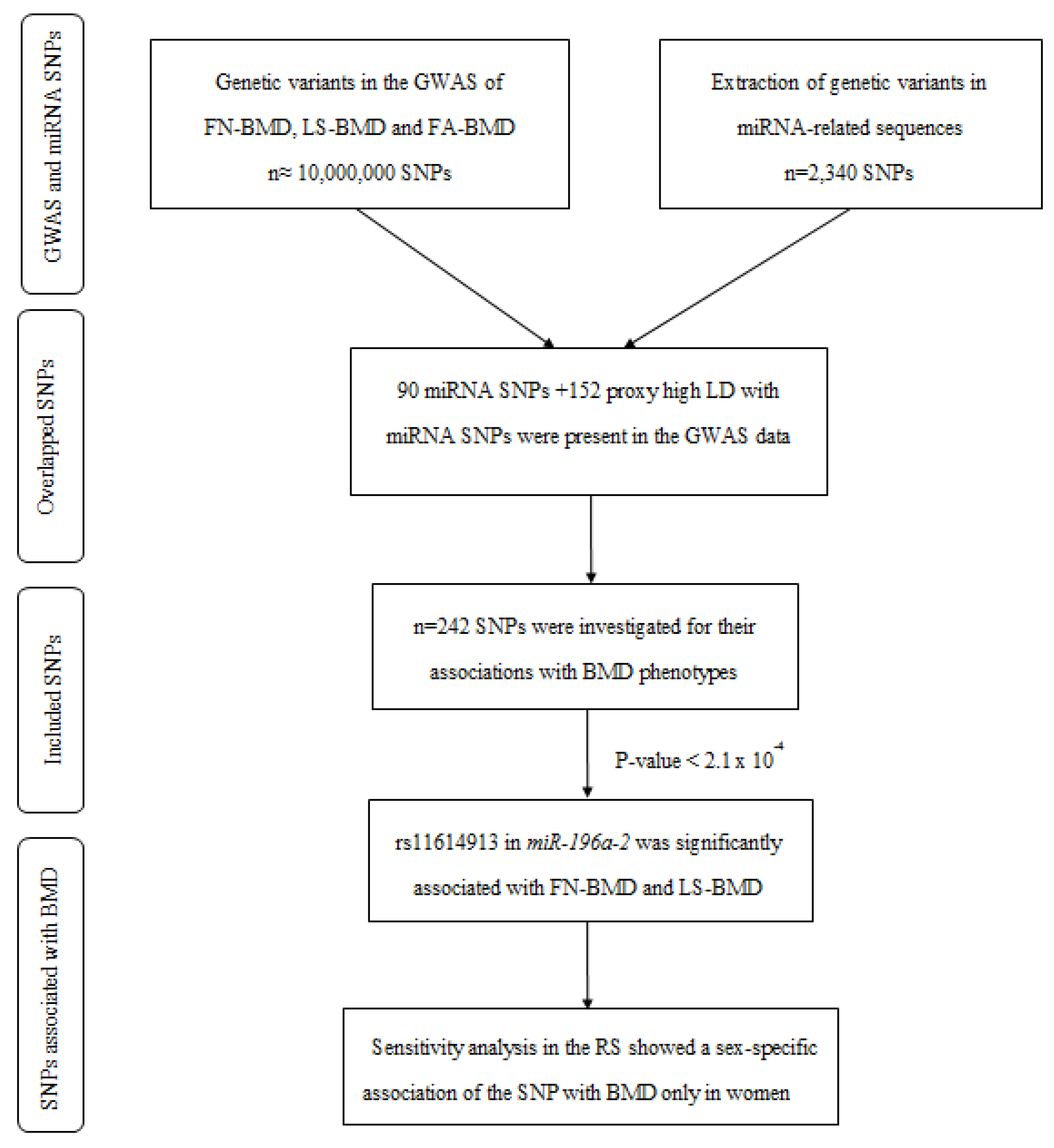
2.1. A Variant in miR-196a-2 Associates with BMD

A total of 2340 variants in miRNA-related sequences were collected by combination of a literature review and miRNASNP database [27]. In parallel, we extracted summary statistics data from the recent GWAS meta-analysis on three BMD phenotypes, including femoral neck (FN-BMD), lumbar spine (LS-BMD) and forearm (FA-BMD), provided by Genetic Factors of Osteoporosis (GEFOS) consortium [7]. Out of 2340 miRNA variants, 90 single-nucleotide polymorphisms (SNPs) were available in the GWAS data. Using the SNAP Web tool, we extracted the proxy SNPs (R2 > 0.8 and distance < 200 kb in 1000 Genomes project) for 152 of the unavailable variants. We studied the association of these 242 miRNA SNPs with BMD phenotypes. One of the SNPs passed the Bonferroni significance threshold of 2.1 × 10⁻⁴ (0.05/242). This includes rs11614913:C > T in miR-196a-2 which is significantly associated with FN-BMD (p-value = 9.9 × 10⁻⁷, β = −0.038) and LS-BMD (p-value = 3.2 × 10⁻¹¹, β = −0.061). This analysis indicated that individuals carrying the rs11614913 minor allele T are more prone to have lower BMD. No significant association was identified between the miRNA variants and FA-BMD. A simplified scheme of the pipeline used for the identification of miRNA SNPs associated with the BMD phenotypes is shown in Figure 1.

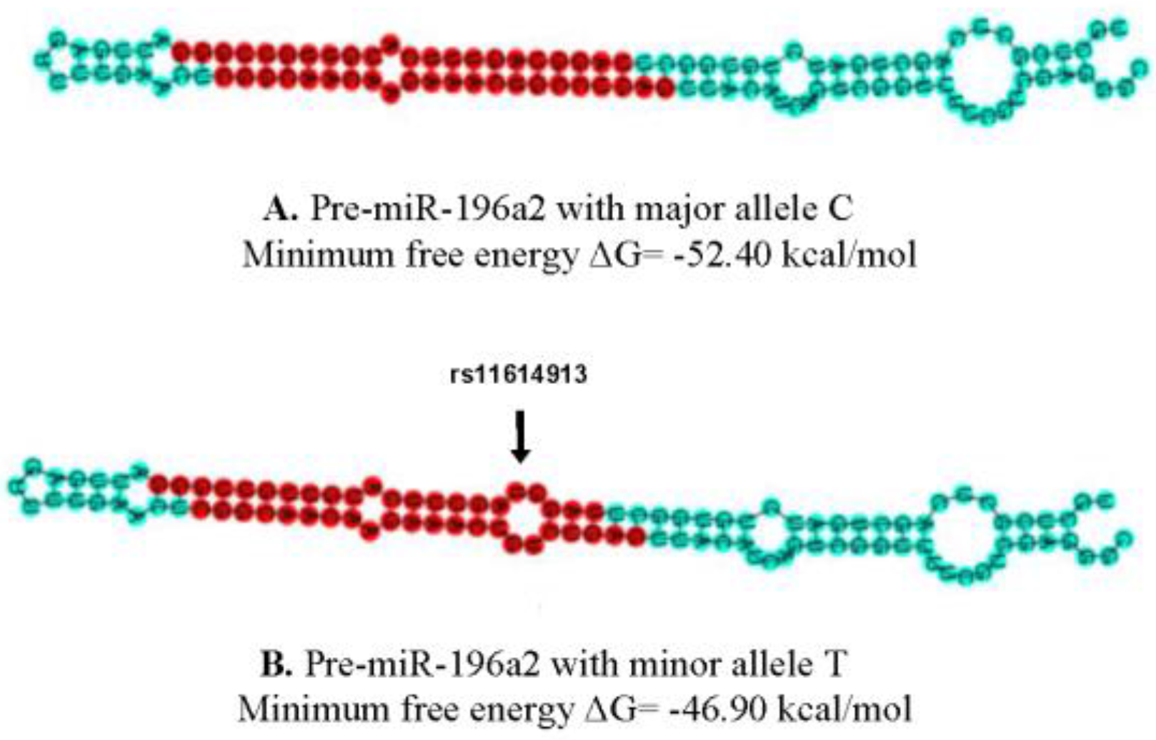
2.2. The Potential Impact of rs11614913 on the miR-196a-2 Structure and Function

We generated the hairpin structures of hsa-miR-196a-2 containing either the major allele C or the minor allele T at rs11614913 site using the Vienna RNAfold algorithm [28]. We observed 4.6 kcal/mol difference in the minimum free energy (MFE) of the thermodynamic predicted structure of pre-miR-196a-2 with the minor allele T compared to the wild type allele C (Figure 2). The analysis suggests that the investigated variant may affect the stability of miR-196a-2. In this line, it has been demonstrated previously that rs11614913-T decreases miR-196a-2 expression in different cell lines [29,30].

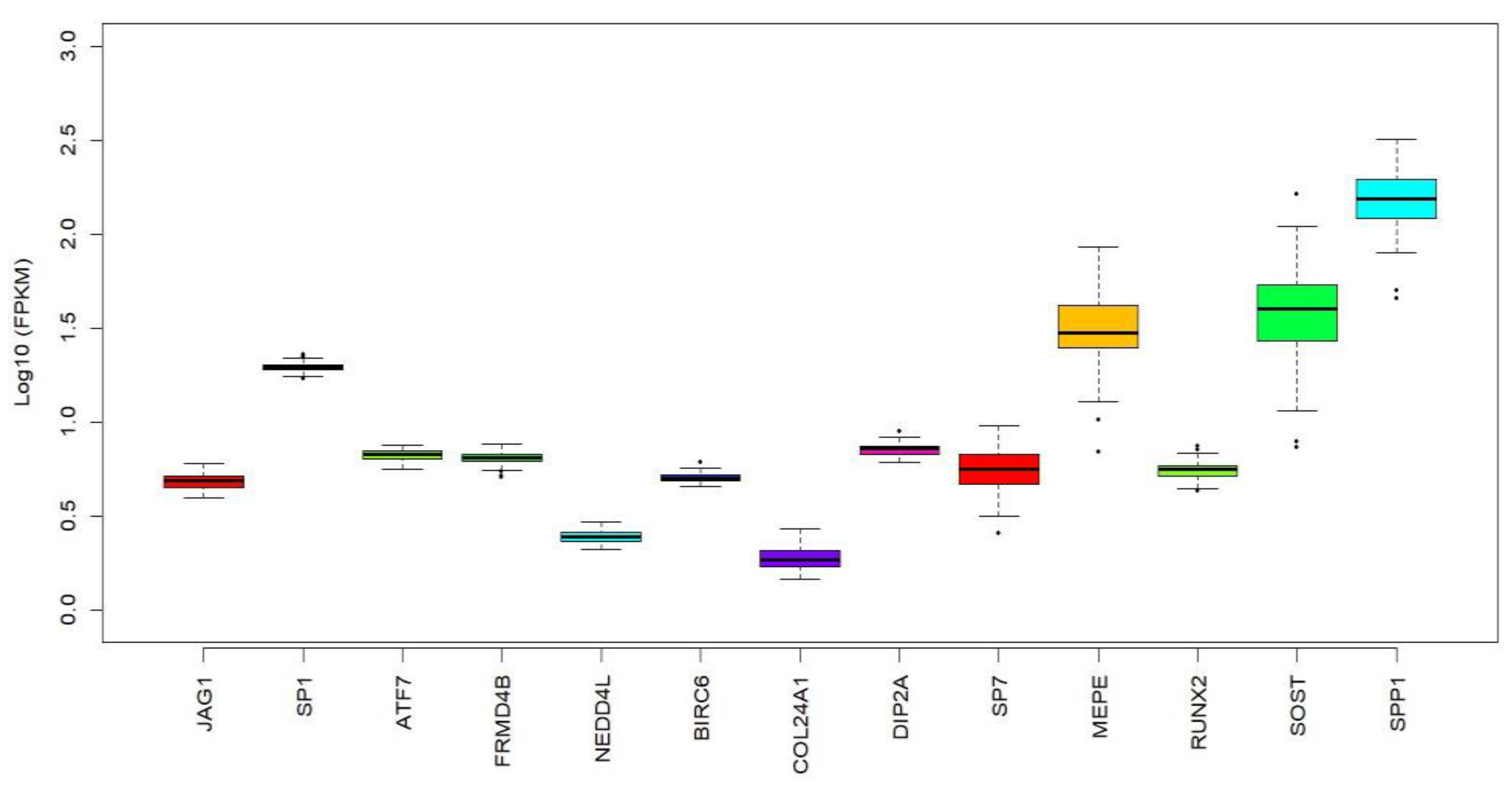
2.3. Associaton of miR-196a-2 Target Genes with BMD

Through leveraging the GEFOS GWAS data and using a candidate gene approach, we tested the association of genetic variants in 457 putative target genes of miR-196a-2 with FN-BMD and LS-BMD. Table 1 shows the top ten target genes of miR-196a-2 with the most significant association with the BMD phenotypes. Using RNA-seq gene expression data of 86 hip bone (iliac crest) biopsies, we found evidence for expression of eight out of the ten highlighted target genes of miR-196a-2 in bone (Figure 3) [12]. Among the bone-expressed targets, JAG1 passed the significance threshold, based on the number of variants in the tested miR-196a-2 target genes (Table 1). This analysis may suggest that JAG1 is more likely to mediate the downstream effect of miR-196a-2 in relation to BMD. Moreover, a number of genes have been demonstrated experimentally (i.e., by luciferase reporter assay, Western blot or qPCR) to be regulated by miR-196a-2. As shown in supplementary Table S1 some of these genes are shown to be involved in either osteogenesis or bone function and may mediate the miR-196a-2 effect on BMD. We checked the correlation of rs11614913 with expression level of its surrounding genes as shown by GTEX portal (http://www.gtexportal.org/home/) and found the association of SNP with expression of HOXC8 and HOXC-AS1 across different tissues.

2.4. Sensitivity Analyses for rs11614913 in miR-196a-2 Using the Rotterdam Study Data

Previous studies have reported sex-specific association of genetic variants with BMD [31,32]. Furthermore, some studies have shown difference in sex response to muscoskeletal cell development, mediated by influence of steroid hormones [33,34]. In order to investigate the potential difference in association between the miR-196a-2 variants and BMD across sexes, we performed a sensitivity analysis using the Rotterdam study (RS) data. The baseline characteristics of the RS participants are shown in Table 2. A total of 6,145 participants (3524 woman and 2621 men) from the three RS cohorts were eligible for this analysis (individuals with data available for rs11614913 and Dual X-ray Absorptiometry (DXA) imaging on FN-BMD and LS-BMD). Mixed linear regression analysis was carried out in sex-stratified data to investigate the association between rs11614913 and the BMD phenotypes (Table 3). In the basic model (adjusting for age, cohort, weight, waist to hip ratio and height) there was a significant association between rs11614913 and FN-BMD only in women (p-value = 0.003; β = 0.009; (95%Confidence Interval, CI) = 0.003, 0.014). The association remained significant for women in the second model (further adjusting for alcohol, smoking status and drugs used for treatment of bone diseases) (p-value = 0.003; β = 0.008; (95%CI) = 0.003, 0.014). We also tested the association between rs11614913 and LS-BMD and found, again, a clear significance only in women in the basic model (p-value = 0.023; β = 0.010; (95%CI) = 0.001, 0.019) and the second model (p-value = 0.026; β = 0.010; (95%CI) = 0.001, 0.018) (Table 3). Next, we further adjusted the second model for sex-hormones to see whether the miRNA variant is linked to sex-hormones (Table 3). The association in females remained significant after further adjustment for five sex-hormones (Model 3) involved in the steroidogenesis pathway. These results suggest that there is sex specificity in the association of miR-196a-2 with BMD.

3. Discussion

Recent studies have shown that miRNAs are important regulators of genes linked to bone remodeling and osteoporosis development [35,36,37,38,39]. Different approaches have been used in previous studies to identify miRNAs involved in osteoporosis, including miRNA expression profiling [38,40] and candidate gene association studies [41]. In this study, we have conducted a genome-wide scan investigating the association of miRNA genetic variants with BMD using GWAS data [7]. This method represents a valuable, extended and complementary approach to previous methods used in the identification of miRNAs associated with BMD.
Our results showed that rs11614913 in the stem region of pre-miR-196a-2 is significantly associated with FN-BMD and LS-BMD. Lack of significant association between rs11614913 within pre-miR-196a-2 and forearm BMD could be attributed to the small sample size in GWAS (n = 8143) compared to FN-BMD (n = 32,735) or LS-BMD (n = 28,498) in the discovery cohorts [7], or differences in bone remodeling between anatomical sites. It has been shown that loaded and unloaded bone (forearm) have distinct transcriptional activities [42,43]. The location of rs11614913 in pre-miR-196a-2 is likely to affect the miRNA processing by enzyme Dicer, and subsequently alter the expression of mature miR-196a-2 [44,45]. Polymorphisms in pre-miRNA sequences have been shown to cause either a destabilization of the interaction due to changes in the free binding energy or a change in target accessibility due to alternations in the miRNA secondary structure [19,46,47]. Our in silico analysis showed differences in the MFE between the predicted structure of pre-miR-196a-2 mutants and the wild type, suggesting the variant’s minor allele may diminish the stability of pre-miR-196a-2. In agreement with this conjecture, previous studies have established the impact of rs11614913 polymorphism (C/T) on the miR-196a-2 expression levels [29,30,44,45,48]. Zhibin Hu et al., have reported that rs11614913 wild-type allele (C) is associated with statistically significant increase in mature miR-196a-2 expression, while studying 23 human lung cancer tissue samples [30]. They also showed that rs11614913 could affect binding of the mature miR-196a-2 to its candidate target mRNA [30]. Furthermore, Zhao Hauanhuan et al., observed the same trend of rs11614913*CC genotype to increase the mature miR-196a-2 expression in different phenotypes of breast cancer [29]. Likewise, Hoffman et al., experimentally demonstrated that rs11614913 mutant allele (T) is associated with statistically significant decrease in miR-196a-2 expression in breast cancer patients [44]. Another study by Vinci et al., presented coherent results of rs11614913*TT decreasing miR-196a-2 expression levels in lung cancer patients [48]. In addition, Xu et al., determined that rs11614913 affects the expression of miR-196a-2 and consequently, expression of its downstream target gene HOXB8 [49]. They hypothesized that the variant might have an impact on miR-196a-HOXB8-Shh signaling pathway, and therefore, be associated with congenital heart disease susceptibility [49]. In other studies, the miR-196a-2 polymorphism rs11614913 has been linked to various phenotypic variations, ranging from several types of cancer [30,44,45,50] to increased risk for cardiovascular disease [49,51,52,53,54]. These data strongly suggest an important functional impact of rs11614913 on miR-196a-2 expression and function that in turn might affect the risk and/or progression of disease.
MiR-196a is shown to be expressed from HOX clusters loci in mammals and HOX genes in turn are shown to be targets of miR-196a [19,55]. The HOX genes play critical roles in limb development and skeletal patterning [56,57]. The miRNA has been also shown to play a role in brown adipogenesis of white fat progenitor cells through targeting HOXC8 [58]. It has been proven that the miRNA regulates HOXC8 at both mRNA and protein levels [55]. In an independent study, Kim et al., observed that adding miR-196-a inhibitors to osteoblast cells in culture causes a significant increase in HOXC8 protein levels, with subsequent increased proliferation and decrease in osteogenic differentiation [59]. These data suggest upregulation of HOXC8 in the miR-196a-2 variant carriers, of significance for osteogenic differentiation. Accordingly, Dong-Li Zhu et al., have recently shown that miR-196a-2 is expressed in osteoblasts and experimentally demonstrated that FGF2, previously identified as a susceptibility gene for osteoporosis in Caucasians [60], is a direct target of miR-196a-2 in the Chinese population [8]. Their experiments proved that miR-196a-2 had an influence on FGF2 mRNA in hFOB1 cells, which is a human fetal osteoblastic cell line [8].
In addition to previously validated targets of miR-196a-2 involved in osteogenesis, we highlighted a number of putative target genes associated with BMD with a potential to mediate the miR-196a-2 effect in BMD. Among them, JAG1 passed the significant threshold to be associated with BMD and is expressed in bone. The JAG1 gene has been previously reported to be associated with increased BMD and suggested as a candidate gene for BMD regulation in diverse ethnic groups [61]. Future experimental studies are needed to explore the postulated miR-196a-2-mediated regulation of the gene in bone tissue or cell lines.
We performed sex-stratified analysis using the Rotterdam study data to get insight into sex specificity for BMD variation on the miR-196a-2 polymorphism. In the sex-combined analysis, we observed significant association of rs11614913 with BMD phenotypes. However, sex-stratified analysis revealed that the association is mainly driven by women. We acknowledge that the observed association in women may have been driven by a lower number of men (our cohort contains 903 more women than men), however, sample size of 6145 should be sustainable to address sex difference. Notably, the miR-196a-2 polymorphism rs11614913 with combination of rs3746444 in miR-499a have been reported previously to be involved in the multiple sclerosis severity, where the association shows only female sex specificity [62]. Multiple sclerosis and osteoporosis share a surprising number of risk factors [63,64,65] and genetics might be one of them, although the interplay of the two miRNA variants and their impacts on gene interaction should be taken in consideration when interpreting the results regarding sex specificity. Considering the sexual dimorphism of bone [31,66], these data might indicate a potential for further clinical and biological investigations regarding the role of miR-196a-2 underlying BMD variation.
This study has some strengths and limitations that need to be considered in interpretation of the reported results. The major strength of this study is leveraging genetic data from the recent GWAS of BMD phenotypes that enabled us legitimate statistical power for detection of miRNA-related variants associated with BMD. The main limitation that needs to be addressed is lack of experimental studies in relevant tissues or cell lines. MiRNA-related SNPs might be only utilitarian if the target mRNA is expressed in the same tissue [67]. Thereby, further biological investigations warrant better insights into the mechanisms by which miR-196a-2 control expression of genes involved in BMD.

4. Materials and Methods

4.1. Genome-Wide Association Studies on BMD Phenotypes

The summary statistics from the recent GWAS meta-analysis on FN-BMD (n = 32,735), LS-BMD (n = 28,498) and FA-BMD (n = 8143) provided by GEFOS consortium were extracted [7]. The GEFOS consortium is a collective effort of numerous research groups combining GWAS data, in order to identify osteoporosis susceptibility alleles that regulate BMD and fracture risk [7]. The GEFOS consortium performed meta-analysis of whole genome sequencing, whole exome sequencing and deep imputation of genotype data in order to determine low-frequency and rare variants associated with risk factors for osteoporosis. The collaboration within the GEFOS has resulted in producing files with summary statistics for approximately 10 million genetics variants (the 1000 Genomes/UK10K reference panel) in 53,236 individuals [7]. More details on datasets and participants are described in detail elsewhere [7].

4.2. Identification of Genetic Variants in miRNA-Encoding Sequences

A dataset of single-nucleotide polymorphisms (SNPs) in miRNA-related sequences was created by combining miRNASNP (http://www.bioguo.org/miRNASNP/) [27] and the literature review (searching in PubMed for miRNA genetic variants). Precursor miRNA sequences (pre-miRNA) undergo cleavage by enzyme Dicer, yielding to mature miRNAs [13], therefore we screened all variants located in human pre-miRNA and mature miRNA sequences. The methodology was explained in details elsewhere [68]. Variants with minor allele frequency (MAF) >0.01 were included. Variants with smaller MAF were illegible due to low imputation quality and issue of being underpowered in further studies. In total, 2340 miRNA variants were extracted. Of these, 242 variants were available in the GEFOS GWAS data and were therefore investigated further for their associations with BMD phenotypes.

4.3. miRNA Target Genes Associated with BMD Phenotypes

Once a miRNA variant was found to be significantly associated with BMD phenotypes, we searched for the miRNA target genes. We postulated that some of the miRNA target genes may mediate the downstream effect of miRNA in relation to BMD phenotypes. In order to identify target genes of miRNAs, putative target genes were extracted from combining TargetScan v7.1 (http://www.targetscan.org/vert_71/) and miRDB (http://mirdb.org/) database [69]. Target genes present in both databases were selected for further investigation. Any supplementary information, such as miRNA conservation between species, host genes, miRNA sequences was collected from TargetScan (v7.1). Both context score and conserved target sites were used to rank the miRNA target genes. In addition, the online database, miRTarBase (http://mirtarbase.mbc.nctu.edu.tw/) provides information on various functional experiments, such as microarrays, western blot, and reported assays performed between miRNAs and their target genes [70]. We used miRTarBase to search for functional experiment confirming the putative interaction between miRNAs of interest and their target genes. A candidate gene approach was performed by leveraging the GWAS data on BMD phenotypes [7] and to investigate the association between genetic variants in the miRNA target genes and BMD. In addition, we evaluated the expression of selected target genes in the bone tissue. Dataset used for gene expression was created out of 86 iliac biopsies [12].

4.4. The Variant Effect on the Pre-miRNA Structure

The secondary structure of pre-miRNA is critical for the miRNA production. The Vienna RNAfold algorithm (ViennaRNA package 2.0) was used to predict the impact of miRNA variants on the hairpin stem-loop structure of pre-miRNAs [28]. The ViennaRNA package 2.0 is available to the public domain and relies on numerous algorithms for prediction and analysis of RNA secondary structures [71]. The program calculates the shift in minimum free energy (MFE) of the thermodynamic ensemble in the hairpin structure of miRNA (wild type and mutant) [72]. The shift in MFE is likely to be related to the function, as it can result in instability of miRNA.

4.5. The Rotterdam Study Data

The Rotterdam study (RS) is a population-based cohort study, with main goal of identifying chronic disabling conditions of the middle aged and elderly people [73]. Participants were interviewed at home and went through an extensive set of examinations, including bone mineral densitometry, sample collections for in-depth molecular and genetic analysis [73]. The RS includes three sub-cohorts. We used the data from the baseline, second and third cohort (RS-I-4, RS-II-2, and RS-III-1). For all participants, DXA-based BMD measurements were collected for FN-BMD and LS-BMD. The RS does not include data on FA-BMD since this site is used for prediction of osteoporosis only when data is not available for FN-BMD or LS-BMD due to numerous reasons (e.g., patients either being obese, men with hyperparathyroidism or receiving androgen-deprivation therapy (ADT) for prostate cancer) [74]. Furthermore, determinants were assessed either by physical examinations, collection of blood samples, or by questionnaires. Participants were included if they had FN-BMD or LS-BMD measurements, which resulted in combination of three cohorts (RS-I-4, RS-II-2, and RS-III-1). We used multiple linear regression in sex-stratified dataset to examine the association between the candidate miRNA variant and BMD phenotypes (separately). Our analysis was adjusted for all potential confounders in three models.

5. Conclusions

The results of this study suggest that miR-196a-2 polymorphism (rs11614913:C > T) is associated with reduced FN-BMD and LS-BMD. We highlighted a number of target genes that may mediate miR-196a-2 function in influencing BMD. The identified miR-196a-2 might have a future implication in the clinical field related to diagnosis and treatment of osteoporosis. Future biological studies will give insight into the mechanisms by which miR-196a-2 may control expression of bone-related genes. Collectively, our study provides further understanding of the miRNA-mediated regulation of BMD.

Supplementary Materials

Supplementary materials can be found at www.mdpi.com/1422-0067/18/12/2529/s1.

Acknowledgments

The Rotterdam Study is supported by Erasmus MC (Erasmus Medical Center Rotterdam), the Erasmus University Rotterdam, the Netherlands Organization for Scientific Research (NWO), the Netherlands Organization for Health Research and Development (ZonMW), the Research Institute for Diseases in the Elderly (RIDE), the Ministry of Education, Culture and Science, and the Ministry of Health, Welfare and Sports. The authors are grateful to the Rotterdam Study participants, the staff involved with the Rotterdam Study and the participating general practitioners and pharmacists. We are also grateful to the GEFOS consortium (EC-FP7-HEALTH- F2-2008-201865-GEFOS) for making the GWAS summary statistics data publicly available. The mobility stimuli plan of the European Union Erasmus Mundus Action program supported Irma Karabegović (ERAWEB) and Maša Zrimšec (Erasmus+HE). The ZonMW Project number: NWO/ZONMW-VIDI-016-136-367 supported Carolina Medina-Gomez, Maša Zrimšek and Fernando Rivadeneira, together with the creation of the RNA-seq expression dataset in collaboration with the Lovisenberg Diakonale Hospital research foundation.

Author Contributions

Mohsen Ghanbari conceived and designed the study. Irma Karabegović, Carolina Medina-Gomez and Mohsen Ghanbari performed the miRNA in-silico analyses. Irma Karabegović and Silvana Maas analyzed the epidemiologic data. André G. Uitterlinden and Fernando Rivadeneira, provided the Rotterdam Study data. Maša Zrimšek, Carolina Medina-Gomez, Sjur Reppe, Kaare M. Gautvik and Fernando Rivadeneira assembled and analyzed the expression data in bone. Irma Karabegović and Mohsen Ghanbari wrote first draft of manuscript. All authors read, commented and approved the manuscript.

Conflicts of Interest

The authors declare no conflict of interest.

Abbreviations

GWASGenome-wide association studies
GEFOSGenetic factors for osteoporosis
BMD
SNP
Bone mineral density
Single nucleotide polymorphism
FN-BMDFemoral neck bone mineral density
LS-BMDLumbar spine bone mineral density
FA-BMDForearm bone mineral density
miRNAmicroRNA
WHRWaist to hip ratio
RSRotterdam Study
DXADual X-ray Absorptiometry
MFEMinimum free energy
LDLinkage disequilibrium
DHEADehydroepiandrosterone
DHEASDehydroepiandrosterone sulfate
IQRInterquartile range

References

  1. Hernlund, E.; Svedbom, A.; Ivergard, M.; Compston, J.; Cooper, C.; Stenmark, J.; McCloskey, E.V.; Jonsson, B.; Kanis, J.A. Osteoporosis in the European Union: Medical management, epidemiology and economic burden. A report prepared in collaboration with the International Osteoporosis Foundation (IOF) and the European Federation of Pharmaceutical Industry Associations (EFPIA). Arch Osteoporos 2013, 8, 136. [Google Scholar] [CrossRef] [PubMed]
  2. Rivadeneira, F.; Makitie, O. Osteoporosis and bone mass disorders: From gene pathways to treatments. Trends Endocrinol. Metab. 2016, 27, 262–281. [Google Scholar] [CrossRef] [PubMed]
  3. Rivadeneira, F.; Styrkarsdottir, U.; Estrada, K.; Halldorsson, B.V.; Hsu, Y.H.; Richards, J.B.; Zillikens, M.C.; Kavvoura, F.K.; Amin, N.; Aulchenko, Y.S.; et al. Twenty bone-mineral-density loci identified by large-scale meta-analysis of genome-wide association studies. Nat. Genet. 2009, 41, 1199–1206. [Google Scholar] [CrossRef] [PubMed] [Green Version]
  4. Kling, J.M.; Clarke, B.L.; Sandhu, N.P. Osteoporosis prevention, screening, and treatment: A review. J. Womens Health 2014, 23, 563–572. [Google Scholar] [CrossRef] [PubMed]
  5. Blake, G.M.; Fogelman, I. The role of DXA bone density scans in the diagnosis and treatment of osteoporosis. Postgrad. Med. J. 2007, 83, 509–517. [Google Scholar] [CrossRef] [PubMed]
  6. Stewart, T.L.; Ralston, S.H. Role of genetic factors in the pathogenesis of osteoporosis. J. Endocrinol. 2000, 166, 235–245. [Google Scholar] [CrossRef] [PubMed]
  7. Zheng, H.F.; Forgetta, V.; Hsu, Y.H.; Estrada, K.; Rosello-Diez, A.; Leo, P.J.; Dahia, C.L.; Park-Min, K.H.; Tobias, J.H.; Kooperberg, C.; et al. Whole-genome sequencing identifies EN1 as a determinant of bone density and fracture. Nature 2015, 526, 112–117. [Google Scholar] [CrossRef] [PubMed]
  8. Zhu, D.L.; Guo, Y.; Zhang, Y.; Dong, S.S.; Xu, W.; Hao, R.H.; Chen, X.F.; Yan, H.; Yang, S.Y.; Yang, T.L. A functional SNP regulated by miR-196a-3p in the 3’ UTR of FGF2 is associated with bone mineral density in the Chinese population. Hum. Mutat. 2017, 38, 725–735. [Google Scholar] [CrossRef] [PubMed]
  9. Dole, N.S.; Kapinas, K.; Kessler, C.B.; Yee, S.P.; Adams, D.J.; Pereira, R.C.; Delany, A.M. A single nucleotide polymorphism in osteonectin 3′ untranslated region regulates bone volume and is targeted by miR-433. J. Bone Miner. Res. 2015, 30, 723–732. [Google Scholar] [CrossRef] [PubMed]
  10. Guo, L.J.; Liao, L.; Yang, L.; Li, Y.; Jiang, T.J. MiR-125a TNF receptor-associated factor 6 to inhibit osteoclastogenesis. Exp. Cell Res. 2014, 321, 142–152. [Google Scholar] [CrossRef] [PubMed]
  11. Jemtland, R.; Holden, M.; Reppe, S.; Olstad, O.K.; Reinholt, F.P.; Gautvik, V.T.; Refvem, H.; Frigessi, A.; Houston, B.; Gautvik, K.M. Molecular disease map of bone characterizing the postmenopausal osteoporosis phenotype. J. Bone Miner. Res. 2011, 26, 1793–1801. [Google Scholar] [CrossRef] [PubMed]
  12. Reppe, S.; Refvem, H.; Gautvik, V.T.; Olstad, O.K.; Hovring, P.I.; Reinholt, F.P.; Holden, M.; Frigessi, A.; Jemtland, R.; Gautvik, K.M. Eight genes are highly associated with BMD variation in postmenopausal Caucasian women. Bone 2010, 46, 604–612. [Google Scholar] [CrossRef] [PubMed]
  13. Ha, M.; Kim, V.N. Regulation of microRNA biogenesis. Nat. Rev. Mol. Cell Biol. 2014, 15, 509–524. [Google Scholar] [CrossRef] [PubMed]
  14. Bushati, N.; Cohen, S.M. microRNA functions. Annu. Rev. Cell Dev. Biol. 2007, 23, 175–205. [Google Scholar] [CrossRef] [PubMed]
  15. Ardekani, A.M.; Naeini, M.M. The role of microRNAs in human diseases. Avicenna J. Med. Biotechnol. 2010, 2, 161–179. [Google Scholar] [PubMed]
  16. Tufekci, K.U.; Oner, M.G.; Meuwissen, R.L.; Genc, S. The role of microRNAs in human diseases. Methods Mol. Biol. 2014, 1107, 33–50. [Google Scholar] [PubMed]
  17. Zhang, Y.; Lu, Y.J.; Yang, B.F. Potential role of microRNAs in human diseases and the exploration on design of small molecule agents. Acta Pharm. Sin. 2007, 42, 1115–1121. [Google Scholar]
  18. Lu, J.; Clark, A.G. Impact of microRNA regulation on variation in human gene expression. Genome Res. 2012, 22, 1243–1254. [Google Scholar] [CrossRef] [PubMed]
  19. Dole, N.S.; Delany, A.M. microRNA variants as genetic determinants of bone mass. Bone 2016, 84, 57–68. [Google Scholar] [CrossRef] [PubMed]
  20. Park, H.; Huang, X.; Lu, C.; Cairo, M.S.; Zhou, X. microRNA-146a and microRNA-146b regulate human dendritic cell apoptosis and cytokine production by targeting TRAF6 and IRAK1 proteins. J. Biol. Chem. 2015, 290, 2831–2841. [Google Scholar] [CrossRef] [PubMed]
  21. Gravallese, E.M.; Galson, D.L.; Goldring, S.R.; Auron, P.E. The role of TNF-receptor family members and other TRAF-dependent receptors in bone resorption. Arthritis Res. 2001, 3, 6–12. [Google Scholar] [CrossRef] [PubMed] [Green Version]
  22. Kim, J.H.; Jin, H.M.; Kim, K.; Song, I.; Youn, B.U.; Matsuo, K.; Kim, N. The mechanism of osteoclast differentiation induced by IL-1. J. Immunol. 2009, 183, 1862–1870. [Google Scholar] [CrossRef] [PubMed]
  23. Chen, P.; Wei, D.; Xie, B.; Ni, J.; Xuan, D.; Zhang, J. Effect and possible mechanism of network between microRNAs and RUNX2 gene on human dental follicle cells. J. Cell. Biochem. 2014, 115, 340–348. [Google Scholar] [CrossRef] [PubMed]
  24. Nakasa, T.; Shibuya, H.; Nagata, Y.; Niimoto, T.; Ochi, M. The inhibitory effect of microRNA-146a expression on bone destruction in collagen-induced arthritis. Arthritis Rheumatol. 2011, 63, 1582–1590. [Google Scholar] [CrossRef] [PubMed]
  25. Polesskaya, A.; Cuvellier, S.; Naguibneva, I.; Duquet, A.; Moss, E.G.; Harel-Bellan, A. Lin-28 binds IGF-2 mRNA and participates in skeletal myogenesis by increasing translation efficiency. Genes Dev. 2007, 21, 1125–1138. [Google Scholar] [CrossRef] [PubMed]
  26. Hassan, M.Q.; Gordon, J.A.; Beloti, M.M.; Croce, C.M.; van Wijnen, A.J.; Stein, J.L.; Stein, G.S.; Lian, J.B. A network connecting Runx2, SATB2, and the miR-23a~27a~24-2 cluster regulates the osteoblast differentiation program. Proc. Natl. Acad. Sci. USA 2010, 107, 19879–19884. [Google Scholar] [CrossRef] [PubMed]
  27. Gong, J.; Liu, C.; Liu, W.; Wu, Y.; Ma, Z.; Chen, H.; Guo, A.Y. An update of miRNASNP database for better SNP selection by GWAS data, miRNA expression and online tools. Database (Oxford) 2015, 2015. [Google Scholar] [CrossRef] [PubMed]
  28. Lorenz, R.; Bernhart, S.H.; Honer Zu Siederdissen, C.; Tafer, H.; Flamm, C.; Stadler, P.F.; Hofacker, I.L. ViennaRNA Package 2.0. Algorithms Mol. Biol. 2011, 6, 26. [Google Scholar] [CrossRef] [PubMed]
  29. Zhao, H.; Xu, J.; Zhao, D.; Geng, M.; Ge, H.; Fu, L.; Zhu, Z. Somatic mutation of the SNP rs11614913 and its association with increased miR-196a2 Expression in Breast Cancer. DNA Cell Biol. 2016, 35, 81–87. [Google Scholar] [CrossRef] [PubMed]
  30. Hu, Z.; Chen, J.; Tian, T.; Zhou, X.; Gu, H.; Xu, L.; Zeng, Y.; Miao, R.; Jin, G.; Ma, H.; et al. Genetic variants of miRNA sequences and non-small cell lung cancer survival. J. Clin. Investig. 2008, 118, 2600–2608. [Google Scholar] [CrossRef] [PubMed]
  31. Karasik, D.; Ferrari, S.L. Contribution of gender-specific genetic factors to osteoporosis risk. Ann. Hum. Genet. 2008, 72, 696–714. [Google Scholar] [CrossRef] [PubMed]
  32. Naganathan, V.; Macgregor, A.; Snieder, H.; Nguyen, T.; Spector, T.; Sambrook, P. Gender differences in the genetic factors responsible for variation in bone density and ultrasound. J. Bone Miner. Res. 2002, 17, 725–733. [Google Scholar] [CrossRef] [PubMed]
  33. Corsi, K.A.; Pollett, J.B.; Phillippi, J.A.; Usas, A.; Li, G.; Huard, J. Osteogenic potential of postnatal skeletal muscle-derived stem cells is influenced by donor sex. J. Bone Miner. Res. 2007, 22, 1592–1602. [Google Scholar] [CrossRef] [PubMed]
  34. Tosi, L.L.; Boyan, B.D.; Boskey, A.L. Does sex matter in musculoskeletal health? The influence of sex and gender on musculoskeletal health. J. Bone Joint Surg. Am. 2005, 87, 1631–1647. [Google Scholar] [PubMed]
  35. Van Wijnen, A.J.; van de Peppel, J.; van Leeuwen, J.P.; Lian, J.B.; Stein, G.S.; Westendorf, J.J.; Oursler, M.J.; Im, H.J.; Taipaleenmaki, H.; Hesse, E.; et al. MicroRNA functions in osteogenesis and dysfunctions in osteoporosis. Curr. Osteoporos Rep. 2013, 11, 72–82. [Google Scholar] [CrossRef] [PubMed]
  36. Zhang, Y.; Gao, Y.; Cai, L.; Li, F.; Lou, Y.; Xu, N.; Kang, Y.; Yang, H. MicroRNA-221 is involved in the regulation of osteoporosis through regulates RUNX2 protein expression and osteoblast differentiation. Am. J. Transl. Res. 2017, 9, 126–135. [Google Scholar] [PubMed]
  37. Sun, M.; Zhou, X.; Chen, L.; Huang, S.; Leung, V.; Wu, N.; Pan, H.; Zhen, W.; Lu, W.; Peng, S. The Regulatory Roles of MicroRNAs in Bone Remodeling and Perspectives as Biomarkers in Osteoporosis. Biomed. Res. Int. 2016, 2016, 1652417. [Google Scholar] [CrossRef] [PubMed]
  38. De-Ugarte, L.; Yoskovitz, G.; Balcells, S.; Guerri-Fernandez, R.; Martinez-Diaz, S.; Mellibovsky, L.; Urreizti, R.; Nogues, X.; Grinberg, D.; Garcia-Giralt, N.; et al. MiRNA profiling of whole trabecular bone: Identification of osteoporosis-related changes in MiRNAs in human hip bones. BMC Med. Genom. 2015, 8, 75. [Google Scholar]
  39. Hackl, M.; Heilmeier, U.; Weilner, S.; Grillari, J. Circulating microRNAs as novel biomarkers for bone diseases—Complex signatures for multifactorial diseases? Mol. Cell. Endocrinol. 2016, 432, 83–95. [Google Scholar] [CrossRef] [PubMed]
  40. Seeliger, C.; Karpinski, K.; Haug, A.T.; Vester, H.; Schmitt, A.; Bauer, J.S.; van Griensven, M. Five freely circulating miRNAs and bone tissue miRNAs are associated with osteoporotic fractures. J. Bone Miner. Res. 2014, 29, 1718–1728. [Google Scholar] [CrossRef] [PubMed]
  41. De-Ugarte, L.; Caro-Molina, E.; Rodriguez-Sanz, M.; Garcia-Perez, M.A.; Olmos, J.M.; Sosa-Henriquez, M.; Perez-Cano, R.; Gomez-Alonso, C.; Del Rio, L.; Mateo-Agudo, J.; et al. SNPs in bone-related miRNAs are associated with the osteoporotic phenotype. Sci. Rep. 2017, 7, 516. [Google Scholar] [CrossRef] [PubMed]
  42. Kalogeropoulos, M.; Varanasi, S.S.; Olstad, O.K.; Sanderson, P.; Gautvik, V.T.; Reppe, S.; Francis, R.M.; Gautvik, K.M.; Birch, M.A.; Datta, H.K. Zic1 transcription factor in bone: Neural developmental protein regulates mechanotransduction in osteocytes. FASEB J. 2010, 24, 2893–2903. [Google Scholar] [CrossRef] [PubMed]
  43. Varanasi, S.S.; Olstad, O.K.; Swan, D.C.; Sanderson, P.; Gautvik, V.T.; Reppe, S.; Francis, R.M.; Gautvik, K.M.; Datta, H.K. Skeletal site-related variation in human trabecular bone transcriptome and signaling. PLoS ONE 2010, 5, e10692. [Google Scholar] [CrossRef] [PubMed]
  44. Hoffman, A.E.; Zheng, T.; Yi, C.; Leaderer, D.; Weidhaas, J.; Slack, F.; Zhang, Y.; Paranjape, T.; Zhu, Y. microRNA miR-196a-2 and breast cancer: A genetic and epigenetic association study and functional analysis. Cancer Res. 2009, 69, 5970–5977. [Google Scholar] [CrossRef] [PubMed]
  45. Song, Z.S.; Wu, Y.; Zhao, H.G.; Liu, C.X.; Cai, H.Y.; Guo, B.Z.; Xie, Y.A.; Shi, H.R. Association between the rs11614913 variant of miRNA-196a-2 and the risk of epithelial ovarian cancer. Oncol. Lett. 2016, 11, 194–200. [Google Scholar] [CrossRef] [PubMed]
  46. Haas, U.; Sczakiel, G.; Laufer, S.D. microRNA-mediated regulation of gene expression is affected by disease-associated SNPs within the 3′-UTR via altered RNA structure. RNA Biol. 2012, 9, 924–937. [Google Scholar] [CrossRef] [PubMed]
  47. Mahen, E.M.; Watson, P.Y.; Cottrell, J.W.; Fedor, M.J. mRNA secondary structures fold sequentially but exchange rapidly in vivo. PLoS Biol. 2010, 8, e1000307. [Google Scholar] [CrossRef] [PubMed]
  48. Vinci, S.; Gelmini, S.; Pratesi, N.; Conti, S.; Malentacchi, F.; Simi, L.; Pazzagli, M.; Orlando, C. Genetic variants in miR-146a, miR-149, miR-196a2, miR-499 and their influence on relative expression in lung cancers. Clin. Chem. Lab. Med. 2011, 49, 2073–2080. [Google Scholar] [CrossRef] [PubMed]
  49. Xu, J.; Hu, Z.; Xu, Z.; Gu, H.; Yi, L.; Cao, H.; Chen, J.; Tian, T.; Liang, J.; Lin, Y.; et al. Functional variant in microRNA-196a2 contributes to the susceptibility of congenital heart disease in a Chinese population. Hum. Mutat. 2009, 30, 1231–1236. [Google Scholar] [CrossRef] [PubMed]
  50. Sun, M.; Liu, X.H.; Li, J.H.; Yang, J.S.; Zhang, E.B.; Yin, D.D.; Liu, Z.L.; Zhou, J.; Ding, Y.; Li, S.Q.; et al. miR-196a is upregulated in gastric cancer and promotes cell proliferation by downregulating p27(kip1). Mol. Cancer Ther. 2012, 11, 842–852. [Google Scholar] [CrossRef] [PubMed]
  51. Zhi, H.; Wang, L.; Ma, G.; Ye, X.; Yu, X.; Zhu, Y.; Zhang, Y.; Zhang, J.; Wang, B. Polymorphisms of miRNAs genes are associated with the risk and prognosis of coronary artery disease. Clin. Res. Cardiol. 2012, 101, 289–296. [Google Scholar] [CrossRef] [PubMed]
  52. Zhou, B.; Rao, L.; Peng, Y.; Wang, Y.; Chen, Y.; Song, Y.; Zhang, L. Common genetic polymorphisms in pre-microRNAs were associated with increased risk of dilated cardiomyopathy. Clin. Chim. Acta 2010, 411, 1287–1290. [Google Scholar] [CrossRef] [PubMed]
  53. Su, Y.M.; Li, J.; Guo, Y.F.; Cai, F.; Cai, X.X.; Pan, H.Y.; Deng, X.T.; Pan, M. A Functional single-nucleotide polymorphism in pre-microRNA-196a2 is associated with atrial fibrillation in han chinese. Clin. Lab. 2015, 61, 1179–1185. [Google Scholar] [CrossRef] [PubMed]
  54. Ghanbari, M.; Sedaghat, S.; de Looper, H.W.; Hofman, A.; Erkeland, S.J.; Franco, O.H.; Dehghan, A. The association of common polymorphisms in miR-196a2 with waist to hip ratio and miR-1908 with serum lipid and glucose. Obesity (Silver Spring) 2015, 23, 495–503. [Google Scholar] [CrossRef] [PubMed]
  55. Yekta, S.; Shih, I.H.; Bartel, D.P. microRNA-directed cleavage of HOXB8 mRNA. Science 2004, 304, 594–596. [Google Scholar] [CrossRef] [PubMed]
  56. Alexander, T.; Nolte, C.; Krumlauf, R. Hox genes and segmentation of the hindbrain and axial skeleton. Annu. Rev. Cell Dev. Biol. 2009, 25, 431–456. [Google Scholar] [CrossRef] [PubMed]
  57. Zakany, J.; Duboule, D. The role of Hox genes during vertebrate limb development. Curr. Opin. Genet. Dev. 2007, 17, 359–366. [Google Scholar] [CrossRef] [PubMed]
  58. Mori, M.; Nakagami, H.; Rodriguez-Araujo, G.; Nimura, K.; Kaneda, Y. Essential role for miR-196a in brown adipogenesis of white fat progenitor cells. PLoS Biol. 2012, 10, e1001314. [Google Scholar] [CrossRef] [PubMed]
  59. Kim, Y.J.; Bae, S.W.; Yu, S.S.; Bae, Y.C.; Jung, J.S. miR-196a regulates proliferation and osteogenic differentiation in mesenchymal stem cells derived from human adipose tissue. J. Bone Miner. Res. 2009, 24, 816–825. [Google Scholar] [CrossRef] [PubMed]
  60. Lei, S.F.; Papasian, C.J.; Deng, H.W. Polymorphisms in predicted miRNA binding sites and osteoporosis. J. Bone Miner. Res. 2011, 26, 72–78. [Google Scholar] [CrossRef] [PubMed]
  61. Kung, A.W.; Xiao, S.M.; Cherny, S.; Li, G.H.; Gao, Y.; Tso, G.; Lau, K.S.; Luk, K.D.; Liu, J.M.; Cui, B.; et al. Association of JAG1 with bone mineral density and osteoporotic fractures: A genome-wide association study and follow-up replication studies. Am. J. Hum. Genet. 2010, 86, 229–239. [Google Scholar] [CrossRef] [PubMed]
  62. Kiselev, I.; Bashinskaya, V.; Kulakova, O.; Baulina, N.; Popova, E.; Boyko, A.; Favorova, O. Variants of microRNA genes: Gender-specific associations with multiple sclerosis risk and severity. Int. J. Mol. Sci. 2015, 16, 20067–20081. [Google Scholar] [CrossRef] [PubMed]
  63. Hearn, A.P.; Silber, E. Osteoporosis in multiple sclerosis. Mult. Scler. J. 2010, 16, 1031–1043. [Google Scholar] [CrossRef] [PubMed]
  64. Sioka, C.; Kyritsis, A.P.; Fotopoulos, A. Multiple sclerosis, osteoporosis, and vitamin D. J. Neurol. Sci. 2009, 287, 1–6. [Google Scholar] [CrossRef] [PubMed]
  65. Gibson, J.C.; Summers, G.D. Bone health in multiple sclerosis. Osteoporos Int. 2011, 22, 2935–2949. [Google Scholar] [CrossRef] [PubMed]
  66. Estrada, K.; Styrkarsdottir, U.; Evangelou, E.; Hsu, Y.H.; Duncan, E.L.; Ntzani, E.E.; Oei, L.; Albagha, O.M.; Amin, N.; Kemp, J.P.; et al. Genome-wide meta-analysis identifies 56 bone mineral density loci and reveals 14 loci associated with risk of fracture. Nat. Genet. 2012, 44, 491–501. [Google Scholar] [CrossRef] [PubMed] [Green Version]
  67. Arnold, M.; Ellwanger, D.C.; Hartsperger, M.L.; Pfeufer, A.; Stumpflen, V. Cis-acting polymorphisms affect complex traits through modifications of microRNA regulation pathways. PLoS ONE 2012, 7, e36694. [Google Scholar] [CrossRef] [PubMed]
  68. Ghanbari, M.; Ikram, M.A.; de Looper, H.W.; Hofman, A.; Erkeland, S.J.; Franco, O.H.; Dehghan, A. Genome-wide identification of microRNA-related variants associated with risk of Alzheimer’s disease. Sci. Rep. 2016, 6, 28387. [Google Scholar] [CrossRef] [PubMed]
  69. Agarwal, V.; Bell, G.W.; Nam, J.W.; Bartel, D.P. Predicting effective microRNA target sites in mammalian mRNAs. Elife 2015, 4, e05005. [Google Scholar] [CrossRef] [PubMed]
  70. Chou, C.H.; Chang, N.W.; Shrestha, S.; Hsu, S.D.; Lin, Y.L.; Lee, W.H.; Yang, C.D.; Hong, H.C.; Wei, T.Y.; Tu, S.J.; et al. miRTarBase 2016: Updates to the experimentally validated miRNA-target interactions database. Nucleic Acids Res. 2016, 44, D239–D247. [Google Scholar] [CrossRef] [PubMed]
  71. Hofacker, I.L. Vienna RNA secondary structure server. Nucleic Acids Res. 2003, 31, 3429–3431. [Google Scholar] [CrossRef] [PubMed]
  72. Will, S.; Jabbari, H. Sparse RNA folding revisited: Space-efficient minimum free energy structure prediction. Algorithms Mol. Biol. 2016, 11, 7. [Google Scholar] [CrossRef] [PubMed]
  73. Ikram, M.A.; Brusselle, G.G.O.; Murad, S.D.; van Duijn, C.M.; Franco, O.H.; Goedegebure, A.; Klaver, C.C.W.; Nijsten, T.E.C.; Peeters, R.P.; Stricker, B.H.; et al. The Rotterdam Study: 2018 update on objectives, design and main results. Eur. J. Epidemiol. 2017, 32, 807–850. [Google Scholar] [CrossRef] [PubMed]
  74. Watts, N.B.; Adler, R.A.; Bilezikian, J.P.; Drake, M.T.; Eastell, R.; Orwoll, E.S.; Finkelstein, J.S.; Endocrine, S. Osteoporosis in men: An Endocrine Society clinical practice guideline. J. Clin. Endocrinol. Metab. 2012, 97, 1802–1822. [Google Scholar] [CrossRef] [PubMed]
Figure 1. A simplified diagram of the pipeline used to identify miRNA genetic variants associated with BMD. FN-BMD: Femoral neck bone mineral density; LS-BMD: Lumbar spine bone mineral density; FA-BMD: Forearm bone mineral density; SNP: Single-nucleotide polymorphism; GWAS: Genome-wide association studies.
Ijms 18 02529 g001
Figure 2. Schematic view of the predicted pre-miR-196a-2 hairpin structure containing the SNP major allele C or minor allele T. The minimum free energy (MFE) change of the thermodynamic ensemble (ΔG) is shown. The red part indicates mature sequence and the blue part shows the rest of pre-miRNA sequence.
Ijms 18 02529 g002
Figure 3. Expression of the highlighted miR-196a-2 target genes and positive controls (SP7, MEPE, RUNX2, SOST and SPP1) in RNA-seq data consisting of 86 hip bone (iliac crest) biopsies. The expression data are shown in the metric Log10 FPKM (fragments per kilobase of transcript per million mapped reads).
Ijms 18 02529 g003
Table 1. Putative target genes of miR-196a-2 (3p and 5p) that are associated with FN-BMD and LS-BMD. Leading SNPs within each target gene associated with BMD in GEFOS GWAS data are shown. Significantly associated genes, after Bonferroni correction for multiple testing (p-value <7.0 × 10⁻⁶), are depicted in bold.
miRNA IDAssociated PhenotypeAssociated Target Genesp-Value in GWAS DataTop SNP
miR-196a-3p Ijms 18 02529 i001JAG11.8 × 10−5rs2235811
MACROD22.0 × 10−6rs365824
SP14.2 × 10−5rs4759334
JAG14.7 × 10−9rs2235811
ATF76.3 × 10−5rs1078358
MACROD28.1 × 10−5rs6110288
miR-196a-5p Ijms 18 02529 i002FRMD4B5.6 × 10−4rs1564757
NEDD4L9.6 × 10−4rs533502
BIRC61.2 × 10−3rs6737916
COL24A12.6 × 10−3rs1359419
RSPO23.1 × 10−3rs446454
DIP2A3.3 × 10−3rs2330593
Table 2. Demographic characteristics of the Rotterdam study cohorts. Values are mean (standard deviation), numbers (percentages) or median (interquartile range (IQR)); used for alcohol only. FN-BMD: Femoral neck bone mineral density; LS-BMD: Lumbar spine bone mineral density; WHR: Waist to hip ratio; Bone drugs: drugs used for treatment of bone diseases; DHEA: dehydroepiandrosterone; DHEAS: dehydroepiandrosterone sulfate.
VariablesMenWomen
FN-BMD (g/cm2) 0.95 (0.14)0.87 (0.14)
LS-BMD (g/cm2) 1.20 (0.19)1.08 (0.19)
Age (years) 65.71 (10.45)66.29 (10.61)
Weight (kg) 85.55 (12.85)73.11 (13.09)
WHR 0.95 (0.07)0.84 (0.07)
Height (cm) 176.41 (7.01)162.73 (6.50)
Alcohol (g/day) 9.29 (3.57–20.00)4.29 (0.54–10.00)
DHEA (nmol/L) 11.82 (7.32)12.31 (7.65)
DHEAS (nmol/L) 3200.18 (1757.16)2099.17 (1337.77)
Androstenedione (nmol/L) 3.24 (1.27)2.70 (1.29)
Testosterone (nmol/L) 17.53 (5.78)0.90 (0.45)
Estradiol (pmol/L) 96.93 (33.82)38.86 (33.18)
Smokingnever smoker1125 (42.9%)2071 (58.8%)
former smoker1039 (39.7%)841 (23.9%)
current smoker456 (17.4%)612 (17.4%)
Bone drugsno2607 (99.5%)3400 (96.5%)
yes13 (0.5%)124 (3.5%)
Table 3. Association between rs11614913 and BMD phenotypes in participants of the Rotterdam Study. Model 1 (M1) is adjusted for age, cohort, weight, waist to hip ratio (WHR) and height. Model 2 (M2) is adjusted for M1 + alcohol, smoking status (current, former and never smoker) and drugs used for treatment of bone diseases. Model 3 (M3) is adjusted for M2 + estradiol, testosterone, androstenedione, DHEA, and DHEAS. “Combined” was additionally adjusted for sex.
PhenotypeModelMenWomenCombined
β95%CIp-Valueβ95%CIp-vβ95%CIp-Value
FN-BMDM10.004−0.003, 0.0110.2570.0090.003, 0.0140.0030.0070.003, 0.0120.002
M20.004−0.003, 0.0110.2670.0080.003, 0.0140.0030.0070.003, 0.0120.002
M30.004−0.004, 0.0110.3190.0080.003, 0.0140.0030.0070.002, 0.0110.003
LS-BMDM10.005−0.006, 0.0160.3800.0100.001, 0.0190.0230.0090.002, 0.0160.011
M20.004−0.007, 0.0150.4230.0100.001, 0.0180.0260.0090.002, 0.0160.012
M30.003−0.008, 0.0140.5730.0090.001, 0.0180.0380.0080.001, 0.0150.020

