Review

Synergistic Anticancer Effect of Tocotrienol Combined with Chemotherapeutic Agents or Dietary Components: A Review

1
Faculty of Applied Life Sciences, Niigata University of Pharmacy and Applied Life Sciences, Niigata 956-8603, Japan
2
Food & Biodynamic Chemistry Laboratory, Graduate School of Agricultural Science, Tohoku University, Sendai 981-8555, Japan
3
Food and Biotechnology Innovation Project, New Industry Creation Hatchery Center (NICHe), Tohoku University, Sendai, Miyagi 980-8579, Japan
4
Food and Health Science Research Unit, Graduate School of Agricultural Science, Tohoku University, Sendai, Miyagi 981-8555, Japan
*
Author to whom correspondence should be addressed.
Int. J. Mol. Sci. 2016, 17(10), 1605; https://doi.org/10.3390/ijms17101605
Submission received: 30 July 2016 / Revised: 29 August 2016 / Accepted: 14 September 2016 / Published: 22 September 2016
(This article belongs to the Special Issue Tocopherols and Tocotrienols: Metabolism and Properties)

Abstract

:
Tocotrienol (T3), unsaturated vitamin E, is gaining a lot of attention owing to its potent anticancer effect, since its efficacy is much greater than that of tocopherol (Toc). Various factors are known to be involved in such antitumor action, including cell cycle arrest, apoptosis induction, antiangiogenesis, anti-metastasis, nuclear factor-κB suppression, and telomerase inhibition. Owing to a difference in the affinity of T3 and Toc for the α-tocopherol transfer protein, the bioavailability of orally ingested T3 is lower than that of Toc. Furthermore, cellular uptake of T3 is interrupted by coadministration of α-Toc in vitro and in vivo. Based on this, several studies are in progress to screen for molecules that can synergize with T3 in order to augment its potency. Combinations of T3 with chemotherapeutic drugs (e.g., statins, celecoxib, and gefitinib) or dietary components (e.g., polyphenols, sesamin, and ferulic acid) exhibit synergistic actions on cancer cell growth and signaling pathways. In this review, we summarize the current status of synergistic effects of T3 and an array of agents on cancer cells, and discuss their molecular mechanisms of action. These combination strategies would encourage further investigation and application in cancer prevention and therapy.

Graphical Abstract

1. Introduction

Vitamin E is divided into two groups, tocopherol (Toc) and tocotrienol (T3), both of which are made up of a chromanol ring with an isoprenoid-derived hydrophobic tail (Figure 1). Toc has a fully saturated phytyl tail, while T3 contains an unsaturated isoprenoid side chain with three double bonds. Both Toc and T3 occur naturally in four different forms: α-, β-, γ-, and δ-isomers, which are distinguished by the numbers and positions of a methyl group on the chromanol ring [1]. Toc is abundant in various foods including nuts, whole grains, green leafy vegetables, and common vegetable oils (e.g., olive, safflower, and sunflower oils). In contrast, T3 is present in a small fraction of plants; however, annatto, palm, and rice bran oils are known to be some of the richest sources of T3 [2].
T3 was first discovered and isolated from the latex of the rubber tree (Hevea brasiliensis) in 1964 [3]. Although the beneficial health effects of T3 were not evident over the following two decades, Qureshi et al. demonstrated for the first time that T3 possesses a cholesterol-lowering effect in 1986 [4]. Moreover, anti-carcinogenic and anti-proliferative actions of T3 were revealed in 1989 and 1995, respectively [5,6]. Since then, a number of researchers have investigated the mechanism of action of T3 against a variety of diseases, to clarify the broad beneficial activities of T3, such as its anti-oxidative [7], anti-tumor [6], anti-diabetic [8], anti-inflammatory [9], immune-stimulatory [10], cardio-protective [11], bone-protective [12], neuro-protective [13], hepato-protective [14], and nephro-protective effects [15]. These bioactivities of T3 are generally superior to those of Toc, since T3 is more efficiently incorporated into the lipid bilayer of the cell membrane compared with Toc, owing to its unsaturated isoprenoid chain [16].
Toc and T3 are absorbed in the small intestine, packaged into chylomicrons, and then secreted into the lymph and blood [17]. In the bloodstream, chylomicron triacylglycerol is hydrolyzed by lipoprotein lipase, forming chylomicron remnants. These are mainly taken up by the liver where α-tocopherol transfer protein (α-TTP) transfers vitamin E to very-low-density lipoproteins. Toc and T3 are then secreted into the blood again, and transported to various tissues. Although α-TTP exhibits the highest affinity for α-Toc among all vitamin E isomers, its binding affinity to α-T3 is 12% of that to α-Toc [18]. Thus, the bioavailability of orally administered T3 is lower than that of Toc. In fact, plasma concentrations of Toc and T3 are shown to reach 11–37 μM and 1 μM, respectively [19]. Moreover, cellular uptake of T3 is interrupted by coadministration of α-Toc in vitro [20] and in vivo [21], indicating that α-Toc decreases the bioavailability of T3.
Considering this, several studies are in progress to screen for molecules that can synergize with T3 in order to augment its potency. Combinations of T3 and certain drugs, such as statins [22], erlotinib/gefitinib [23], celecoxib [24], SU11274 [25], GW966/T0070907 [26], oridonin [27], and baicalein [28], have synergistic actions on cancer cell growth and signaling pathways. In addition, co-treatment with T3 and dietary components, including epigallocatechin gallate (EGCG)/resveratrol [29], sesamin [30], and ferulic acid [31], also exhibit synergistic effects. In this review, we present an overview of the agents that can potentiate the anticancer effects of T3 and their synergistic mechanisms of action.

2. Synergistic Anticancer Actions of T3 and Chemotherapeutic Drugs
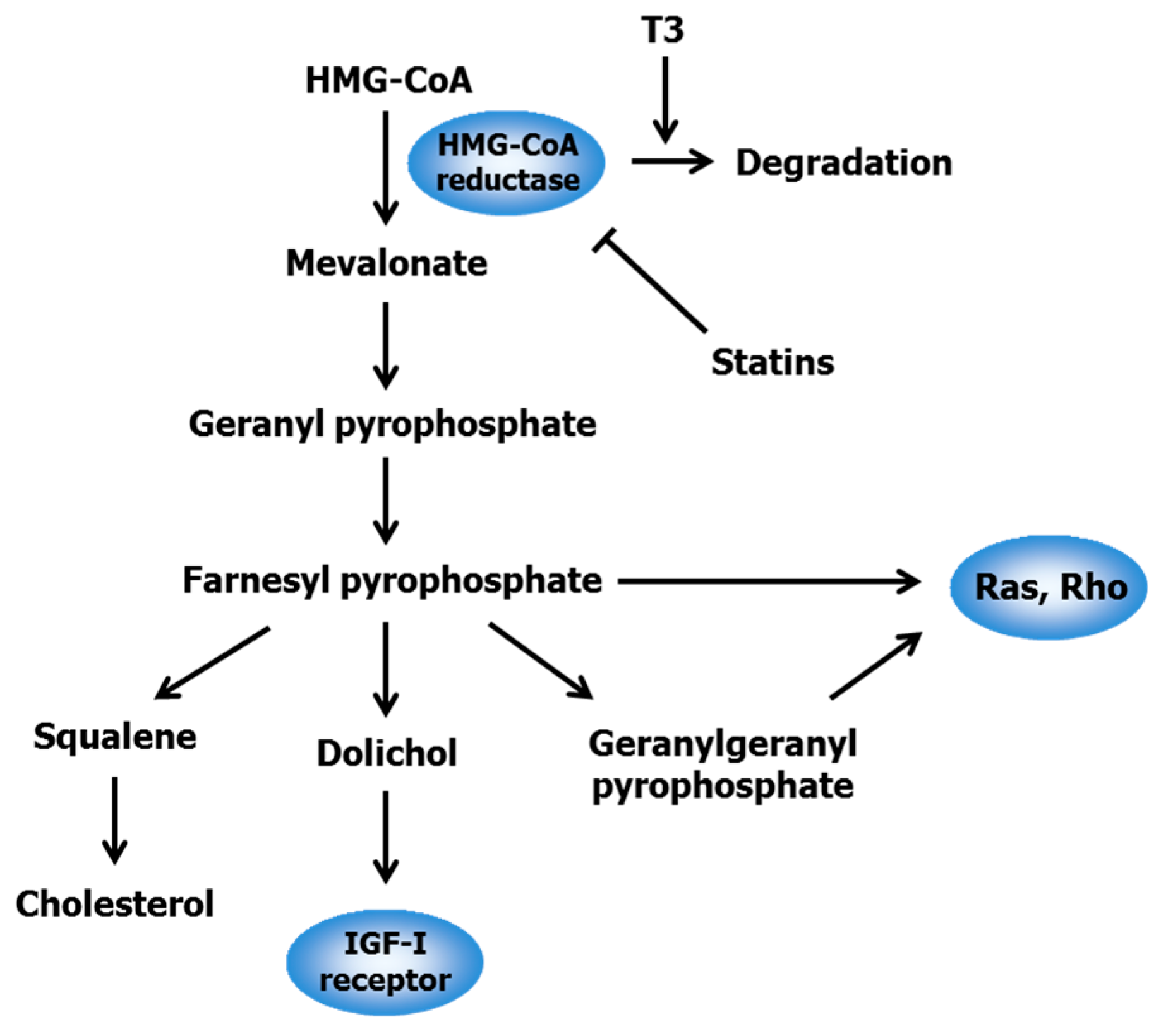
2.1. Statins

Statins, a class of drugs including lovastatin, simvastatin, mevastatin, and atorvastatin, can lower high blood cholesterol levels through competitive inhibition of 3-hydroxy-3-methylglutaryl-coenzyme A (HMG-CoA) reductase, the rate-limiting enzyme of the mevalonate pathway for cholesterol synthesis [32]. This pathway provides various isoprenoid intermediates including farnesyl pyrophosphate, geranylgeranyl pyrophosphate, and dolichol, all of which play an important role in cell survival and growth (Figure 2). Farnesylation and geranylgeranylation of Ras- and Rho-family proteins lead to their membrane anchorage, which is essential to their activation and initiation of downstream signaling pathways [33]. Ras proteins participate in regulating cell proliferation and survival, while Rho proteins are involved in the control of cell motility and cell-cell adhesion. Dolichol is responsible for the N-linked glycosylation and translocation of insulin-like growth factor I (IGF-I) receptors to the cell surface, thereby leading to cell proliferation [34]. Hence, the mevalonate pathway contributes to posttranslational modification and maturation among Ras, Rho, and IGF-I receptor proteins that regulate cell cycles, apoptosis, and metastasis. Intriguingly, HMG-CoA activity is dysregulated and up-regulated in various tumor tissues [35]. Statins can, therefore, suppress the proliferation of several types of cancer cells by inducing G1 arrest and/or apoptosis [35], indicating that statins exhibit not only cholesterol-lowering but also anticancer effects.
T3, particularly the γ- and δ-isomers, has been shown to attenuate cholesterol synthesis through posttranscriptional down-regulation of HMG-CoA reductase [36,37]. Although δ-T3 dose-dependently suppressed the proliferation of MIA PaCa-2, BxPC-3, and PANC-1 cells, supplementation with mevalonate, the product of HMG-CoA reductase, diminished the δ-T3-mediated growth inhibition of these cells [38]. Given that statins and T3 attenuate HMG-CoA reductase activity via a different mechanism of action, co-treatment with these agents is thought to exert additive or synergistic inhibitory effects on tumor growth. Mo and Elson [22] discovered for the first time that co-treatment with γ-T3 and lovastatin resulted in a synergistic decrease in the growth of DU145 and LNCaP human prostate cancer cells. McAnally et al. [39] reported that combined treatment with T3 and lovastatin synergistically suppressed the proliferation of B16 (murine melanoma), DU145 (human prostate carcinoma), and A549 (human lung carcinoma) cells. Furthermore, coadministration of T3 and lovastatin synergistically suppressed the growth of B16 cells implanted in C57BL6 mice. Wali and Sylvester [40] revealed that a single treatment with 3–4 μM γ-T3 or 2–8 μM statins (i.e., simvastatin, lovastatin, or mevastatin) significantly decreased the growth of neoplastic +SA mouse mammary epithelial cells, whereas for combinations of 0.25–2 μM γ-T3 and 0.25 μM each statin exhibited a synergistic inhibitory effect via suppression of mitogen-activated protein kinase (MAPK), c-Jun N-terminal kinase JNK, p38, and protein kinase B (Akt). Moreover, the growth-inhibitory effect of γ-T3 on HT29 and HCT116 colon cancer cells was enhanced by atorvastatin through cell cycle arrest at G1 phase and apoptosis [41], and co-treatment with δ-T3 and lovastatin caused a synergistic decrease in the growth of A2058 melanoma cells [42].

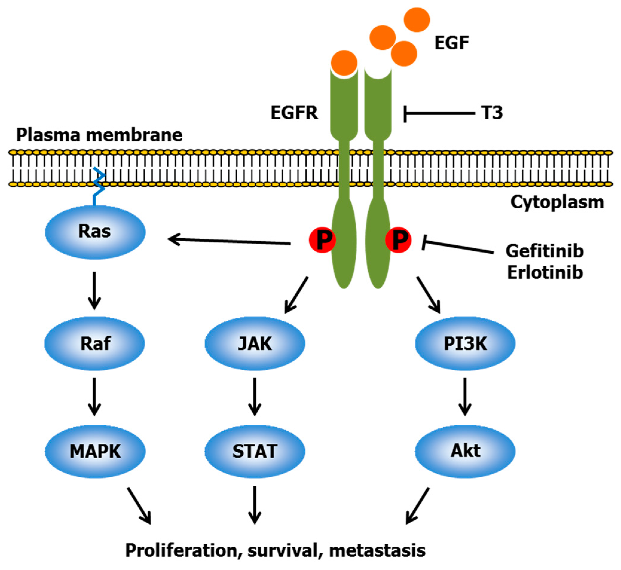
2.2. Gefitinib and Erlotinib
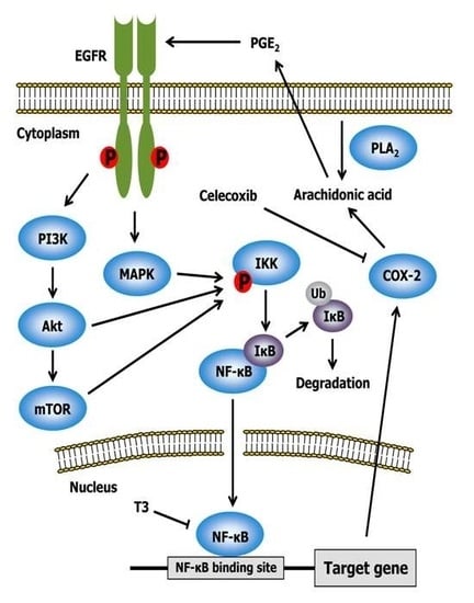
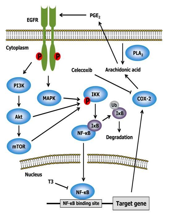
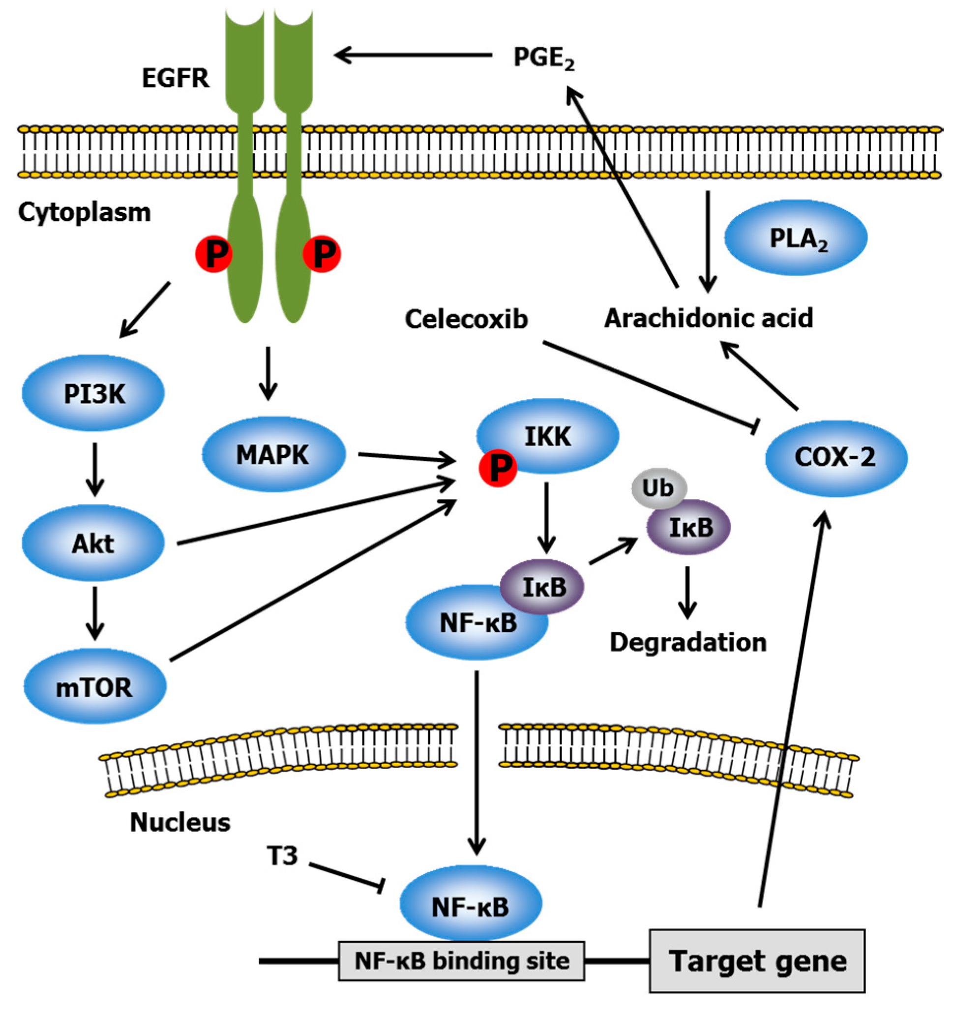
Gefitinib and erlotinib are well-known epidermal growth factor receptor (EGFR) tyrosine kinase inhibitors [43]. EGFR, a plasma membrane glycoprotein, is a member of the ErbB receptor family, and is composed of an extracellular ligand-binding domain, a lipophilic transmembrane domain, and an intracellular cytoplasmic domain with tyrosine kinase [44]. EGFR is activated by some ligands including epidermal growth factor (EGF), transforming growth factor-α, heparin-binding EGF, and amphiregulin. Ligand binding to the extracellular domain of EGFR induces a conformational change in the intracellular cytoplasmic domain, which promotes homodimerization or heterodimerization with the other ErbB family members, leading to the autophosphorylation of tyrosine residues and phosphorylation and the activation of downstream signaling pathways (Figure 3) [45]. These pathways include Ras/Raf/MAPK, phospholipase C/phosphatidylinositol 3-kinase (PI3K)/Akt, and Janus kinase/signal transducer and activator of transcription (JAK/STAT) [46]. The MAPK pathway is implicated in cell proliferation and survival. The PI3K/Akt pathway is involved in cell proliferation and migration. The JAK/STAT pathway participates in the transcription of genes involved in oncogenesis. Abnormal activation and overexpression of EGFR occur in various types of tumor cells, resulting in the enhancement of cell growth, survival and metastasis [47]. Gefitinib and erlotinib can competitively inhibit the binding of adenosine triphosphate to EGFR kinase, which causes the inhibition of autophosphorylation and downstream pathways [43]. Therefore, both drugs exert potent antitumor activity, and have been approved for first-line treatment in patients with lung adenocarcinoma with mutated EGFR.
T3 down-regulates the ErbB family receptors and downstream pathways. Samant and Sylvester [48] showed that γ-T3 dose-dependently inhibited +SA cell growth, and revealed that its inhibitory effect was involved in the suppression of the PI3K/Akt pathway through a significant decrease in ErbB3 receptor tyrosine phosphorylation. T3, but not Toc, attenuated PI3K/Akt and MAPK pathways via downregulation of ErbB2 expression, thereby leading to the induction of apoptosis in pancreatic cancer cells [49]. Pierpaoli et al. [50] examined the effect of dietary supplementation with T3 extracts from annatto seeds (10% γ- and 90% δ-T3 mixture) on the spontaneous development of mammary tumors in ErbB2 transgenic mice. Oral administration of annatto-T3 diminished the size of mammary tumors, and induced apoptosis and senescence-like growth arrest of tumor cells. In a cell culture experiment, the suppression of breast cancer cell growth, increased apoptosis and senescence molecular markers (p53, p21, p16, and p27), and a decreased expression of ErbB2 was observed in the cells treated with annatto-T3. Alawin et al. [51] revealed that γ-T3 interfered with the dimerization and phosphorylation of ErbB2 via its accumulation in the lipid raft microdomain, resulting in the inhibition of the proliferation in SKBR3 and BT474 human breast cancer cells.
There are limited reports that have investigated whether EGFR inhibitors (i.e., gefitinib and erlotinib) can synergize with T3. Treatment with 3.5 μM γ-T3, 0.5 μM erlotinib or 1.0 μM gefitinib significantly repressed the proliferation of +SA cells. The combination of 0.5–3 μM γ-T3 with 0.5 μM erlotinib or 1.0 μM gefitinib synergistically inhibited cell growth and elicited apoptosis through the activation of caspase-3. These synergistic inhibitory effects were mediated by a marked reduction in ErbB2-4 levels [23]. Bachawal et al. [52] also clarified that co-treatment with γ-T3 and erlotinib/gefitinib resulted in a synergistic decrease in +SA cell growth through down-regulation of ErbB receptors and downstream signaling of Akt and STAT.

2.3. Celecoxib

Celecoxib, a non-steroidal anti-inflammatory drug, is a specific inhibitor of cyclooxygenase-2 (COX-2) [53]. Aberrant activation of COX-2 is observed in gastric, hepatocellular, esophageal, pancreatic, colorectal, breast, bladder, cervical, endometrial, skin, and lung cancers, and is involved in promoting cell survival, angiogenesis, and metastasis [54]. Therefore, the elucidation of downstream signaling of COX-2 is important for understanding cancer progression. COX-2 metabolizes arachidonic acid to prostaglandin H2, which can be isomerized to prostaglandin E2 (PGE2) by PGE synthase. Interestingly, PGE2 activates EGFR via a Src-dependent mechanism [55]. This finding indicates that COX-2 inhibitors have cancer-fighting activity, since EGFR is recognized as a target for cancer therapy as described above. It is well established that EGFR phosphorylation induces nuclear factor-κB (NF-κB) activation (Figure 4) [56]. NF-κB is one of the major pro-inflammatory transcriptional factors. In unstimulated cells, inhibitors of κB (IκB) bind to NF-κB, resulting in its cytosolic location in an inactive form. In response to cell stimulation, IκB proteins are phosphorylated by IκB kinase (IKK), leading to IκB ubiquitination and degradation, thereby allowing active NF-κB to translocate to the nucleus. Various kinases, such as MAPK, Akt and mammalian target of rapamycin (mTOR), can phosphorylate IKK, which eventually causes NF-κB activation [57]. NF-κB target genes participate in the regulation of inflammation (e.g., COX-2, tumor necrosis factor-α, and interleukin-1), cell proliferation (e.g., Cyclin D1, Cyclin E, and c-Myc), antiapoptosis (e.g., Bcl-xL, inhibitor of apoptosis protein, and tumor necrosis factor receptor-associated factor), metastasis (e.g., matrix metalloproteinase and urokinase plasminogen activator), and angiogenesis (e.g., interleukin-1 and vascular endothelial growth factor) [58]. The findings noted above indicate that celecoxib can inhibit NF-κB as well as COX-2. In addition, celecoxib prevents NF-κB activation via the inhibition of IKK and Akt, resulting in the repression of COX-2 synthesis and various genes required for inflammation and proliferation [59].
The effects of T3 on NF-κB have been extensively studied in several types of tumor cells such as mammary epithelial [60], myeloma [61], prostate [62], colon [63], melanoma [64], breast [65], pancreatic [66], lung [67], gastric [68], and oral cancer cells [69]. Ahn et al. [61] found that γ-T3 but not γ-Toc decreased NF-κB activation through the repression of the phosphorylation and degradation of IκB, inhibition of IKK activation by blocking activation of transforming growth factor-β-activated kinase 1, and attenuation of the phosphorylation and nuclear translocation of p65, an NF-κB family member. Its inhibitory action was correlated with the down-regulation of NF-κB target gene expression related to proliferation, antiapoptosis, invasion, and angiogenesis. Ji et al. [67] showed that treatment of lung cancer cells with δ-T3 caused a reduction in NF-κB-DNA binding activity and down-regulation of NF-κB-dependent gene expression including genes coding for surviving, matrix metalloproteinase-9, vascular endothelial growth factor, and Bcl-xL. Wang et al. [70] revealed that γ-T3 inhibited NF-κB activation via induction of A20 and/or Cezanne, both of which act as strong inhibitors of NF-κB activity.
A synergistic effect between T3 and celecoxib was discovered by Shirode and Sylvester. Although 3–4 μM γ-T3 or 7.5–10 μM celecoxib alone significantly inhibited +SA cell growth in a dose-dependent manner, co-treatment with 0.25 μM γ-T3 and 2.5 μM celecoxib had synergistic action [24]. The anti-proliferative effect was mediated by a decrease in PGE2 synthesis and reduced levels of COX-2, Akt, and NF-κB. They also clarified that the synergy between γ-T3 and celecoxib was due to the suppression of ErbB2-4 receptor levels and following reduction of downstream Akt and NF-κB signaling [71].

2.4. Other Drugs

Mesenchymal epithelial transition factor (Met), also known as hepatocyte growth factor (HGF) receptor, is the cell surface receptor for HGF, which possesses tyrosine kinase activity [72]. The binding of HGF induces homodimerization of the Met receptor and phosphorylation of a tyrosine residue within the catalytic site, leading to the activation of downstream signaling pathways, including MAPK, Akt, and STAT [73]. Met is known to interact with EGFR, thereby inducing a diverse series of signaling cascades [74]. Several types of tumor cells exhibit sustained Met stimulation, overexpression, or mutation [73]. Hence, Met plays a key role in cancer cell growth and survival. Ayoub et al. revealed that γ-T3 repressed HGF-induced Met tyrosine kinase activation and signaling in breast cancer cells (+SA, MCF-7, and MDA-MB-231) [25,75]. A combination of γ-T3 and SU11274 (a Met inhibitor) synergistically suppressed cell proliferation through the reduction in Akt, STAT, and NF-κB activation.
The peroxisome proliferator-activated receptor γ (PPARγ) belongs to the nuclear hormone receptor superfamily, which acts as a transcription factor after it heterodimerizes with the retinoid X receptor [76]. PPARγ can be activated by ligands such as 15-deoxy-Δ12,14-prostaglandin J2 and some unsaturated fatty acids [77]. The receptor is present in various tissues and cell types throughout the body, including monocytes, macrophages, adipocytes, liver, skeletal muscle, breast, prostate, colon, as well as cancer cells [77]. Most of the target genes of PPARγ participate in the control of lipid metabolism and transport. In addition, PPARγ can attenuate the expression of pro-inflammatory transcription factors such as NF-κB and activator protein-1 (AP-1) [78]. Synthetic PPARγ ligands inhibit an array of cancer cells in vitro and in vivo [77], suggesting an important role of PPARγ as a tumor suppressor; however, the precise mechanism is still unclear. Campbell et al. reported that T3, especially γ- and δ-isomers, repressed the proliferation of prostate cancer cells (PC-3 and LNCaP) more effectively compared with Toc, while PPARγ knockdown diminished the anti-proliferative effect of T3 [79]. T3 induced 15-lipoxygenase-2 that is responsible for the production of 15-S-hydroxyeicosatrienoic acid, a PPARγ-activating ligand. These results suggested that T3 inhibited prostate cancer cell growth through, in part, PPARγ-dependent mechanisms. Malaviya and Sylvester examined the anti-proliferative effect of γ-T3 in combination with PPARγ agonists or antagonists on breast cancer cells (MCF-7 and MDA-MB-231) [26]. Single treatment with γ-T3, PPARγ agonists (rosiglitazone or troglitazone) or antagonists (GW9662 or T0070907) resulted in a dose-dependent growth inhibition of breast cancer cells. Unexpectedly, a combination with γ-T3 and the agonists promoted cancer cell proliferation, whereas co-treatment of γ-T3 and the antagonists synergistically suppressed the cell growth. The contradictory results might be derived from the cancer cell types used in this experiment; the authors [26] employed breast cancer cells while Campbell et al. [79] used prostate cancer cells. They concluded that the synergistic effect between γ-T3 and PPARγ antagonists was mediated through PPARγ-independent mechanisms [80].
Autophagy is a cellular self-catabolic process by which dysfunctional or unnecessary cytoplasmic components are degraded by lysosomal enzymes [81]. The process is initiated by the formation of the isolation membrane (also known as the phagophore) to engulf damaged protein aggregates and intracellular organelles. The isolation membrane expands and closes to form a double-membrane vesicle (i.e., autophagosome), leading to the fusion of autophagosomes with lysosomes. Finally, the inner contents are degraded and recycled. Although autophagy maintains intracellular homeostasis, excessive autophagy disturbs normal cellular function and induces cell death [82]. The autophagic pathway includes mTOR, class I PI3K, Akt, class III PI3K, Beclin-1, Atg family member proteins, and p53 [83]. γ-T3 has been shown to induce apoptosis and autophagy in prostate and breast cancer cells [84,85]. Tiwari et al. investigated the synergistic action of γ-T3 and oridonin, an autophagy inducer [86], against +SA mammary cancer cells. Co-treatment with γ-T3 and oridonin synergistically decreased cell viability via the elevation of autophagy markers (e.g., Beclin-1 and Atg), suppression of Akt/mTOR signaling, and up-regulation of apoptotic markers (e.g., caspase-3 and Bax/Bcl-2 ratio) [27].
The aryl hydrocarbon receptor (AhR) is a ligand-activated transcription factor, which regulates cell differentiation, proliferation, immune response, and epidermal barrier function [87]. Ligand-bound AhR complexes translocate to the cell nucleus to interact with AhR nuclear translocator (ARNT), forming a functional AhR-ARNT heterodimer, resulting in the transcriptional activation of several target genes (e.g., p21 [88] and Bax [89]) by binding to the xenobiotic responsive element. In B16F10 mouse melanoma cells, stable knockdown of AhR enhanced tumorigenesis and metastatic potential to the lungs, while constitutive AhR activation potently inhibited melanoma progression [90]. Baicalein, one of flavones, is a component of the traditional herbal remedy known as Chinese skullcap (Scutellaria baicalensis). Baicalein can act as a ligand of AhR, and exhibits anticancer action through, in part, AhR activation [91]. Yamashita et al. found that γ-T3 dose-dependently induced the expression of AhR in B16 mouse melanoma cells [28]. Thus, combination treatment with γ-T3 and baicalein synergistically inhibited the cell growth via induction of p21 and Bax expressions.

3. Synergistic Anti-Proliferative Effects of T3 and Dietary Components

3.1. Epigallocatechin Gallate (EGCG) and Resveratrol
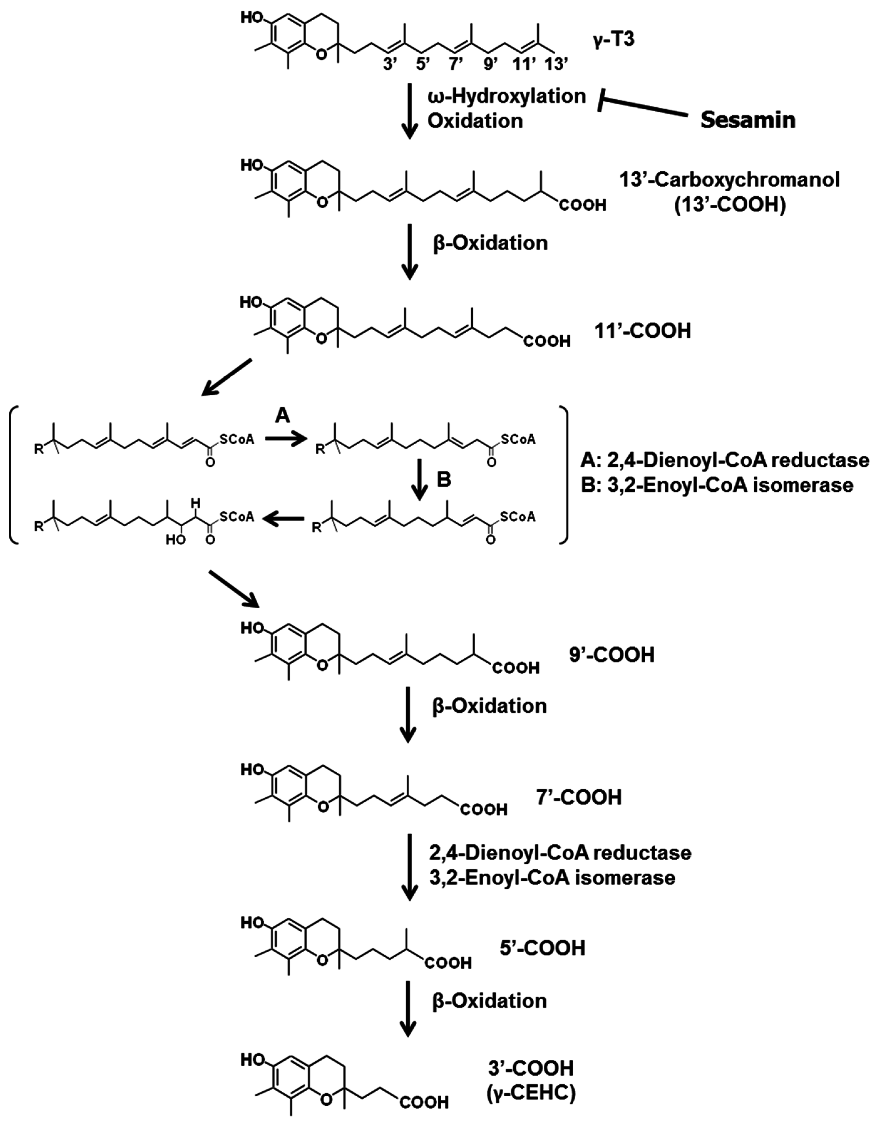
EGCG and resveratrol are well-known dietary polyphenols. EGCG is present in tea, grapes, and certain seeds of leguminous plants [92], while resveratrol is naturally found in grapes, peanuts, berries, and red wine [93]. Both polyphenols have diverse health-promoting effects, particularly anticancer action [94,95], similar to T3. The molecular mechanisms of the cancer-fighting properties of these three dietary compounds overlap each other. For example, EGFR is one of the critical targets of EGCG in the repression of cancer proliferation [96]. EGCG attenuates the activation of ErbB family receptors via the inhibition of tyrosine kinase activity and EGF binding to the EGFR, leading to a down-regulation of the MAPK and Akt signaling pathways. EGCG also down-regulates NF-κB and AP-1, thereby modulating the expression of target genes involved in apoptosis and cell cycle regulation. Similarly, resveratrol can suppress EGFR activation [97]. In addition, resveratrol down-regulates the mRNA expression of HMG-CoA reductase [98], and combined treatment with resveratrol and simvastatin synergistically reduces cell proliferation [99].
Hsieh and Mu [29] tested the synergistic anti-proliferative effect of EGCG, resveratrol, and γ-T3 on breast cancer cells. Treatment with resveratrol or γ-T3 alone (10 μM each) significantly inhibited cell growth, but treatment with EGCG did not show any effect. Resveratrol potentiated γ-T3-induced inhibition of cell proliferation. However, effective suppression of the cell growth was not observed when the three phytochemicals were concomitantly used. Co-treatment with γ-T3 with either EGCG or resveratrol caused a significant additive effect in reducing cyclin D1 and Bcl-2 expression. The triple combination of EGCG, resveratrol and γ-T3 synergistically induced NAD(P)H quinone dehydrogenase 1 (NQO1). NQO1 is involved in phase II detoxification, and its expression is regulated by nuclear factor erythroid 2–related factor 2 (Nrf2) [100]. Nrf2, a transcriptional factor, is maintained in an inactive form in the cytoplasm by binding to Kelch-like ECH-associated protein 1 (Keap1). During redox imbalance, Nrf2 dissociates from Keap1, resulting in the translocation of Nrf2 to the nucleus and heterodimerization with Maf to bind to antioxidant response elements in various promoter regions, which increases the transcription of a variety of cytoprotective genes (e.g., glutathione S-transferases, heme oxygenase-1, and NQO1) [101]. Hence, Nrf2 acts as a central regulator of the adaptive response to oxidative stress. EGCG, resveratrol, and γ-T3 can individually up-regulate Nrf2 [102,103,104]. However, it is unclear how the three phytochemicals synergistically increase NQO1 expression [29].

3.2. Sesamin

Sesamin, one of the major lignans in sesame seed and flax, possesses various beneficial functions, including anti-oxidative [105], lipid-lowering (arachidonic acid [106] and cholesterol [107] levels), anti-hypertensive [108], neuroprotective [109], anti-tumor [110], and anti-inflammatory [111] actions. Sesamin also plays a unique role in improving the bioavailability of T3 in vitro [112] and in vivo [113].
All vitamin E isoforms are catabolized through the oxidative degradation of their side chains to form water-soluble metabolites known as carboxyethyl hydroxychromans (CEHCs). The degradation begins with cytochrome P450 (CYP)-catalyzed ω-hydroxylation. In humans, CYP4F2 [114] has been reported to mediate this metabolic process, and CYP3A [115] might also be involved. These CYPs catalyze the hydroxylation of one of the terminal methyl groups on the hydrophobic side chain. The ω-hydroxylated Toc and T3 are then oxidized to the corresponding carboxylic acid, followed by five cycles of β-oxidation to eliminate a two-carbon moiety from the side chain at each cycle, ultimately leading to the formation of CEHCs (Figure 5) [116,117]. Most CEHCs are excreted in the urine [17]. Some CYP inhibitors can markedly elevate the levels of vitamin E and lower the levels of vitamin E metabolites in various tissues. Ketoconazole is a common inhibitor of CYP3A and 4F. Coadministration of ketoconazole and vitamin E (Toc and T3) decreased urinary excretion of CEHC and increased Toc and T3 concentrations in the serum and various tissues of rats [118]. In addition, sesamin strongly inhibits CYP4F2 activity, thereby elevating vitamin E concentrations in rat and human liver microsomes [114]. Thus, the bioavailability of Toc and T3 is improved by the addition of sesamin.
Akl et al. [30] investigated the synergistic inhibition of +SA mammary cancer cell proliferation with a combination of γ-T3 and sesamin. Treatment with 3.5–5 μM γ-T3 or 60–120 μM sesamin alone caused a significant inhibition of the cell growth. Combined treatment with 1–5 μM γ-T3 and 20 μM sesamin resulted in a synergistic suppression of +SA proliferation, indicating that an increase in T3 bioavailability by sesamin caused an enhancement of its anticancer activity. Co-treatment of γ-T3 and sesamin markedly prevented the activation of the ErbB receptor and its downstream signaling molecules (i.e., Raf, PI3K, Akt, NF-κB, JAK, and STAT), indicating that the synergistic action was associated with the EGF-dependent pathway. Akl et al. [119] also revealed that the combination of γ-T3 and sesamin synergistically repressed the proliferation of breast cancer cells (+SA, MCF-7, and MDA-MB-231) via the induction of G1 cell cycle arrest; however, this had no effects on normal epithelial cell growth. The combined treatment efficiently influenced cell cycle regulators of the G1/S phase transition; cyclin D1, cyclin-dependent kinase (CDK) 2, CDK4, CDK6, phospho-retinoblastoma, and E2F1 levels were reduced, and p27 and p16 levels were increased.

3.3. Ferulic Acid

The bran fraction of rice contains a variety of bioactive components with chemopreventive activity, including T3, ferulic acid, γ-oryzanol, β-sitosterol, and squalene [120]. Numerous studies have been performed to investigate the anticancer properties of dietary rice bran. Tantamango et al. [121] reported that the consumption of certain foods was associated with the decreased risk of development of polyps in a prospective study. In addition to the protective effect of green vegetables, dried fruit, and legumes, this study found that consumption of brown rice had the strongest correlation with a reduced risk of polyp formation.
We showed that T3 can act as an effective anti-tumor compound (i.e., antiangiogenesis [122] and telomerase inhibition [123]), and demonstrated that δ-T3 exhibits the most potent anti-cancer property among the four T3 isomers. Ferulic acid is receiving attention owing to its wide range of therapeutic effects against cancer [124], diabetes [125], cardiovascular diseases [126], and neurodegenerative disorders [127]. Although rice bran has a broad spectrum of beneficial activity for human health, little is known about the synergistic effect of rice bran components on cancer cell proliferation to date. We, therefore, investigated the potential role of its components, particularly δ-T3 and ferulic acid, in synergistic growth-inhibitory activity against an array of cancer cells, such as DU-145 human prostate carcinoma, MCF-7 human breast adenocarcinoma, and PANC-1 human pancreatic carcinoma cells [31].
δ-T3 dose-dependently inhibited the proliferation of these three cell lines, while ferulic acid exhibited no growth inhibition even at a concentration of 50 μM. Combined treatment with 10–12.5 μM δ-T3 and 5–20 μM ferulic acid markedly reduced cell growth compared with treatment with δ-T3 alone, although 20 μM ferulic acid had no inhibitory effect at all. Ferulic acid enhanced δ-T3-induced G1 phase arrest through the up-regulation of p21, a negative regulator of G1 progression.
To elucidate the reason for the synergistic inhibition of cell proliferation by co-treatment with δ-T3 and ferulic acid, the intracellular content of δ-T3 was analyzed by high performance liquid chromatography (HPLC). Co-treatment with δ-T3 and ferulic acid increased the cellular concentration of δ-T3 in PANC-1 cells compared to treatment with δ-T3 alone. The increment of cellular δ-T3 levels in PANC-1 implies that ferulic acid either suppresses the intracellular metabolism of δ-T3 or facilitates cellular uptake of δ-T3 from the cell culture medium. To address the latter possibility, we evaluated δ-T3 concentrations in a culture medium using HPLC, and found that ferulic acid treatment did not influence the amount of δ-T3 in the culture medium [31]. It seems, therefore, unlikely that ferulic acid affects cellular δ-T3 incorporation. These findings suggest that sesamin and ferulic acid may synergize with T3 through a similar mode of action (i.e., increasing the concentration of T3). Zhao et al. [128] investigated the bioavailability of ferulic acid in rats administered 70 μmol/kg of ferulic acid, and revealed that the plasma concentration of ferulic acid was 25.3 ± 10.1 μM at 5 min after administration. These observations indicate that physiological concentrations of ferulic acid can potentiate the growth-inhibitory effects of δ-T3 on several types of cancer cells, and suggest that ferulic acid may be a promising candidate for augmenting the anti-cancer activity of δ-T3.
Moreover, we hypothesized that combined treatment with δ-T3 and ferulic acid would not only inhibit cancer cell growth but would also enhance various physiological activities of δ-T3. We additionally investigated whether the combination of δ-T3 and ferulic acid synergistically down-regulated telomerase activity in DLD-1 colorectal cancer cells [129]. As expected, co-treatment with δ-T3 and ferulic acid resulted in a synergistic reduction of cellular telomerase activity via a decreased expression of human telomerase reverse transcriptase, the catalytic subunit of telomerase. Taken together, these results indicate that ferulic acid improves the bioavailability of T3, thereby synergistically suppressing cancer cell proliferation [31] and cellular telomerase activity [129].

4. Conclusions

Toc is widely present in various foods; however, T3-containing foods are limited. The daily intake of T3 in a Japanese population was estimated at 1.86–2.15 mg/day/person, which appeared relatively low compared with that of Toc (9 mg/day/person) [130]. Little is known about the influence of Toc on the anticancer effect of T3. In an attempt to clarify whether Toc affects the anti-proliferative activity of T3, DLD-1 cells were treated with both Toc isomers and δ-T3. All Toc isomers, especially α-Toc, diminished δ-T3-induced cytotoxicity to DLD-1 cells [20]. Co-treatment with α-Toc dose-dependently decreased δ-T3 uptake into the cells. These results indicate that α-Toc is not only less cytotoxic to cancer cells, but also reduces the cytotoxicity of δ-T3 by interfering with its cellular uptake. Findings of this in vitro study are supported by other in vivo work that found that α-T3 concentrations in various tissues and plasma decreased by the addition of dietary α-Toc, in the rats fed both α-T3 and α-Toc [21]. These findings raise concerns about the chemopreventive activity of T3 in vivo, since α-Toc is ubiquitously present as the dominant vitamin E isomer in animals. Hiura et al. [131] reported that dietary supplementation of pure T3 effectively suppressed tumor growth in a mouse xenograft model, suggesting that a high dose of T3 might overcome the inhibitory effects of endogenous α-Toc.
As we described above, chemotherapeutic drugs (i.e., HMG-CoA reductase inhibitors, EGFR tyrosine kinase inhibitors, a COX-2 inhibitor, a Met inhibitor, PPARγ antagonists, an autophagy inducer, and an AhR modulator) or dietary components (polyphenols, sesamin, and ferulic acid) potentiate the anticancer properties of T3. Combination therapy has an advantage in reducing the toxic adverse side effects of drugs associated with high-dose monotherapy. The clinical application of dietary compounds in cancer prevention is attractive, because they are non-toxic at physiological doses. Of note, a clinical trial conducted in humans resulted in no toxicities when up to 800 mg/day of δ-T3 was administered for several months [132]. Nesaretnam et al. [133] performed a pilot clinical trial to examine the effect of a T3-rich fraction (200 mg/day) and tamoxifen (20 mg/day) for 5 years in women with early breast cancer. Although the risk of dying due to breast cancer was lowered by 60% in patients treated with the combination of T3-rich fraction and tamoxifen compared to patients treated with placebo and tamoxifen, this was not statistically significant. Estrogen receptor (ER) status is a very important factor in planning breast cancer treatment. Two ERs, ERα and ERβ, are expressed in normal breast tissue, but the ratio of ERα to ERβ is elevated in breast tumors [134]. Therefore, selective ER modulators such as tamoxifen are currently used to treat breast cancer. In ERα-positive T47-D and MCF-7 human breast cancer cells, overexpression of ERβ not only attenuated Akt signaling via down-regulation of ErbB2/ErbB3 but also improved the sensitivity of these cancer cells to tamoxifen [135]. Comitato et al. [136,137] reported that T3-rich fraction from palm oil promoted ERβ translocation into nucleus, leading to the induction of apoptosis in MDA-MB-231 and MCF-7 breast cancer cells. These findings suggest that ER might be a promising target for breast cancer therapy by T3.
In this review, we summarized current research on the synergistic anti-tumor effect of T3 and various agents, and discussed their related mechanisms. Further studies, particularly animal and clinical tests, on the combination therapy of T3 and certain agents will contribute to their applications in cancer treatment and prevention.

Acknowledgments

Some of the work presented in this review was partly supported by a grant from The Tojuro Iijima Foundation for Food Science and Technology.

Author Contributions

Takahiro Eitsuka wrote the manuscript; Naoto Tatewaki, Hiroshi Nishida, Kiyotaka Nakagawa and Teruo Miyazawa revised the manuscript.

Conflicts of Interest

The authors declare no conflict of interest.

References

  1. Mustacich, D.J.; Bruno, R.S.; Traber, M.G. Vitamin E. Vitam. Horm. 2007, 76, 1–21. [Google Scholar] [PubMed]
  2. Packer, L.; Weber, S.U.; Rimbach, G. Molecular aspects of α-tocotrienol antioxidant action and cell signalling. J. Nutr. 2001, 131, 369S–373S. [Google Scholar] [PubMed]
  3. Pennock, J.F.; Hemming, F.W.; Kerr, J.D. A reassessment of tocopherol chemistry. Biochem. Biophys. Res. Commun. 1964, 17, 542–548. [Google Scholar] [CrossRef]
  4. Qureshi, A.A.; Burger, W.C.; Peterson, D.M.; Elson, C.E. The structure of an inhibitor of cholesterol biosynthesis isolated from barley. J. Biol. Chem. 1986, 261, 10544–10550. [Google Scholar] [PubMed]
  5. Sundram, K.; Khor, H.T.; Ong, A.S.; Pathmanathan, R. Effect of dietary palm oils on mammary carcinogenesis in female rats induced by 7,12-dimethylbenz(a)anthracene. Cancer Res. 1989, 49, 1447–1451. [Google Scholar] [PubMed]
  6. Nesaretnam, K.; Guthrie, N.; Chambers, A.F.; Carroll, K.K. Effect of tocotrienols on the growth of a human breast cancer cell line in culture. Lipids 1995, 30, 1139–1143. [Google Scholar] [CrossRef] [PubMed]
  7. Serbinova, E.; Kagan, V.; Han, D.; Packer, L. Free radical recycling and intramembrane mobility in the antioxidant properties of α-tocopherol and α-tocotrienol. Free Radic. Biol. Med. 1991, 10, 263–275. [Google Scholar] [CrossRef]
  8. Wan Nazaimoon, W.M.; Khalid, B.A. Tocotrienols-rich diet decreases advanced glycosylation end-products in non-diabetic rats and improves glycemic control in streptozotocin-induced diabetic rats. Malays. J. Pathol. 2002, 24, 77–82. [Google Scholar] [PubMed]
  9. Wu, S.J.; Liu, P.L.; Ng, L.T. Tocotrienol-rich fraction of palm oil exhibits anti-inflammatory property by suppressing the expression of inflammatory mediators in human monocytic cells. Mol. Nutr. Food Res. 2008, 52, 921–929. [Google Scholar] [CrossRef] [PubMed]
  10. Ren, Z.; Pae, M.; Dao, M.C.; Smith, D.; Meydani, S.N.; Wu, D. Dietary supplementation with tocotrienols enhances immune function in C57BL/6 mice. J. Nutr. 2010, 140, 1335–1341. [Google Scholar] [CrossRef] [PubMed]
  11. Vasanthi, H.R.; Parameswari, R.P.; Das, D.K. Multifaceted role of tocotrienols in cardioprotection supports their structure: Function relation. Genes Nutr. 2012, 7, 19–28. [Google Scholar] [CrossRef] [PubMed]
  12. Norazlina, M.; Lee, P.L.; Lukman, H.I.; Nazrun, A.S.; Ima-Nirwana, S. Effects of vitamin E supplementation on bone metabolism in nicotine-treated rats. Singap. Med. J. 2007, 48, 195–199. [Google Scholar]
  13. Khanna, S.; Roy, S.; Slivka, A.; Craft, T.K.S.; Chaki, S.; Rink, C.; Notestine, M.A.; DeVries, A.C.; Parinandi, N.L.; Sen, C.K. Neuroprotective properties of the natural vitamin E α-tocotrienol. Stroke 2005, 36, 2258–2264. [Google Scholar] [CrossRef] [PubMed]
  14. Ong, F.B.; Wan Ngah, W.Z.; Shamaan, N.A.; Md Top, A.G.; Marzuki, A.; Khalid, A.K. Glutathione S-transferase and γ-glutamyl transpeptidase activities in cultured rat hepatocytes treated with tocotrienol and tocopherol. Comp. Biochem. Physiol. C 1993, 106, 237–240. [Google Scholar] [CrossRef]
  15. Gupta, A.; Chopra, K. Effect of tocotrienols on iron-induced renal dysfunction and oxidative stress in rats. Drug Chem. Toxicol. 2009, 32, 319–325. [Google Scholar] [CrossRef] [PubMed]
  16. Suzuki, Y.J.; Tsuchiya, M.; Wassall, S.R.; Choo, Y.M.; Govil, G.; Kagan, V.E.; Packer, L. Structural and dynamic membrane properties of α-tocopherol and α-tocotrienol: Implication to the molecular mechanism of their antioxidant potency. Biochemistry 1993, 32, 10692–10699. [Google Scholar] [CrossRef] [PubMed]
  17. Traber, M.G. Vitamin E regulatory mechanisms. Annu. Rev. Nutr. 2007, 27, 347–362. [Google Scholar] [CrossRef] [PubMed]
  18. Hosomi, A.; Arita, M.; Sato, Y.; Kiyose, C.; Ueda, T.; Igarashi, O.; Arai, H.; Inoue, K. Affinity for α-tocopherol transfer protein as a determinant of the biological activities of vitamin E analogs. FEBS Lett. 1997, 409, 105–108. [Google Scholar] [CrossRef]
  19. O’Byrne, D.; Grundy, S.; Packer, L.; Devaraj, S.; Baldenius, K.; Hoppe, P.P.; Kraemer, K.; Jialal, I.; Traber, M.G. Studies of LDL oxidation following α-, γ-, or δ-tocotrienyl acetate supplementation of hypercholesterolemic humans. Free Radic. Biol. Med. 2000, 29, 834–845. [Google Scholar] [CrossRef]
  20. Shibata, A.; Nakagawa, K.; Sookwong, P.; Tsuduki, T.; Asai, A.; Miyazawa, T. α-Tocopherol attenuates the cytotoxic effect of δ-tocotrienol in human colorectal adenocarcinoma cells. Biochem. Biophys. Res. Commun. 2010, 397, 214–219. [Google Scholar] [CrossRef] [PubMed]
  21. Ikeda, S.; Tohyama, T.; Yoshimura, H.; Hamamura, K.; Abe, K.; Yamashita, K. Dietary α-tocopherol decreases α-tocotrienol but not γ-tocotrienol concentration in rats. J. Nutr. 2003, 133, 428–434. [Google Scholar] [PubMed]
  22. Mo, H.; Elson, C.E. Studies of the isoprenoid-mediated inhibition of mevalonate synthesis applied to cancer chemotherapy and chemoprevention. Exp. Biol. Med. 2004, 229, 567–585. [Google Scholar]
  23. Bachawal, S.V.; Wali, V.B.; Sylvester, P.W. Enhanced antiproliferative and apoptotic response to combined treatment of γ-tocotrienol with erlotinib or gefitinib in mammary tumor cells. BMC Cancer 2010. [Google Scholar] [CrossRef] [PubMed]
  24. Shirode, A.B.; Sylvester, P.W. Synergistic anticancer effects of combined γ-tocotrienol and celecoxib treatment are associated with suppression in Akt and NFκB signaling. Biomed. Pharmacother. 2010, 64, 327–332. [Google Scholar] [CrossRef] [PubMed]
  25. Ayoub, N.M.; Bachawal, S.V.; Sylvester, P.W. γ-Tocotrienol inhibits HGF-dependent mitogenesis and Met activation in highly malignant mammary tumour cells. Cell Prolif. 2011, 44, 516–526. [Google Scholar] [CrossRef] [PubMed]
  26. Malaviya, A.; Sylvester, P.W. Mechanisms mediating the effects of γ-tocotrienol when used in combination with PPARγ agonists or antagonists on MCF-7 and MDA-MB-231 breast cancer cells. Int. J. Breast Cancer 2013, 2013, 101705. [Google Scholar] [CrossRef] [PubMed]
  27. Tiwari, R.V.; Parajuli, P.; Sylvester, P.W. Synergistic anticancer effects of combined γ-tocotrienol and oridonin treatment is associated with the induction of autophagy. Mol. Cell. Biochem. 2015, 408, 123–137. [Google Scholar] [CrossRef] [PubMed]
  28. Yamashita, S.; Baba, K.; Makio, A.; Kumazoe, M.; Huang, Y.; Lin, I.C.; Bae, J.; Murata, M.; Yamada, S.; Tachibana, H. γ-Tocotrienol upregulates aryl hydrocarbon receptor expression and enhances the anticancer effect of baicalein. Biochem. Biophys. Res. Commun. 2016, 473, 801–807. [Google Scholar] [CrossRef] [PubMed]
  29. Hsieh, T.C.; Wu, J.M. Suppression of cell proliferation and gene expression by combinatorial synergy of EGCG, resveratrol and γ-tocotrienol in estrogen receptor-positive MCF-7 breast cancer cells. Int. J. Oncol. 2008, 33, 851–859. [Google Scholar] [PubMed]
  30. Akl, M.R.; Ayoub, N.M.; Sylvester, P.W. Mechanisms mediating the synergistic anticancer effects of combined γ-tocotrienol and sesamin treatment. Planta Med. 2012, 78, 1731–1739. [Google Scholar] [CrossRef] [PubMed]
  31. Eitsuka, T.; Tatewaki, N.; Nishida, H.; Kurata, T.; Nakagawa, K.; Miyazawa, T. Synergistic inhibition of cancer cell proliferation with a combination of δ-tocotrienol and ferulic acid. Biochem. Biophys. Res. Commun. 2014, 453, 606–611. [Google Scholar] [CrossRef] [PubMed]
  32. Goldstein, J.L.; Brown, M.S. Regulation of the mevalonate pathway. Nature 1990, 343, 425–430. [Google Scholar] [CrossRef] [PubMed]
  33. Zhang, F.L.; Casey, P.J. Protein prenylation: Molecular mechanisms and functional consequences. Annu. Rev. Biochem. 1996, 65, 241–269. [Google Scholar] [CrossRef] [PubMed]
  34. Dricu, A.; Carlberg, M.; Wang, M.; Larsson, O. Inhibition of N-linked glycosylation using tunicamycin causes cell death in malignant cells: Role of down-regulation of the insulin-like growth factor 1 receptor in induction of apoptosis. Cancer Res. 1997, 57, 543–548. [Google Scholar] [PubMed]
  35. Demierre, M.F.; Higgins, P.D.; Gruber, S.B.; Hawk, E.; Lippman, S.M. Statins and cancer prevention. Nat. Rev. Cancer 2005, 5, 930–942. [Google Scholar] [CrossRef] [PubMed]
  36. Parker, R.A.; Pearce, B.C.; Clark, R.W.; Gordon, D.A.; Wright, J.J. Tocotrienols regulate cholesterol production in mammalian cells by post-transcriptional suppression of 3-hydroxy-3-methylglutaryl-coenzyme A reductase. J. Biol. Chem. 1993, 268, 11230–11238. [Google Scholar] [PubMed]
  37. Song, B.L.; DeBose-Boyd, R.A. Insig-dependent ubiquitination and degradation of 3-hydroxy-3-methylglutaryl coenzyme a reductase stimulated by δ- and γ-tocotrienols. J. Biol. Chem. 2006, 281, 25054–25061. [Google Scholar] [CrossRef] [PubMed]
  38. Hussein, D.; Mo, H. d-δ-Tocotrienol-mediated suppression of the proliferation of human PANC-1, MIA PaCa-2, and BxPC-3 pancreatic carcinoma cells. Pancreas 2009, 38, e124–e136. [Google Scholar] [CrossRef] [PubMed]
  39. McAnally, J.A.; Gupta, J.; Sodhani, S.; Bravo, L.; Mo, H. Tocotrienols potentiate lovastatin-mediated growth suppression in vitro and in vivo. Exp. Biol. Med. 2007, 232, 523–531. [Google Scholar]
  40. Wali, V.B.; Sylvester, P.W. Synergistic antiproliferative effects of γ-tocotrienol and statin treatment on mammary tumor cells. Lipids 2007, 42, 1113–1123. [Google Scholar] [CrossRef] [PubMed]
  41. Yang, Z.; Xiao, H.; Jin, H.; Koo, P.T.; Tsang, D.J.; Yang, C.S. Synergistic actions of atorvastatin with γ-tocotrienol and celecoxib against human colon cancer HT29 and HCT116 cells. Int. J. Cancer 2010, 126, 852–863. [Google Scholar] [CrossRef] [PubMed]
  42. Fernandes, N.V.; Guntipalli, P.K.; Mo, H. d-δ-Tocotrienol-mediated cell cycle arrest and apoptosis in human melanoma cells. Anticancer Res. 2010, 30, 4937–4944. [Google Scholar] [PubMed]
  43. Yuan, Y.; Li, X.F.; Chen, J.Q.; Dong, C.X.; Weng, S.S.; Huang, J.J. Critical appraisal of the role of gefitinib in the management of locally advanced or metastatic non-small cell lung cancer. OncoTargets Ther. 2014, 7, 841–852. [Google Scholar] [CrossRef] [PubMed]
  44. Ciardiello, F.; Tortora, G. EGFR antagonists in cancer treatment. N. Engl. J. Med. 2008, 358, 1160–1174. [Google Scholar] [CrossRef] [PubMed]
  45. Hynes, N.E.; Lane, H.A. ERBB receptors and cancer: The complexity of targeted inhibitors. Nat. Rev. Cancer 2005, 5, 341–354. [Google Scholar] [CrossRef] [PubMed]
  46. Laurent-Puig, P.; Lievre, A.; Blons, H. Mutations and response to epidermal growth factor receptor inhibitors. Clin. Cancer Res. 2009, 15, 1133–1139. [Google Scholar] [CrossRef] [PubMed]
  47. Olayioye, M.A.; Neve, R.M.; Lane, H.A.; Hynes, N.E. The ErbB signaling network: Receptor heterodimerization in development and cancer. EMBO J. 2000, 19, 3159–3167. [Google Scholar] [CrossRef] [PubMed]
  48. Samant, G.V.; Sylvester, P.W. γ-Tocotrienol inhibits ErbB3-dependent PI3K/Akt mitogenic signalling in neoplastic mammary epithelial cells. Cell Prolif. 2006, 39, 563–574. [Google Scholar] [CrossRef] [PubMed]
  49. Shin-Kang, S.; Ramsauer, V.P.; Lightner, J.; Chakraborty, K.; Stone, W.; Campbell, S.; Reddy, S.A.; Krishnan, K. Tocotrienols inhibit AKT and ERK activation and suppress pancreatic cancer cell proliferation by suppressing the ErbB2 pathway. Free Radic. Biol. Med. 2011, 51, 1164–1174. [Google Scholar] [CrossRef] [PubMed]
  50. Pierpaoli, E.; Viola, V.; Barucca, A.; Orlando, F.; Galli, F.; Provinciali, M. Effect of annatto-tocotrienols supplementation on the development of mammary tumors in HER-2/neu transgenic mice. Carcinogenesis 2013, 34, 1352–1360. [Google Scholar] [CrossRef] [PubMed]
  51. Alawin, O.A.; Ahmed, R.A.; Ibrahim, B.A.; Briski, K.P.; Sylvester, P.W. Antiproliferative effects of γ-tocotrienol are associated with lipid raft disruption in HER2-positive human breast cancer cells. J. Nutr. Biochem. 2016, 27, 266–277. [Google Scholar] [CrossRef] [PubMed]
  52. Bachawal, S.V.; Wali, V.B.; Sylvester, P.W. Combined γ-tocotrienol and erlotinib/gefitinib treatment suppresses Stat and Akt signaling in murine mammary tumor cells. Anticancer Res. 2010, 30, 429–437. [Google Scholar] [PubMed]
  53. Thun, M.J.; Henley, S.J.; Patrono, C. Nonsteroidal anti-inflammatory drugs as anticancer agents: Mechanistic, pharmacologic, and clinical issues. J. Natl. Cancer Inst. 2002, 94, 252–266. [Google Scholar] [CrossRef] [PubMed]
  54. Brown, J.R.; DuBois, R.N. Cyclooxygenase as a target in lung cancer. Clin. Cancer Res. 2004, 10, 4266s–4269s. [Google Scholar] [CrossRef] [PubMed]
  55. Pai, R.; Soreghan, B.; Szabo, I.L.; Pavelka, M.; Baatar, D.; Tarnawski, A.S. Prostaglandin E2 transactivates EGF receptor: A novel mechanism for promoting colon cancer growth and gastrointestinal hypertrophy. Nat. Med. 2002, 8, 289–293. [Google Scholar] [CrossRef] [PubMed]
  56. Shostak, K.; Chariot, A. EGFR and NF-κB: Partners in cancer. Trends Mol. Med. 2015, 21, 385–393. [Google Scholar] [CrossRef] [PubMed]
  57. Ozes, O.N.; Mayo, L.D.; Gustin, J.A.; Pfeffer, S.R.; Pfeffer, L.M.; Donner, D.B. NF-κB activation by tumour necrosis factor requires the Akt serine-threonine kinase. Nature 1999, 401, 82–85. [Google Scholar] [CrossRef] [PubMed]
  58. Gupta, S.C.; Sundaram, C.; Reuter, S.; Aggarwal, B.B. Inhibiting NF-κB activation by small molecules as a therapeutic strategy. Biochim. Biophys. Acta 2010, 1799, 775–787. [Google Scholar] [CrossRef] [PubMed]
  59. Shishodia, S.; Koul, D.; Aggarwal, B.B. Cyclooxygenase (COX)-2 inhibitor celecoxib abrogates TNF-induced NF-κB activation through inhibition of activation of IκB α kinase and Akt in human non-small cell lung carcinoma: Correlation with suppression of COX-2 synthesis. J. Immunol. 2004, 173, 2011–2022. [Google Scholar] [CrossRef] [PubMed]
  60. Shah, S.J.; Sylvester, P.W. Γ-tocotrienol inhibits neoplastic mammary epithelial cell proliferation by decreasing Akt and nuclear factor κB activity. Exp. Biol. Med. 2005, 230, 235–241. [Google Scholar]
  61. Ahn, K.S.; Sethi, G.; Krishnan, K.; Aggarwal, B.B. Γ-tocotrienol inhibits nuclear factor-κB signaling pathway through inhibition of receptor-interacting protein and TAK1 leading to suppression of antiapoptotic gene products and potentiation of apoptosis. J. Biol. Chem. 2007, 282, 809–820. [Google Scholar] [CrossRef] [PubMed]
  62. Yap, W.N.; Chang, P.N.; Han, H.Y.; Lee, D.T.; Ling, M.T.; Wong, Y.C.; Yap, Y.L. Γ-tocotrienol suppresses prostate cancer cell proliferation and invasion through multiple-signalling pathways. Br. J. Cancer 2008, 99, 1832–1841. [Google Scholar] [CrossRef] [PubMed]
  63. Xu, W.L.; Liu, J.R.; Liu, H.K.; Qi, G.Y.; Sun, X.R.; Sun, W.G.; Chen, B.Q. Inhibition of proliferation and induction of apoptosis by γ-tocotrienol in human colon carcinoma HT-29 cells. Nutrition 2009, 25, 555–566. [Google Scholar] [CrossRef] [PubMed]
  64. Chang, P.N.; Yap, W.N.; Lee, D.T.; Ling, M.T.; Wong, Y.C.; Yap, Y.L. Evidence of γ-tocotrienol as an apoptosis-inducing, invasion-suppressing, and chemotherapy drug-sensitizing agent in human melanoma cells. Nutr. Cancer 2009, 61, 357–366. [Google Scholar] [CrossRef] [PubMed]
  65. Yap, W.N.; Zaiden, N.; Tan, Y.L.; Ngoh, C.P.; Zhang, X.W.; Wong, Y.C.; Ling, M.T.; Yap, Y.L. Id1, inhibitor of differentiation, is a key protein mediating anti-tumor responses of γ-tocotrienol in breast cancer cells. Cancer Lett. 2010, 291, 187–199. [Google Scholar] [CrossRef] [PubMed]
  66. Kunnumakkara, A.B.; Sung, B.; Ravindran, J.; Diagaradjane, P.; Deorukhkar, A.; Dey, S.; Koca, C.; Yadav, V.R.; Tong, Z.; Gelovani, J.G.; et al. Γ-tocotrienol inhibits pancreatic tumors and sensitizes them to gemcitabine treatment by modulating the inflammatory microenvironment. Cancer Res. 2010, 70, 8695–8705. [Google Scholar] [CrossRef] [PubMed]
  67. Ji, X.; Wang, Z.; Geamanu, A.; Sarkar, F.H.; Gupta, S.V. Inhibition of cell growth and induction of apoptosis in non-small cell lung cancer cells by δ-tocotrienol is associated with notch-1 down-regulation. J. Cell. Biochem. 2011, 112, 2773–2783. [Google Scholar] [CrossRef] [PubMed]
  68. Manu, K.A.; Shanmugam, M.K.; Ramachandran, L.; Li, F.; Fong, C.W.; Kumar, A.P.; Tan, P.; Sethi, G. First evidence that γ-tocotrienol inhibits the growth of human gastric cancer and chemosensitizes it to capecitabine in a xenograft mouse model through the modulation of NF-κB pathway. Clin. Cancer Res. 2012, 18, 2220–2229. [Google Scholar] [CrossRef] [PubMed]
  69. Kani, K.; Momota, Y.; Harada, M.; Yamamura, Y.; Aota, K.; Yamanoi, T.; Takano, H.; Motegi, K.; Azuma, M. γ-Tocotrienol enhances the chemosensitivity of human oral cancer cells to docetaxel through the downregulation of the expression of NF-κB-regulated anti-apoptotic gene products. Int. J. Oncol. 2013, 42, 75–82. [Google Scholar] [PubMed]
  70. Wang, Y.; Park, N.Y.; Jang, Y.; Ma, A.; Jiang, Q. Vitamin E γ-tocotrienol inhibits cytokine-stimulated NF-κB activation by induction of anti-inflammatory A20 via stress adaptive response due to modulation of sphingolipids. J. Immunol. 2015, 195, 126–133. [Google Scholar] [CrossRef] [PubMed]
  71. Shirode, A.B.; Sylvester, P.W. Mechanisms mediating the synergistic anticancer effects of combined γ-tocotrienol and celecoxib treatment. J. Bioanal. Biomed. 2011, 3, 1–7. [Google Scholar] [CrossRef] [PubMed]
  72. Bottaro, D.P.; Rubin, J.S.; Faletto, D.L.; Chan, A.M.; Kmiecik, T.E.; Vande Woude, G.F.; Aaronson, S.A. Identification of the hepatocyte growth factor receptor as the c-Met proto-oncogene product. Science 1991, 251, 802–804. [Google Scholar] [CrossRef] [PubMed]
  73. Eder, J.P.; Vande Woude, G.F.; Boerner, S.A.; LoRusso, P.M. Novel therapeutic inhibitors of the c-Met signaling pathway in cancer. Clin. Cancer Res. 2009, 15, 2207–2214. [Google Scholar] [CrossRef] [PubMed]
  74. Guo, A.; Villén, J.; Kornhauser, J.; Lee, K.A.; Stokes, M.P.; Rikova, K.; Possemato, A.; Nardone, J.; Innocenti, G.; Wetzel, R.; et al. Signaling networks assembled by oncogenic EGFR and c-Met. Proc. Natl. Acad. Sci. USA 2008, 105, 692–697. [Google Scholar] [CrossRef] [PubMed]
  75. Ayoub, N.M.; Akl, M.R.; Sylvester, P.W. Combined γ-tocotrienol and Met inhibitor treatment suppresses mammary cancer cell proliferation, epithelial-to-mesenchymal transition and migration. Cell Prolif. 2013, 46, 538–553. [Google Scholar] [CrossRef] [PubMed]
  76. Mangelsdorf, D.J.; Evans, R.M. The RXR heterodimers and orphan receptors. Cell 1995, 83, 841–850. [Google Scholar] [CrossRef]
  77. Koeffler, H.P. Peroxisome proliferator-activated receptor γ and cancers. Clin. Cancer Res. 2003, 9, 1–9. [Google Scholar] [PubMed]
  78. Ricote, M.; Li, A.C.; Willson, T.M.; Kelly, C.J.; Glass, C.K. The peroxisome proliferator-activated receptor-γ is a negative regulator of macrophage activation. Nature 1998, 391, 79–82. [Google Scholar] [CrossRef] [PubMed]
  79. Campbell, S.E.; Rudder, B.; Phillips, R.B.; Whaley, S.G.; Stimmel, J.B.; Leesnitzer, L.M.; Lightner, J.; Dessus-Babus, S.; Duffourc, M.; Stone, W.L.; et al. γ-Tocotrienol induces growth arrest through a novel pathway with TGFβ2 in prostate cancer. Free Radic. Biol. Med. 2011, 50, 1344–1354. [Google Scholar] [CrossRef] [PubMed]
  80. Malaviya, A.; Sylvester, P.W. Synergistic antiproliferative effects of combined γ-tocotrienol and PPARγ antagonist treatment are mediated through PPAR γ-independent mechanisms in breast cancer cells. PPAR Res. 2014, 2014, 439146. [Google Scholar] [CrossRef] [PubMed]
  81. Rabinowitz, J.D.; White, E. Autophagy and metabolism. Science 2010, 330, 1344–1348. [Google Scholar] [CrossRef] [PubMed]
  82. Lin, L.; Baehrecke, E.H. Autophagy, cell death, and cancer. Mol. Cell. Oncol. 2015, 2, e985913. [Google Scholar] [CrossRef] [PubMed]
  83. Su, Z.; Yang, Z.; Xu, Y.; Chen, Y.; Yu, Q. Apoptosis, autophagy, necroptosis, and cancer metastasis. Mol. Cancer 2015, 14, 48. [Google Scholar] [CrossRef] [PubMed]
  84. Jiang, Q.; Rao, X.; Kim, C.Y.; Freiser, H.; Zhang, Q.; Jiang, Z.; Li, G. γ-Tocotrienol induces apoptosis and autophagy in prostate cancer cells by increasing intracellular dihydrosphingosine and dihydroceramide. Int. J. Cancer 2012, 130, 685–693. [Google Scholar] [CrossRef] [PubMed]
  85. Tiwari, R.V.; Parajuli, P.; Sylvester, P.W. γ-Tocotrienol-induced autophagy in malignant mammary cancer cells. Exp. Biol. Med. 2014, 239, 33–44. [Google Scholar] [CrossRef] [PubMed]
  86. Cui, Q.; Tashiro, S.; Onodera, S.; Minami, M.; Ikejima, T. Autophagy preceded apoptosis in oridonin-treated human breast cancer MCF-7 cells. Biol. Pharm. Bull. 2007, 30, 859–864. [Google Scholar] [CrossRef] [PubMed]
  87. Safe, S.; Lee, S.O.; Jin, U.H. Role of the aryl hydrocarbon receptor in carcinogenesis and potential as a drug target. Toxicol. Sci. 2013, 135, 1–16. [Google Scholar] [CrossRef] [PubMed]
  88. Pang, P.H.; Lin, Y.H.; Lee, Y.H.; Hou, H.H.; Hsu, S.P.; Juan, S.H. Molecular mechanisms of p21 and p27 induction by 3-methylcholanthrene, an aryl-hydrocarbon receptor agonist, involved in antiproliferation of human umbilical vascular endothelial cells. J. Cell. Physiol. 2008, 215, 161–171. [Google Scholar] [CrossRef] [PubMed]
  89. Matikainen, T.; Perez, G.I.; Jurisicova, A.; Pru, J.K.; Schlezinger, J.J.; Ryu, H.Y.; Laine, J.; Sakai, T.; Korsmeyer, S.J.; Casper, R.F.; et al. Aromatic hydrocarbon receptor-driven Bax gene expression is required for premature ovarian failure caused by biohazardous environmental chemicals. Nat. Genet. 2001, 28, 355–360. [Google Scholar] [CrossRef] [PubMed]
  90. Contador-Troca, M.; Alvarez-Barrientos, A.; Barrasa, E.; Rico-Leo, E.M.; Catalina-Fernández, I.; Menacho-Márquez, M.; Bustelo, X.R.; García-Borrón, J.C.; Gómez-Durán, A.; Sáenz-Santamaría, J.; et al. The dioxin receptor has tumor suppressor activity in melanoma growth and metastasis. Carcinogenesis 2013, 34, 2683–2693. [Google Scholar] [CrossRef] [PubMed]
  91. Cheng, Y.H.; Li, L.A.; Lin, P.; Cheng, L.C.; Hung, C.H.; Chang, N.W.; Lin, C. Baicalein induces G1 arrest in oral cancer cells by enhancing the degradation of cyclin D1 and activating AhR to decrease Rb phosphorylation. Toxicol. Appl. Pharmacol. 2012, 263, 360–367. [Google Scholar] [CrossRef] [PubMed]
  92. Manach, C.; Scalbert, A.; Morand, C.; Remesy, C.; Jimenez, L. Polyphenols: Food sources and bioavailability. Am. J. Clin. Nutr. 2004, 79, 727–747. [Google Scholar] [PubMed]
  93. Burns, J.; Yokota, T.; Ashihara, H.; Lean, M.E.; Crozier, A. Plant foods and herbal sources of resveratrol. J. Agric. Food Chem. 2002, 50, 3337–3340. [Google Scholar] [CrossRef] [PubMed]
  94. Singh, B.N.; Shankar, S.; Srivastava, R.K. Green tea catechin, epigallocatechin-3-gallate (EGCG): Mechanisms, perspectives and clinical applications. Biochem. Pharmacol. 2011, 82, 1807–1821. [Google Scholar] [CrossRef] [PubMed]
  95. Singh, C.K.; Ndiaye, M.A.; Ahmad, N. Resveratrol and cancer: Challenges for clinical translation. Biochim. Biophys. Acta 2015, 1852, 1178–1185. [Google Scholar] [CrossRef] [PubMed]
  96. Shimizu, M.; Shirakami, Y.; Moriwaki, H. Targeting receptor tyrosine kinases for chemoprevention by green tea catechin, EGCG. Int. J. Mol. Sci. 2008, 9, 1034–1049. [Google Scholar] [CrossRef] [PubMed]
  97. Provinciali, M.; Re, F.; Donnini, A.; Orlando, F.; Bartozzi, B.; di Stasio, G.; Smorlesi, A. Effect of resveratrol on the development of spontaneous mammary tumors in HER-2/neu transgenic mice. Int. J. Cancer 2005, 115, 36–45. [Google Scholar] [CrossRef] [PubMed]
  98. Cho, I.J.; Ahn, J.Y.; Kim, S.; Choi, M.S.; Ha, T.Y. Resveratrol attenuates the expression of HMG-CoA reductase mRNA in hamsters. Biochem. Biophys. Res. Commun. 2008, 367, 190–194. [Google Scholar] [CrossRef] [PubMed]
  99. Wong, D.H.; Villanueva, J.A.; Cress, A.B.; Sokalska, A.; Ortega, I.; Duleba, A.J. Resveratrol inhibits the mevalonate pathway and potentiates the antiproliferative effects of simvastatin in rat theca-interstitial cells. Fertil. Steril. 2011, 96, 1252–1258. [Google Scholar] [CrossRef] [PubMed]
  100. Dinkova-Kostova, A.T.; Talalay, P. NAD(P)H:quinone acceptor oxidoreductase 1 (NQO1), a multifunctional antioxidant enzyme and exceptionally versatile cytoprotector. Arch. Biochem. Biophys. 2010, 501, 116–123. [Google Scholar] [CrossRef] [PubMed]
  101. Kensler, T.W.; Wakabayashi, N. Nrf2: Friend or foe for chemoprevention? Carcinogenesis 2010, 31, 90–99. [Google Scholar] [CrossRef] [PubMed]
  102. Sriram, N.; Kalayarasan, S.; Sudhandiran, G. Epigallocatechin-3-gallate augments antioxidant activities and inhibits inflammation during bleomycin-induced experimental pulmonary fibrosis through Nrf2-Keap1 signaling. Pulm. Pharmacol. Ther. 2009, 22, 221–236. [Google Scholar] [CrossRef] [PubMed]
  103. Hsieh, T.C.; Lu, X.; Wang, Z.; Wu, J.M. Induction of quinone reductase NQO1 by resveratrol in human K562 cells involves the antioxidant response element ARE and is accompanied by nuclear translocation of transcription factor Nrf2. Med. Chem. 2006, 2, 275–285. [Google Scholar] [PubMed]
  104. Hsieh, T.C.; Elangovan, S.; Wu, J.M. Differential suppression of proliferation in MCF-7 and MDA-MB-231 breast cancer cells exposed to α-, γ- and delta-tocotrienols is accompanied by altered expression of oxidative stress modulatory enzymes. Anticancer Res. 2010, 30, 4169–4176. [Google Scholar] [PubMed]
  105. Cao, J.; Chen, J.; Xie, L.; Wang, J.; Feng, C.; Song, J. Protective properties of sesamin against fluoride-induced oxidative stress and apoptosis in kidney of carp (Cyprinus carpio) via JNK signaling pathway. Aquat. Toxicol. 2015, 167, 180–190. [Google Scholar] [CrossRef] [PubMed]
  106. Shimizu, S.; Akimoto, K.; Shinmen, Y.; Kawashima, H.; Sugano, M.; Yamada, H. Sesamin is a potent and specific inhibitor of δ5 desaturase in polyunsaturated fatty acid biosynthesis. Lipids 1991, 26, 512–516. [Google Scholar] [CrossRef] [PubMed]
  107. Hirose, N.; Inoue, T.; Nishihara, K.; Sugano, M.; Akimoto, K.; Shimizu, S.; Yamada, H. Inhibition of cholesterol absorption and synthesis in rats by sesamin. J. Lipid Res. 1991, 32, 629–638. [Google Scholar] [PubMed]
  108. Nakano, D.; Itoh, C.; Ishii, F.; Kawanishi, H.; Takaoka, M.; Kiso, Y.; Tsuruoka, N.; Tanaka, T.; Matsumura, Y. Effects of sesamin on aortic oxidative stress and endothelial dysfunction in deoxycorticosterone acetate-salt hypertensive rats. Biol. Pharm. Bull. 2003, 26, 1701–1705. [Google Scholar] [CrossRef] [PubMed]
  109. Fujikawa, T.; Kanada, N.; Shimada, A.; Ogata, M.; Suzuki, I.; Hayashi, I.; Nakashima, K. Effect of sesamin in Acanthopanax senticosus HARMS on behavioral dysfunction in rotenone-induced parkinsonian rats. Biol. Pharm. Bull. 2005, 28, 169–172. [Google Scholar] [CrossRef] [PubMed]
  110. Yokota, T.; Matsuzaki, Y.; Koyama, M.; Hitomi, T.; Kawanaka, M.; Enoki-Konishi, M.; Okuyama, Y.; Takayasu, J.; Nishino, H.; Nishikawa, A.; et al. Sesamin, a lignan of sesame, down-regulates cyclin D1 protein expression in human tumor cells. Cancer Sci. 2007, 98, 1447–1453. [Google Scholar] [CrossRef] [PubMed]
  111. Monteiro, E.M.; Chibli, L.A.; Yamamoto, C.H.; Pereira, M.C.; Vilela, F.M.; Rodarte, M.P.; Pinto, M.A.; do Amaral Mda, P.; Silverio, M.S.; Araujo, A.L.; et al. Antinociceptive and anti-inflammatory activities of the sesame oil and sesamin. Nutrients 2014, 6, 1931–1944. [Google Scholar] [CrossRef] [PubMed]
  112. You, C.S.; Sontag, T.J.; Swanson, J.E.; Parker, R.S. Long-chain carboxychromanols are the major metabolites of tocopherols and tocotrienols in A549 lung epithelial cells but not HepG2 cells. J. Nutr. 2005, 135, 227–232. [Google Scholar] [PubMed]
  113. Ikeda, S.; Toyoshima, K.; Yamashita, K. Dietary sesame seeds elevate α- and γ-tocotrienol concentrations in skin and adipose tissue of rats fed the tocotrienol-rich fraction extracted from palm oil. J. Nutr. 2001, 131, 2892–2897. [Google Scholar] [PubMed]
  114. Sontag, T.J.; Parker, R.S. Cytochrome P450 ω-hydroxylase pathway of tocopherol catabolism. Novel mechanism of regulation of vitamin E status. J. Biol. Chem. 2002, 277, 25290–25296. [Google Scholar] [CrossRef] [PubMed]
  115. Parker, R.S.; Sontag, T.J.; Swanson, J.E. Cytochrome P4503A-dependent metabolism of tocopherols and inhibition by sesamin. Biochem. Biophys. Res. Commun. 2000, 277, 531–534. [Google Scholar] [CrossRef] [PubMed]
  116. Birringer, M.; Pfluger, P.; Kluth, D.; Landes, N.; Brigelius-Flohé, R. Identities and differences in the metabolism of tocotrienols and tocopherols in HepG2 cells. J. Nutr. 2002, 132, 3113–3118. [Google Scholar] [PubMed]
  117. Freiser, H.; Jiang, Q. Γ-tocotrienol and γ-tocopherol are primarily metabolized to conjugated 2-(β-carboxyethyl)-6-hydroxy-2,7,8-trimethylchroman and sulfated long-chain carboxychromanols in rats. J. Nutr. 2009, 139, 884–889. [Google Scholar] [CrossRef] [PubMed]
  118. Abe, C.; Uchida, T.; Ohta, M.; Ichikawa, T.; Yamashita, K.; Ikeda, S. Cytochrome P450-dependent metabolism of vitamin E isoforms is a critical determinant of their tissue concentrations in rats. Lipids 2007, 42, 637–645. [Google Scholar] [CrossRef] [PubMed]
  119. Akl, M.R.; Ayoub, N.M.; Abuasal, B.S.; Kaddoumi, A.; Sylvester, P.W. Sesamin synergistically potentiates the anticancer effects of γ-tocotrienol in mammary cancer cell lines. Fitoterapia 2013, 84, 347–359. [Google Scholar] [CrossRef] [PubMed]
  120. Henderson, A.J.; Ollila, C.A.; Kumar, A.; Borresen, E.C.; Raina, K.; Agarwal, R.; Ryan, E.P. Chemopreventive properties of dietary rice bran: Current status and future prospects. Adv. Nutr. 2012, 3, 643–653. [Google Scholar] [CrossRef] [PubMed]
  121. Tantamango, Y.M.; Knutsen, S.F.; Beeson, W.L.; Fraser, G.; Sabate, J. Foods and food groups associated with the incidence of colorectal polyps: The Adventist Health Study. Nutr. Cancer 2011, 63, 565–572. [Google Scholar] [CrossRef] [PubMed]
  122. Miyazawa, T.; Shibata, A.; Sookwong, P.; Kawakami, Y.; Eitsuka, T.; Asai, A.; Oikawa, S.; Nakagawa, K. Antiangiogenic and anticancer potential of unsaturated vitamin E (tocotrienol): A review. J. Nutr. Biochem. 2009, 20, 79–86. [Google Scholar] [CrossRef] [PubMed]
  123. Eitsuka, T.; Nakagawa, K.; Miyazawa, T. Down-regulation of telomerase activity in DLD-1 human colorectal adenocarcinoma cells by tocotrienol. Biochem. Biophys. Res. Commun. 2006, 348, 170–175. [Google Scholar] [CrossRef] [PubMed]
  124. Kawabata, K.; Yamamoto, T.; Hara, A.; Shimizu, M.; Yamada, Y.; Matsunaga, K.; Tanaka, T.; Mori, H. Modifying effects of ferulic acid on azoxymethane-induced colon carcinogenesis in F344 rats. Cancer Lett. 2000, 157, 15–21. [Google Scholar] [CrossRef]
  125. Ohnishi, M.; Matuo, T.; Tsuno, T.; Hosoda, A.; Nomura, E.; Taniguchi, H.; Sasaki, H.; Morishita, H. Antioxidant activity and hypoglycemic effect of ferulic acid in STZ-induced diabetic mice and KK-Ay mice. Biofactors 2004, 21, 315–319. [Google Scholar] [CrossRef] [PubMed]
  126. Xu, X.; Xiao, H.; Zhao, J.; Zhao, T. Cardioprotective effect of sodium ferulate in diabetic rats. Int. J. Med. Sci. 2012, 9, 291–300. [Google Scholar] [CrossRef] [PubMed]
  127. Yan, J.J.; Cho, J.Y.; Kim, H.S.; Kim, K.L.; Jung, J.S.; Huh, S.O.; Suh, H.W.; Kim, Y.H.; Song, D.K. Protection against beta-amyloid peptide toxicity in vivo with long-term administration of ferulic acid. Br. J. Pharmacol. 2001, 133, 89–96. [Google Scholar] [CrossRef] [PubMed]
  128. Zhao, Z.; Egashira, Y.; Sanada, H. Ferulic acid sugar esters are recovered in rat plasma and urine mainly as the sulfoglucuronide of ferulic acid. J. Nutr. 2003, 133, 1355–1361. [Google Scholar] [PubMed]
  129. Eitsuka, T.; Tatewaki, N.; Nishida, H.; Nakagawa, K.; Miyazawa, T. A combination of δ-tocotrienol and ferulic acid synergistically inhibits telomerase activity in DLD-1 human colorectal adenocarcinoma cells. J. Nutr. Sci. Vitaminol. 2016, 62, 281–287. [Google Scholar]
  130. Sookwong, P.; Nakagawa, K.; Yamaguchi, Y.; Miyazawa, T.; Kato, S.; Kimura, F.; Miyazawa, T. Tocotrienol distribution in foods: Estimation of daily tocotrienol intake of Japanese population. J. Agric. Food Chem. 2010, 58, 3350–3355. [Google Scholar] [CrossRef] [PubMed]
  131. Hiura, Y.; Tachibana, H.; Arakawa, R.; Aoyama, N.; Okabe, M.; Sakai, M.; Yamada, K. Specific accumulation of γ- and δ-tocotrienols in tumor and their antitumor effect in vivo. Nutr. Biochem. 2009, 20, 607–613. [Google Scholar] [CrossRef] [PubMed]
  132. Yap, S.P.; Yuen, K.H.; Wong, J.W. Pharmacokinetics and bioavailability of α-, γ- and δ-tocotrienols under different food status. J. Pharm. Pharmacol. 2001, 53, 67–71. [Google Scholar] [CrossRef] [PubMed]
  133. Nesaretnam, K.; Selvaduray, K.R.; Abdul Razak, G.; Veerasenan, S.D.; Gomez, P.A. Effectiveness of tocotrienol-rich fraction combined with tamoxifen in the management of women with early breast cancer: A pilot clinical trial. Breast Cancer Res. 2010, 12, R81. [Google Scholar] [CrossRef] [PubMed]
  134. Rice, S.; Whitehead, S.A. Phytoestrogens oestrogen synthesis and breast cancer. J. Steroid Biochem. Mol. Biol. 2008, 108, 186–195. [Google Scholar] [CrossRef] [PubMed]
  135. Lindberg, K.; Helguero, L.A.; Omoto, Y.; Gustafsson, J.Å.; Haldosén, L.A. Estrogen receptor β represses Akt signaling in breast cancer cells via downregulation of HER2/HER3 and upregulation of PTEN: Implications for tamoxifen sensitivity. Breast Cancer Res. 2011, 13, R43. [Google Scholar] [CrossRef] [PubMed]
  136. Comitato, R.; Nesaretnam, K.; Leoni, G.; Ambra, R.; Canali, R.; Bolli, A.; Marino, M.; Virgili, F. A novel mechanism of natural vitamin E tocotrienol activity: Involvement of ERβ signal transduction. Am. J. Physiol. Endocrinol. Metab. 2009, 297, E427–E437. [Google Scholar] [CrossRef] [PubMed]
  137. Comitato, R.; Leoni, G.; Canali, R.; Ambra, R.; Nesaretnam, K.; Virgili, F. Tocotrienols activity in MCF-7 breast cancer cells: Involvement of ERβ signal transduction. Mol. Nutr. Food Res. 2010, 54, 669–678. [Google Scholar] [CrossRef] [PubMed]
Figure 1. Chemical structure of vitamin E.
Ijms 17 01605 g001
Figure 2. Mevalonate pathway and intermediates necessary for the posttranslational modification of Ras, Rho, and insulin-like growth factor I (IGF-I) receptor. T3, tocotrienol; HMG-CoA, 3-hydroxy-3-methylglutaryl-coenzyme A. Arrows and perpendicular lines indicate activation/induction and inhibition/suppression, respectively.
Ijms 17 01605 g002
Figure 3. Epidermal growth factor receptor (EGFR) and its downstream signaling proteins. EGF, epidermal growth factor; EGFR, epidermal growth factor receptor; JAK, Janus kinase; STAT, signal transducer and activator of transcription; PI3K, phosphatidylinositol 3-kinase; MAPK, mitogen-activated protein kinase; Akt, protein kinase B; P, phosphorylation. Arrows and perpendicular lines indicate activation/induction and inhibition/suppression, respectively.
Ijms 17 01605 g003
Figure 4. Crosstalk between EGFR and nuclear factor-κB (NF-κB) signaling pathway. PGE2, prostaglandin E2; IκB, inhibitors of κB; IKK, IκB kinase; COX-2, cyclooxygenase-2; mTOR, mammalian target of rapamycin; Ub, ubiquitylation; PLA2, phosphorlipase A2; P, phosphorylation. Arrows and perpendicular lines indicate activation/induction and inhibition/suppression, respectively.
Ijms 17 01605 g004
Figure 5. Metabolic pathway of γ-T3. The second and fourth cycles of β-oxidation are needed for 2,4-dienoyl-CoA reductase (A) and 3,2-enoyl-CoA isomerase (B). Arrows and perpendicular lines indicate activation/induction and inhibition/suppression, respectively.
Ijms 17 01605 g005

