
Hypothyroidism Exacerbates Thrombophilia in Female Rats Fed with a High Fat Diet

 ,
,
Abstract
:1. Introduction
2. Results

| Parameters | Normal Diet Group (n = 30) | High Fat Diet Group (n = 27) | ||||
|---|---|---|---|---|---|---|
| Control (n = 10) | Hypothyroid (n = 10) | Hyperthyroid (n = 10) | Control (n = 10) | Hypothyroid (n = 10) | Hyperthyroid (n = 7) | |
| Body weight change (g) | 36 ± 24 | −35 ± 8 *** | 31 ± 7 | 56 ± 24 | −42 ± 16 *** | 49 ± 17 †† |
| Body weight change (%) | 14 ± 10 | −13 ± 2 *** | 12 ± 3 | 21 ± 9 | −15 ± 5 *** | 19 ± 7 |
| Food intake/rate/day (g) | 19 | 10 | 30 | 13 | 8 | 18 |
| Calorie intake/rat/day (kcal) | 49 | 26 | 80 | 61 | 38 | 87 |
| Total T3 (pg/mL) | 417 ± 85 | 269 ± 74 ** | 951 ± 454 ** | 450 ± 58 | 446 ± 176 † | 1708 ± 1447 |
| Triglyceride (mg/dL) | 62 ± 7.7 | 46 ± 4.5 *** | 61 ± 36.5 | 50 ± 12.4 º | 32 ± 6.1 ††† | 88 ± 14.1 |
| Free fatty acids (mmol/L) | 1.10 ± 0.17 | 0.70 ± 0.18 ** | 1.01 ± 0.29 | 0.83 ± 0.11 ºº | 0.53 ± 0.14 | 1.20 ± 0.51 |
| HDL (mg/dL) | 44 ± 5.4 | 39 ± 6.0 | 41 ± 7.4 | 44 ± 5.2 | 66 ± 4.6 ††† | 40 ± 8.7 |
| Cholesterol (mg/dL) | 86 ± 13 | 87 ± 14 | 69 ± 17 * | 81 ± 14 | 185 ± 23 ††† | 64 ± 13 |
| Free cholesterol (mg/dL) | 24 ± 3.6 | 26 ± 5.1 | 19 ± 6.5 * | 20 ± 5.2 | 59 ± 8.5 ††† | 18 ± 6.7 |
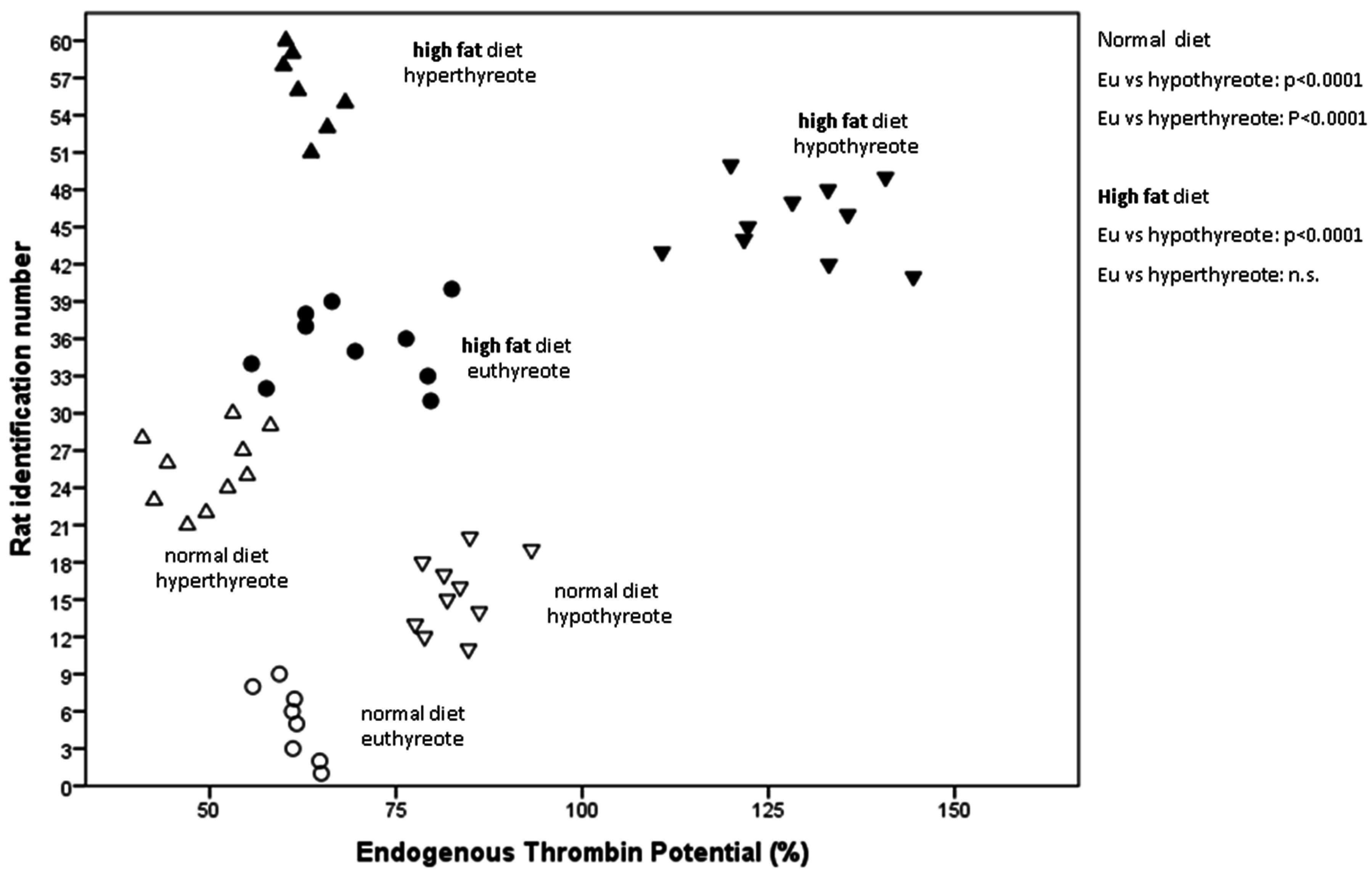
| ETP (%) | 61.3 ± 2.9 | 82.8 ± 4.5 *** | 49.5 ± 5.6 *** | 69.3 ± 9.7 | 129 ± 10.4 ††† | 62.9 ± 3.1 |
| PZ INR | 0.83 ± 0.02 | 0.9 ± 0.2 | 0.83 ± 0.07 | 0.96 ± 0.3 | 0.83 ± 0.05 | 0.81 ± 0.05 |
| APTT (s) | 24 ± 8 | 38 ± 10 ** | 21 ± 2 | 31.2 ± 8 | 52 ± 5 ††† | 26.2 ± 6 |
| Fibrinogen (g/L) | 0.87 ± 0.1 | 1.39 ± 0.4 *** | 0.9 ± 0.1 | 0.94 ± 0.08 | 1.74 ± 0.4 ††† | 1.25 ± 0.5 |
| Antithrombin (%) | 124 ± 4 | 117 ± 15 | 116 ± 28 | 108 ± 21 | 91 ± 23 | 106 ± 15 |
3. Discussion
4. Experimental Section
4.1. Animals
4.2. Experimental Design and Treatment
4.3. Blood Collection
4.4. Laboratory Procedures
4.5. Statistical Analysis
Acknowledgments
Author Contributions
Conflicts of Interest
References
- Bubber, P.; Chauhan, A.; Sharma, A.; Bubber, N.; Bansal, D.D. Effect of thyroxine on fibrinolytic system in rat. Indian J. Physiol. Pharmacol. 2012, 56, 267–272. [Google Scholar] [PubMed]
- Duntas, L.H.; Wartofsky, L. Cardiovascular risk and subclinical hypothyroidism: Focus on lipids and new emerging risk factors. What is the evidence? Thyroid 2007, 17, 1075–1084. [Google Scholar] [CrossRef] [PubMed]
- Biondi, B.; Klein, I. Hypothyroidism as a risk factor for cardiovascular disease. Endocrine 2004, 24, 1–13. [Google Scholar] [CrossRef]
- Horacek, J.; Maly, J.; Svilias, I.; Smolej, L.; Cepkova, J.; Vizda, J.; Sadilek, P.; Fatorova, I.; Zak, P. Prothrombotic changes due to an increase in thyroid hormone levels. Eur. J. Endocrinol. 2015, 172, 537–542. [Google Scholar] [CrossRef] [PubMed]
- Chen, Q.; Yan, Y.; Zhang, L.; Cheng, K.; Liu, Y.; Zhu, W. Effect of hyperthyroidism on the hypercoagulable state and thromboembolic events in patients with atrial fibrillation. Cardiology 2014, 127, 176–182. [Google Scholar] [CrossRef] [PubMed]
- Lippi, G.; Franchini, M.; Targher, G.; Montagnana, M.; Salvagno, G.L.; Guidi, G.C.; Favaloro, E.J. Hyperthyroidism is associated with shortened APTT and increased fibrinogen values in a general population of unselected outpatients. J. Thromb. Thrombolysis 2009, 28, 362–365. [Google Scholar] [CrossRef] [PubMed]
- Squizzato, A.; Romualdi, E.; Buller, H.R.; Gerdes, V.E. Clinical review: Thyroid dysfunction and effects on coagulation and fibrinolysis: A systematic review. J. Clin. Endocrinol. Metab. 2007, 92, 2415–2420. [Google Scholar] [CrossRef] [PubMed]
- Jabbar, A.; Razvi, S. Thyroid disease and vascular risk. Clin. Med. 2014, 14 (Suppl. 6), S29–S32. [Google Scholar] [CrossRef] [PubMed]
- Pruller, F.; Raggam, R.B.; Posch, V.; Almer, G.; Truschnig-Wilders, M.; Horejsi, R.; Moller, R.; Weghuber, D.; Ille, R.; Schnedl, W.; et al. Trunk weighted obesity, cholesterol levels and low grade inflammation are main determinants for enhanced thrombin generation. Atherosclerosis 2012, 220, 215–218. [Google Scholar] [CrossRef] [PubMed]
- Sanchez, C.; Poggi, M.; Morange, P.E.; Defoort, C.; Martin, J.C.; Tanguy, S.; Dutour, A.; Grino, M.; Alessi, M.C. Diet modulates endogenous thrombin generation, a biological estimate of thrombosis risk, independently of the metabolic status. Arterioscler. Thromb. Vasc. Biol. 2012, 32, 2394–2404. [Google Scholar] [CrossRef] [PubMed]
- Schneider, J.G.; Isermann, B.; Kleber, M.E.; Wang, H.; Boehm, B.O.; Grammer, T.B.; Prueller, F.; Nawroth, P.P.; Maerz, W. Inverse association of the endogenous thrombin potential (ETP) with cardiovascular death: The Ludwigshafen Risk and Cardiovascular Health (LURIC) study. Int. J. Cardiol. 2014, 176, 139–144. [Google Scholar] [CrossRef] [PubMed]
- Tobias, D.K.; Pan, A.; Jackson, C.L.; O’Reilly, E.J.; Ding, E.L.; Willett, W.C.; Manson, J.E.; Hu, F.B. Body-mass index and mortality among adults with incident type 2 diabetes. N. Engl. J. Med. 2014, 370, 233–244. [Google Scholar] [CrossRef] [PubMed]
- Seehaus, S.; Shahzad, K.; Kashif, M.; Vinnikov, I.A.; Schiller, M.; Wang, H.; Madhusudhan, T.; Eckstein, V.; Bierhaus, A.; Bea, F.; et al. Hypercoagulability inhibits monocyte transendothelial migration through protease-activated receptor-1-, phospholipase-Cβ-, phosphoinositide 3-kinase-, and nitric oxide-dependent signaling in monocytes and promotes plaque stability. Circulation 2009, 120, 774–784. [Google Scholar] [CrossRef] [PubMed]
- Boyle, J.J.; Johns, M.; Lo, J.; Chiodini, A.; Ambrose, N.; Evans, P.C.; Mason, J.C.; Haskard, D.O. Heme induces heme oxygenase 1 via NRF2: Role in the homeostatic macrophage response to intraplaque hemorrhage. Arterioscler. Thromb. Vasc. Biol. 2011, 31, 2685–2691. [Google Scholar] [CrossRef] [PubMed]
- Sadrzadeh, S.M.; Graf, E.; Panter, S.S.; Hallaway, P.E.; Eaton, J.W. Hemoglobin. A biologic Fenton reagent. J. Biol. Chem. 1984, 259, 14354–14356. [Google Scholar] [PubMed]
- Ardissino, D.; Merlini, P.A.; Bauer, K.A.; Galvani, M.; Ottani, F.; Franchi, F.; Bertocchi, F.; Rosenberg, R.D.; Mannucci, P.M. Coagulation activation and long-term outcome in acute coronary syndromes. Blood 2003, 1, 2731–2735. [Google Scholar] [CrossRef] [PubMed]
- Ragginer, C.; Bernecker, C.; Ainoedhofer, H.; Pailer, S.; Kieslinger, P.; Truschnig-Wilders, M.; Gruber, H.J. Treatment with the nitric oxide donor SNP increases triiodothyronine levels in hyper- and hypothyroid Sprague-Dawley rats. Horm. Metab. Res. 2013, 45, 808–812. [Google Scholar] [CrossRef] [PubMed]
- McAllister, R.M.; Albarracin, I.; Price, E.M.; Smith, T.K.; Turk, J.R.; Wyatt, K.D. Thyroid status and nitric oxide in rat arterial vessels. J. Endocrinol. 2005, 185, 111–119. [Google Scholar] [CrossRef] [PubMed]
- Messarah, M.; Saoudi, M.; Boumendjel, A.; Boulakoud, M.S.; Feki, A.E. Oxidative stress induced by thyroid dysfunction in rat erythrocytes and heart. Environ. Toxicol. Pharmacol. 2011, 31, 33–41. [Google Scholar] [CrossRef] [PubMed]
- Weltman, N.Y.; Ojamaa, K.; Savinova, O.V.; Chen, Y.F.; Schlenker, E.H.; Zucchi, R.; Saba, A.; Colligiani, D.; Pol, C.J.; Gerdes, A.M. Restoration of cardiac tissue thyroid hormone status in experimental hypothyroidism: A dose-response study in female rats. Endocrinology 2013, 154, 2542–2552. [Google Scholar] [CrossRef] [PubMed]
- Li, W.; Wang, D.; Song, G.; Zuo, C.; Qiao, X.; Qin, S. The effect of combination therapy of allicin and fenofibrate on high fat diet-induced vascular endothelium dysfunction and liver damage in rats. Lipids Health Dis. 2010, 9, 131. [Google Scholar] [CrossRef] [PubMed]
- Bhandari, U.; Kumar, V.; Khanna, N.; Panda, B.P. The effect of high-fat diet-induced obesity on cardiovascular toxicity in Wistar albino rats. Hum. Exp. Toxicol. 2011, 30, 1313–1321. [Google Scholar] [CrossRef] [PubMed]
- Nadal-Casellas, A.; Proenza, A.M.; Gianotti, M.; Llad, I. Brown adipose tissue redox status in response to dietary-induced obesity-associated oxidative stress in male and female rats. Stress 2011, 14, 174–184. [Google Scholar] [PubMed]
© 2015 by the authors; licensee MDPI, Basel, Switzerland. This article is an open access article distributed under the terms and conditions of the Creative Commons Attribution license (http://creativecommons.org/licenses/by/4.0/).
Share and Cite
Mangge, H.; Prüller, F.; Zelzer, S.; Ainödhofer, H.; Pailer, S.; Kieslinger, P.; Haybaeck, J.; Obermayer-Pietsch, B.; Cvirn, G.; Gruber, H.-J. Hypothyroidism Exacerbates Thrombophilia in Female Rats Fed with a High Fat Diet. Int. J. Mol. Sci. 2015, 16, 15776-15784. https://doi.org/10.3390/ijms160715776
Mangge H, Prüller F, Zelzer S, Ainödhofer H, Pailer S, Kieslinger P, Haybaeck J, Obermayer-Pietsch B, Cvirn G, Gruber H-J. Hypothyroidism Exacerbates Thrombophilia in Female Rats Fed with a High Fat Diet. International Journal of Molecular Sciences. 2015; 16(7):15776-15784. https://doi.org/10.3390/ijms160715776
Chicago/Turabian StyleMangge, Harald, Florian Prüller, Sieglinde Zelzer, Herwig Ainödhofer, Sabine Pailer, Petra Kieslinger, Johannes Haybaeck, Barbara Obermayer-Pietsch, Gerhard Cvirn, and Hans-Jürgen Gruber. 2015. "Hypothyroidism Exacerbates Thrombophilia in Female Rats Fed with a High Fat Diet" International Journal of Molecular Sciences 16, no. 7: 15776-15784. https://doi.org/10.3390/ijms160715776