Article

VEGFR-1 Overexpression Identifies a Small Subgroup of Aggressive Prostate Cancers in Patients Treated by Prostatectomy

1
Institute of Pathology, University Medical Center Hamburg-Eppendorf, Martinistrasse 52, D-210246 Hamburg, Germany
2
Martini-Clinic, Prostate Cancer Center, University Medical Center Hamburg-Eppendorf, Martinistrasse 52, D-210246 Hamburg, Germany
3
Department of Urology, Section for translational Prostate Cancer Center, University Medical Center Hamburg-Eppendorf, Martinistrasse 52, D-210246 Hamburg, Germany
*
Author to whom correspondence should be addressed.
These authors contributed equally to this work.
Int. J. Mol. Sci. 2015, 16(4), 8591-8606; https://doi.org/10.3390/ijms16048591
Submission received: 16 February 2015 / Revised: 27 March 2015 / Accepted: 10 April 2015 / Published: 16 April 2015
(This article belongs to the Special Issue Advances in Molecular Oncology)

Abstract

:
The VEGFR-1 is suggested to promote tumor progression. In the current study we analyzed prevalence and prognostic impact of the VEGFR-1 by immunohistochemistry on a tissue microarray containing more than 3000 prostate cancer specimens. Results were compared to tumor phenotype, ETS-related gene (ERG) status, and biochemical recurrence. Membranous VEGFR-1 expression was detectable in 32.6% of 2669 interpretable cancers and considered strong in 1.7%, moderate in 6.7% and weak in 24.2% of cases. Strong VEGFR-1 expression was associated with TMPRSS2:ERG fusion status as determined by fluorescence in situ hybridization (FISH) and immunohistochemistry (p < 0.0001 each). Elevated VEGFR-1 expression was linked to high Gleason grade and advanced pT stage in TMPRSS2:ERG negative cancers (p = 0.0008 and p = 0.001), while these associations were absent in TMPRSS2:ERG positive cancers. VEGFR-1 expression was also linked to phosphatase and tensin homolog (PTEN) deletions. A comparison with prostate specific antigen (PSA) recurrence revealed that the 1.7% of prostate cancers with the highest VEGFR-1 levels had a strikingly unfavorable prognosis. This could be seen in all cancers, in the subsets of TMPRSS2:ERG positive or negative, PTEN deleted or undeleted carcinomas (p < 0.0001 each). High level VEGFR-1 expression is infrequent in prostate cancer, but identifies a subgroup of aggressive cancers, which may be candidates for anti-VEGFR-1 targeted therapy.

Graphical Abstract

1. Introduction

Prostate cancer is the most prevalent cancer in men in Western societies [1]. Although the majority of prostate cancers behave in an indolent manner, a small subset is highly aggressive and requires extensive treatment [2,3]. Established pre-therapeutic prognostic parameters are limited to Gleason grade and tumor extent on biopsies, preoperative prostate specific antigen (PSA), and clinical stage. Because these data are statistically powerful, but often insufficient for optimal individual treatment decisions, it is hoped that a better understanding of disease biology will eventually lead to the identification of clinically applicable molecular markers that enable a more reliable prediction of prostate cancer aggressiveness. At the same time, there is a need for alternative anticancer drugs targeting specific structures of prostate cancer cells.
Vascular endothelial growth factor receptor 1 (VEGFR-1) is a candidate for both a prognostic biomarker and representing a suitable target molecule for specific cancer therapy [4]. The VEGF family is composed of five structurally related factors (VEGF-A, -B, -C, -D and phosphatidylinositol-glycan biosynthesis class F (PIGF)), which act as the primary activators of angiogenesis by binding to tyrosine kinase receptors VEGF-receptor 1, 2 and 3 (VEGFR1–3). Whereas the roles of VEGFR-2 and VEGFR-3 as direct stimulators of angiogenesis (VEGFR-2) and lymphangiogenesis (VEGFR-3) have been characterized thoroughly, the function of VEGFR-1 is less clear. The VEGFR-1 gene encodes two different proteins: membrane-bound FGFR1 (Flt-1) and a soluble form termed sVEGFR-1. Both forms have been shown to negatively regulate VEGFR-2 through high-affinity binding of VEGFs, which consequently become unavailable for VEGFR-2 (reviewed in [5]). However, it has been suggested that VEGFR-1 might indirectly promote tumor cell growth by activation of monocytes and macrophages, which invade the tumor and produce VEGFs and cytokines, leading to angiogenesis and lymphangiogenesis via activation of VEGFR-2 and VEGFR-3 [6,7].
VEGFR-1 is described to be expressed on tumor cells in many tumor types, including lymphoma [8], leukemia [9], multiple myeloma [10], melanoma [11], non-small cell lung cancer [12], colon cancer [13], pancreatic cancer [14] and breast cancer [4]. In several cancer entities, such as renal cell cancer [15], squamous cell cancer [16,17], non-small cell lung cancer [18], colon cancer [19] and breast cancer [20], VEGFR-1 was shown to be linked to disease outcome. In prostate, differential expression between cancerous and normal epithelium was described [21,22,23,24]. In addition, studies on 113, 40, and 79 cancers had suggested a possible link between high VEGFR-1 expression levels and unfavorable tumor phenotype and poor disease outcome [21,22,25]. However, this observation was not confirmed by others analyzing cancers from 15 patients [23].
To further clarify the clinical relevance of VEGFR-1 expression in prostate cancer by immunohistochemistry (IHC), we took advantage of our preexisting tissue microarray (TMA) containing >3000 prostate cancer specimens connected to a database with extensive clinical follow up. Our findings demonstrate that high levels of VEGFR-1 protein expression are strongly linked to an adverse phenotype and early PSA recurrence of prostate cancer.

2. Results

2.1. Technical Issues

A total of 2669 (82%) tumor samples were interpretable in our TMA analysis. Reason for non-informative cases (592 spots, 18%) included lack of tissue samples or absence of unequivocal cancer tissue in the TMA spot.

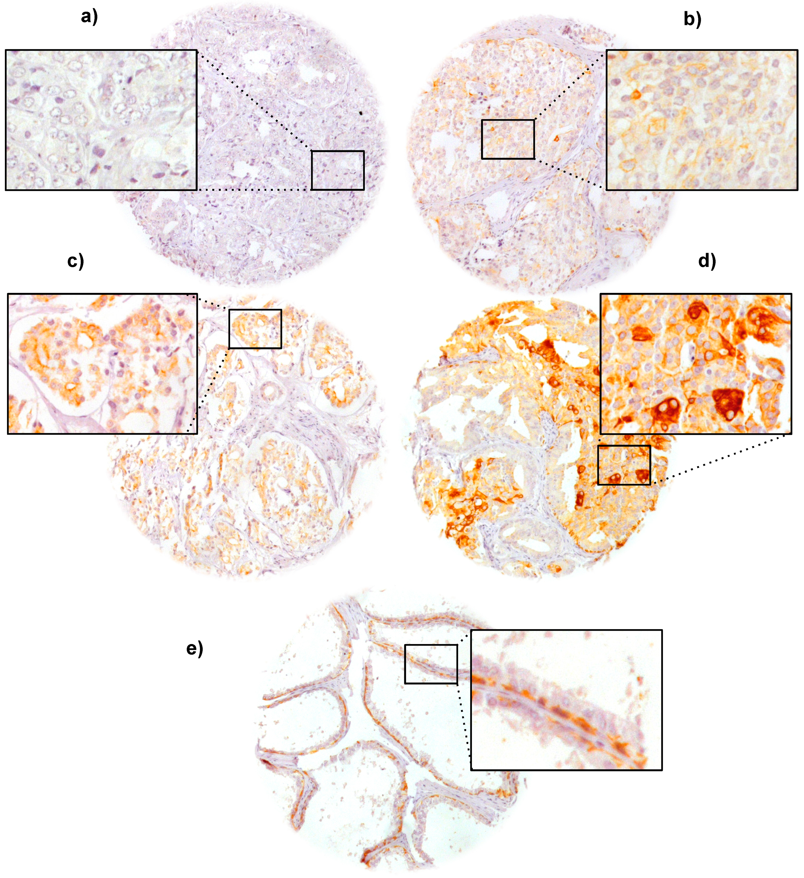
2.2. VEGFR-1 Expression in Prostate Cancer

VEGFR-1 immunostaining was localized in the membrane and the cytoplasm. Positive VEGFR-1 staining was also regularly recorded in basal cells of non-neoplastic prostate epithelium and in basal cells of prostatic intraepithelial neoplasia (PIN). Positive VEGFR-1 staining was seen in 869 of our 2669 (32.6%) interpretable prostate cancers and was considered weak in 24.2%, moderate in 6.7% and strong in 1.7% of cancers, while 1800 (67.4%) did not show any membranous VEGFR-1 reactivity. Representative images of positive and negative VEGFR-1 immunostainings are given in Figure 1.

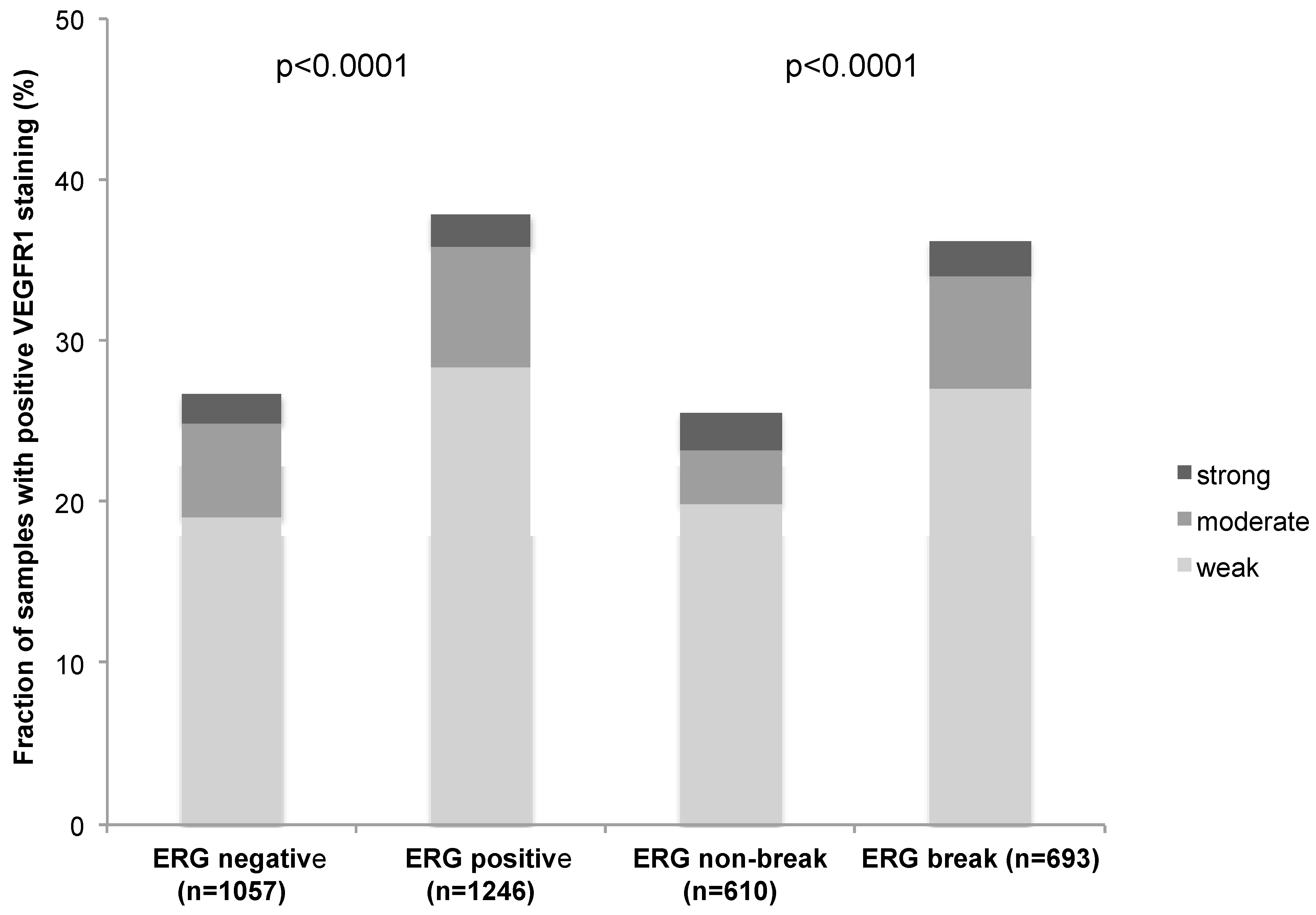
2.3. Association with TMPRSS2:ERG Fusion Status and ERG Protein Expression

To evaluate whether VEGFR-1 expression is associated with ERG status in prostate cancers, we used data from previous studies [26]. Data on TMPRSS2:ERG fusion status obtained by fluorescence in-situ hybridization were available from 1303 patients and by immunohistochemistry from 2303 patients. VEGFR-1 expression was significantly linked to TMPRSS2:ERG rearrangement and ERG expression (p < 0.0001 each, Figure 2).

2.4. Association with Tumor Phenotype

Increased VEGFR-1 expression was significantly linked to high Gleason grade (p = 0.03) and advanced pathological tumor stage (p < 0.0001), if all tumors were jointly analyzed (Table 1).
Figure 1. (a) Negative; (b) weak; (c) moderate; and (d) strong VEGFR-1 immunostaining in prostate cancer as well as (e) in a normal prostate. Magnification 70× and 240× for the insets.
Ijms 16 08591 g001
Figure 2. Correlation between VEGFR-1 expression and ERG-fusion probed with IHC and FISH.
Ijms 16 08591 g002
Table 1. Association between VEGFR-1 expression and cancer phenotype.
ParameterEvaluable (N)Immunostaining (%)p Value
NegativeWeakModerate Strong
All cancers266967246.71.7
Tumor stage
pT2172869237.01.0<0.0001
pT3a55368237.02.0
pT3b32663294.63.7
pT43450295.915
Gleason grade
≤3 + 3115669237.31.10.03
3 + 4116566266.81.7
4 + 327369234.83.7
≥4 + 44768232.16.4
Lymph node metastasis
N0131667265.31.60.05
N+8457324.86.0
PSA preoperative
<440163278.02.20.23
4–10143567247.31.3
11–2056469235.72.5
>2020472224.41.5
Subgroup analysis of ERG positive and ERG negative cancers revealed, that these associations were largely retained in the subsets of ERG negative and positive cancers (Supplementary Tables S1 and S2).

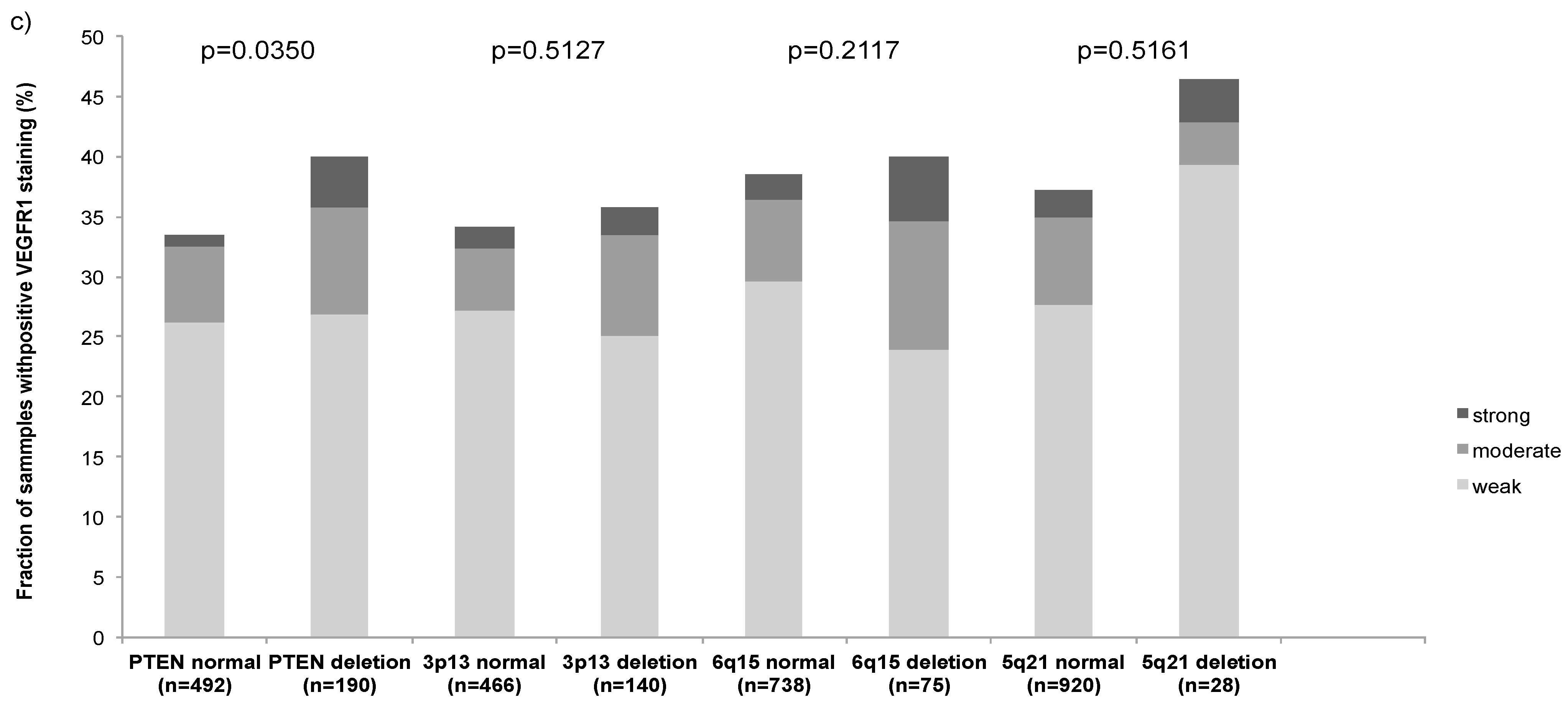
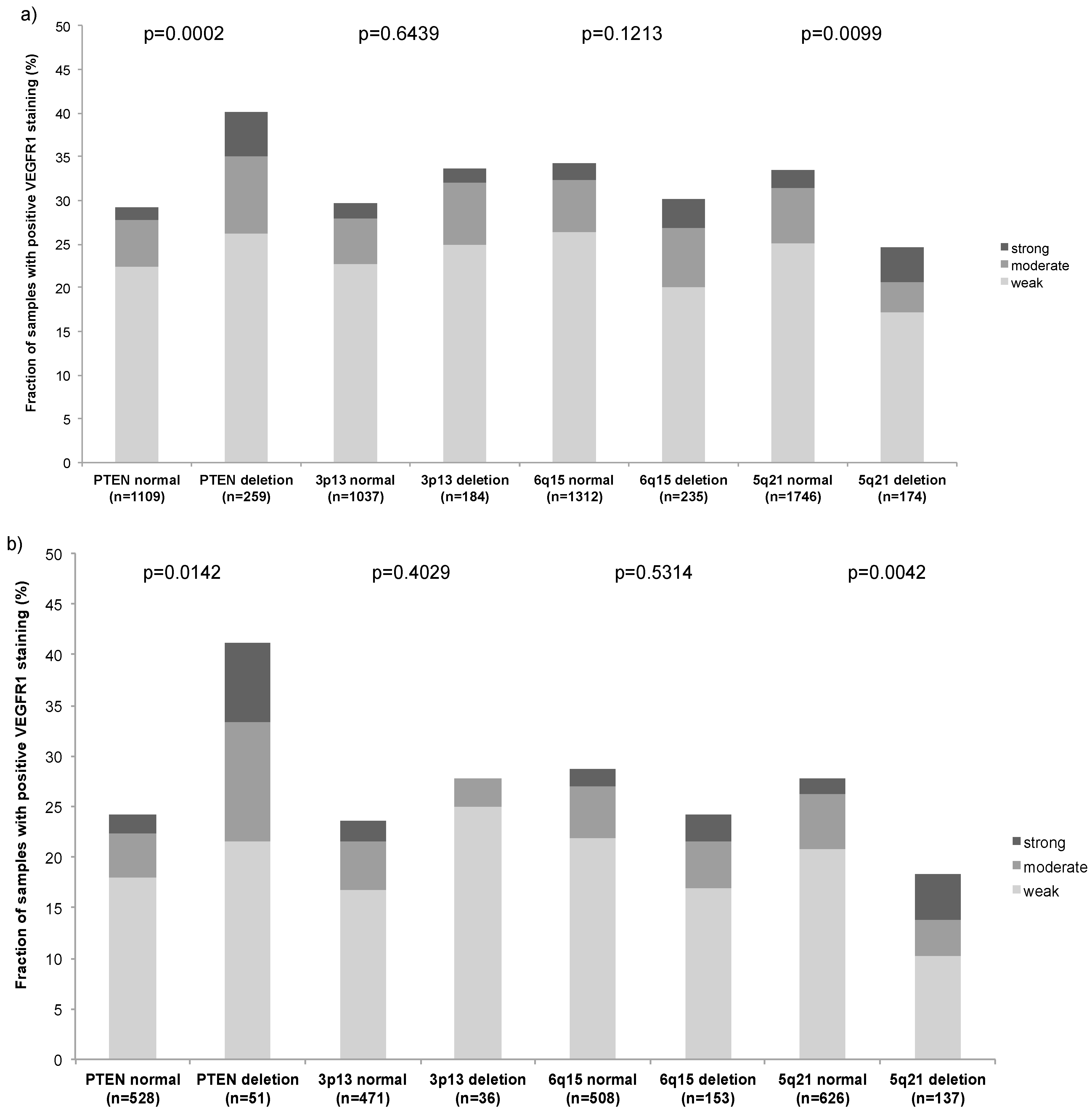
2.5. Association with Other Key Genomic Deletions

Earlier studies had provided evidence for distinct molecular subgroups of prostate cancers defined by TMPRSS2:ERG fusions and several genomic deletions. We as well as others described a strong link of PTEN and 3p13 deletion to ERG positivity and of 5q21 and 6q15 deletions to ERG negativity [27,28,29,30]. To study, whether VEGFR-1 data expression might be particularly linked to one of these genomic deletions, VEGFR-1 data were compared to preexisting findings on PTEN (10q23), FOXP1 (3p13), MAP3K7 (6q15) and CHD1 (5q21) deletions. High VEGFR-1 staining was linked to PTEN deletions in the analysis of all tumors (p = 0.0002; Figure 3a), but also in the subgroup of ERG negative (p = 0.0142; Figure 3b) and ERG positive cancers (p = 0.0350; Figure 3c).
Figure 3. VEGFR-1 expression by IHC versus PTEN, FOXP1, MAP3K7 and CHD1 deletions by FISH in (a) All cancers; (b) ERG negative; and (c) ERG positive.
Ijms 16 08591 g003aIjms 16 08591 g003b

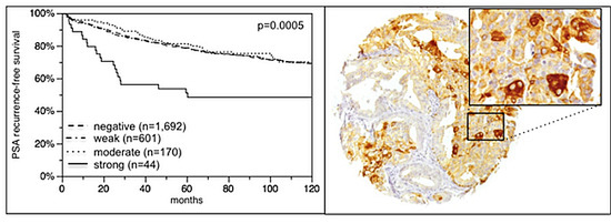
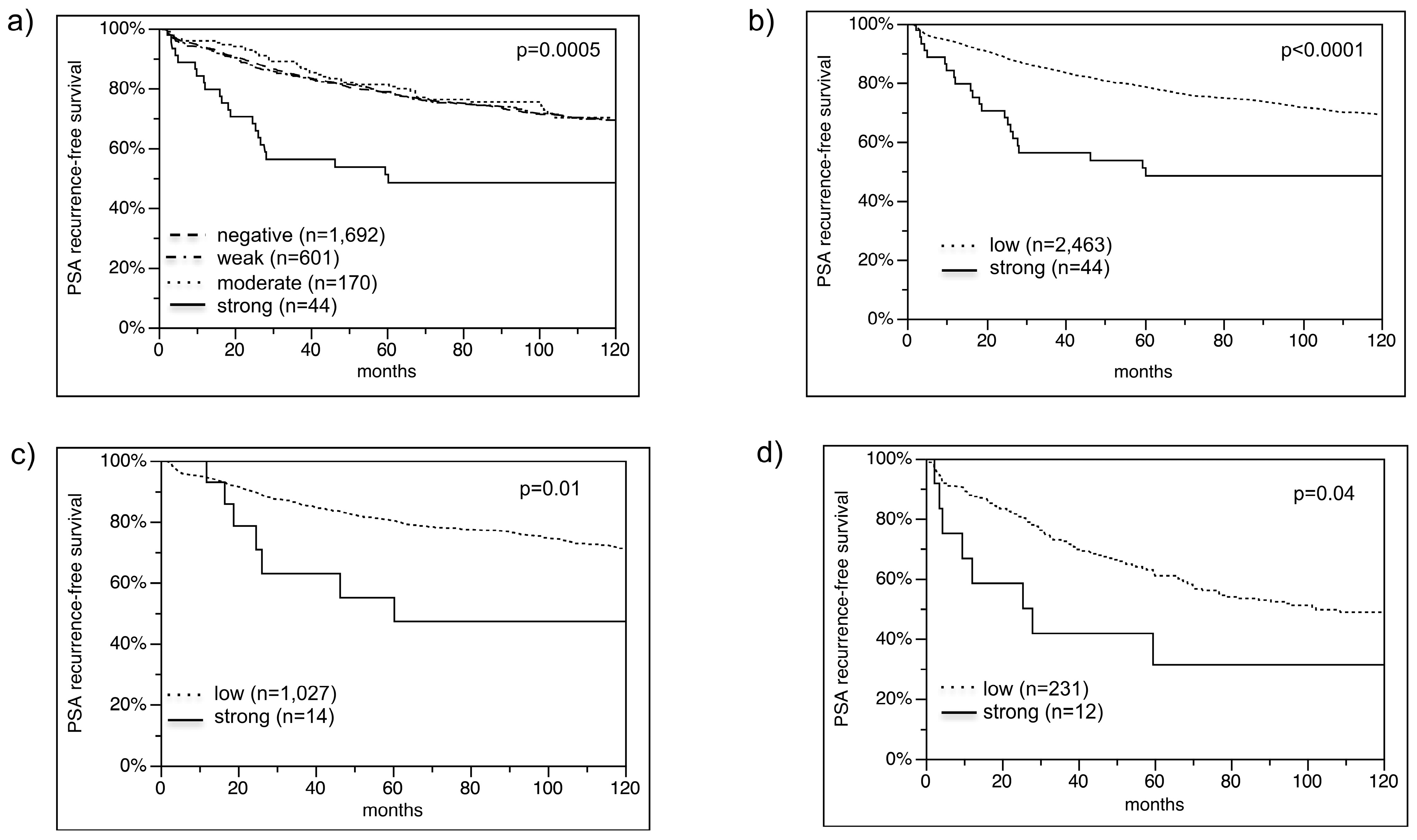
2.6. Association with PSA Recurrence

Follow-up data were available for 2507 patients with interpretable VEGFR-1 immunostaining on the TMA. There was a statistically significant association between high VEGFR-1 expression and early PSA recurrence if all tumors were analyzed (p = 0.0005; Figure 4a). The analysis revealed that unfavorable disease outcome was seen in particular in cancers with very high VEGFR-1 expression levels. Because of their similar clinical behavior, cancers with negative, weak and moderate VEGFR-1 expression, were subsequently combined as “low” for further analyses (Figure 4b). Separate analyses of molecularly defined cancer subgroups such as PTEN deleted and non-deleted prostate cancers revealed, that high level VEGFR-1 expression remained to be strongly associated with early PSA recurrence in these subsets (Figure 4c,d). Multivariate analysis revealed that strong VEGFR-1 expression remained a significant predictor of disease outcome (p = 0.02), if pathological tumor stage (p < 0.0001), Gleason grade (p < 0.0001), nodal stage (p < 0.0001), resection margin (p < 0.0001) and pre-operative PSA (p = 0.05) were included into the analysis (Table 2).

3. Discussion

In this study, IHC was applied to a TMA to evaluate VEGFR-1 expression in prostate cancer and normal prostatic epithelium. The TMA approach is optimal for IHC studies, because TMAs enable maximal experimental standardization. In this study more than 3000 prostate cancer specimens were analyzed in one day in one experiment using one set of reagents at identical concentrations, temperatures and exposure times. Moreover, all TMA sections were cut within hours on a single day immediately before staining to avoid unequal tissue reactivity to antibody binding [31,32]. Finally one pathologist interpreted all IHCs on one day to optimally standardize staining interpretation.
Figure 4. (a) Biochemical recurrence is associated with strong VEGFR-1 staining; (b) negative, weak and moderate grouped in low; (c) PTEN normal subset; and (d) PTEN deleted subset.
Ijms 16 08591 g004
Table 2. Multivariate analysis of established prognostic parameters and VEGFR-1 expression status.
ParameterRR95% CIp Value
Tumor stage
pT3a vs. pT21.91.5–2.4<0.0001
pT3b vs. pT3a1.81.4–2.2
pT4 vs. pT3b1.40.9–2.1
Gleason grade
3 + 4 vs. ≤3 + 32.41.8–3.3<0.0001
4 + 3 vs. 3 + 42.31.8–2.8
≥4 + 4 vs. 4 + 31.41.0–2.1
Nodal stage
pN1 vs. pN01.91.5–2.5< 0.0001
Resection margin status
R1 vs. R01.51.2–1.8<0.0001
Pre-operative PSA (ng/mL)
4–10 vs. <41.10.8–1.60.05
11–20 vs. 4–101.10.9–1.4
>20 vs. 11–201.20.9–1.5
VEGFR-1
strong vs. low1.91.1–3.00.02
Our analysis revealed that VEGFR-1 was consistently and strongly expressed in basal cells of normal prostate epithelium and PIN. Secretory epithelial cells were negative. In cancer cells, VEGFR-1 expression was found in 32.6% of 2669 interpretable prostate cancers, indicating that VEGFR-1 can be activated in a fraction of secretory cells as a consequence of malignant transformation. The frequency of detectable VEGFR-1 staining is substantially lower in our study than in four earlier publications reporting VEGFR-1 expression in 97.5%, 100% and 100% analyzing 40, 15 and 113 prostate cancers and in 100% of 16 pelvic lymph node metastasis [21,22,23,25]. It is most likely, that these differences are due to the use of different antibodies and staining procedures. For our study, we selected the rabbit polyclonal antibody ab2350 raised against the C-terminal polypeptide not present in the soluble form of the VEGFR-1 protein, which does not react with the phosphorylated form of the protein. In contrast, Mao et al. [22] and Woolard et al. [24] used antibodies with a presumable broader binding spectrum, raised against the recombinant VEGFR-1 Ser27-His687 in goats. That varying antibody conditions lead to significant changes in the rate of positive cases was previously shown. For example the frequency of positive p53 immunostaining varied between 4% and 61% based on the experimental protocol [33]. A high variation of immunostaining is all the more expected if antibodies are compared that bind to different epitopes of the target protein.
The analysis of our large tumor cohort revealed that high VEGFR-1 expression was significantly linked to advanced tumor stage and high Gleason grade. However, the difference between the groups was rather small. For example, moderate to strong VEGFR-1 expression was detectable in 8% of pT2, 9% of pT3a and 8% of pT3b cancers. However, the majority of published investigations found associations with Gleason grade [21] and/or pT stage [21,22,25], while some investigations failed to reveal associations with pT stage [23] and/or Gleason grade [22,23].
Only 1.7% of 2669 interpretable cancers were considered to have a strong VEGFR-1 expression in this study. Only these highest expression levels were strongly linked to unfavorable prognosis, while cancers with low or moderate VEGFR-1 expression did behave as cancers without detectable VEGFR-1 expression. It is obvious that studies analyzing substantial smaller patient cohorts would not be able to identify such a small fraction of cancers as a relevant subgroup. Accordingly, two earlier studies failed to find associations between VEGFR-1 expression and disease outcome [21,22]. However, one study on 79 patients reported a prognostic impact of VEGFR-1 expression in pelvic lymph node negative prostate cancer [25]. The striking prognostic impact of high VEGFR-1 expression is consistent with studies suggesting an oncogenic role of this protein. For example, VEGFR-1 was found to promote migration of tumor cells through an Src-dependent pathway linked to activation of focal adhesion [34]. VEGFR-1 is also hypothesized to enable the development of cancers metastases by activation and pre-metastatic localization in distant organs of bone marrow-derived hematopoietic progenitor cells expressing VEGFR-1 (reviewed in [35]). Moreover, several studies suggest that VEGFR-1 might indirectly promote tumor cell growth by activation of monocytes and macrophages, which invade the tumor and produce VEGFs and cytokines, leading to angiogenesis and lymphangiogenesis via activation of VEGFR-2 and VEGFR-3 [6,7].
The mechanisms for VEGFR-1 overexpression applying to prostate cancer are not completely understood. The strong association particularly between low-level (weak to moderate) VEGFR-1 expression and ERG positivity suggests that overexpression of the ERG transcription factor might contribute to VEGFR-1 activation in prostate epithelial cells. In fact, a strong interaction with ERG was earlier demonstrated for VEGFR-2. In a study on transgenic Xenopus embryos and COS-7 cells, it was shown that ERG together with KLF2 (a member of the Krüppel-like factor transcription regulator family) synergistically activated transcription of VEGFR-2 [36]. PTEN deletions and other alterations of the PI3K/AKT signaling pathway may also contribute to high level VEGFR-1 expression in prostate cancer. Both PTEN and VEGFR-1 negatively regulate PI3K/AKT signaling activity in a feedback control loop [37], with intact PTEN being an inhibitor of VEGFR1 expression [38]. This explains the strong link between high level VEGFR-1 expression and PTEN deletions in our cancers. That high level VEGFR-1 expression is associated with poor disease outcome both in PTEN deleted and in PTEN undeleted cancers is further suggesting that a more complex dysregulation of PI3K/AKT signaling by multiple independent alterations of pathway components may have a clinically relevant additive impact.
The striking success of new targeted drugs raise the hope that prostate cancer patients might eventually also benefit from such an approach [39]. However, the few clinical trials in prostate cancer using drugs targeting Epidermal Growth Factor Receptor (EGFR) [40,41], VEGFR-2 [42] and HER2 [43,44,45] were discouraging so far. VEGFR-1 is another potential target molecule for specific cancer therapy [4]. IMC-18F1 (icrucumab), a human monoclonal antibody against VEGFR-1 tested in a Phase I study is described to have antitumor activity [46]. Our data suggest that at least 1.7% of prostate cancer patients might be excellent candidates for benefiting from such a therapy, once the drug should prove to be effective. Although a fraction of 1.7% may appear low, prostate cancer may become a relevant application of such new drugs given its high prevalence.

4. Experimental Section

4.1. Patients

Radical prostatectomy specimens were available from 3261 patients, undergoing surgery between 1992 and 2005 at the Department of Urology and the Martini Clinics at the University Medical Center Hamburg–Eppendorf (Table 3). Analysis of patient and corresponding histopathological data for research purposes, as well as construction of tissue microarrays from archived diagnostic left-over tissues, was approved by local laws (HmbKHG, §12,1) and by the local ethics committee (Ethics commission Ärztekammer Hamburg, WF-049/09 and PV3652). According to local laws, informed consent was not required for this study. Patient records/information was anonymized and de-identified prior to analysis. All work was carried out in compliance with the Helsinki Declaration. Follow-up data were available for a total of 3058, ranging from 1 to 228 months (mean 72.1). Prostate specific antigen values were measured following surgery and PSA recurrence was defined as the time point when postoperative PSA was at least 0.2 ng/mL and increasing at subsequent measurements. All prostate specimens were analyzed according to a standard procedure, including a complete embedding of the entire prostate for histological analysis [33]. The TMA manufacturing process was described before [47]. In short, one 0.6 mm core was taken from a representative tissue block from each patient. The tissues were distributed among 7 TMA blocks, each containing 144 to 522 tumor samples. For internal controls, each TMA block also contained various control tissues, including normal prostate. The molecular database attached to this TMA contained results on ERG expression in 2539, ERG break apart FISH analysis in 1474 [26] and deletion status of CHD1 [30] in 2197, MAP3K7 (6q15) in 1754 [27], PTEN (10q23) in 1447 [29] and FOXP1 (3p13) in 1290 [48] cancers.
Table 3. Clinico-pathological features of arrayed prostate cancers.
Parametern = 3261 on TMAn = 2891 with Clinical Follow-up
Follow-up (months)
Mean72.1
Median68.9
Range1–219
Age (years)
<508378
50–60998912
60–7018071699
>70175169
Pretreatment PSA (ng/mL)
<4513478
4–1016731544
11–20641608
>20225212
pT category (AJCC 2002)
pT220801907
pT3a609579
pT3b372361
pT44242
Gleason grade
≤3 + 314261307
3 + 413111238
4 + 3313297
≤4 + 45549
pN category
pN015441492
pN+9693
pNx14571298
Surgical margin
Negative24752295
Positive627594

4.2. Immunochemistry

Freshly cut TMA sections were immunostained on one day and in one experiment. Primary antibody specific for VEGFR-1 (polyclonal rabbit ab2350, Abcam, Cambridge, UK; dilution 1:450) was applied, slides were deparaffinized and exposed to heat-induced antigen retrieval for 5 min in an autoclave at 121 °C in pH 7.8. Tris-EDTA-Citrate buffer. Bound antibody was then visualized using the EnVision Kit (Dako, Glostrup, Denmark). The staining intensity (0, 1+, 2+, 3+) and the fraction of positive tumor cells were recorded for each tissue spot. A final score was built from the two variables as previously described [26,49]: negative scores had staining intensity of 0, weak ones had a staining intensity of 1+ in ≤70% of tumor cells or 2+ in ≤30% of tumor cells; moderate had staining intensity of 1+ in >70% of tumor cells, staining intensity of 2+ in >30% and ≤70% of tumor cells or staining intensity of 3+ in ≤30% of tumor cells and strong scores had staining intensity of 2+ in >70% of tumor cells or staining intensity of 3+ in >30% of tumor cells.

4.3. Statistics

Statistical calculations were performed using JMP9 statistical software (SAS Institute, Inc., Cary, NC, USA). Contingency tables were calculated with the chi2-test to search for associations between molecular parameters and tumor phenotype. Survival curves were calculated according to Kaplan–Meier. The Log-Rank test was applied to detect significant survival differences between groups. Cox proportional hazards regression analysis was performed to test the statistical independence and significance between pathological, molecular and clinical variables.

5. Conclusions

Our study shows that strong VEGFR-1 expression identifies a small subgroup of highly aggressive prostate cancers. Given its role as a possible drug target, VEGFR-1 has strong potential both as a prognostic and therapeutic target in prostate cancer.

Supplementary Materials

Supplementary materials can be found at https://www.mdpi.com/1422-0067/16/04/8591/s1.

Acknowledgments

We thank Julia Schumann, Sünje Seekamp and Inge Brandt for excellent technical assistance.

Author Contributions

Maria Christina Tsourlakis, Stefan Steurer, Ronald Simon, and Guido Sauter designed the study, and drafted the manuscript. Hans Heinzer, Markus Graefen, Waldemar Wilczak and Till Krech participated in study design. Maria Christina Tsourlakis, Stefan Steurer, Waldemar Wilczak, Sarah Minner, Corinna Wittmer performed IHC analysis and scoring. Puya Khosrawi and Philipp Weigand participated in pathology data analysis. Claudia Hube-Magg and Ronald Simon performed statistical analysis. Thorsten Schlomm, Christina Koop, Martina Kluth, Maria Christina Tsourlakis, Puya Khosrawi and Corinna Wittmer participated in data interpretation, and helped to draft the manuscript. All authors read and approved the final manuscript.

Conflicts of Interest

The authors declare no conflict of interest.

References

  1. Jemal, A.; Bray, F.; Center, M.M.; Ferlay, J.; Ward, E.; Forman, D. Global cancer statistics. CA Cancer J. Clin. 2011, 61, 69–90. [Google Scholar] [CrossRef]
  2. Wilt, T.J.; Brawer, M.K.; Jones, K.M.; Barry, M.J.; Aronson, W.J.; Fox, S.; Gingrich, J.R.; Wei, J.T.; Gilhooly, P.; Grob, B.M.; et al. Radical prostatectomy versus observation for localized prostate cancer. N. Engl. J. Med. 2012, 367, 203–213. [Google Scholar]
  3. Thompson, I.M., Jr.; Tangen, C.M. Prostate cancer—Uncertainty and a way forward. N. Engl. J. Med. 2012, 367, 270–271. [Google Scholar] [CrossRef] [PubMed]
  4. Wu, Y.; Hooper, A.T.; Zhong, Z.; Witte, L.; Bohlen, P.; Rafii, S.; Hicklin, D.J. The vascular endothelial growth factor receptor (vegfr-1) supports growth and survival of human breast carcinoma. Int. J. Cancer 2006, 119, 1519–1529. [Google Scholar] [CrossRef] [PubMed]
  5. Shibuya, M. Vascular endothelial growth factor and its receptor system: Physiological functions in angiogenesis and pathological roles in various diseases. J. Biochem. 2013, 153, 13–19. [Google Scholar] [CrossRef] [PubMed]
  6. Lin, E.Y.; Li, J.F.; Gnatovskiy, L.; Deng, Y.; Zhu, L.; Grzesik, D.A.; Qian, H.; Xue, X.N.; Pollard, J.W. Macrophages regulate the angiogenic switch in a mouse model of breast cancer. Cancer Res. 2006, 66, 11238–11246. [Google Scholar] [CrossRef] [PubMed]
  7. Murakami, M.; Zheng, Y.; Hirashima, M.; Suda, T.; Morita, Y.; Ooehara, J.; Ema, H.; Fong, G.H.; Shibuya, M. Vegfr1 tyrosine kinase signaling promotes lymphangiogenesis as well as angiogenesis indirectly via macrophage recruitment. Arterioscler. Thromb. Vasc. Biol. 2008, 28, 658–664. [Google Scholar] [CrossRef] [PubMed]
  8. Wang, E.S.; Teruya-Feldstein, J.; Wu, Y.; Zhu, Z.; Hicklin, D.J.; Moore, M.A. Targeting autocrine and paracrine vegf receptor pathways inhibits human lymphoma xenografts in vivo. Blood 2004, 104, 2893–2902. [Google Scholar] [CrossRef] [PubMed]
  9. Fragoso, R.; Pereira, T.; Wu, Y.; Zhu, Z.; Cabecadas, J.; Dias, S. Vegfr-1 (flt-1) activation modulates acute lymphoblastic leukemia localization and survival within the bone marrow, determining the onset of extramedullary disease. Blood 2006, 107, 1608–1616. [Google Scholar] [CrossRef] [PubMed]
  10. Vincent, L.; Jin, D.K.; Karajannis, M.A.; Shido, K.; Hooper, A.T.; Rashbaum, W.K.; Pytowski, B.; Wu, Y.; Hicklin, D.J.; Zhu, Z.; et al. Fetal stromal-dependent paracrine and intracrine vascular endothelial growth factor-a/vascular endothelial growth factor receptor-1 signaling promotes proliferation and motility of human primary myeloma cells. Cancer Res. 2005, 65, 3185–3192. [Google Scholar]
  11. Lacal, P.M.; Failla, C.M.; Pagani, E.; Odorisio, T.; Schietroma, C.; Falcinelli, S.; Zambruno, G.; D’Atri, S. Human melanoma cells secrete and respond to placenta growth factor and vascular endothelial growth factor. J. Investig. Dermatol. 2000, 115, 1000–1007. [Google Scholar] [CrossRef] [PubMed]
  12. Decaussin, M.; Sartelet, H.; Robert, C.; Moro, D.; Claraz, C.; Brambilla, C.; Brambilla, E. Expression of vascular endothelial growth factor (vegf) and its two receptors (vegf-r1-flt1 and vegf-r2-flk1/kdr) in non-small cell lung carcinomas (nsclcs): Correlation with angiogenesis and survival. J. Pathol. 1999, 188, 369–377. [Google Scholar] [CrossRef] [PubMed]
  13. Fan, F.; Wey, J.S.; McCarty, M.F.; Belcheva, A.; Liu, W.; Bauer, T.W.; Somcio, R.J.; Wu, Y.; Hooper, A.; Hicklin, D.J.; et al. Expression and function of vascular endothelial growth factor receptor-1 on human colorectal cancer cells. Oncogene 2005, 24, 2647–2653. [Google Scholar]
  14. Wey, J.S.; Fan, F.; Gray, M.J.; Bauer, T.W.; McCarty, M.F.; Somcio, R.; Liu, W.; Evans, D.B.; Wu, Y.; Hicklin, D.J.; et al. Vascular endothelial growth factor receptor-1 promotes migration and invasion in pancreatic carcinoma cell lines. Cancer 2005, 104, 427–438. [Google Scholar]
  15. Laird, A.; O’Mahony, F.C.; Nanda, J.; Riddick, A.C.; O'Donnell, M.; Harrison, D.J.; Stewart, G.D. Differential expression of prognostic proteomic markers in primary tumour, venous tumour thrombus and metastatic renal cell cancer tissue and correlation with patient outcome. PLoS ONE 2013, 8, e60483. [Google Scholar] [CrossRef] [PubMed]
  16. Pentheroudakis, G.; Nicolaou, I.; Kotoula, V.; Fountzilas, E.; Markou, K.; Eleftheraki, A.G.; Fragkoulidi, A.; Karasmanis, I.; Tsigka, A.; Angouridakis, N.; et al. Prognostic utility of angiogenesis and hypoxia effectors in patients with operable squamous cell cancer of the larynx. Oral Oncol. 2012, 48, 709–716. [Google Scholar]
  17. Pajares, M.J.; Agorreta, J.; Larrayoz, M.; Vesin, A.; Ezponda, T.; Zudaire, I.; Torre, W.; Lozano, M.D.; Brambilla, E.; Brambilla, C.; et al. Expression of tumor-derived vascular endothelial growth factor and its receptors is associated with outcome in early squamous cell carcinoma of the lung. J. Clin. Oncol. 2012, 30, 1129–1136. [Google Scholar]
  18. Carrillo de Santa Pau, E.; Arias, F.C.; Caso Pelaez, E.; Munoz Molina, G.M.; Sanchez Hernandez, I.; Muguruza Trueba, I.; Moreno Balsalobre, R.; Sacristan Lopez, S.; Gomez Pinillos, A.; del Val Toledo Lobo, M. Prognostic significance of the expression of vascular endothelial growth factors a, b, c, and d and their receptors r1, r2, and r3 in patients with nonsmall cell lung cancer. Cancer 2009, 115, 1701–1712. [Google Scholar] [CrossRef] [PubMed]
  19. Takahashi, Y.; Kitadai, Y.; Bucana, C.D.; Cleary, K.R.; Ellis, L.M. Expression of vascular endothelial growth factor and its receptor, kdr, correlates with vascularity, metastasis, and proliferation of human colon cancer. Cancer Res. 1995, 55, 3964–3968. [Google Scholar] [PubMed]
  20. Ieni, A.; Giuffre, G.; Adamo, V.; Tuccari, G. Prognostic impact of cd133 immunoexpression in node-negative invasive breast carcinomas. Anticancer Res. 2011, 31, 1315–1320. [Google Scholar] [PubMed]
  21. Talagas, M.; Uguen, A.; Garlantezec, R.; Fournier, G.; Doucet, L.; Gobin, E.; Marcorelles, P.; Volant, A.; de Braekeleer, M. Vegfr1 and nrp1 endothelial expressions predict distant relapse after radical prostatectomy in clinically localized prostate cancer. Anticancer Res. 2013, 33, 2065–2075. [Google Scholar] [PubMed]
  22. Mao, K.; Camparo, P.; Badoual, C.; Peyromaure, M.; Delongchamps, N.B.; Vieillefond, A.; Dinh-Xuan, A.T. The association of vascular endothelial growth factor receptor-1 with the risk of cancer progression following radical prostatectomy. Oncol. Rep. 2008, 19, 171–175. [Google Scholar] [PubMed]
  23. Ferrer, F.A.; Miller, L.J.; Lindquist, R.; Kowalczyk, P.; Laudone, V.P.; Albertsen, P.C.; Kreutzer, D.L. Expression of vascular endothelial growth factor receptors in human prostate cancer. Urology 1999, 54, 567–572. [Google Scholar] [CrossRef] [PubMed]
  24. Woollard, D.J.; Opeskin, K.; Coso, S.; Wu, D.; Baldwin, M.E.; Williams, E.D. Differential expression of vegf ligands and receptors in prostate cancer. Prostate 2013, 73, 563–572. [Google Scholar] [CrossRef] [PubMed]
  25. Fujita, K.; Nakayama, M.; Nakai, Y.; Takayama, H.; Nishimura, K.; Ujike, T.; Nishimura, K.; Aozasa, K.; Okuyama, A.; Nonomura, N. Vascular endothelial growth factor receptor 1 expression in pelvic lymph nodes predicts the risk of cancer progression after radical prostatectomy. Cancer Sci. 2009, 100, 1047–1050. [Google Scholar] [CrossRef] [PubMed]
  26. Minner, S.; Enodien, M.; Sirma, H.; Luebke, A.M.; Krohn, A.; Mayer, P.S.; Simon, R.; Tennstedt, P.; Muller, J.; Scholz, L.; et al. Erg status is unrelated to psa recurrence in radically operated prostate cancer in the absence of antihormonal therapy. Clin. Cancer Res. 2011, 17, 5878–5888. [Google Scholar]
  27. Kluth, M.; Hesse, J.; Heinl, A.; Krohn, A.; Steurer, S.; Sirma, H.; Simon, R.; Mayer, P.S.; Schumacher, U.; Grupp, K.; et al. Genomic deletion of map3k7 at 6q12–22 is associated with early psa recurrence in prostate cancer and absence of tmprss2:Erg fusions. Mod. Pathol. 2013, 26, 975–983. [Google Scholar]
  28. Berger, M.F.; Lawrence, M.S.; Demichelis, F.; Drier, Y.; Cibulskis, K.; Sivachenko, A.Y.; Sboner, A.; Esgueva, R.; Pflueger, D.; Sougnez, C.; et al. The genomic complexity of primary human prostate cancer. Nature 2011, 470, 214–220. [Google Scholar] [Green Version]
  29. Krohn, A.; Diedler, T.; Burkhardt, L.; Mayer, P.S.; de Silva, C.; Meyer-Kornblum, M.; Kotschau, D.; Tennstedt, P.; Huang, J.; Gerhauser, C.; et al. Genomic deletion of pten is associated with tumor progression and early psa recurrence in erg fusion-positive and fusion-negative prostate cancer. Am. J. Pathol. 2012, 181, 401–412. [Google Scholar]
  30. Burkhardt, L.; Fuchs, S.; Krohn, A.; Masser, S.; Mader, M.; Kluth, M.; Bachmann, F.; Huland, H.; Steuber, T.; Graefen, M.; et al. Chd1 is a 5q21 tumor suppressor required for erg rearrangement in prostate cancer. Cancer Res. 2013, 73, 2795–2805. [Google Scholar]
  31. Simon, R.; Mirlacher, M.; Sauter, G. Immunohistochemical analysis of tissue microarrays. Methods Mol. Biol. 2010, 664, 113–126. [Google Scholar] [PubMed]
  32. Simon, R.; Mirlacher, M.; Sauter, G. Tissue microarrays. Methods Mol. Med. 2005, 114, 257–268. [Google Scholar] [PubMed]
  33. Schlomm, T.; Iwers, L.; Kirstein, P.; Jessen, B.; Kollermann, J.; Minner, S.; Passow-Drolet, A.; Mirlacher, M.; Milde-Langosch, K.; Graefen, M.; et al. Clinical significance of p53 alterations in surgically treated prostate cancers. Mod. Pathol. 2008, 21, 1371–1378. [Google Scholar]
  34. Lesslie, D.P.; Summy, J.M.; Parikh, N.U.; Fan, F.; Trevino, J.G.; Sawyer, T.K.; Metcalf, C.A.; Shakespeare, W.C.; Hicklin, D.J.; Ellis, L.M.; et al. Vascular endothelial growth factor receptor-1 mediates migration of human colorectal carcinoma cells by activation of src family kinases. Br. J. Cancer 2006, 94, 1710–1717. [Google Scholar]
  35. Schwartz, J.D.; Rowinsky, E.K.; Youssoufian, H.; Pytowski, B.; Wu, Y. Vascular endothelial growth factor receptor-1 in human cancer: Concise review and rationale for development of imc-18f1 (human antibody targeting vascular endothelial growth factor receptor-1). Cancer 2010, 116, 1027–1032. [Google Scholar] [CrossRef] [PubMed]
  36. Meadows, S.M.; Salanga, M.C.; Krieg, P.A. Kruppel-like factor 2 cooperates with the ets family protein erg to activate flk1 expression during vascular development. Development 2009, 136, 1115–1125. [Google Scholar] [CrossRef] [PubMed]
  37. Zeng, H.; Dvorak, H.F.; Mukhopadhyay, D. Vascular permeability factor (vpf)/vascular endothelial growth factor (vegf) peceptor-1 down-modulates vpf/vegf receptor-2-mediated endothelial cell proliferation, but not migration, through phosphatidylinositol 3-kinase-dependent pathways. J. Biol. Chem. 2001, 276, 26969–26979. [Google Scholar] [CrossRef] [PubMed]
  38. Zhiyong, C.; Wentong, L.; Xiaoyang, Y.; Ling, P. Pten’s regulation of vegf and vegfr1 expression and its clinical significance in myeloid leukemia. Med. Oncol. 2012, 29, 1084–1092. [Google Scholar] [CrossRef] [PubMed]
  39. Benson, J.D.; Chen, Y.N.; Cornell-Kennon, S.A.; Dorsch, M.; Kim, S.; Leszczyniecka, M.; Sellers, W.R.; Lengauer, C. Validating cancer drug targets. Nature 2006, 441, 451–456. [Google Scholar] [CrossRef] [PubMed]
  40. Canil, C.M.; Moore, M.J.; Winquist, E.; Baetz, T.; Pollak, M.; Chi, K.N.; Berry, S.; Ernst, D.S.; Douglas, L.; Brundage, M.; et al. Randomized phase ii study of two doses of gefitinib in hormone-refractory prostate cancer: A trial of the national cancer institute of canada-clinical trials group. J. Clin. Oncol. 2005, 23, 455–460. [Google Scholar]
  41. Pezaro, C.; Rosenthal, M.A.; Gurney, H.; Davis, I.D.; Underhill, C.; Boyer, M.J.; Kotasek, D.; Solomon, B.; Toner, G.C. An open-label, single-arm phase two trial of gefitinib in patients with advanced or metastatic castration-resistant prostate cancer. Am. J. Clin. Oncol. 2009, 32, 338–341. [Google Scholar] [CrossRef] [PubMed]
  42. Stadler, W.M.; Cao, D.; Vogelzang, N.J.; Ryan, C.W.; Hoving, K.; Wright, R.; Karrison, T.; Vokes, E.E. A randomized phase II trial of the antiangiogenic agent su5416 in hormone-refractory prostate cancer. Clin. Cancer Res. 2004, 10, 3365–3370. [Google Scholar] [CrossRef] [PubMed]
  43. Lara, P.N., Jr.; Chee, K.G.; Longmate, J.; Ruel, C.; Meyers, F.J.; Gray, C.R.; Edwards, R.G.; Gumerlock, P.H.; Twardowski, P.; Doroshow, J.H.; et al. Trastuzumab plus docetaxel in her-2/neu-positive prostate carcinoma: Final results from the california cancer consortium screening and phase ii trial. Cancer 2004, 100, 2125–2131. [Google Scholar]
  44. Morris, M.J.; Reuter, V.E.; Kelly, W.K.; Slovin, S.F.; Kenneson, K.; Verbel, D.; Osman, I.; Scher, H.I. Her-2 profiling and targeting in prostate carcinoma. Cancer 2002, 94, 980–986. [Google Scholar] [CrossRef] [PubMed]
  45. Ziada, A.; Barqawi, A.; Glode, L.M.; Varella-Garcia, M.; Crighton, F.; Majeski, S.; Rosenblum, M.; Kane, M.; Chen, L.; Crawford, E.D. The use of trastuzumab in the treatment of hormone refractory prostate cancer; phase ii trial. Prostate 2004, 60, 332–337. [Google Scholar] [CrossRef] [PubMed]
  46. Lorusso, P.M.; Krishnamurthi, S.; Youssoufian, H.; Hall, N.; Fox, F.; Dontabhaktuni, A.; Grebennik, D.; Remick, S. Icrucumab, a fully human monoclonal antibody against the vascular endothelial growth factor receptor-1, in the treatment of patients with advanced solid malignancies: A phase 1 study. Investig. New Drugs 2013, 32, 303–311. [Google Scholar] [CrossRef]
  47. Kononen, J.; Bubendorf, L.; Kallioniemi, A.; Barlund, M.; Schraml, P.; Leighton, S.; Torhorst, J.; Mihatsch, M.J.; Sauter, G.; Kallioniemi, O.P. Tissue microarrays for high-throughput molecular profiling of tumor specimens. Nat. Med. 1998, 4, 844–847. [Google Scholar] [CrossRef] [PubMed]
  48. Krohn, A.; Seidel, A.; Burkhardt, L.; Bachmann, F.; Mader, M.; Grupp, K.; Eichenauer, T.; Becker, A.; Adam, M.; Graefen, M.; et al. Recurrent deletion of 3p13 targets multiple tumour suppressor genes and defines a distinct subgroup of aggressive erg fusion-positive prostate cancers. J. Pathol. 2013, 231, 130–141. [Google Scholar]
  49. Tsourlakis, M.C.; Walter, E.; Quaas, A.; Graefen, M.; Huland, H.; Simon, R.; Sauter, G.; Steurer, S.; Schlomm, T.; Minner, S. High nr-cam expression is associated with favorable phenotype and late psa recurrence in prostate cancer treated by prostatectomy. Prostate Cancer Prostatic Dis. 2013, 16, 159–164. [Google Scholar] [PubMed]

