Article

Antitumor Effects of Vitamin D Analogs on Hamster and Mouse Melanoma Cell Lines in Relation to Melanin Pigmentation

by
Tomasz Wasiewicz
1,
Paulina Szyszka
1,2,
Miroslawa Cichorek
3,
Zorica Janjetovic
4,
Robert C. Tuckey
5,
Andrzej T. Slominski
4 and
Michal A. Zmijewski
1,*
1
Department of Histology, Medical University of Gdańsk, Dębinki 1a, 80-211 Gdańsk, Poland
2
Department of Endocrinology and Internal Medicine, Medical University of Gdańsk, Dębinki 1a, 80-211 Gdańsk, Poland
3
Department of Embryology, Medical University of Gdańsk, Dębinki 1a, 80-211 Gdańsk, Poland
4
Department of Dermatology, University of Alabama Birmingham, VA Medical Center, Birmingham, AL 35294, USA
5
School of Chemistry and Biochemistry, the University of Western Australia, Crawley, Perth, WA 6009, Australia
*
Author to whom correspondence should be addressed.
Int. J. Mol. Sci. 2015, 16(4), 6645-6667; https://doi.org/10.3390/ijms16046645
Submission received: 13 January 2015 / Revised: 5 March 2015 / Accepted: 12 March 2015 / Published: 24 March 2015
(This article belongs to the Section Biochemistry)

Abstract

:
Deregulated melanogenesis is involved in melanomagenesis and melanoma progression and resistance to therapy. Vitamin D analogs have anti-melanoma activity. While the hypercalcaemic effect of the active form of Vitamin D (1,25(OH)2D3) limits its therapeutic use, novel Vitamin D analogs with a modified side chain demonstrate low calcaemic activity. We therefore examined the effect of secosteroidal analogs, both classic (1,25(OH)2D3 and 25(OH)D3), and novel relatively non-calcemic ones (20(OH)D3, calcipotriol, 21(OH)pD, pD and 20(OH)pL), on proliferation, colony formation in monolayer and soft-agar, and mRNA and protein expression by melanoma cells. Murine B16-F10 and hamster Bomirski Ab cell lines were shown to be effective models to study how melanogenesis affects anti-melanoma treatment. Novel Vitamin D analogs with a short side-chain and lumisterol-like 20(OH)pL efficiently inhibited rodent melanoma growth. Moderate pigmentation sensitized rodent melanoma cells towards Vitamin D analogs, and altered expression of key genes involved in Vitamin D signaling, which was opposite to the effect on heavily pigmented cells. Interestingly, melanogenesis inhibited ligand-induced Vitamin D receptor translocation and ligand-induced expression of VDR and CYP24A1 genes. These findings indicate that melanogenesis can affect the anti-melanoma activity of Vitamin D analogs in a complex manner.

1. Introduction

Besides its classical role in calcium homeostasis and bone metabolism [1], the active form of Vitamin D3, 1α,25-dihydroxyvitamin D3 (1,25(OH)2D3), also known as calcitriol, demonstrates high therapeutic and prophylactic potential in osteoporosis, psoriasis and autoimmune diseases, to name just a few [2,3]. It also inhibits growth and induces the differentiation of multiple cell types [4,5]. Pleiotropic effects of 1,25(OH)2D3 include enhancement of DNA repair processes, protection against reactive oxygen species (ROS) and immunomodulation [6,7,8]. In humans, the majority of Vitamin D3 is synthesized by skin exposed to sunlight [9], where ultraviolet radiation B (280–320 nm) induces the photolysis of the B ring in 7-dehydrocholesterol (7-DHC). The resulting previtamin D3 isomerizes via a non-enzymatic reaction producing Vitamin D3 (cholecalciferol, (3β,5Z,7E)-9,10-secocholesta-5,7,10(19)-trien-3-ol). Further UV-irradiation of previtamin D3 produces lumisterol3 (9β,10α-cholesta-5,7-diene-3β-ol, L3), and tachysterol3 (6E-9,10-secocholesta-5(10),6,8-trien-3β-ol, T3). Vitamin D3 formation is influenced by both skin phenotype and the UVB dose [10,11,12]. UV radiation is also the main environmental factor involved in the development of skin cancer, including melanoma. The synthesis of melanin pigment in the skin represents a natural protective mechanism against UV-induced damage and carcinogenesis [13], but also limits Vitamin D3 synthesis [14].
Vitamin D3 is metabolized to the active form by the consecutive action of CYP2R1 or CYP27A1, catalyzing formation of 25(OH)D3 (25-hydroxyvitamin D3, calcifediol) in the liver, and CYP27B1, producing 1,25(OH)2D3 in the kidney [15,16,17,18]. 1,25(OH)2D3 is also synthesized locally by cells expressing the above enzymes, such as keratinocytes, dendritic cells, melanocytes, lymphocytes and different types of cancer cells [19,20,21,22,23,24]. The level of 1,25(OH)2D3 in the circulation is tightly regulated by 24-hydroxylase (CYP24A1), encoded by the CYP24A1 gene [25]. Hydroxylation of 1,25(OH)2D3 at C24 results in a dramatic decrease in its biological activity and leads to further oxidation by CYP24A1 producing calcitroic acid which is excreted in the urine (for review see [26,27]).
To exert its biological activity, 1,25(OH)2D3 binds to the Vitamin D receptor (VDR). The VDR then heterodimerizes with the retinoid X receptor (RXR) and translocates to the nucleus where it regulates the expression of more than 900 genes, including ones involved in cell cycle progression, differentiation and apoptosis [28,29,30,31].
Despite its antiproliferative properties, the use of 1,25(OH)2D3 as a therapeutic agent at supra-physiological concentrations (above 50,000 units a day) is limited due to its hypercalcemic effects [4,12]. However, shortening or removal of the cholesterol-type side chain significantly reduces or abolishes the calcemic effects [32,33,34,35,36,37]. Furthermore, more than 3000 analogs of Vitamin D3 have been synthesized, with the biological activity of many of these still being extensively investigated, both as single agents (35) and in combination with other cytostatic compounds [38,39]. Recently, we discovered a new metabolic pathway of Vitamin D activation producing novel analogs, including 20-hydroxyvitamin D3 (20(OH)D3) (reviewed in [40,41]), which can operate in vivo [42], with the products showing strong antileukemic and anticancer activities in vitro (reviewed in [19,34,35,40]) while being noncalcemic and nontoxic in vivo [43,44]. We have already tested the in vitro activity of the major product of this pathway, 20(OH)D3, and its metabolites, finding that they show antiproliferative activity against cultured melanoma cells, acting as biased agonists on the VDR [43,45,46]. Importantly, 20(OH)D3 is noncalcemic and nontoxic in vivo at pharmacologically relevant doses [44], and is produced in vivo defining it as an endogenous/natural product [40,42].
Melanin pigmentation, a marker of melanocyte differentiation, affects cellular metabolism and behavior [13,47,48]. Although limited literature is available on the influence of melanin pigmentation on melanoma therapy, it has been demonstrated that melanogenesis shortens overall- and disease free-survival of patients with stage III and IV disease [49,50]. Furthermore, the presence and type of melanin pigment (eumelanin vs. pheomelanin) affects melanoma development in animal models [51]. In addition, inhibition of melanogenesis can sensitize human melanoma cells towards chemo-, immuno- and radio-therapy [52,53,54,55], as well as to treatment with Vitamin D analogs [35,36]. In highly pigmented melanomas, the efficacy of Vitamin D analogs decreases, probably as a result of diminished VDR expression [36,46,50].
In the current study, we investigated the cellular responses to classic and novel Vitamin D analogs in pigmented and non-pigmented hamster Ab, AbC1 and murine B16-F10 melanoma cell lines. These models were selected due to the ability to tightly regulate their melanin pigment production by the concentration l-tyrosine in the culture medium. Cells from primary cultures of transplantable tumors (Ab melanoma) and from established in vitro lines (AbC1 and B16-F10) were used for experiments.

2. Results

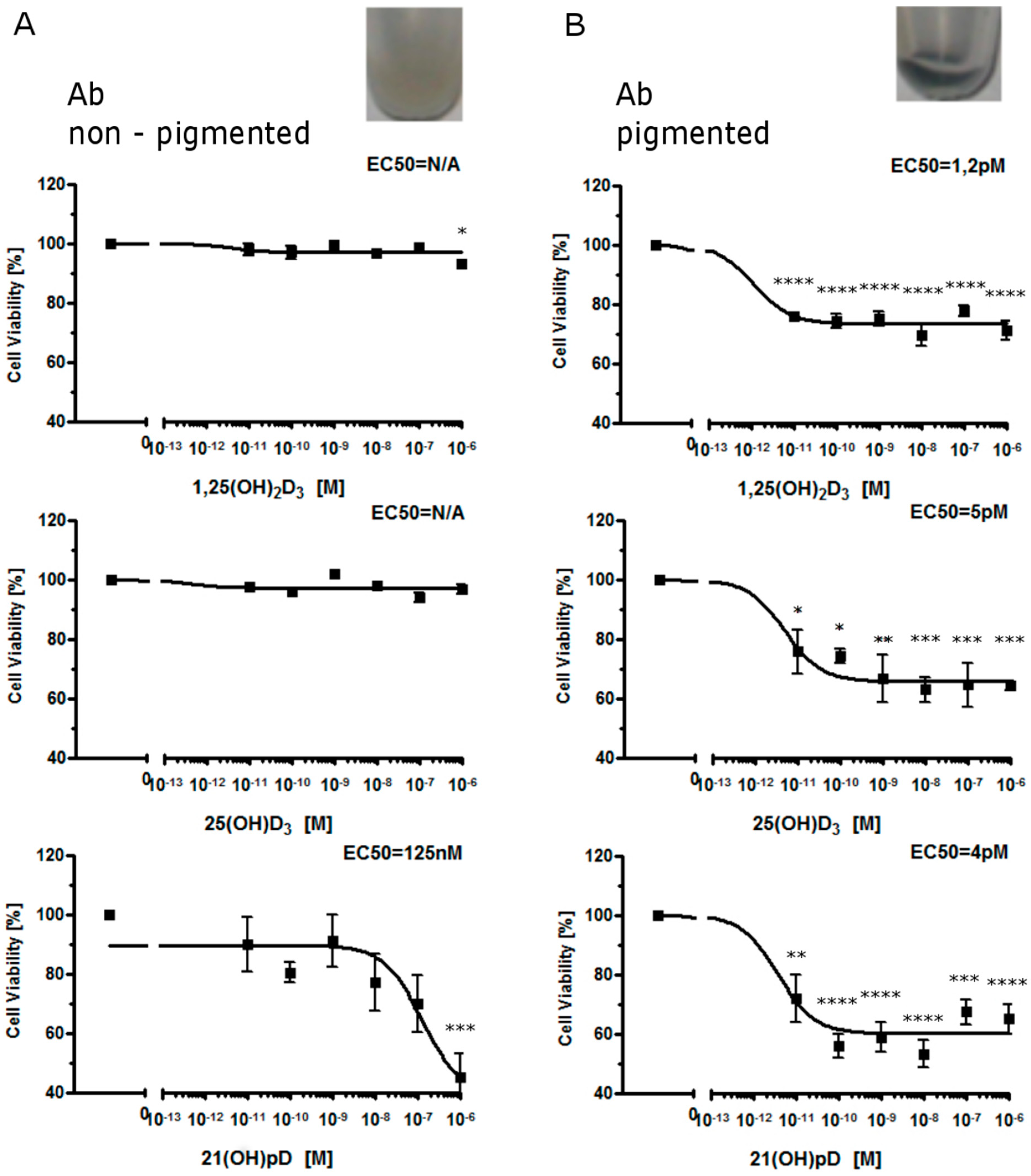
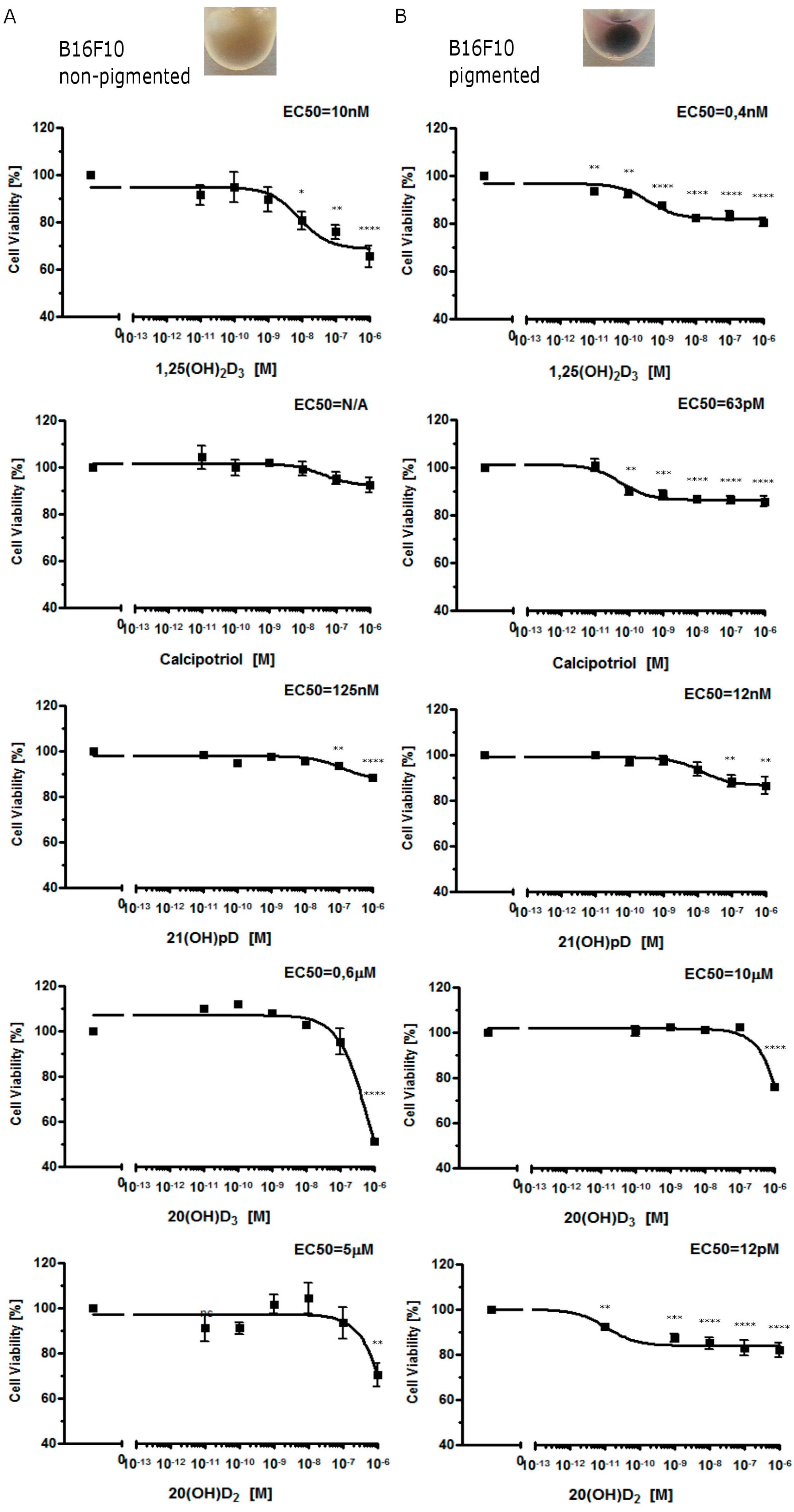
2.1. Moderate Pigmentation Enhances the Antiproliferative Effect of Vitamin D Analogs in Ab and B16-F10 Cells

Previous studies on human melanoma SKMEL-188 cells demonstrated that enhanced melanogenesis attenuated the antiproliferative activity of 20(OH)D3 which was associated with downregulation of VDR expression [46]. It had no effect on the antiproliferative effects of pD metabolites but enhanced the effects of lumisterol-like and tachysterol-like compounds [36]. To better understand the mechanism of differential action of Vitamin D and lumisterol metabolites in relation to the melanogenic pathway, we used two rodent (mouse and hamster) melanoma models in which melanin synthesis is inducible by melanin precursors [47,56,57].
In primary cultures of Ab cells freshly isolated from solid tumors, moderate melanin pigmentation sensitized cells towards 1,25(OH)2D3, 25(OH)D3 and the short side-chained 21(OH)pD analog. Interestingly, only 21(OH)pD demonstrated antiproliferative activity towards non-pigmented Ab primary cultures (Figure 1). In contrast, all compounds tested, including 1,25(OH)2D3, calcipotriol, 20(OH)D3, and 21(OH)pD, inhibited growth of non-pigmented murine B16-F10 cells (Figure 2). The sensitivity of pigmented Ab and B16-F10 cells towards Vitamin D analogs was higher in comparison to non-pigmented cells (Figure 1 and Figure 2) with one exception, the effect of 20(OH)D3 was similar for both pigmented and non-pigmented B16-F10 melanoma cells (Figure 2).
Figure 1. Inhibition of growth of hamster Ab melanoma cells by 1,25(OH)2D3, 25(OH)D3 and 21(OH)pD. Non-pigmented (A) and pigmented (B) Ab cells (grown as described in Materials and Methods section) were treated with serial dilutions of Vitamin D analogs (0.01–1000 nM). The experiment was repeated with similar results. The amelanotic and melanotic phenotypes are shown as inserts. Data are presented as mean ± SEM (n = 6). Statistical significance was estimated using t-test and presented as * p < 0.05, ** p < 0.005, *** p < 0.0005, **** p < 0.0001 vs. control.
Ijms 16 06645 g001
Figure 2. Growth inhibition of B16-F10 mouse melanoma cells by 1,25(OH)2D3, calcipotriol, 21(OH)pD, 20(OH)D3 and 20(OH)D2. Non–pigmented (A) and pigmented (B) B16-F10 cells were treated with serial dilutions of Vitamin D analogs (0.01–1000 nM). After 48 h the cells were subjected to a SRB test. The experiment was repeated with similar results. Examples of amelanotic and melanotic cells are shown as inserts above graphs. 1,25(OH)2D3 was used as a positive control. Data are presented as mean ± SEM for six independent measurements. Statistical significance was estimated using the t-test and presented as * p < 0.05, ** p < 0.005, *** p < 0.0005, **** p < 0.0001 vs. control.
Ijms 16 06645 g002

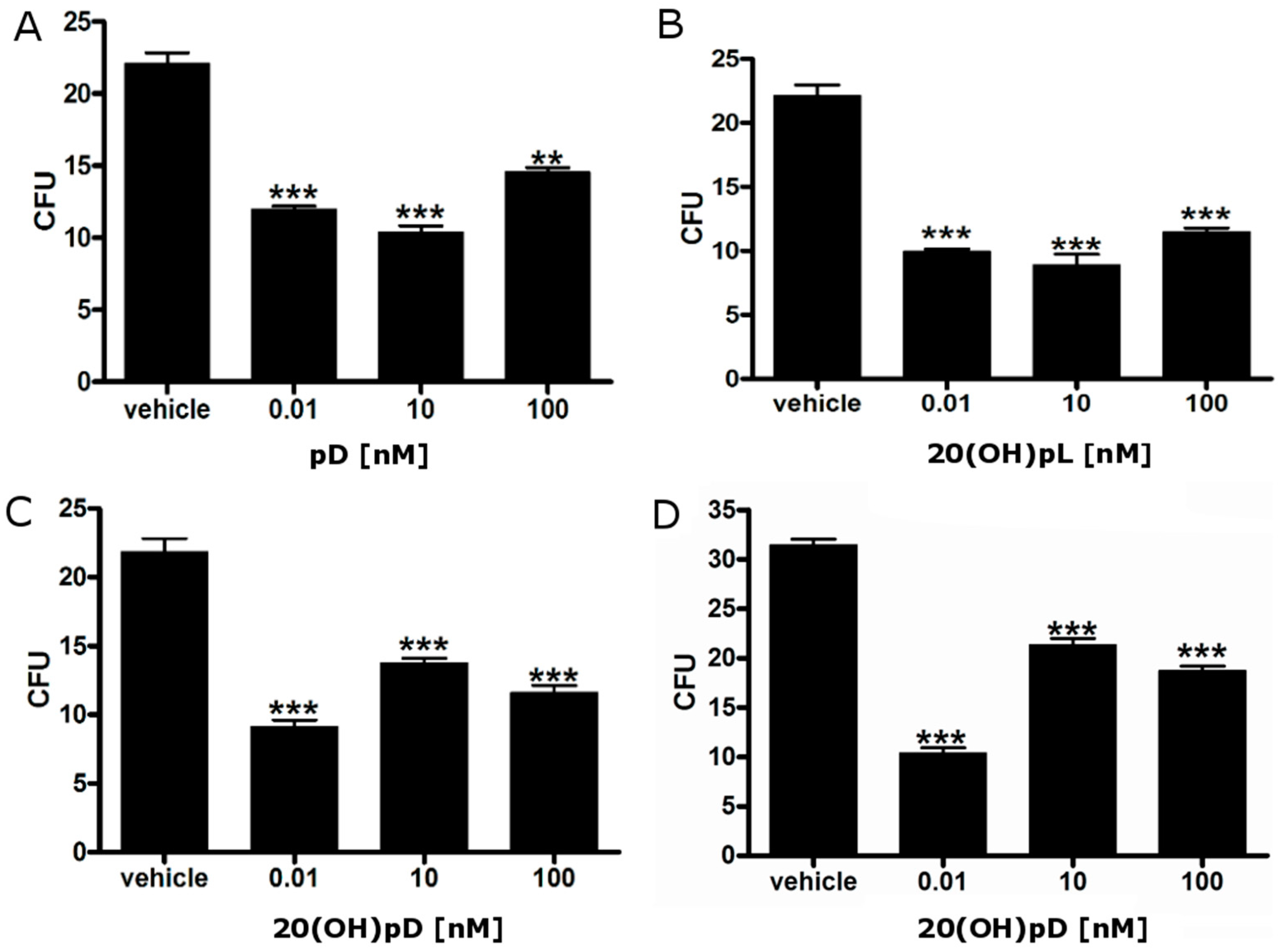
2.2. Vitamin D Analogs Inhibit the Growth of AbC1 Cells

We tested the inhibitory effects of Vitamin D and lumisterol analogs with a short side-chain, including pD, 20(OH)pL and 20(OH)pD, on colony formation in soft agar. All three analogs inhibited colony formation in soft agar at all concentrations tested (Figure 3). In addition, the detailed effect of 20(OH)pD on anchorage-independent growth, where colonies of different sizes (0.2 or 0.5 mm) were measured, is shown in Figure 3C,D.
Figure 3. Short side-chain vitamin D analogs inhibit the ability of AbC1 hamster melanoma cells to form colonies in soft agar. Cells were grown in soft agar in the presence or absence of pD (A); 20(OH)pL (B); or 20(OH)pD (C,D). After three weeks, colonies with a diameter larger than 0.2 mm (C) or 0.5 mm (D) were counted. Data are shown as means ± SD (n = 4), statistical significance was estimated using t-test and presented as ** p < 0.01, *** p < 0.001.
Ijms 16 06645 g003

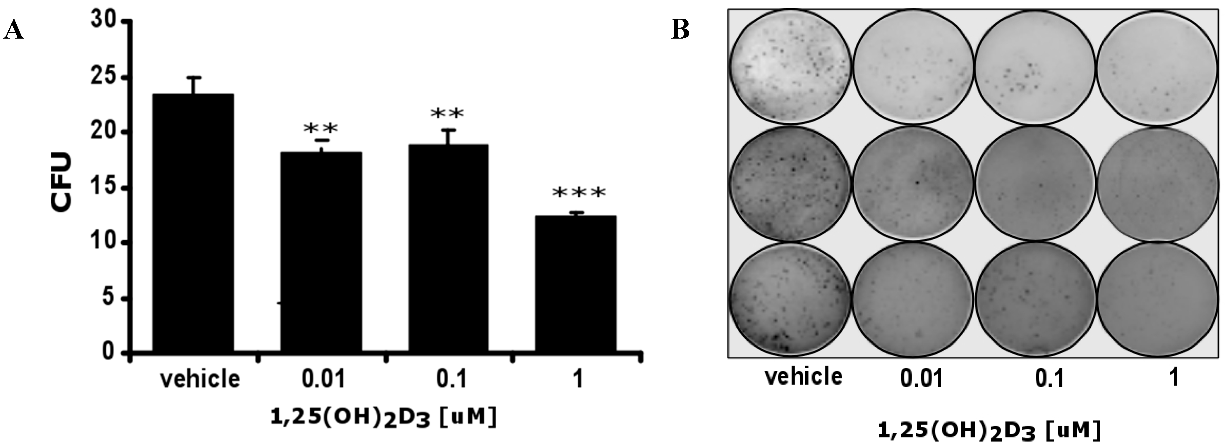
2.3. 1,25(OH)2D3 Decreases the Number and Size of Colonies Formed by Hamster Ab Melanoma Cells Freshly Isolated from Solid Tumors

The AbC1 line represents a continuous line derived from Ab melanoma that has been propagated in vitro since 1987 [56]. Therefore, further detailed testing was performed on primary cell cultures using freshly isolated melanoma cells from transplantable Ab amelanotic tumors. 1,25(OH)2D3 decreased the ability of primary Ab cells to form colonies in soft agar assays, in a dose-dependent manner, and also decreased their size (Figure 4). Although the AbC1 cell line was isolated directly from an Ab hamster melanoma, it should be treated as a separate continuous cell line. Therefore, future in vivo studies on short side chain Vitamin D and lumisterol analogues are required to compare Ab melanoma sensitivity with classical 1,25(OH)2D3, although differences are suggested by studies on the AbC1 line (Figure 3 and Figure 4).
Figure 4. 1,25(OH)2D3 inhibits Ab hamster melanoma colony formation in soft agar. Ab cells were grown in soft agar in the presence or absence of 1,25(OH)2D3. After three weeks, colonies were counted. Statistical significance was estimated using the t-test and data are presented as ** p < 0.01, *** p < 0.001, ±SD (A); Representative plate (n = 3) showing inhibition of Ab melanoma colony formation (B).
Ijms 16 06645 g004

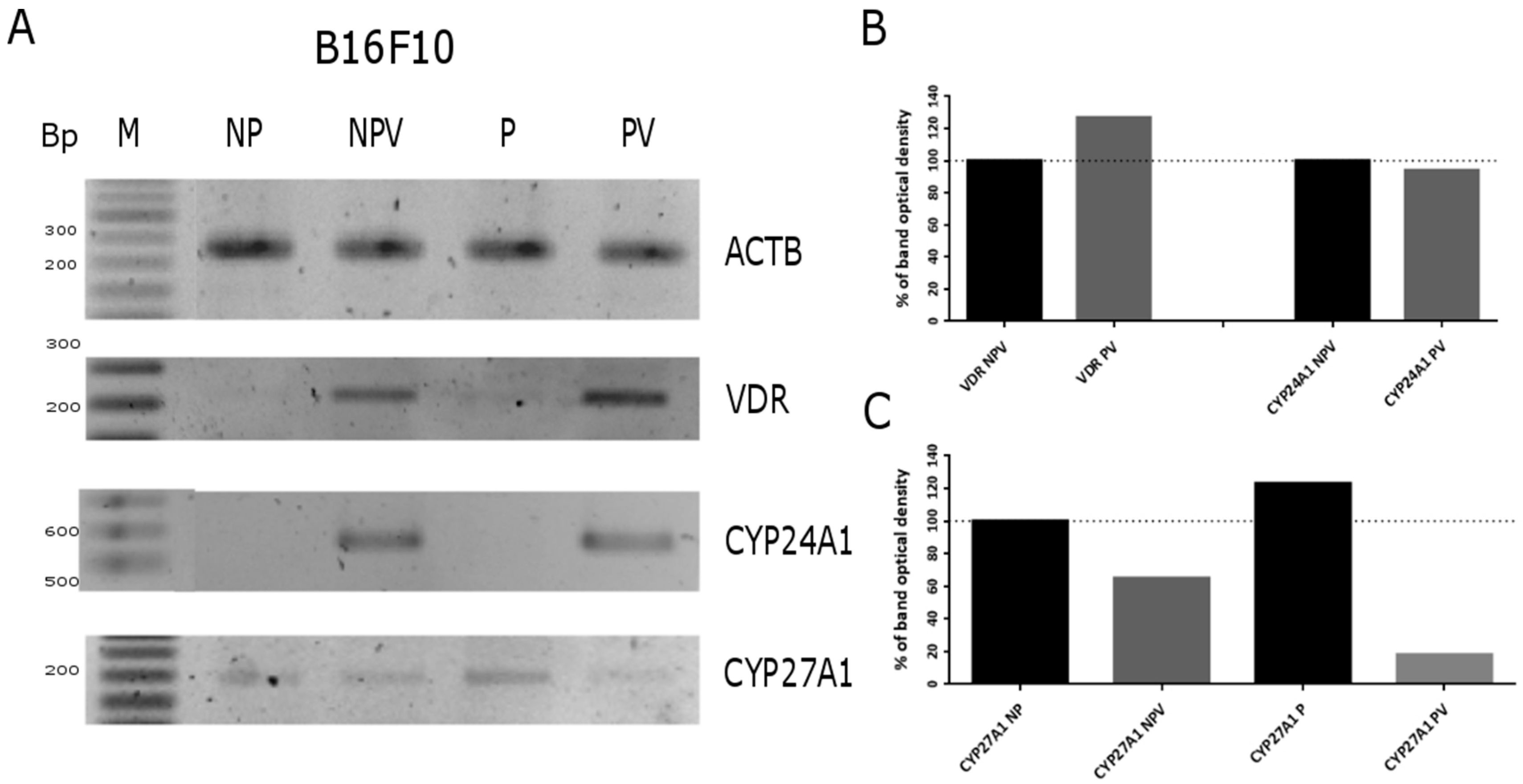
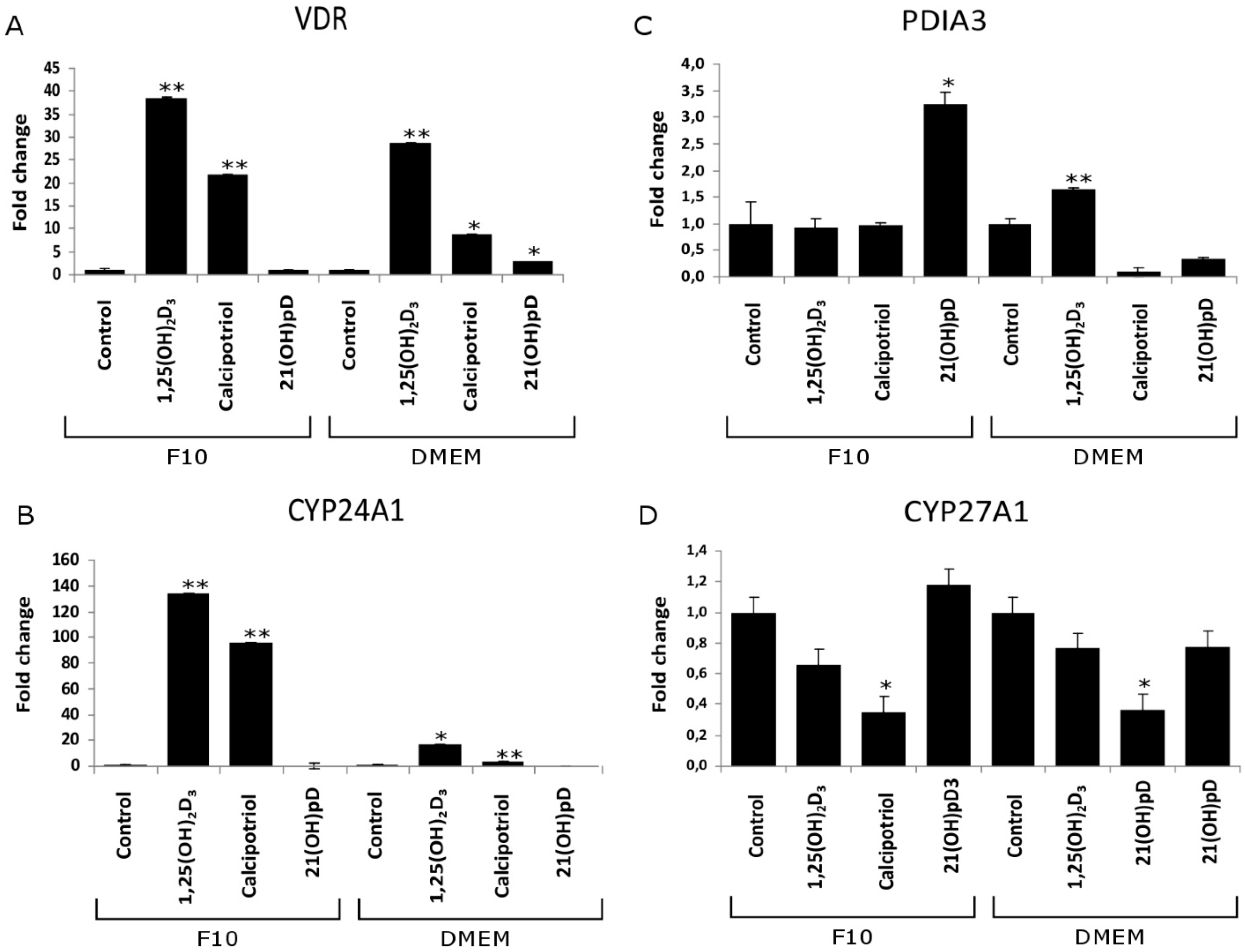
2.4. Expression of Vitamin D Related Genes Is Modulated by Melanin Pigmentation

As reported previously, media rich in tyrosine, such as DMEM, activates the pigmentary system in Ab hamster melanoma cells [57,58] with an accompanying increase in tyrosinase activity [59,60]. These results were confirmed in the current study (see inserts in Figure 1 and Figure 5). Since sensitivity of cells to Vitamin D depends on expression of several genes involved in regulation of its activity and metabolism, we investigated how production of melanin pigment influences their expression. It was shown that pigmentation dramatically increased the expression of VDR and its co-receptor RXR at the mRNA level (Figure 5A), while expression of 25-hydoxylase (CYP27A1) mRNA was only slightly elevated by pigmentation. Interestingly, expression of the potential alternative Vitamin D receptor PDIA3 decreased in pigmented Ab cells. The increases in VDR and 24-hydroxylase CYP24A1 mRNA levels after 1,25(OH)2D3 treatment were more pronounced in pigmented Ab cells than in non-pigmented ones (Figure 5B).
In order to confirm proper PCR amplification of fragments of hamster TYR, PDIA3 and CYP24A1 mRNAs, partial DNA sequencing of the PCR fragments was performed (Figure S1). In silico translation of TYR, PDIA3 and CYP24A1 PCR fragments, additionally confirmed the correct identification of the amplicons (Figure S2).
Figure 5. Melanogenesis and 1,25(OH)2D3 modulate the expression of tyrosinase (TYR), Vitamin D receptor (VDR), retinoid X receptor (RXR), disulfide isomerase (PDIA3), 25-hydoxylase (CYP27A1) and 1α-hydroxylase (CYP27B1) genes in hamster Ab melanoma cells. The effects of pigmentation (A) and 1,25(OH)2D3 on pigmented and non-pigmented cells (B) on the expression of selected genes were measured by semi-quantitative PCR. PCR analysis was performed as described in Materials and Methods. Glyceraldehyde 3-phosphate dehydrogenase (GAPDH) and β-actin (ACTB) were used as control genes. Length of amplicon fragments (in base pairs): GAPDH (496), Actin (353), TYR (205), VDR (234), RXR (210), CYP27A1(209), CYP27B1 (189), CYP24A1 (201). Densitometric analysis of PCR results (from panel A and B) obtained using TotalLab Quant v 11.4 (C,D). M, DNA marker; NP, non-pigmented cells (F10); NPV, non-pigmented cells treated with 1,25(OH)2D3; P, pigmented cells (DMEM); PV, pigmented cells treated with 1,25(OH)2D3; and C, control. Panels C and D showed quantification of band density from panels A and B, respectively.
Ijms 16 06645 g005
As shown in Figure 6, in murine B16-F10 cells, the switch of medium from F-10 (low tyrosine) to DMEM:F-10 mixture (50:50, high in tyrosine) initially stimulated expression of VDR mRNAs (moderate melanin pigmentation) but then it inhibited its expression when the cells became heavily pigmented (not shown). Administration of 1 μM 1,25(OH)2D3 to B16-F10 cells increased VDR and CYP24A1 mRNA levels in both pigmented and non-pigmented melanoma cells (Figure 6). Unfortunately, quantification of band intensity of VDR and CYP24A1 PCR products was not possible due to lack of visible product in untreated cells (both pigmented and non-pigmented B16-F10). In the case of CYP27A1, its expression was slightly increased by pigmentation while treatment of pigmented B16-F10 cells with 1,25(OH)2D3 attenuated its expression. The relative levels of VDR, CYP24A1, CYP27A1 and PDIA3 mRNA in control and ligand stimulated cells was analyzed further by real time PCR. Interestingly, the induction of the expression of VDR and its downstream CYP24A1 genes by 1,25(OH)2D3 and calcipotriol was attenuated in pigmented cells in comparison to the non-pigmented ones (Figure 7A,B). For example, pigmented B16-F10 cells showed eight-fold lower CYP24A1 expression after treatment with 1,25(OH)2D3 and a 25-fold lower expression after calcipotriol treatment, compared to non-pigmented cells (Figure 7B). In addition, 1,25(OH)2D3 stimulated expression of VDR and CYP24A1 genes to a higher degree than calcipotriol in both pigmented and non-pigmented cells (Figure 7A,B). Interestingly, treatment of both pigmented and non-pigmented B16-F10 cells with 1,25(OH)2D3 and calcipotriol resulted in reduction of CYP27A1 expression and the effect was more pronounced in cells treated with calcipotriol than with 1,25(OH)2D3 (Figure 7C).
Figure 6. Melanogenesis and 1,25(OH)2D3 modulate VDR, CYP24A1 and CYP27A1 gene expression in B16-F10 melanoma cells. 1,25(OH)2D3 was used at a concentration of 1 μM. PCR analysis was performed as described in Materials and Methods, β-actin (ACTB) was used as a control (A); The lengths of amplicons fragments (in base pairs) were: Actin (353), VDR (234), CYP24A1 (201), CYP27A1 (209). Densitometric analysis of PCR results obtained using TotalLab Quant v 11.4 (B,C). NP, non-pigmented cells (F10); NPV, non-pigmented cells treated with 1,25(OH)2D3; P, pigmented cells (DMEM); PV, pigmented cells treated with 1,25(OH)2D3. Data are from the most representative measurement.
Ijms 16 06645 g006
Treatment with 21(OH)pD, a short side-chain analog of Vitamin D (36), increased PDIA3 mRNA levels in non-pigmented B16-F10 cells (Figure 7D), but had no effect on VDR, CYP27A1 or CYP24A1 mRNA. However, mRNA levels of PDIA3 decreased in pigmented B16-F10 cells treated with calcipotriol and 21OHpD, while slightly increasing in cells treated with 1,25(OH)2D3 (Figure 7C).
Figure 7. Influence of 1,25(OH)2D3, calcipotriol and 21(OH)pD on the expression of VDR, PDIA3, CYP24A1 and CYP27A1 genes in pigmented and non-pigmented B16-F10 melanoma cells. VDR (A); PDIA3 (B); CYP24A1 (C) and CYP27A1 (D) mRNA levels were determined by quantitative PCR as described in Material and Methods. The expression of the genes was normalized by the comparative ΔΔ−Ct method, using β-actin as a housekeeping gene. Statistical significance was determined using the t-test and data are presented as * p < 0.05, ** p < 0.01 ±SD.
Ijms 16 06645 g007

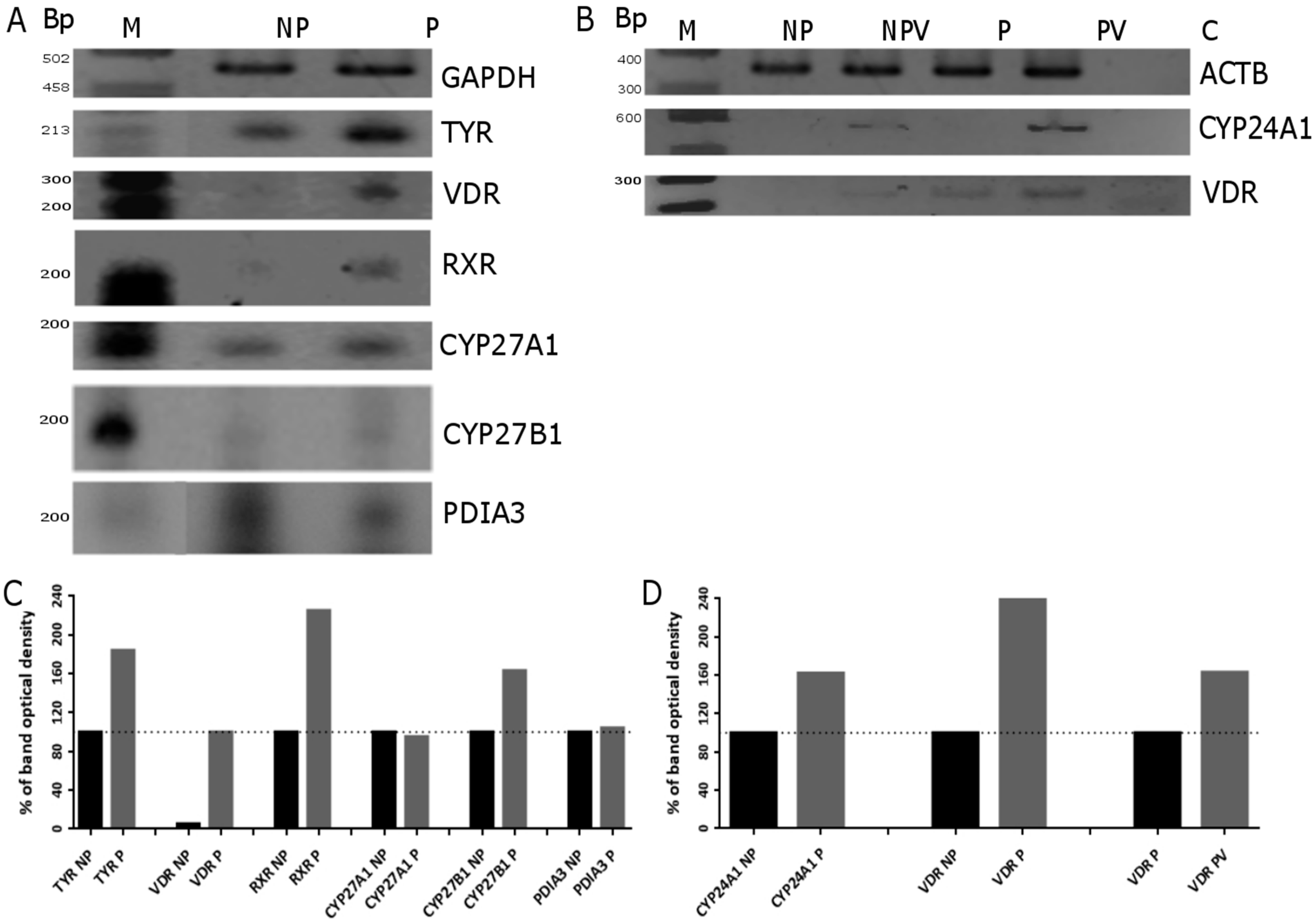
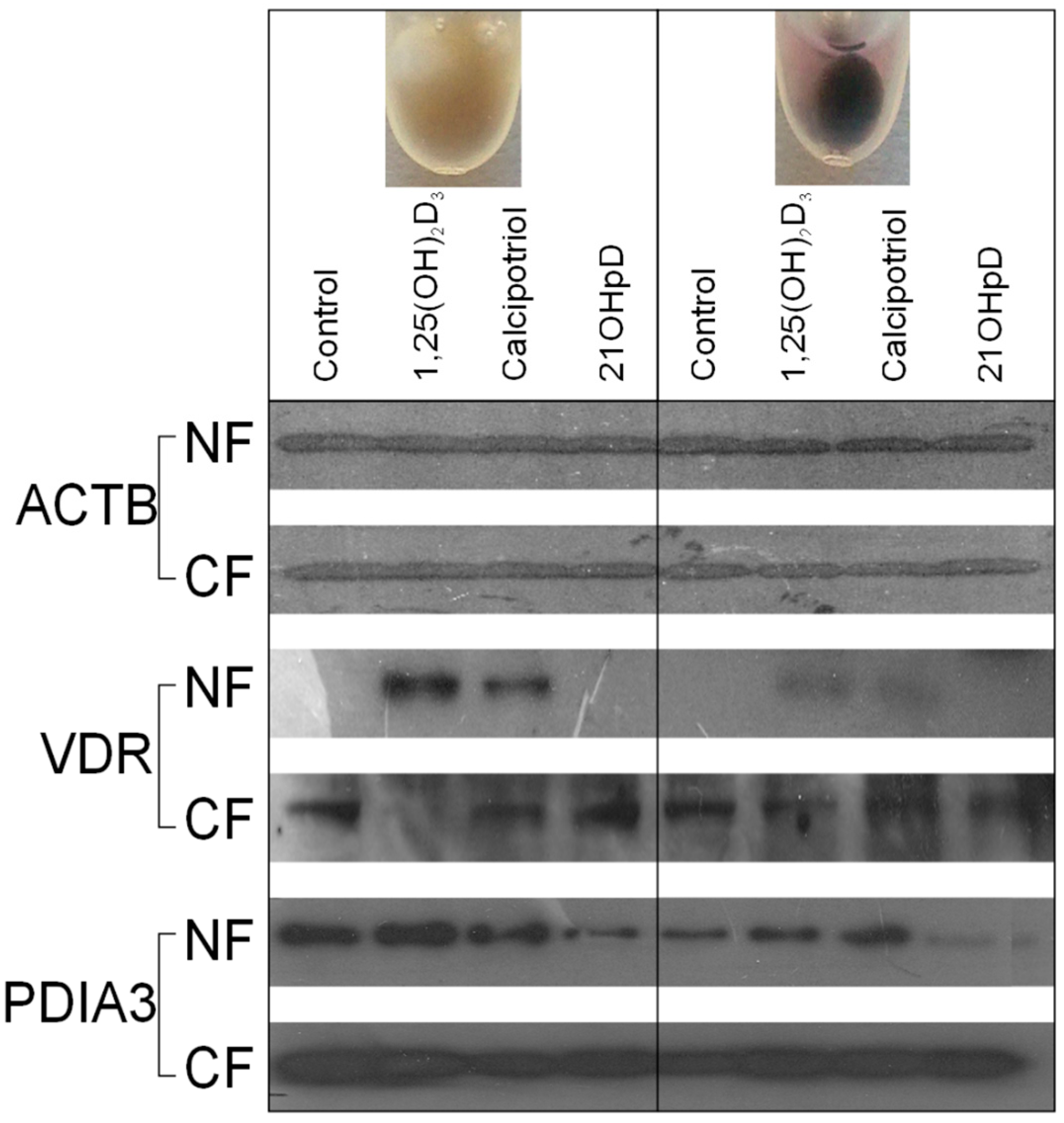
2.5. VDR Receptor Translocation to the Nucleus Is Stimulated by 1,25(OH)2D3

Western blotting analysis showed that the VDR is synthesized in B16-F10 mouse melanoma cells and its translocation to the nucleus is stimulated by treatment with 1,25(OH)2D3 or calcipotriol (Figure 8). Interestingly, ligand induced translocation of the VDR to the nucleus is attenuated in pigmented cells in comparison to the non-pigmented ones. However, 21(OH)pD had no effect on VDR protein expression nor its translocation to the nucleus. In addition, Western blot analysis demonstrated a negative correlation between 21(OH)pD treatment and the amount of PDIA3 in the nuclear fraction, while nuclear localization of PDIA3 was slightly increased by 1,25(OH)2D3 or by calcipotriol treatment of pigmented B16-F10 melanoma cells.

3. Discussion

In this paper, we report the differential anti-proliferative responses of pigmented and non-pigmented rodent melanoma cells to classical metabolites of Vitamin D (1,25(OH)2D3 and 25(OH)D3) and novel 20(OH)D3 [61] and calcipotriol [62], as well as short-side chain secosteroids, pD [63], 20(OH)pD, 20(OH)pL [64] and 21(OH)pD [36]. It should be noted that 20(OH)D3, pD, 20(OH)pD and 21(OH)pD are non- or low- calcemic secosteroids that may represent therapeutic alternatives to 1,25(OH)2D3, which is highly calcemic [32,33,34,35,36,37,38,39,40,41,42,43,44]. These compounds belong to a new class of endogenously produced Vitamin D analogs, with their 5,7-diene-like precursors potentially being made in the skin or other tissues [34,40,41,42,65]. Furthermore, formation of 21(OH)pD from 21(OH)7DHP occurs in skin subjected to UVB irradiation under ex vivo conditions [36]. Interestingly, the short side-chain lumisterol-like derivative, 20(OH)pL, which is generated together with 20(OH)pD after UVB irradiation of 20(OH)7DHP [36], was also found to inhibit murine melanoma cell growth, broadening the spectrum of biologically active photo-derivatives of 5,7-dienes.
An additional goal of this study was to characterize the effects of inducible pigmentation on the response to Vitamin D analogs in two melanoma cell lines, murine B16-F10 and hamster Ab. Both cell lines are fully synergistic with animal models (mouse and hamster, respectively), enabling their future direct use in animal studies.
Production of melanin has been shown to influence Vitamin D production in the skin [14]. In human melanoma cells, melanin pigment attenuates their responsiveness to gamma radiation, the cytotoxic effects of chemotherapeutics and immunotoxic activities towards tumor cells [52,53,54]. Melanogenesis also decreases the sensitivity of human melanoma cells to treatment with Vitamin D derivatives in vitro [46]. Finally, pigmentation of human melanomas in vivo shows a reverse correlation with the expression of VDR [50,66] and CYP27B1 [67]. These studies indicate that melanin pigmentation can impair both the metabolism and activity of classical Vitamin D derivatives in human melanomas.
The sensitivity of murine B16-F10 melanoma to Vitamin D analogs has been widely investigated with contradictory results, with the responsiveness to the treatment usually being related to VDR expression (see for review and discussion: [35]). Current studies on rodent melanomas show that expression of VDR is below the level of detectability in non-pigmented cells by conventional PCR. However, treatment with 1,25(OH)2D3 and calcipotriol resulted in a significant increase in the level of the VDR transcript, with 1,25(OH)2D3 being the more effective of the two secosteroids (Figure 5, Figure 6, and Figure 7). The increased expression of VDR transcript was observed in both pigmented and non-pigmented cells and correlated with induction of the expression of its main downstream target gene, CYP24A1. Interestingly, induction of melanin pigmentation attenuated 1,25(OH)2D3 and calcipotriol induced expression of VDR and CYP24A1 genes (Figure 7A,B). This finding is consistent with attenuation of 1,25(OH)2D3 induced translocation of VDR to the nucleus in pigmented melanoma cells (Figure 8), which again explains the reduction by eight-fold of the stimulation of CYP24A1 gene expression by 1,25(OH)2D3 (Figure 7B). In contrast, treatment with the short side-chain Vitamin D analog, 21(OH)pD, did not stimulate expression of either VDR or CYP24A1 mRNAs. Based on the presented data, we suggest that very low expression of VDR is sufficient for activation of Vitamin D genomic responses, and VDR itself is one of the first targets. Various mechanisms may trigger VDR expression with the induction of VDR-regulated genes being a secondary effect.
Figure 8. Western blot analysis of the effects of 1,25(OH)2D3, calcipotriol and 21(OH)pD on the cytoplasmic and nuclear location of the VDR and the PDIA3 receptor. Pigmented and non-pigmented B16-F10 melanoma cells were treated with 10 nM 1,25(OH)2D3, calcipotriol or 21(OH)pD, and VDR and PDIA3 receptors in cytoplasmic (CF) and nuclear fractions (NF) measured by western blotting, as described in the Materials and Methods section. Actin (ACTB) was used as a loading control.
Ijms 16 06645 g008
Our data indicate that moderate pigmentation sensitizes murine melanoma cells to Vitamin D analogs, including 1,25(OH)2D3, 25(OH)D3, calcipotriol and 21(OH)pD. The inhibitory effect of 1,25(OH)2D3 on proliferation is consistent with previous reports (for review see [19,34,35]). For example, an increased sensitivity of pigmented human SKMEL-188 melanoma cells towards 1,25(OH)2D3, 21(OH)pL, 21(OH)7DHP or its oxidized analog 21(OH)oxy-piT was shown recently [36]. In the same study, the activity of 21(OH)pD was not affected by pigmentation. In the current study, pigmented murine B16-F10 cells were only slightly more sensitive than non-pigmented ones. This response contrasted with that of pigmented hamster Ab melanoma cells, which were several fold more sensitive to 21(OH)pD than non-pigmented cells. Interestingly, an induction of heavy pigmentation in human SKMEL-188 cells attenuated the anti-proliferative effects of 20(OH)D3, which was accompanied by decreased expression of VDR at the protein level [46]. With B16-F10 murine melanoma in the current study, 20(OH)D3 exerted an inhibitory effect only at the highest concentration tested (10−6 M) in both pigmented and non-pigmented cells. These data indicate that the sensitivity of melanoma cells to Vitamin D derivatives can be influenced by the intensity of melanin pigmentation in a species-, cell type- and condition-dependent manner. Furthermore, it can be speculated that production of pigment influences the metabolism and stability of Vitamin D analogs, and thus this phenomenon requires further in depth investigation. However, observed changes in the level of CYP27A1 mRNA may suggest enhanced 25-hydoxylase activity. Interestingly, production of melanin is also accompanied by reactive oxygen species (ROS) generation. Although the presence of ROS is usually linked to Vitamin D degradation, recently we described a new oxidized photo-derivative of 21(OH)7-dehydropregnonolene (21(OH)oxy-piT) as a potent inhibitor of human SKMEL-188 melanoma proliferation [36].
The treatment of murine B16-F10 cells with 21(OH)pD caused antiproliferative effects, but did not lead to an increase in VDR or CYP24A1 mRNA levels. It is well established that increased CYP24A1 expression leads to increased inactivation of Vitamin D analogs [68], thus the high CYP24A1 mRNA expression after treatment may, at least partially, explain the observed limited growth inhibition of non-pigmented mouse B16-F10 cells. It has to be noted that short side-chain analogs, such as 21(OH)pD, are not a target for 24-hydroxylation because they lack a sterol side-chain with C24. The lack of stimulation of CYP24A1 expression in murine B16-F10 melanoma cells by 21(OH)pD may be explained by the impaired interaction with the VDR. Indeed, our study shows that the secosteroids tested do differ in their ability to induce the translocation of the VDR into the nucleus of B16-F10 cells. In comparison to 1,25(OH)2D3, calcipotriol showed only partial activity, while treatment with 21(OH)pD did not result in any detectable nuclear localization of the VDR. Recent molecular modeling studies by Kim et al. [69], indicated that a range of Vitamin D analogs differed in their predicted VDR binding capacity in a manner that correlated with their ability to stimulate VDR translocation to the nucleus. According to this in silico modeling, 21(OH)pD showed the lowest VDR docking score (weakest binding) among the Vitamin D analogs tested [69], and this corresponded to a poor ability to translocate the VDR from the cytoplasm to the nucleus [36,69]. In contrast to murine B16-F10 cells, treatment of human SKMEL-188 melanoma cells with 21(OH)pD resulted in partial translocation of VDR to the nucleus [36,69], suggesting possible species- or cell line-dependent differences in the response. Thus, the effects of pigmentation on the activity of Vitamin D analogs appear to depend not only on VDR expression and its translocation to the nucleus, but also on the structural properties of the secosteroid and possibly other cell-specific factors or receptors yet to be identified. The antiproliferative potential of 21(OH)pD, in conjunction with its low-calcemic activity and limited requirement for genomic activity of the VDR, warrants further in vivo testing against advanced, pigmented melanomas. This secosteroid could be of great value for the treatment of melanomas which display decreased expression of the VDR and enhanced expression of CYP24A1.
It has been suggested that PDIA3 (ERp57) is a Vitamin D-binding protein, which could also act as a transcription factor after stimulation [70,71]. The effects of Vitamin D analogs on PDIA3 mRNA levels and translocation to the nucleus were tested in B16-F10 cells. Only treatment with 21(OH)pD resulted in an elevated level of PDIA3 mRNA in non-pigmented cells, while 21(OH)pD and calcipotriol decreased PDIA3 mRNA expression in pigmented cells. 1,25(OH)2D3 had only a minor, but statistically significant, stimulatory effect on the PDIA3 mRNA level in pigmented B16-F10 cells. Interestingly, while 1,25(OH)2D3 and calcipotriol had only minor effects on the translocation of PDIA3 to the nucleus, 21(OH)pD inhibited nuclear translocation in both pigmented and non-pigmented B16-F10 cells. Therefore, PDIA3 might be considered as an alternative target for analogs with a short side-chain but the mechanism of their action requires further studies. In addition, it remains to be tested whether retinoic acid orphan receptors (RORs) can serve as a target for 21(OH)pD, since Vitamin D analogs with a full-length side chain affect the activity of RORα and γ [72].
When analyzing the melanin pigmentary system, it is important to note that an enhanced inhibitory effect of Vitamin D analogs is only observed immediately after induction of melanogenesis, i.e., in mildly pigmented melanoma cells. Prolonged exposure to high levels of tyrosine resulted in heavy pigmentation of both Ab and B16-F10 melanoma cells, followed by inhibition of proliferation and cell detachment. Hyperpigmentation of melanoma cells is also associated with a decrease of the VDR, RXR and PDIA3 mRNA levels in B16-F10 cells. Furthermore, pigmented B16 melanoma cells show attenuation of responsiveness to ligand stimulation of VDR translocation to nucleus (Figure 8), and expression of VDR and CYP24A1 genes (Figure 7A,B), indicating that VDR signaling is malfunctioning in melanotic cells. These findings are consistent with a report showing that a high level of pigmentation of human melanomas was correlated with low expression of the VDR and CYP27B1 in vivo [50,67] and downregulation of VDR protein expression in pigmented human melanma line [50,66]. Since pigmentation affected overall disease-free survival in melanoma patients [49], further studies on the complex relationship between Vitamin D response, pigmentation and melanoma behavior are mandatory.

4. Materials and Methods

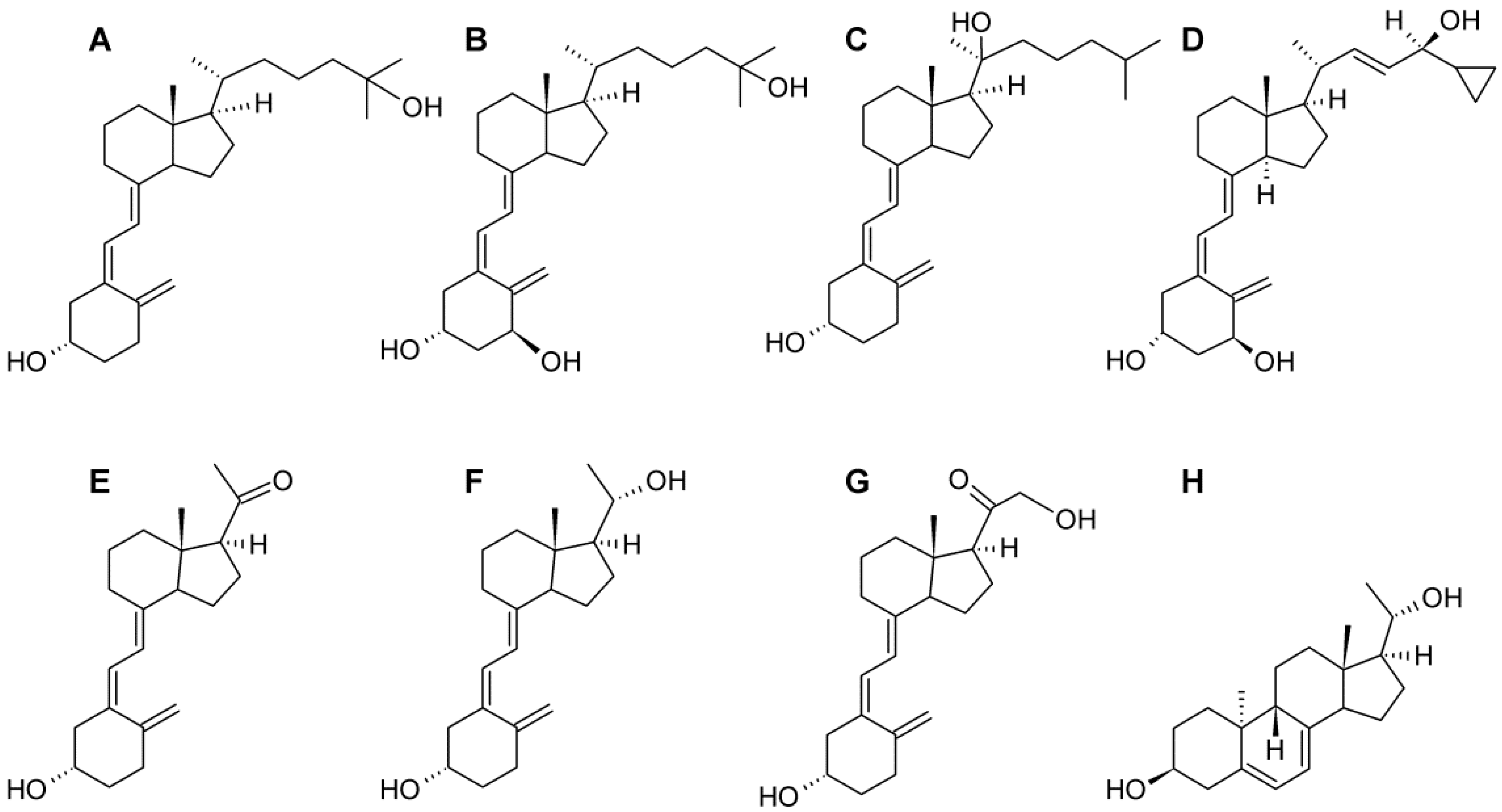
4.1. Cell Lines and Vitamin D Analogs

The Bomirski hamster Ab melanoma line originated in 1963 from the transplantable Ma melanotic melanoma of the Syrian hamster, through a process of tumor progression that included an increase in the growth rate, loss of melanin pigmentation and significant shortening of the overall survival of animals bearing the melanomas [73]. Since then, the Ab hamster melanoma line has been maintained in vivo by consecutive subcutaneous transplantations of tumor material [73,74,75]. The hamster Ab melanoma cells were obtained for each experiment from solid tumors using a non-enzymatic method of cell isolation, as previously described [76]. The resection of the tumor was performed after 10–11 days of growth after the transplantation. The cell suspensions used in the experiments consisted of 95%–98% viable cells, as estimated by the trypan blue exclusion assay. The transplantation procedure was approved by the Animal Ethics Committee at the Medical University of Gdansk and was in accordance with the National Health and Medical Research Council’s guide for the care and use of laboratory animals. Experiments were performed on freshly isolated cells defined as a primary melanoma cell culture.
Hamster Ab primary cells and the mouse B16-F10 cell line were cultured, in RPMI and Ham’s F-10 media, respectively, supplemented with 10% fetal bovine serum (Sigma, Poznan, Poland) and antibiotic-antimycotic solution (Sigma, Poznan, Poland). Culturing Ab melanoma cells in a medium high in tyrosine induced rapid melanin production with attendant changes in the differentiation program of the cells [57,58]. The tyrosine concentrations in media used were: DMEM, 423 μM; F-10, 11 μM; 50:50 DMEM:F-10, 217 μM; RPMI, 118 μM; 50:50 DMEM:RPMI, 270 μM.
The hamster AbC1 melanoma cell line, cloned from a primary culture of Bomirski Ab melanoma [56], was cultured in Ham’s F-10 medium supplemented with glucose, L-glutamine, pyridoxine hydrochloride (Cellgrow, Manassas, VA, USA), 5% fetal bovine serum (FBS) (Sigma, Poznan, Poland) and 1% penicillin/streptomycin/amphotericin antibiotic solution (Sigma, Poznan, Poland), as described previously [56,77]. For experimental purposes, 5% charcoal-stripped FBS (GE Healthcare Life Sciences, Warsaw, Poland) was used. The AbC1 cell line is an excellent model for pigmentation studies, as increasing the concentration of tyrosine in the medium induces melanin pigmentation of these cells and changes their biological activities [56,75,78,79].
1,25(OH)2D3, 25(OH)D3 and calcipotriol were acquired from Pharmaceutical Research Institute, Warsaw, Poland. Other Vitamin D analogs: pD, 20(OH)pD, 20(OH)pL and 21(OH)pD were synthesized and purified as described previously [36,64]. 20(OH)D2 and 20(OH)D3 analogs were also synthesized and purified as described previously [80,81,82,83]. The chemical structures of these secosteroids are presented in Figure 9.
Figure 9. Chemical structures of the Vitamin D analogs tested. (A) 25(OH)D3; (B) 1,25(OH)2D3; (C) 20(OH)D3; (D) calcipotriol; (E) pD; (F) 20(OH)pD; (G) 21(OH)pD; (H) 20(OH)pL.
Ijms 16 06645 g009

4.2. MTT Assay

Hamster melanoma cells were seeded at a density of 8000 cells per well into 96-well plates in RPMI or a mixture of RPMI:DMEM (50:50, v/v) media (Sigma, Poznan, Poland), supplemented with 2% charcoal-stripped fetal bovine serum (Biological Industries, Israel) and antibiotic-antimycotic solution (Sigma, Poland). After incubation for 24 h, the medium was replaced with a fresh one containing serial dilutions of 1,25(OH)2D3 or its analogs (at 0.01–1000 nM concentrations). After incubation for 48 h, 20 μL of MTT (3-(4,5-dimethylthiazol-2-yl)-2,5-diphenyltetrazoliumbromide; 5 mg/mL in PBS; Sigma, Poland) were added to each well and the plates were incubated for a further 3 h. Media were discarded and 100 μL of acidified isopropanol (containing 0.1 M hydrochloric acid) were added. The absorbance was measured at 570 nm.

4.3. SRB Assay

B16-F10 cells were seeded at a density of 8000–10,000 per well in 96-well plates in F-10 or DMEM medium (Sigma, Poland), supplemented with 2% charcoal-stripped fetal bovine serum (Biological Industries, Israel) and antibiotic-antimycotic solution (Sigma, Poznan, Poland). After incubation for 24 h, medium was replaced with a fresh one containing vehicle, 1,25(OH)2D3 or its analogs at concentrations of 1 pM to 1 μM. Following incubation for 48 h, 100 μL of 20% TCA were added and cells incubated for 1 h in 4 °C. The medium was then removed and 100 μL of SRB (sulforhodamine B) were added and the plates incubated for 15 min at room temperature. The SRB was discarded, cells washed and 150 μL of 10 mM Tris (pH 10.5) were added. The absorbance was measured at 570 nm.

4.4. PCR-Based Expression Analysis

RNA was isolated using a Total RNA kit (A&A Biotechnology, Gdynia, Poland). Reverse transcription (500 ng RNA/reaction) was performed with a First Strand cDNA Synthesis Kit (Fermentas, Lithuania). PCR reactions were performed using cDNA (diluted 10-fold in sterile water) and PCR MIX PLUS (A&A Biotechnology, Gdynia, Poland). Quantitative PCR analysis was performed using cDNA (diluted 5-fold in sterile water) and 2× PCR Master Mix SYBR (A&A Biotechnology). The primers for both lines were designed based on the mice and rat sequences using Primer Quest software (Integrated Device Technology, San Jose, CA, USA). The primers for the housekeeping gene, GAPDH, were obtained from the First Strand cDNA Synthesis Kit (Fermentas, Lithuania). The sequences of the primers are shown in Table S1. Standard PCR reactions were performed using a MJ Mini Bio-Rad cycler. Following agarose gel electrophoresis, the amplicons were visualized by ethidium bromide staining. For Ab cells, PCR fragments encoding tyrosinase, PDIA3 and CYP24A1 were sequenced by the Institute of Biochemistry and Biophysics (Polish Academy of Science, Warsaw, Poland). The resulting DNA sequences were compared with mouse and rat genes by multiple alignment algorithm using ClustalW2 software (Figure S1). After in silico translation of Ab sequenced fragments, predicted sequences of hamster proteins were aligned with the corresponding mouse and rat protein sequences (Figure S2).

4.5. Colony Formation Assay

The ability of hamster melanoma cells to form colonies in soft agar, as an equivalent of tumorigenicity, was determined as described previously [45], with some modifications. The bottom of the wells of a 12-well plate was covered with 0.5 mL of solubilized 0.8% agar solution in RPMI medium (base-mix). After solidification of the base, a top-mix of 0.4% agar solution in RPMI medium containing 500 Ab Bomirski cells or 1000 AbC1 cells, was placed on the top. The solidified soft agar cell layer was covered with 100 μL of liquid RPMI medium containing serial dilutions of Vitamin D and its analogs. 1,25(OH)2D3 was used at final concentrations of 10 nM, 100 nM or 1 µM for Ab cells, while 20(OH)pD, pD and 20(OH)pL were tested at concentrations of 0.01 nM, 10 nM or 100 nM, and 100 µM for AbC1 cells. Cells were grown under 5% CO2 at 37 °C for 2–3 weeks with the 100 µL of medium containing the secosteroids being changed every 2–3 days. Ab melanoma colonies were identified using ethidium bromide (0.2 mL/well) and visualized under a UV transilluminator. AbC1 melanoma colonies were stained with 500 µL/well of MTT (0.5 mg/mL) (Promega, Mannheim, Germany). Colonies were photographed and counted using ImageJ software.

4.6. Western Blotting

Pigmented and non-pigmented B16-F10 cells were treated for 24 h with 10 µM 1,25(OH)2D3, calcipotriol ((1S,3R,5Z,7E,22E,24S)-26,27-Cyclo-9,10-secocholesta-5,7,10,22-tetraene-1,3,24-triol) or 21(OH)pD. The Nuclear Extract Kit (Active Motif, La Hulpe, Belgium) was used for preparation of nuclear and cytoplasmic fractions. The protein concentration was measured by the Bradford method and samples were stored at −80 °C. The detection of VDR, PDIA3 and actin in nuclear and cytoplasmic extracts was performed using previously described methodology (46). Anti-VDR (1:100, Abcam, Cambridge, Great Britain), anti-PDIA3 (1:250, Sigma-Aldrich, Poznan, Poland) and anti-AKTB (1:200, Santa Cruz Biotechnology, CA, USA) antibodies were used to detect the antigens. The appropriate secondary antibody coupled to horseradish peroxidase (1:2000, Santa Cruz Biotechnology, Heidelberg, Germany) was then applied and proteins were visualized using Super Signal West Pico (Pierce Biotechnology, Rockford, IL, USA).

5. Conclusions

Our study suggests that complex mechanisms are involved in the antiproliferative activity of Vitamin D analogs on melanoma cells, which include a modulatory role of melanin pigmentation. First, low calcemic analogs of Vitamin D are potent inhibitors of rodent melanoma cells proliferation; Second, mildly pigmented melanoma cells are more sensitive to several Vitamin D analogs than non-pigmented ones; Third, pigmented cells not only show attenuation of ligand-induced translocation of VDR to the nucleus but also ligand-induced expression of VDR and CYP24A1 genes is reduced in comparison to non-pigmented cells; Last, nuclear localization of VDR and activation of the classical (genomic) Vitamin D response pathway may not be necessary for growth inhibition of murine cells. Other mechanisms should be taken into consideration, including but not limited to involvement of Vitamin D analogs in the regulation of PDIA3, Wnt/β-catenin, Hedgehog, Notch, RORα and RORγ related pathways [35,71,84]. We also speculate that elevated levels of reactive oxygen species (ROS) associated with active melanogenesis may stimulate formation of oxidized derivatives of Vitamin D analogs thereby modulating their activity. A similar effect was shown recently following prolonged UVB irradiation of 21-hydroxydehydropregnenolone, the 21(OH)pD precursor [36]. Thus, active melanogenesis may decrease the potency of some Vitamin D analogs by ROS-mediated deactivation while melanin, by sequestrating Vitamin D compounds, may reduce their availability to interact with the corresponding receptors. This hypothesis requires further testing.

Supplementary Materials

Supplementary materials can be found at https://www.mdpi.com/1422-0067/16/04/6645/s1.

Acknowledgments

The study was supported by a grant of the Polish Ministry of Science and Higher Education, Project No. N405 623238 to Michal A. Zmijewski and grants R01A6R052190, R21AR066505 and 1R01AR056666-01A2 from the NIH/NAIMS (USA) and a West Clinic grant to Andrzej T. Slominski.

Author Contributions

Tomasz Wasiewicz and Paulina Szyszka performed the main experiments, took part in data analyzes and preparation of the manuscript. Miroslawa Cichorek contributed to Ab melanoma studies, while Zorica Janjetovic performed colony formation assay on AbC1 hamster melanoma; Robert C. Tuckey and Andrzej T. Slominski provided crucial vitamin D analogs; Michal A. Zmijewski and Andrzej T. Slominski design experiments; Michal A. Zmijewski, Tomasz Wasiewicz, Robert C. Tuckey and Andrzej T. Slominski analyzed data and prepared final version of the manuscript.

Conflicts of Interest

The authors declare no conflict of interest.

References

  1. Haussler, M.R.; Haussler, C.A.; Jurutka, P.W.; Thompson, P.D.; Hsieh, J.C.; Remus, L.S.; Selznick, S.H.; Whitfield, G.K. The vitamin D hormone and its nuclear receptor: Molecular actions and disease states. J. Endocrinol. 1997, 154, S57–S73. [Google Scholar] [CrossRef] [PubMed]
  2. Carlberg, C.; Seuter, S. The vitamin D receptor. Dermatol. Clin. 2007, 25, 515–523. [Google Scholar] [CrossRef] [PubMed]
  3. Wierzbicka, J.; Piotrowska, A.; Żmijewski, M.A. The renaissance of vitamin D. Acta Biochim. Pol. 2014, 61, 679–686. [Google Scholar] [PubMed]
  4. Holick, M.F. Vitamin D: A millenium perspective. J. Cell Biochem. 2003, 88, 296–307. [Google Scholar] [CrossRef] [PubMed]
  5. DeLuca, H.F. Overview of general physiologic features and functions of vitamin D. Am. J. Clin. Nutr. 2004, 80, 1689S–1696S. [Google Scholar] [PubMed]
  6. Fleet, J.C.; DeSmet, M.; Johnson, R.; Li, Y. Vitamin D and cancer: A review of molecular mechanisms. Biochem. J. 2012, 441, 61–76. [Google Scholar] [CrossRef] [PubMed]
  7. Holick, M.F. Vitamin D deficiency. N. Engl. J. Med. 2007, 357, 266–281. [Google Scholar] [CrossRef] [PubMed]
  8. Slominski, A.; Zbytek, B.; Nikolakis, G.; Manna, P.R.; Skobowiat, C.; Zmijewski, M.; Li, W.; Janjetovic, Z.; Postlethwaite, A.; Zouboulis, C.C.; et al. Steroidogenesis in the skin: Implications for local immune functions. J. Steroid Biochem. Mol. Biol. 2013, 137, 107–123. [Google Scholar] [CrossRef] [PubMed]
  9. Holick, M.F. Resurrection of vitamin D deficiency and rickets. J. Clin. Investig. 2006, 116, 2062–2072. [Google Scholar] [CrossRef] [PubMed]
  10. Holick, M.F. The photobiology of vitamin D and its consequences for humans. Ann. N. Y. Acad. Sci. 1985, 453, 1–13. [Google Scholar] [CrossRef] [PubMed]
  11. Holick, M.F. The cutaneous photosynthesis of previtamin D3: A unique photoendocrine system. J. Investig. Dermatol. 1981, 77, 51–58. [Google Scholar] [CrossRef] [PubMed]
  12. Holick, M.F.; Clark, M.B. The photobiogenesis and metabolism of vitamin D. Fed. Proc. 1978, 37, 2567–2574. [Google Scholar] [PubMed]
  13. Slominski, A.; Tobin, D.J.; Shibahara, S.; Wortsman, J. Melanin pigmentation in mammalian skin and its hormonal regulation. Physiol. Rev. 2004, 84, 1155–1228. [Google Scholar] [CrossRef] [PubMed]
  14. Wacker, M.; Holick, M.F. Sunlight and Vitamin D: A global perspective for health. Dermatoendocrinology 2013, 5, 51–108. [Google Scholar] [CrossRef]
  15. Zehnder, D.; Bland, R.; Williams, M.C. Extrarenal expression of 25-hydroxyvitamin D3-1α-hydroxylase. J. Clin. Endocrinol. Metab. 2001, 86, 888–894. [Google Scholar] [PubMed]
  16. Flanagan, J.N.; Wang, L.; Tangpricha, V.; Reichrath, J.; Chen, T.C.; Holick, M.F. Regulation of the 25-hydroxyvitamin d-1α-hydroxylase gene and its splice variant. Recent Results Cancer Res. 2003, 164, 157–167. [Google Scholar] [PubMed]
  17. Wang, L.; Whitlatch, L.W.; Flanagan, J.N.; Holick, M.F.; Chen, T.C. Vitamin D autocrine system and prostate cancer. Recent Results Cancer Res. 2003, 164, 223–237. [Google Scholar] [PubMed]
  18. Cheng, J.B.; Motola, D.L.; Mangelsdorf, D.J.; Russell, D.W. De-orphanization of cytochrome P450 2R1: A microsomal vitamin D 25-hydroxilase. J. Biol. Chem. 2003, 278, 38084–38093. [Google Scholar] [CrossRef] [PubMed]
  19. Slominski, A.T.; Zmijewski, M.A.; Semak, I.; Zbytek, B.; Pisarchik, A.; Li, W.; Zjawiony, J. Cytochromes P450 and skin cancer: Role of local endocrine pathways. Anticancer Agents Med. Chem. 2014, 14, 77–96. [Google Scholar] [CrossRef] [PubMed]
  20. Adams, J.S.; Hewison, M. Extrarenal expression of the 25-hydroxyvitamin d-1-hydroxylase. Arch. Biochem. Biophys. 2012, 523, 95–102. [Google Scholar] [CrossRef] [PubMed]
  21. Adams, J.S.; Singer, F.R.; Gacad, M.A.; Sharma, O.P.; Hayes, M.J.; Vouros, P.; Holick, M.F. Isolation and structural identification of 1,25-dihydroxyvitamin D3 produced by cultured alveolar macrophages in sarcoidosis. J. Clin. Endocrinol. Metab. 1985, 60, 960–966. [Google Scholar] [CrossRef] [PubMed]
  22. Bikle, D.D.; Nemanic, M.K.; Gee, E.; Elias, P. 1,25-Dihydroxyvitamin D3 production by human keratinocytes. Kinetics and regulation. J. Clin. Investig. 1986, 78, 557–566. [Google Scholar] [CrossRef] [PubMed]
  23. Reichel, H.; Koeffler, H.P.; Norman, A.W. Production of 1 α,25-dihydroxyvitamin D3 by hematopoietic cells. Prog. Clin. Biol. Res. 1990, 332, 81–97. [Google Scholar] [PubMed]
  24. Schwartz, G.G.; Whitlatch, L.W.; Chen, T.C.; Lokeshwar, B.L.; Holick, M.F. Human prostate cells synthesize 1,25-dihydroxyvitamin D3 from 25-hydroxyvitamin D3. Cancer Epidemiol. Biomarkers Prev. 1998, 7, 391–395. [Google Scholar] [PubMed]
  25. Annalora, A.J.; Goodin, D.B.; Hong, W.X.; Zhang, Q.; Johnson, E.F.; Stout, C.D. Crystal structure of CYP24A1, a mitochondrial cytochrome P450 involved in vitamin D metabolism. J. Mol. Biol. 2010, 396, 441–451. [Google Scholar] [CrossRef] [PubMed]
  26. Sakaki, T.; Kagawa, N.; Yamamoto, K.; Inouye, K. Metabolism of vitamin D3 by cytochromes P450. Front. Biosci. 2005, 10, 119–134. [Google Scholar] [CrossRef] [PubMed]
  27. Prosser, D.E.; Jones, G. Enzymes involved in the activation and inactivation of vitamin D. Trends Biochem. Sci. 2004, 29, 664–673. [Google Scholar] [CrossRef] [PubMed]
  28. Kerry, D.M.; Dwivedi, P.P.; Hahn, C.N.; Morris, H.A.; Omdahl, J.L.; May, B.K. Transcriptional synergism between vitamin D-responsive elements in the rat 25-hydroxyvitamin D3 24-hydroxylase (CYP24) promoter. J. Biol. Chem. 1996, 271, 29715–29721. [Google Scholar] [CrossRef] [PubMed]
  29. Wang, T.T.; Tavera-Mendoza, L.E.; Laperriere, D.; Libby, E.; MacLeod, N.B.; Nagai, Y.; Bourdeau, V.; Konstorum, A.; Lallemant, B.; Zhang, R.; et al. Large-scale in silico and microarray-based identification of direct 1,25-dihydroxyvitamin D3 target genes. Mol. Endocrinol. 2005, 19, 2685–2695. [Google Scholar] [CrossRef] [PubMed]
  30. Hansen, C.M.; Binderup, L.; Hamberg, K.J.; Carlberg, C. Vitamin D and cancer: Effects of 1,25(OH)2D3 and its analogs on growth control and tumorigenesis. Front. Biosci. 2001, 6, D820–D848. [Google Scholar] [CrossRef] [PubMed]
  31. Reichrath, J. Will analogs of 1,25-dihydroxyvitamin D3 (calcitriol) open a new era in cancer therapy? Onkologie 2001, 24, 128–133. [Google Scholar] [CrossRef] [PubMed]
  32. Murari, M.P.; Londowski, J.M.; Bollman, S.; Kumar, R. Synthesis and biological activity of 3 β-hydroxy-9,10-secopregna-5,7,10[19]-triene-20-one: A side chain analogue of vitamin D3. J. Steroid Biochem. 1982, 17, 615–619. [Google Scholar] [CrossRef] [PubMed]
  33. Plum, L.A.; Prahl, J.M.; Ma, X.; Sicinski, R.R.; Gowlugari, S.; Clagett-Dame, M.; DeLuca, H.F. Biologically active noncalcemic analogs of 1α,25-dihydroxyvitamin D with an abbreviated side chain containing no hydroxyl. Proc. Natl. Acad. Sci. USA 2004, 101, 6900–6904. [Google Scholar] [CrossRef] [PubMed]
  34. Slominski, A.; Kim, T.K.; Zmijewski, M.A.; Anjetovic, Z.; Li, W.; Chen, J.; Kusniatsova, E.I.; Semak, I.; Postlethwaite, A.; Miller, D.D.; et al. Novel vitamin D photoproducts and their precursors in the skin. Dermatoendocrinology 2013, 5, 7–19. [Google Scholar] [CrossRef]
  35. Szyszka, P.; Zmijewski, M.A.; Slominski, A.T. New vitamin D analogs as potential therapeutics in melanoma. Expert Rev. Anticancer Ther. 2012, 12, 585–599. [Google Scholar] [CrossRef] [PubMed]
  36. Zmijewski, M.A.; Li, W.; Chen, J.; Kim, T.-K.; Zjawiony, J.K.; Sweatman, T.W.; Miller, D.D.; Slominski, A.T. Synthesis and photochemical transformation of 3 β,21-dihydroxypregna-5,7-dien-20-one to novel secosteroids that show anti-melanoma activity. Steroids 2011, 76, 193–203. [Google Scholar] [CrossRef] [PubMed]
  37. Janjetovic, Z.; Zmijewski, M.A.; Tuckey, R.C.; DeLeon, D.A.; Nguyen, M.N.; Pfeffer, L.M.; Slominski, A.T. 20-Hydroxycholecalciferol, product of vitamin D3 hydroxylation by P450scc, decreases NF-κB activity by increasing IκB α levels in human keratinocytes. PLoS ONE 2009, 4, e5988. [Google Scholar] [CrossRef] [PubMed]
  38. Pelczynska, M.; Switalska, M.; Maciejewska, M.; Jaroszewicz, I.; Kutner, A.; Opolski, A. Antiproliferative activity of vitamin D compounds in combination with cytostatics. Anticancer Res. 2006, 26, 2701–2705. [Google Scholar] [PubMed]
  39. Pelczynska, M.; Wietrzyk, J.; Jaroszewicz, I.; Nevozhay, D.; Switalska, M.; Kutner, A.; Zabel, M.; Opolski, A. Correlation between VDR expression and antiproliferative activity of vitamin D3 compounds in combination with cytostatics. Anticancer Res. 2005, 25, 2235–2240. [Google Scholar] [PubMed]
  40. Slominski, A.T.; Kim, T.K.; Li, W.; Yi, A.K.; Postlethwaite, A.; Tuckey, R.C. The role of CYP11A1 in the production of vitamin D metabolites and their role in the regulation of epidermal functions. J. Steroid Biochem. Mol. Biol. 2014, 144, 28–39. [Google Scholar] [CrossRef] [PubMed]
  41. Slominski, A.T.; Li, W.; Kim, T.; Semak, I.; Wang, J.; Zjawiony, J.; Tuckey, R.C. Novel activities of CYP11A1 and their potential physiological significance. J. Steroid Biochem Mol. Biol. 2014, 14, 265–269. [Google Scholar]
  42. Slominski, A.T.; Kim, T.K.; Shehabi, H.Z.; Semak, I.; Tang, E.K.; Nguyen, M.N.; Benson, H.A.; Korik, E.; Janjetovic, Z.; Chen, J.; et al. In vivo evidence for a novel pathway of vitamin D3 metabolism initiated by P450scc and modified by CYP27B1. FASEB J. 2012, 26, 3901–3915. [Google Scholar] [CrossRef] [PubMed]
  43. Wang, J.; Slominski, A.; Tuckey, R.C.; Janjetovic, Z.; Kulkarni, A.; Chen, J.; Postlethwaite, A.E.; Miller, D.; Li, W. 20-hydroxyvitamin D3 inhibits proliferation of cancer cells with high efficacy while being non-toxic. Anticancer Res. 2012, 32, 739–746. [Google Scholar] [PubMed]
  44. Slominski, A.T.; Janjetovic, Z.; Fuller, B.E.; Zmijewski, M.A.; Tuckey, R.C.; Nguyen, M.N.; Sweatman, T.; Li, W.; Zjawiony, J.; Miller, D.; et al. Products of vitamin D3 or 7-dehydrocholesterol metabolism by cytochrome p450scc show anti-leukemia effects, having low or absent calcemic activity. PLoS ONE 2010, 5, e9907. [Google Scholar] [CrossRef] [PubMed]
  45. Slominski, A.T.; Janjetovic, Z.; Kim, T.K.; Wright, A.C.; Grese, L.N.; Riney, S.J.; Nguyen, M.N.; Tuckey, R.C. Novel vitamin D hydroxyderivatives inhibit melanoma growth and show differential effects on normal melanocytes. Anticancer Res. 2012, 32, 3733–3742. [Google Scholar] [PubMed]
  46. Janjetovic, Z.; Brozyna, A.A.; Tuckey, R.C.; Kim, T.K.; Nguyen, M.N.; Jozwicki, W.; Pfeffer, S.R.; Pfeffer, L.M.; Slominski, A.T. High basal NF-κB activity in nonpigmented melanoma cells is associated with an enhanced sensitivity to vitamin D3 derivatives. Br. J. Cancer 2011, 105, 1874–1884. [Google Scholar] [CrossRef] [PubMed]
  47. Slominski, A.; Zmijewski, M.A.; Pawelek, J. l-tyrosine and l-dihydroxyphenylalanine as hormone-like regulators of melanocyte functions. Pigment Cell Melanoma Res. 2012, 25, 14–27. [Google Scholar] [CrossRef]
  48. Slominski, A.; Kim, T.K.; Brozyna, A.A.; Janjetovic, Z.; Brooks, D.L.; Schwab, L.P.; Skobowiat, C.; Jóźwicki, W.; Seagroves, T.N. The role of melanogenesis in regulation of melanoma behavior: Melanogenesis leads to stimulation of HIF-1α expression and HIF-dependent attendant pathways. Arch. Biochem. Biophys. 2014, 563, 79–93. [Google Scholar] [CrossRef] [PubMed]
  49. Brożyna, A.A.; Jóźwicki, W.; Carlson, J.A.; Slominski, A.T. Melanogenesis affects overall and disease-free survival in patients with stage III and IV melanoma. Hum. Pathol. 2013, 44, 2071–2074. [Google Scholar] [CrossRef] [PubMed]
  50. Brożyna, A.A.; Jozwicki, W.; Janjetovic, Z.; Slominski, A.T. Expression of vitamin D receptor decreases during progression of pigmented skin lesions. Hum. Pathol. 2011, 42, 618–631. [Google Scholar] [CrossRef] [PubMed]
  51. Lo, J.A.; Fisher, D.E. The melanoma revolution: From UV carcinogenesis to a new era in therapeutics. Science 2014, 346, 945–949. [Google Scholar] [CrossRef] [PubMed]
  52. Slominski, A.; Paus, R.; Mihm, M.C. Inhibition of melanogenesis as an adjuvant strategy in the treatment of melanotic melanomas: Selective review and hypothesis. Anticancer Res. 1998, 18, 3709–3715. [Google Scholar] [PubMed]
  53. Brozyna, A.A.; vanMiddlesworth, L.; Slominski, A.T. Inhibition of melanogenesis as a radiation sensitizer for melanoma therapy. Int. J. Cancer 2008, 123, 1448–1456. [Google Scholar] [CrossRef] [PubMed]
  54. Slominski, A.; Zbytek, B.; Slominski, R. Inhibitors of melanogenesis increase toxicity of cyclophosphamide and lymphocytes against melanoma cells. Int. J. Cancer 2009, 124, 1470–1477. [Google Scholar] [CrossRef]
  55. Slominski, A.T.; Carlson, J.A. Melanoma resistance: A bright future for academicians and a challenge for patient advocates. Mayo Clin. Proc. 2014, 89, 429–433. [Google Scholar] [CrossRef] [PubMed]
  56. Slominski, A.; Moellmann, G.; Kuklinska, E.; Bomirski, A.; Pawelek, J. Positive regulation of melanin pigmentation by two key substrates of the melanogenic pathway, l-tyrosine and l-dopa. J. Cell Sci. 1998, 89, 287–296. [Google Scholar]
  57. Slominski, A. Some properties of Bomirski Ab amelanotic melanoma cells, which underwent spontaneous melanization in primary cell culture. Growth kinetics, cell morphology, melanin content and tumorigenicity. J. Cancer Res. Clin. Oncol. 1985, 109, 29–37. [Google Scholar] [CrossRef] [PubMed]
  58. Slominski, A. Rapid melanization of Bomirski amelanotic melanoma cells in cell culture. Biosci. Rep. 1983, 3, 189–194. [Google Scholar] [CrossRef] [PubMed]
  59. Slominski, A.; Scislowski, P.W.; Bomirski, A. Tyrosinase activity in primary cell culture of amelanotic melanoma cells. Biosci. Rep. 1983, 3, 1027–1034. [Google Scholar] [CrossRef] [PubMed]
  60. Slominski, A.; Bomirski, A.; Scislowski, P.W.; Zolnierowicz, S. Effects of actinomycin D and cycloheximide on the increase in tyrosinase activity of hamster amelanotic melanoma cells in vitro. Biosci. Rep. 1984, 4, 1059–1064. [Google Scholar] [CrossRef] [PubMed]
  61. Zbytek, B.; Janjetovic, Z.; Tuckey, R.C.; Zmijewski, M.A.; Sweatman, T.W.; Jones, E.; Nguyen, M.N.; Slominski, A.T. 20-hydroxyvitamin D3, a product of vitamin D3 hydroxylation by cytochrome P450scc, stimulates keratinocyte differentiation. J. Investig. Dermatol. 2008, 128, 2271–2280. [Google Scholar] [CrossRef] [PubMed]
  62. Milczarek, M.; Filip-Psurska, B.; Swiętnicki, W.; Kutner, A.; Wietrzyk, J. Vitamin D analogs combined with 5-fluorouracil in human HT-29 colon cancer treatment. Oncol. Rep. 2014, 32, 491–504. [Google Scholar] [PubMed]
  63. Zmijewski, M.A.; Li, W.; Zjawiony, J.K.; Sweatman, T.W.; Chen, J.; Miller, D.D.; Slominski, A.T. Synthesis and photo-conversion of androsta- and pregna-5,7-dienes to vitamin D3-like derivatives. J. Photochem. Photobiol. B 2008, 7, 1570–1576. [Google Scholar] [CrossRef]
  64. Zmijewski, M.A.; Li, W.; Zjawiony, J.K.; Sweatman, T.W.; Chen, J.; Miller, D.D.; Slominski, A.T. Photo-conversion of two epimers (20R and 20S) of pregna-5,7-diene-3β, 17α, 20-triol and their bioactivity in melanoma cells. Steroids 2009, 74, 218–228. [Google Scholar] [CrossRef] [PubMed]
  65. Slominski, A.T.; Kim, T.K.; Shehabi, H.Z.; Tang, E.K.; Benson, H.A.; Semak, I.; Lin, Z.; Yates, C.R.; Wang, J.; Li, W.; et al. In vivo production of novel vitamin D2 hydroxy-derivatives by human placentas, epidermal keratinocytes, Caco-2 colon cells and the adrenal gland. Mol. Cell. Endocrinol. 2014, 383, 181–192. [Google Scholar] [CrossRef]
  66. Brożyna, A.A.; Jóźwicki, W.; Slominski, A.T. Decreased VDR expression in cutaneous melanomas as marker of tumor progression: New data and analyses. Anticancer Res. 2014, 34, 2735–2743. [Google Scholar] [PubMed]
  67. Brożyna, A.A.; Jóźwicki, W.; Janjetovic, Z.; Slominski, A.T. Expression of the vitamin D-activating enzyme 1α-hydroxylase (CYP27B1) decreases during melanoma progression. Hum. Pathol. 2013, 44, 374–387. [Google Scholar] [CrossRef] [PubMed]
  68. Jones, G.; Prosser, D.E.; Kaufmann, M. 25-Hydroxyvitamin D-24-hydroxylase (CYP24A1): Its important role in the degradation of vitamin D. Arch. Biochem. Biophys. 2012, 523, 9–18. [Google Scholar] [CrossRef] [PubMed]
  69. Kim, T.K.; Wang, J.; Janjetovic, Z.; Chen, J.; Tuckey, R.C.; Nguyen, M.N.; Tang, E.K.; Miller, D.; Li, W.; Slominski, A.T. Correlation between secosteroid-induced vitamin D receptor activity in melanoma cells and computer-modeled receptor binding strength. Mol. Cell. Endocrinol. 2012, 361, 143–152. [Google Scholar] [CrossRef] [PubMed]
  70. Sequeira, V.B.; Rybchyn, M.S.; Tongkao-On, W.; Gordon-Thomson, C.; Malloy, P.J.; Nemere, I.; Norman, A.W.; Reeve, V.E.; Halliday, G.M.; et al. The role of the vitamin D receptor and ERp57 in photoprotection by 1α,25-dihydroxyvitamin D3. Mol. Endocrinol. 2012, 26, 574–582. [Google Scholar] [CrossRef] [PubMed]
  71. Khanal, R.C.; Nemere, I. The ERp57/GRp58/1,25D3-MARRS receptor: Multiple functional roles in diverse cell systems. Curr. Med. Chem. 2007, 14, 1087–1093. [Google Scholar] [CrossRef] [PubMed]
  72. Slominski, A.T.; Kim, T.K.; Takeda, Y.; Janjetovic, Z.; Brozyna, A.A.; Skobowiat, C.; Wang, J.; Postlethwaite, A.; Li, W.; Tuckey, R.C.; et al. RORα and RORγ are expressed in human skin and serve as receptors for endogenously produced noncalcemic 20-hydroxy- and 20,23-dihydroxyvitamin D. FASEB J. 2014, 28, 2775–2789. [Google Scholar] [CrossRef] [PubMed]
  73. Bomirski, A.; Słominski, A.; Bigda, J. The natural history of a family of transplantable melanomas in hamsters. Cancer Metastasis Rev. 1988, 7, 95–118. [Google Scholar] [CrossRef] [PubMed]
  74. Cichorek, M. Camptothecin-induced death of amelanotic and melanotic melanoma cells in different phases of cell cycle. Neoplasma 2011, 58, 227–234. [Google Scholar] [CrossRef]
  75. Slominski, A.; Paus, R. Bomirski melanomas—A versatile and powerful model for pigment cell and melanoma research (review). Int. J. Oncol. 1993, 2, 221–228. [Google Scholar] [PubMed]
  76. Kozłowska, K.; Zarzeczna, M.; Cichorek, M. Sensitivity of transplantable melanoma cells to cytokines with regard to their spontaneous apoptosis. Pathobiology 2001, 69, 249–257. [Google Scholar] [CrossRef] [PubMed]
  77. Slominski, A.; Moellmann, G.; Kuklinska, E. MSH inhibits growth in a line of amelanotic hamster melanoma cells and induces increases in cyclic AMP levels and tyrosinase activity without inducing melanogenesis. J. Cell Sci. 1989, 92, 551–559. [Google Scholar] [PubMed]
  78. Slominski, A.; Moellmann, G.; Kuklinska, E. l-Tyrosine, l-dopa, and tyrosinase as positive regulators of the subcellular apparatus of melanogenesis in Bomirski Ab amelanotic melanoma cells. Pigment. Cell Res. 1989, 2, 109–116. [Google Scholar] [CrossRef] [PubMed]
  79. Slominski, A.; Jastreboff, P.; Pawelek, J. l-Tyrosine stimulates induction of tyrosinase activity by MSH and reduces cooperative interactions between MSH receptors in hamster melanoma cells. Biosci. Rep. 1989, 9, 579–586. [Google Scholar] [CrossRef] [PubMed]
  80. Slominski, A.; Semak, I.; Zjawiony, J.; Wortsman, J.; Li, W.; Szczesniewski, A.; Tuckey, R.C. The cytochrome P450scc system opens an alternate pathway of vitamin D3 metabolism. FEBS J. 2005, 272, 4080–4090. [Google Scholar] [CrossRef] [PubMed]
  81. Slominski, A.; Semak, I.; Wortsman, J.; Zjawiony, J.; Li, W.; Zbytek, B.; Tuckey, R.C. An alternative pathway of vitamin D metabolism. Cytochrome P450scc (CYP11A1)-mediated conversion to 20-hydroxyvitamin D2 and 17,20-dihydroxyvitamin D2. FEBS J. 2006, 273, 2891–2901. [Google Scholar] [CrossRef] [PubMed]
  82. Nguyen, M.N.; Slominski, A.; Li, W.; Ng, Y.R.; Tuckey, R.C. Metabolism of vitamin D2 to 17,20,24-trihydroxyvitamin D2 by cytochrome p450scc (CYP11A1). Drug Metab. Dispos. 2009, 37, 761–767. [Google Scholar] [CrossRef] [PubMed]
  83. Tuckey, R.C.; Li, W.; Zjawiony, J.K.; Zmijewski, M.A.; Nguyen, M.N.; Sweatman, T.; Miller, D.; Slominski, A. Pathways and products for the metabolism of vitamin D3 by cytochrome P450scc. FEBS J. 2008, 275, 2585–2596. [Google Scholar] [CrossRef] [PubMed]
  84. Larriba, M.J.; González-Sancho, J.M.; Bonilla, F.; Muñoz, A. Interaction of vitamin D with membrane-based signaling pathways. Front. Physiol. 2014, 5. [Google Scholar] [CrossRef] [PubMed]

