Correction

Correction: Xiong, Z., et al. Different Roles of GRP78 on Cell Proliferation and Apoptosis in Cartilage Development. Int. J. Mol. Sci. 2015, 16, 21153–21176
1
Department of Cell Biology and Genetics, Core Facility of Development Biology, Chongqing Medical University, Chongqing 400016, China
2
Laboratory of Stem Cells and Tissue Engineering, Chongqing Medical University, Chongqing 400016, China
*
Author to whom correspondence should be addressed.
Int. J. Mol. Sci. 2015, 16(12), 30103-30104; https://doi.org/10.3390/ijms161226222
Submission received: 9 November 2015 / Revised: 9 November 2015 / Accepted: 23 November 2015 / Published: 17 December 2015
(This article belongs to the Section Biochemistry)
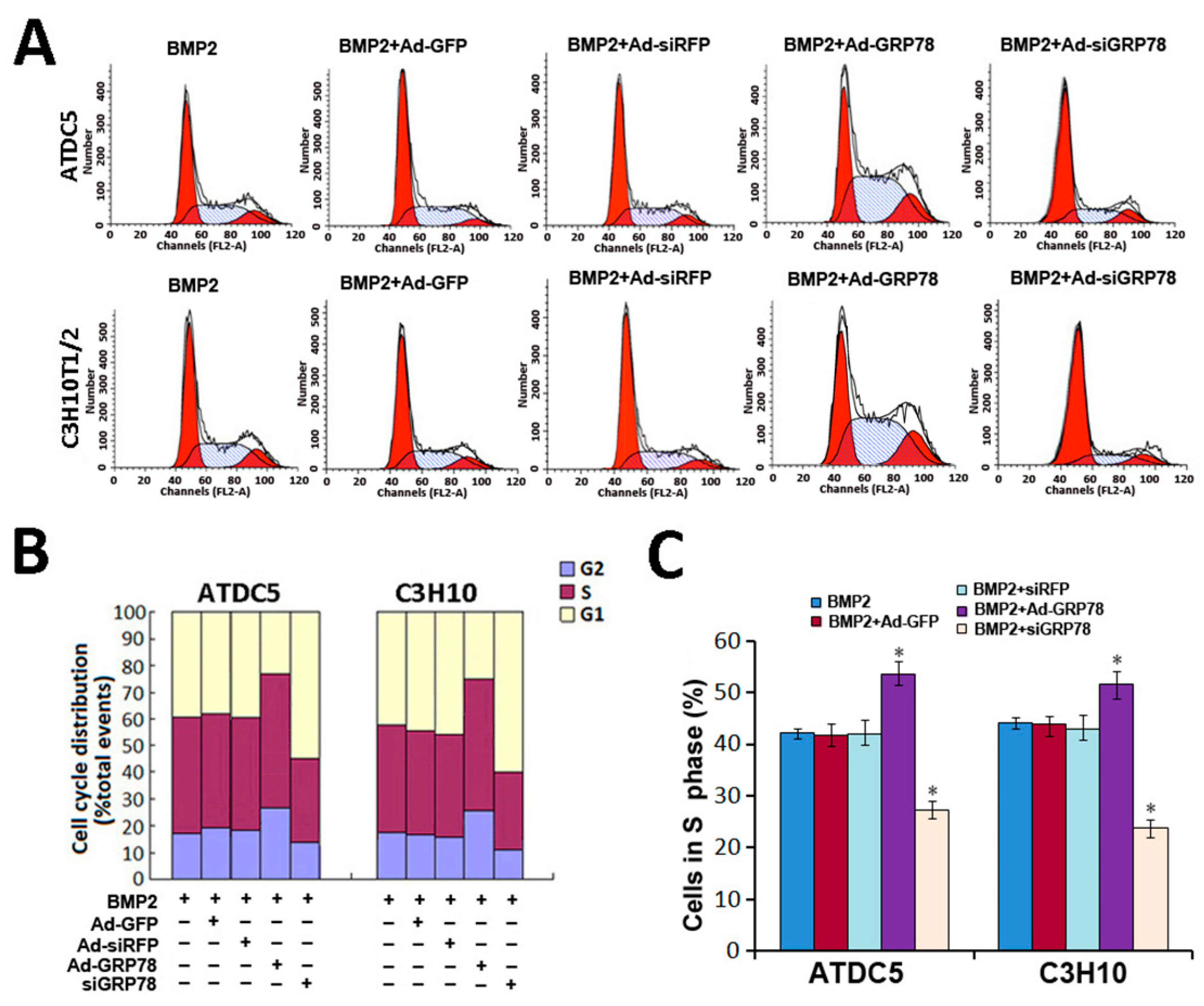
The authors wish to replace Figure 4A on Page 21161 of their paper published in IJMS [1]. There was an error in the representative image of BMP2 + Ad-GRP78 group of C3H10T1/2 in the Figure 4A. The images related to ATDC5 was repeated to show the BMP2 + Ad-GRP78 effects on C3H10T1/2. Please see the corrected Figure 4 here. The authors apologize for any inconvenience. The manuscript will be updated and the original will remain online at the article webpage.
Figure 4. Cellular proliferation analysis by FCM. (A) Flow cytometry images with propidium iodide staining and analysis on cell cycle distribution. Micromass culture of ATDC5 cells and C3H10T1/2 were treated with BMP2 (300 ng/mL) / BMP2 + Ad-GFP/BMP2 + Ad-siRFP / BMP2 + Ad-GRP78 / BMP2 + Ad-siGRP78. Flow cytometry analysis showed that the percentage of the BMP2 + Ad-GRP78 ATDC5 cells in S phase were increased significantly compared to those in BMP2 controls, whereas the percentage of the BMP2 + Ad-siGRP78 ATDC5 cells in S phase were dramatically decreased compared with BMP2 control. The result of C3H10T1/2 is the same. Experiments were repeated three times, and samples were analyzed by Student’s t-test and statistical significance with p < 0.05. Representative images were shown; (B) Flow cytometry assay on the percentages of the ATDC5 and C3H10T1/2 cells in G2/M phase after treatment with BMP2 (300 ng/mL)/BMP2 + Ad-GFP / BMP2 + Ad-siRFP / BMP2 + Ad-GRP78 / BMP2 + Ad-siGRP78; (C) Flow cytometry analysis showed that the percentages of the ATDC5 and C3H10T1/2 Ad-GRP78 cells in S phase were increased significantly, whereas the percentages of the ATDC5 and C3H10T1/2 Ad-siGRP78 cells in S phase were decreased compared with those in their controls. * p < 0.05 compared with control.
Ijms 16 26222 g004

Reference

  1. Xiong, Z.; Jiang, R.; Li, X.; Liu, Y.; Guo, F. Different roles of GRP78 on cell proliferation and apoptosis in cartilage development. Int. J. Mol. Sci. 2015, 16, 21153–21176. [Google Scholar] [CrossRef] [PubMed]