Review

Regulatory Proteolysis in Arabidopsis-Pathogen Interactions

Plant Protection Institute, Centre for Agricultural Research, Hungarian Academy of Sciences, Herman Ottó út 15, 1022 Budapest, Hungary
*
Author to whom correspondence should be addressed.
Int. J. Mol. Sci. 2015, 16(10), 23177-23194; https://doi.org/10.3390/ijms161023177
Submission received: 2 August 2015 / Revised: 7 September 2015 / Accepted: 15 September 2015 / Published: 24 September 2015
(This article belongs to the Special Issue Plant Microbe Interaction)

Abstract

:
Approximately two and a half percent of protein coding genes in Arabidopsis encode enzymes with known or putative proteolytic activity. Proteases possess not only common housekeeping functions by recycling nonfunctional proteins. By irreversibly cleaving other proteins, they regulate crucial developmental processes and control responses to environmental changes. Regulatory proteolysis is also indispensable in interactions between plants and their microbial pathogens. Proteolytic cleavage is simultaneously used both by plant cells, to recognize and inactivate invading pathogens, and by microbes, to overcome the immune system of the plant and successfully colonize host cells. In this review, we present available results on the group of proteases in the model plant Arabidopsis thaliana whose functions in microbial pathogenesis were confirmed. Pathogen-derived proteolytic factors are also discussed when they are involved in the cleavage of host metabolites. Considering the wealth of review papers available in the field of the ubiquitin-26S proteasome system results on the ubiquitin cascade are not presented. Arabidopsis and its pathogens are conferred with abundant sets of proteases. This review compiles a list of those that are apparently involved in an interaction between the plant and its pathogens, also presenting their molecular partners when available.

Graphical Abstract

1. Introduction

Proteolytic enzymes have been proven to possess crucial housekeeping and regulatory functions in cells of intact or pathogen-exposed plant tissues. Enzyme-catalysed proteolysis in living cells, however, is performed by an extremely diverse group of enzymes. In the genome of the dicotyledonous model plant Arabidopsis thaliana, the number of protease coding genes and their homologs is between 800 and 900, representing approximately 2.5% of all protein-coding genes [1,2,3]. In humans, proteases also represent about 2% of protein coding genes [4].
Proteases (peptidases or proteolytic enzymes) cleave peptide bonds between amino acid residues of proteins, oligo- or polypeptides. Aminopeptidases detach N-terminal amino acid residues, carboxypeptidases split C-terminal amino acid residues and endopeptidases cleave peptide bonds between amino acids in internal positions. Proteases in Arabidopsis hydrolyze peptide bonds in five ways, which gives the names to five catalytic classes: aspartic proteases, cysteine proteases, serine proteases, metalloproteases and threonine proteases [1,3,5].
A systematic classification of peptidases is offered by the MEROPS database (release 9.13) [1] including 745 known and putative peptidases and their 124 homologs for Arabidopsis thaliana. These Arabidopsis proteases are distributed over 30 clans and subdivided into 60 families [1,2].
The aim of this article, on one hand, is to provide an organised overview on the proteolytic enzymes produced by Arabidopsis cells whose functions in interactions with pathogens have been confirmed. Pathogen-derived proteolytic factors are also discussed when they apparently play regulatory roles during interactions with their host. The nature of interaction is typically proteolytic cleavage of host metabolites. However, besides their catalytic domains, a great number of proteases contain numerous additional domains or modules [4], which enable them to establish a wide range of interactions.
Host-derived Arabidopsis proteolytic enzymes discussed in the article are listed in Table 1. Proteolytic enzymes secreted by pathogens of Arabidopsis are summarized in Table 2.
It has to be emphasized that reviewing functional aspects of the highly sophisticated ubiquitin-26S proteasome system (UPS) in Arabidopsis pathogenesis was beyond the scope of this article. Although UPS is an extremely important cellular proteolytic machinery, and our knowledge on the involvement of the UPS in plant-pathogen interactions has exploded in the last 10 years, there have been several excellent review papers written in this particular field recently [6,7,8,9,10]. Our primary goal with the current article was to compile published data on proteins with experimentally proven or computationally inferred peptidase activity in the context of Arabidopsis microbial pathogenesis. The vast majority of UPS components do not fall into this category. However, the Arabidopsis proteasome β1 subunit PBA1 is still discussed here because it apparently possesses caspase-3-like proteolytic activity [11].
Some clearly-defined types and aspects of Arabidopsis-pathogen interactions are discussed throughout the paper. These are (i) pattern-triggered or basal immunity, when conserved microbe-associated molecular patterns (MAMPs) are recognized by host cell surface pattern-recognition receptors (PRR); (ii) susceptibility, when virulent or compatible pathogens deliver effectors or virulence factors into the host cells overcoming host basal immunity and thereby causing disease; (iii) effector-triggered or hypersensitive response (HR)-type immunity, when effectors released by an avirulent or incompatible pathogen strain are detected in the host cells by intracellular nucleotide-binding leucine-rich repeat (NB-LRR) immune receptor R proteins leading to programmed cell death (PCD) or HR and (iv) systemic acquired resistance (SAR), when an initial pathogen infection is able to induce a primed condition in local (infected) and distal tissues of host plants to make them respond in a more rapid and robust manner to even lower levels of pathogenic cues than control plants do [12,13].
Table 1. Arabidopsis thaliana proteases whose functions in interactions with pathogens have been confirmed.
GeneFull NameAGI CodeUniprot AccessionMEROPS IdentifierReference
AtCDR1Constitutive Disease Resistance 1At5g33340Q6XBF8A01.069[14,15]
AtAED1Apoplastic EDS1-Dependent 1At5g10760Q9LEW3A01.A14[16]
AtRD21aResponsive to Dehydration 21aAt1g47128P43297C01.064[17,18,19,20]
AtRD21bResponsive to Dehydration 21bAt5g43060Q0WM94C01.A12[20]
AtXCP1Xylem Cysteine Proteinase 1At4g35350O65493C01.065[17]
AtXCP2Xylem Cysteine Proteinase 2At1g20850Q9LM66C01.120[17,20]
AtCPR1Probable Cysteine ProteinaseAt3g19400Q9LT77C01.A12[17]
AtALEUAleurainAt5g60360Q8H166C01.163[17]
AtALEULAleurain-LikeAt3g45310Q8RWQ9C01.162
AtRD19aResponsive to Dehydration 19aAt4g39090P43296C01.022[21]
AtCathB1Cathepsin B1, B2, B3At1g02300Q56XY7C01.A10[22]
AtCathB2At1g02305Q93VC9C01.144
AtCathB3At4g01610Q9ZSI0C01.144
AtMC1Metacaspase 1At1g02170Q7XJE6C14.047[23]
AtMC2Metacaspase 2At4g25110Q7XJE5C14.A04[23]
AtMC4Metacaspase 4At1g79340O64517C14.033[24]
AtαVPEα, β, δ, or γ Vacuolar Processing EnzymeAt2g25940P49047C13.002[25,26,27,28]
AtβVPEAt1g62710Q39044C13.001
AtδVPEAt3g20210Q9LJX8C13.A01
AtγVPEAt4g32940Q39119C13.006
AtCEP1KDEL Cysteine Endopeptidase 1At5g50260Q9FGR9C01.A03[29]
AtSBT3.3Subtilase 3.3At1g32960Q9MAP5S08.A35[30]
AtPBA126S Proteasome β1 SubunitAt4g31300Q8LD27T01.010[31]
Table 2. Pathogen-secreted proteases whose functions in interactions with Arabidopsis have been partially elucidated.
ProteaseSpeciesUniprot AccessionMEROPS IdentifierReference
AvrPphBP. syringae pv. phaseolicolaQ52430C58.002[32,33,34]
AvrRpt2P. syringae pv. tomatoQ6LAD6C70.001[35,36,37,38,39,40,41]
XopDX. campestris pv. vesicatoriaQ3BYJ5C48.023[42,43]
HopX1P. syringae pv. tabaciQ83YM6N/A[44]
protease IVP. aeruginosaQ02SZ7S01.281[45]
AprAP. syringae pv. tomatoQ87ZU2M10.060[46]

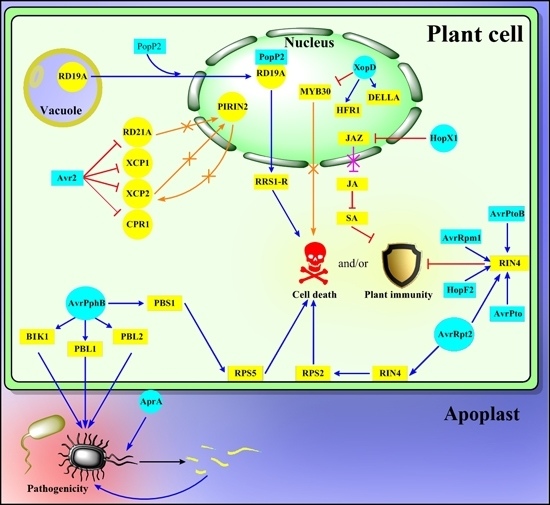
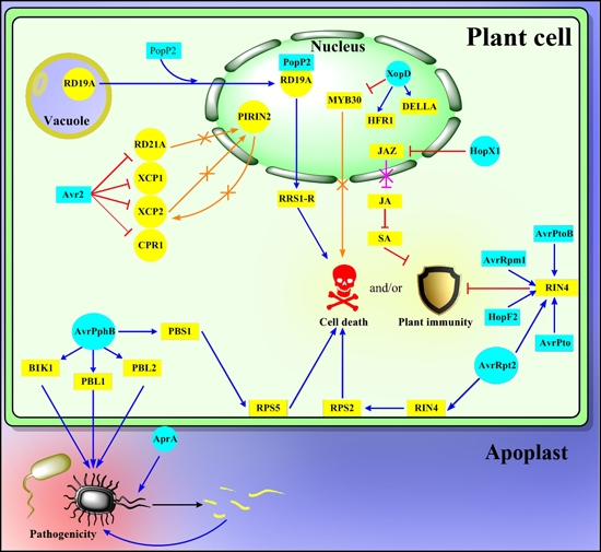
2. Functions of Proteolytic Enzymes in Various Arabidopsis thaliana Pathosystems

2.1. Host-Derived Aspartic Proteases

The majority of proteases discussed in this review are cysteine proteases. Nevertheless, one of the first Arabidopsis proteases proven to function in the immune system was an exception. Overexpression of the aspartic protease Constitutive Disease Resistance 1 (CDR1) in a gain-of-function mutant resulted in resistance to normally virulent Pseudomonas syringae. Antisense CDR1 plants with reduced levels of CDR1 protein were compromised for resistance to avirulent P. syringae and more susceptible to virulent strains than wild type. Levels of salicylic acid were significantly elevated in CDR1 overexpressor compared with wild-type plants and CDR1 activation apparently induced a salicylic acid-dependent disease resistance response in Arabidopsis [14,15]. CDR1 encodes an apoplastic protein that shares significant sequence similarity to aspartic proteases. It was also shown that the proteolytic activity of CDR1 was necessary for its biological functions. Like other eukaryotic aspartic proteases, CDR1 possesses two active sites with the conserved motifs aspartic acid-threonine-glycine-serine and aspartic acid-serine-glycine-threonine, respectively. These data indicate that CDR1 encodes an aspartic protease that functions biologically by the proteolytic cleavage of its endogenous target. CDR1 might process a cell surface protein that could be a component of the basal host defense complex or alternatively it may release an extracellular mobile peptide elicitor that activates host basal defense responses [14,15]. Although the natural substrates for CDR1 are still missing, recombinant CDR1 activity is considerably increased by redox-dependent, disulfide-mediated dimerization [47].
Another aspartic protease, Apoplastic EDS1-Dependent 1 (AED1) was recently described [16], searching for systemic acquired resistance (SAR) regulatory proteins and using two-dimensional PAGE. AED1 accumulation together with some other proteins was reduced in the eds1 mutant expressing the P. syringae effector AvrRpm1 in comparison with wild type background AvrRpm1 expressing plants. Although the mode of AED1 action (e.g., its cellular targets) is still missing, it was concluded that it functions as a negative regulator of systemic acquired resistance acting downstream of salicylic acid. By cleaving apoplastic proteinaceous substrates, it might be part of a homeostatic mechanism to limit SAR signaling and thus regulating the resource allocation in the tradeoff between defense and plant growth [16].

2.2. Host-Derived Cysteine Proteases

Cysteine proteases contain a cysteine nucleophilic residue in their active site that performs a nucleophilic attack in the first step of proteolysis resulting in an intermediate state where the enzyme is covalently attached to its substrate [48]. Known and putative cysteine protease sequences compose almost 16% of all listed peptidase sequences in the latest release of the MEROPS database [1]. Twenty-three out of the 29 proteases discussed in this current review also belong to the group of cysteine proteases. Protease activity profiling [48,49,50] was applied to investigate whether the fungal effector Avr2 produced by Cladosporium fulvum is able to inhibit Arabidopsis cysteine proteases [17]. Protein extracts prepared from Arabidopsis plants were treated with DCG-04, a biotinylated derivative of the irreversible cysteine protease inhibitor E-64. DCG-04 reacts with the catalytic cysteine residue of cysteine proteases and locks the cleavage mechanism in the covalent intermediate state. The biotinylated cysteine proteases were subsequently detected on protein gel blots using a conjugate of streptavidin with horseradish peroxidase. Avr2 was previously shown to bind and inhibit the tomato cysteine protease Rcr3 [51]. Indeed, Avr2 exhibited marked inhibitory effect on most detected Arabidopsis cysteine proteases except Cathepsin B3. Xylem Cysteine Proteinase 1 (XCP1), Xylem Cysteine Proteinase 2 (XCP2) and a Probable Cysteine Proteinase CPR1 showed high Avr2 affinity, whereas Responsive to Dehydration 21A (RD21A) and thiol proteases Aleurain and Aleurain-Like had lower but still apparent affinity to Avr2 [17]. Lack of RD21A cysteine protease activity in Arabidopsis T-DNA mutants led to increased susceptibility to the necrotrophic fungus Botrytis cinerea [18]. RD21A was also shown to be a partner of cytochrome c during hydrogen peroxide-induced programmed cell death in cultured Arabidopsis cells [19]. XCP2, RD21A and Responsive to Dehydration 21B (RD21B) were also independently identified by yeast two-hybrid assays as interacting partners of the Arabidopsis PIRIN2 protease inhibitor encoded by locus At2g43120 [20]. Functional aspects of the XCP2-PIRIN2 interaction were further investigated revealing that PIRIN2 inhibits the autolytic degradation of XCP2. This stabilization of XCP2 by PIRIN2 results in accumulation of XCP2 and in increased overall XCP2 activity. It was also presented that the XCP2-PIRIN2 interaction is necessary for full susceptibility to the xylem-colonizing bacterial pathogen Ralstonia solanacearum. It is conceivable, that XCP2-mediated autolysis of cellular contents in leaves or vessel elements facilitates R. solanacearum pathogenesis [20].
The cysteine-type endopeptidase Responsive to Dehydration 19A (RD19A) was identified as an interacting partner of R. solanacearum PopP2 effector [21]. RD19A encodes a drought-inducible cysteine protease [52] whose transcript levels increase strongly after R. solanacearum infection [21]. PopP2 elicits a disease resistance response in Arabidopsis mediated by its cognate R protein RRS1-R and functional RD19A is required for efficient RRS1-R-dependent defense against R. solanacearum. As far as the intracellular position of RD19A, without PopP2 it localizes in mobile prevacuolar vesicles, showing perfect colocalization with another cysteine protease Aleurain. In the presence of PopP2 effector, however, RD19A is recruited to the nucleus where it physically associates with PopP2 as it was confirmed by FLIM (Fluorescence Lifetime Imaging) approach [53]. The FLIM approach is a quantitative, noninvasive method that monitors the Förster resonance energy transfer between the donor and acceptor molecules fused to PopP2 and RD19A, respectively. Collectively, these findings suggest that RD19A is an important Arabidopsis factor for PopP2-triggered RRS1-R–mediated disease resistance.
Cathepsins are lysosomal cysteine and aspartic proteases that (along with caspases) participate in mammalian apoptosis [54]. Functions of three Arabidopsis orthologs of mammalian Cathepsin B (CathB) were analyzed to describe their contributions to various forms of disease resistance [22]. AtCathB1, AtCathB2 and AtCathB3 encode Cathepsin B-like cysteine proteases. Basal immunity to the virulent bacterial strain P. syringae pv. tomato DC3000 was not affected in atcathb single or double mutants, where only one or two AtCathB isoforms were knocked out. Triple mutants, on the other hand, exhibited impaired resistance to the virulent P. syringae strain, indicating that the three AtCathB isoforms in Arabidopsis act redundantly (they fulfill nearly the same role) to confer basal defense to the plants. (Triple mutants were generated by crossing atcathb1 and atcathb3 single mutants and the resulting double mutant was transformed with a CathB2:RNAi hairpin silencing construct.) Using an incompatible Arabidopsis-P. syringae pv. tomato system (RPM1-AvrB), it was also presented that AtCathB genes do not contribute to R gene mediated resistance, but they are redundantly required for programmed cell death (PCD) during the hypersensitive response (HR) triggered by P. syringae pv. tomato DC3000 (AvrB) [22].
Metacaspases are distant orthologs of animal caspases in plants, which are also present in protozoa and fungi. They belong to the CD cysteine protease clan [1]. Nine metacaspase genes (AtMC1AtMC9) have been found in the Arabidopsis genome [3]. The role of metacaspases in pathogen-induced (as well as in other forms of) programmed cell death in Arabidopsis is now well established. AtMC1 is a crucial pro-death protein in Arabidopsis during the hypersensitive response mediated by intracellular NB-LRR immune receptor R proteins. T-DNA insertional mutation in AtMC1 resulted in markedly reduced cell death upon elicitation by the avirulent bacterial strain P. syringae pv. tomato DC3000 (avrRPM1). In contrast, AtMC2 negatively regulated the same cell death phenomenon and it antagonized AtMC1 function. AtMC1 activity required conserved caspase-like catalytic residues, whereas AtMC2 function appeared to be independent of the putative catalytic residues [23].
AtMC4 was also confirmed as a positive mediator of cell death in Arabidopsis tissues challenged by avirulent bacteria or treated with a fungal toxin [24]. Plants carrying mutation in atmc4 showed not only decreased sensitivity to the mycotoxin fumonisin B1 produced by the fungal pathogen Fusarium moniliforme, but they also developed reduced hypersensitive cell death symptoms when they were inoculated with the avirulent bacterial strain P. syringae pv. maculicola (avrRpt2). Subcellular localization studies revealed that mature AtMC4 resides mainly in the cytoplasm [24]. Another Arabidopsis metacaspase, AtMC9 was currently described as a proteolytic enzyme responsible for cleavage and activation of the GRIM REAPER protein that controls superoxide-induced cell death in Arabidopsis. Both GRIM REAPER and AtMC9 show extracellular localization [55].
Vacuolar processing enzyme (VPE) is a cysteine proteinase originally identified as the proteinase responsible for the maturation and activation of vacuolar proteins in plants [56]. Unlike metacaspases, that lack aspartic acid specificity of caspases and cleave their substrates after arginine and lysine residues, VPEs cleave peptide bonds at the C-terminal sides of asparagine or aspartic acid residues [49]. The Arabidopsis genome encodes four VPE genes: αVPE, βVPE, γVPE, and δVPE [25]. A VPE quadruple mutant (deficient in transcribing functional mRNA for all four VPE isoforms simultaneously) showed greatly diminished fumonisin B1-induced cell death and analysis of single mutants revealed that γVPE possessed the most essential role in the fungal toxin-induced cell death [25]. In accord with these results, plants overexpressing γVPE exhibited increased ion leakage (a marker of hypersensitive cell death) after inoculation with the avirulent P. syringae pv. tomato DC3000 (AvrRpm1). This result suggests that γVPE regulates cell death progression during plant-pathogen interaction. Compromised resistance to Botrytis cinerea, P. syringae pv. tomato DC3000 (AvrRpm1) and turnip mosaic virus observed in a γVPE single mutant was also reported here [26].
Using VPE activity profiling, enhanced VPE activity in Arabidopsis plants challenged by a virulent strain (Noco2) of the oomycete pathogen Hyaloperonospora arabidopsidis was reported [27]. By contrast, an avirulent isolate (Cala2) of the same pathogen was unable to trigger VPE activation. Sporulation of H. arabidopsidis (virulent strain) was quantified in a VPE quadruple mutant and significant reduction in the spore count was observed in comparison with wild type plants, demonstrating that VPEs are needed to promote H. arabidopsidis virulence. These data indicate that H. arabidopsidis as an obligate biotroph may take advantage of increased protein turnover and nutrient release mediated by host VPE activation [27]. Interestingly, VPEs also promote mutualistic interaction with the fungus Piriformospora indica in Arabidopsis roots [28].
The involvement of an Arabidopsis KDEL cysteine endopeptidase (AtCEP1) in pathogen defense has been recently published [29]. KDEL cysteine peptidases are ubiquitous in plants and are characterized by a C-terminal lysine-aspartic acid-glutamic acid-leucine (KDEL) motif that serves as an endoplasmic reticulum retention signal [57]. AtCEP1 apparently accumulated in the endoplasmic reticulum of epidermal cells that were penetrated by the biotrophic powdery mildew fungus Erysiphe cruciferarum. AtCEP1 labeling was particularly strong around established fungal haustoria. Microscopic examination of plant-fungal interaction sites in an atcep1 mutant revealed that epidermal cell death was suppressed, whereas the number of haustoria increased in infected leaves of the mutant genotype compared to wild type leaves. Taken together, these findings suggest roles for AtCEP1 in the appearance of late stages of host cell death during E. cruciferarum pathogenesis and potentially in plant basal resistance to restrict parasitic growth of a compatible powdery mildew [29].

2.3. Host-Derived Serine Protease

Subtilases (or subtilisin-like serine proteases) are a large family of serine proteases universal to all kingdoms of life but found most extensively in plants compared to other organisms [58]. The family type peptidase is subtilisin that was originally purified from strains of Bacillus subtilis and related bacteria [1,59]. The Arabidopsis genome contains 56 subtilase encoding loci, divided into six subfamilies [58]. A recently published set of results have underscored the importance of an extracellular Arabidopsis subtilase SBT3.3 in induced disease resistance [30]. Independent SBT3.3 mutants with impaired SBT3.3 activity showed enhanced susceptibility to P. syringae pv. tomato DC3000 and to H. arabidopsidis (isolate WACO9). Conversely, overexpression of SBT3.3 conferred enhanced resistance to bacterial and oomycete pathogens. When the SBT3.3 overexpression phenotype was examined in a sid2 or npr1 mutant background (affecting salicylic acid biosynthesis or signaling), the enhanced disease resistance was abrogated, indicating that SBT3.3 operates upstream of the salicylic acid pathway. SBT3.3 overexpression also poised salicylic acid-mediated defense genes for enhanced activation upon inoculation with P. syringae pv. tomato DC3000. It has been concluded, that SBT3.3 functions as a major component and positive regulator of salicylic acid-dependent immune priming, keeping cells in a sustained sensitized mode following a prior pathogen attack [30]. Intriguingly, tobacco and rice subtilisin-like serine proteases (named phytaspases) that are phylogenetically related to subtilases listed in Arabidopsis subtilase subfamily 1 were shown to regulate programmed cell death during abiotic stress or virus infection. Phytaspase is secreted from the cell and stored in the apoplast but is uniquely relocalized into the cytoplasm upon induction of programmed cell death, where it contributes to the cellular suicide machinery, presumably by cleaving its intracellular targets [60,61].

2.4. Arabidopsis PBA1, the β1 Subunit of the 26S Proteasome

The β1 subunit of the 26S proteasome in Arabidopsis is encoded by the PBA1 gene (At4g31300) [62]. When Arabidopsis leaves were inoculated with avirulent phytopathogenic bacterial strains, leaf cells developed the fusion of membranes of their large central vacuoles with the plasma membranes, leading to the discharge of vacuolar proteins to the intercellular space [31]. This discharge of vacuolar fluid prevented proliferation of bacterial pathogens and activated programmed cell death. The described cellular response could be precisely connected to the activity of the β1 catalytic subunit (PBA1) of the 26S proteasome system, which system selectively breaks down proteins targeted for degradation by modification with polymers of ubiquitin through an ubiquitin-activating (E1) to ubiquitin-conjugating (E2) to ubiquitin ligase (E3) enzymatic cascade. The 26S proteasome is formed by two distinct particles: the 20S core proteasome and the 19S regulatory particle. The 20S core proteasome possesses two peripheral and two central rings and the two central rings are composed of seven β subunits, including β1, β2 and β5. These three subunits are responsible for the three proteolytic activities of the 26S proteasome (peptidylglutamyl peptide-hydrolyzing, trypsin-like, and chymotrypsin-like activities) that can selectively digest target proteins to short peptides [9,62,63]. The Arabidopsis 26S β1 subunit (PBA1) exhibits caspase-3-like or DEVDase proteolytic activity [31], recognizing aspartic acid-glutamic acid-valine-aspartic acid (DEVD) tetrapeptide sequences and hydrolyzing peptide bonds on the carboxy side of the second aspartic acid residue. This protease belongs to the threonine protease T1 (proteasome) family [1]. Depletion of PBA1 in three independent RNAi lines was able to suppress membrane fusion, vacuole discharge and hypersensitive cell death in tissues challenged by the avirulent bacterial strain P. syringae pv. tomato DC3000 (AvrRpm1). Monitoring growth of bacterial cells in the RNAi lines revealed that reduced PBA1 activity resulted in increased bacterial growth. Inoculation with the virulent P. syringae pv. tomato DC3000, on the contrary, did not cause different ultrastructural responses or markedly altered pathogen growth between wild type and pba1 RNAi plants, suggesting that PBA1 activity is mostly required for R gene-mediated immunity and cell death in Arabidopsis. Results with a second avirulent Arabidopsis-P. syringae pv. tomato DC3000 (avrRpt2) interaction also corroborated conclusions described above [31].
These findings indicate that the PBA1 proteasome subunit acts as a caspase-3-like enzyme in Arabidopsis, regulating membrane fusion of the vacuolar and plasma membranes. This PBA1-mediated cellular response leads to hypersensitive cell death and resistance to avirulent bacterial pathogens [31]. Caspase-3 is involved in the execution of apoptosis in animal cells [64].

2.5. Cysteine Protease Effectors Secreted by Pathogens of Arabidopsis

AvrPphB (earlier designated as AvrPph3) is a bacterial type III effector, originally identified from P. syringae pv. phaseolicola. It triggers a disease resistance response and hypersensitive cell death in Arabidopsis plants expressing a corresponding immune receptor R protein RPS5 [65]. AvrPphB is a cysteine protease that cleaves the Arabidopsis protein kinase PBS1. Proteolytic cleavage of PBS1 by AvrPphB elicits an RPS5-mediated immune response and HR-type cell death. It has been shown by using a coimmunoprecipitation assay, that AvrPphB and PBS1 physically interact [32]. PBS1 cleavage was suggested to change the ATP versus ADP binding functions of RPS5 serving as a molecular switch to activate R-gene-mediated molecular pathway [33]. In accord with the guard model of R protein activation, RPS5 does not recognize the Pseudomonas syringae effector AvrPphB directly, but rather perceives the conformational modification of its cellular target (PBS1) as a result of AvrPphB proteolytic activity. It has been also discovered later that besides PBS1, there are other PBS1-like cellular targets of AvrPphB in Arabidopsis, such as BIK1, PBL1 or PBL2 receptor-like protein kinases [34]. These are also proteolytically cleaved by AvrPphB for inhibition of host pattern-triggered immunity.
AvrRpt2, another P. syringae type III effector with cysteine protease activity triggers a disease resistance response in Arabidopsis plants carrying the R protein RPS2 [35,36]. AvrRpt2 cleaves the Arabidopsis RIN4 protein, which leads to its elimination monitored by RPS2 [37,38,39]. Interestingly, RIN4 is attacked by several P. syringae effectors (AvrB, AvrRpm1, AvrRpt2, AvrPto, AvrPtoB, HopF2), highlighting its key role as a regulator of plant immunity [40,66]. Degradation of RIN4 by AvrRpt2 may prevent detection of RIN4 modification caused by other effectors, such as AvrB or AvrRpm1 in the presence of another immune receptor R protein RPM1 [39]. It was recently shown, that AvrRpt2 also promotes an auxin response in Arabidopsis by stimulating the degradation of auxin transcription repressor proteins (e.g., AXR2 or AXR3), and this function requires the cysteine protease activity of AvrRpt2 [41]. This AvrRpt2-dependent turnover of auxin transcription repressor proteins supports virulence of the compatible P. syringae pv. tomato DC3000 strain in susceptible Arabidopsis host.
An Xanthomonas campestris pv. vesicatoria type III effector, XopD exhibits small ubiquitin-like modifier (SUMO) protease activity, due to a cysteine protease domain located at its C-terminus [67]. XopD is a potent suppressor of Arabidopsis defense responses by accumulating in subnuclear structures called nuclear bodies in the nucleus of host cells and recruiting and binding Arabidopsis transcription factor MYB30 there, establishing a physical interaction with it [42]. MYB30 is a positive regulator of pathogen defense and hypersensitive response-related genes [68,69,70]. XopD was shown to repress the transcriptional activity of MYB30 and the defense of Arabidopsis plants exhibited against Xanthomonas campestris. These functions were, however, independent of the effector’s cysteine protease domain but dependent on an XopD helix–loop–helix (HLH) domain, suggesting that the cysteine protease domain might be involved in targeting host defense-related factors other than MYB30 [42].
In accord with this suggested model, a recent work presented Arabidopsis HFR1, a basic helix–loop–helix transcription factor as a potential host target regulated specifically by SUMO protease activity of XopD [43]. A truncated version of XopD (XopDXcc8004), which lacks the N-terminal domain that is crucial in MYB30 repression, but carries the C-terminal cysteine protease domain, was investigated. When XopDXcc8004 (lacking the N-terminal domain) was ectopically expressed in Arabidopsis plants, it resulted in plants showing lesion mimic phenotype and exhibiting transcriptional induction of salicylic acid-regulated genes. This finding reveals that impairment in the N-terminal domain of XopD not only eliminates the ability of XopD to suppress host immunity, but it actually converts XopD into an elicitor of host defense responses. It was also discovered that the SUMO protease activity of XopDXcc8004 was required for these phenotypic responses. Using yeast two-hybrid assay, the Arabidopsis transcription factor HFR1 (Long Hypocotyl in Far-Red 1) showed positive interaction with XopDXcc8004 and the two proteins were colocalized in nuclear bodies of plant cells. Finally, a HFR1 mutant Arabidopsis line exhibited elevated levels of defense-related transcripts and reduced susceptibility to Xanthomonas campestris. These results indicate that HFR1 can be a potential nuclear substrate of XopD, modified by its SUMO protease activity and HFR1 represses defense responses in Arabidopsis [43]. Biological activity of XopDXcc8004 has also been linked in Arabidopsis to gibberellic acid signaling through the transcriptional regulator DELLA proteins [71]. DELLA proteins are localized in the nucleus and they carry a 17-amino acid-long, highly conserved, N-terminal sequence, the DELLA motif (named after the first five amino acids in this sequence), which plays a crucial regulatory role in sensing the gibberellic acid signal [72,73,74]. Functional aspects of the gibberellin-DELLA interaction and signaling have been summarized here [75]. Currently, Arabidopsis DELLA proteins were shown to be nuclear targets of the XopDXcc8004 cysteine protease domain of XopD type III effector [71]. In this study, XopDXcc8004 delayed the development of disease symptoms in P. syringae pv. tomato DC3000-infected Arabidopsis leaves, whereas exogenous gibberellic acid treatment was able to reverse this effect to some extent. XopDXcc8004 also delayed gibberellin-mediated degradation of the RGA DELLA protein, and it was suggested that XopDXcc8004 might promote plant disease tolerance by partially stabilizing DELLA proteins [71]. DELLA proteins were shown to affect responses of Arabidopsis plants to biotrophic and necrotrophic pathogens in an antagonistic manner and they seem to possess integrator roles of salicylic acid and jasmonic acid signaling [76].
Bacterial effector proteins from the HopX1 family (previously AvrPphE) are produced predominantly but not exclusively by various P. syringae pathovars [77,78]. When virulence effectors released by a strain of P. syringae pv. tabaci that does not produce the crucial P. syringae toxin coronatine were analyzed, one type III effector, HopX1 of this particular bacterial strain was identified based on its capability to compromise accumulation of the key jasmonic acid pathway repressors the JAZ proteins [44]. The toxin coronatine helps entry of bacteria into the host plant by inducing the opening of stomata [79] and facilitates bacterial growth by inhibiting salicylic acid-related defense pathways through activation of the antagonistic jasmonic acid-dependent pathway [80,81]. It was shown that HopX1 is a cysteine protease and its enzymatic activity is needed for the degradation of JAZ proteins. It was also presented that HopX1 eliminates JAZ proteins in a pathway independent of the jasmonic acid receptor COI1 (Coronatine-insensitive 1) which would also perceive the presence of coronatine. Instead, HopX1 compromises the accumulation of the JAZ family in a specific manner by directly interacting with JAZ proteins through their central ZIM domain in the cytoplasm and nuclei of host cells. Similar to coronatine, HopX1 also activates the jasmonic acid pathway and suppresses salicylic acid-dependent gene expression. By using coronatine deficient pathogen or COI1 mutant host genotypes, it was presented that HopX1 can complement the deficiency in coronatine production or signaling in order to activate the Arabidopsis jasmonate pathway, induce the opening of stomata and promote bacterial pathogenicity. These results suggest that HopX1 contributes to bacterial pathogenicity by mimicking coronatine-induced host cellular responses to trigger plant susceptibility and bacterial HopX1 and coronatine may function redundantly [44].

2.6. Protease IV, a Bacterial Lysyl Class Serine Protease Effector

A recent secretome analysis of a P. aeruginosa strain (PA14) led to the identification of protease IV, a previously unknown lysyl class serine protease effector and a corresponding novel immune pathway in Arabidopsis [45]. Protease IV treatment elicited an immune response comparable to the effect of the MAMP flg22 (a conserved N-terminal epitope of flagellin), characterized by the activation of mitogen-activated protein kinases (MPK3 and MPK6), induction of an oxidative burst, deposition of callose and protection from P. syringae pv. tomato DC3000 infection. The transcriptomic signature of protease IV treatment was also similar to those gene expression changes that were elicited by flg22. In a search for mechanisms by which protease IV activates an immune response, components of the heterotrimeric G-protein complex were investigated. Indeed, protease IV-triggered immune responses, including the induction of a mitogen-activated protein kinase (MAPK) cascade, were markedly compromised in G-protein mutants, indicating that the G-protein complex may function upstream of a MAPK cascade. Considering further potential signaling components, Receptor for Activated C Kinase 1 (RACK1) emerged as a candidate. The three Arabidopsis RACK1 homologs apparently interacted with one subunit (Gβ) of the G-protein complex and also with several members of a MAPK cascade. RACK1 was suggested to function as a scaffold that binds upstream G-protein signaling to downstream activation of a MAPK cascade. Knockdown of RACK1 genes by using stable RNAi transgenic lines blocked protease IV-mediated defense gene induction and protection from P. syringae pv. tomato DC3000. Therefore, this novel protease IV-mediated immune pathway is distinct from the previously known flg22 pathway because G-proteins act upstream of a MAPK cascade and the RACK1 protein is uniquely involved here [45].

2.7. Alkaline Protease AprA, a Bacterial Zinc Metalloprotease

AprA is an alkaline protease belonging to the serralysin family of zinc metalloproteases. It has been identified in the opportunistic pathogen P. aeruginosa and its orthologs have been found in the human pathogen Serratia marcescens and the plant pathogens Dickeya dadantii or P. syringae pv. tomato DC3000 [82,83,84,85,86]. In fact, P. aeruginosa (and other bacterial species) also express a peptide designated AprI (present in the same operon as AprA) with unclear biological function that acts as an inhibitor of AprA protease [87]. It was shown recently that AprA protease actively degrades monomers of the crucial bacterial MAMP flagellin and the aprA operon appears in a highly divergent group of bacterial species [46,88]. An AprA-deficient P. syringae pv. tomato DC3000 bacterial strain exhibited reduced pathogenicity to Arabidopsis when leaves were pressure-infiltrated with the inoculum and this response was dependent on flagellin recognition mediated by the FLS2 receptor. Defense-related transcriptional changes were also markedly higher after inoculation with the AprA-deficient P. syringae strain compared to a treatment with the wild type strain. When the bacterial AprA inhibitor AprI was ectopically expressed in Arabidopsis the transgenic lines showed reduced susceptibility to wild type P. syringae pv. tomato DC3000. These results together suggest a sophisticated bacterial virulence mechanism, where AvrA protease is secreted by pathogenic (or even mutualistic) bacteria to eliminate their spilled flagellin molecules by proteolytic degradation and to evade flagellin-mediated apoplastic recognition by the host immune system, helping to establish a beneficial interaction with the host organism [46].
It is worth to mention that Arabidopsis cells also encode and synthesize protease inhibitors that inactivate proteases and their roles in plant-pathogen interactions are increasingly understood and appreciated [89,90,91,92,93].

3. Conclusions

Classification of protease functions discussed in this work presents a picture where host-derived protease activities can be distinguished as proteolytic functions that contribute to full basal immunity in various Arabidopsis-pathogen interactions (CDR1, CathB, CEP1), some that are rather needed for R gene-mediated (effector-triggered) defense (RD19A, PBA1, γVPE), some that regulate systemic immunity and salicylic acid-dependent priming (AED1, SBT3.3) and some that are clearly required for pathogen-elicited PCD (AtMC1, AtMC4, VPEs, CathB, PBA1). XCP2 (in an interaction with PIRIN2) and VPEs were also utilized by R. solanacearum or H. arabidopsidis, respectively, to establish full microbial pathogenicity in Arabidopsis. In these two cases, host-derived proteases contributed to pathogen susceptibility.
Concerning the known functions of pathogen-secreted proteases that are listed here, AvrPphB and AvrRpt2 cysteine protease effectors trigger R gene-mediated immunity in Arabidopsis in the presence of their cognate immune receptor R proteins (RPS5 and RPS2). AvrRpt2 by degrading RIN4 and AvrPphB by cleaving PBS1 then perceived by RPS2 or RPS5, respectively, represent effector-target interactions that both exemplify the guard model of effector-receptor recognition. In the absence of corresponding host R proteins, they function as typical virulence effectors. XopD and HopX1 seem to be involved in the suppression of basal immunity in Arabidopsis, whereas AprA might participate in bacterial evasion from being perceived by pattern-recognition receptors of the host basal immune system.
It can be also concluded that our knowledge on the cellular targets of pathogen-secreted proteases is rapidly growing, whereas cellular interacting partners of host proteases are still largely elusive [3,94]. The significance of cellular proteolysis and protein metabolism in the regulation of plant biotic stress responses is also emphasized by transcriptomic and proteomic datasets published recently, which show remarkable enrichment of proteolytic factors upon pathogen or salicylic acid treatments [95,96]. The apparent magnitude of Arabidopsis or pathogen non-proteasomal proteolytic machineries and their expected functional complexity suggest exciting new future discoveries in the field of regulatory proteases in plant-pathogen interactions.

Acknowledgments

Research in the authors’ laboratories has been funded by the Hungarian Scientific Research Fund (OTKA K 104730) and the Bolyai Scholarship (BO 609 12), which are gratefully acknowledged.

Author Contributions

The idea of the article was conceived by Miklós Pogány and Zoltán Bozsó; literature search was performed by Tamás Dankó, Evelin Kámán-Tóth and Ildikó Schwarczinger; structure of manuscript was designed by Miklós Pogány and Zoltán Bozsó; tables and graphical work were created by Tamás Dankó; references were compiled and checked by Ildikó Schwarczinger and Evelin Kámán-Tóth; conclusions were drawn and written by Zoltán Bozsó and Miklós Pogány; Miklós Pogány was responsible for preparation of the manuscript, submission and revisions. All authors have read and approved the final manuscript.

Conflicts of Interest

The authors declare no conflict of interest.

References

  1. Rawlings, N.D.; Waller, M.; Barrett, A.J.; Bateman, A. Merops: The database of proteolytic enzymes, their substrates and inhibitors. Nucleic Acids Res. 2014, 42, D503–D509. [Google Scholar] [CrossRef] [PubMed]
  2. Van der Hoorn, R.A.L. Plant proteases: From phenotypes to molecular mechanisms. Annu. Rev. Plant Biol. 2008, 59, 191–223. [Google Scholar] [CrossRef] [PubMed]
  3. Tsiatsiani, L.; van Breusegem, F.; Gallois, P.; Zavialov, A.; Lam, E.; Bozhkov, P.V. Metacaspases. Cell Death Differ. 2011, 18, 1279–1288. [Google Scholar] [CrossRef] [PubMed]
  4. Turk, B.; Turk, D.; Turk, V. Protease signalling: The cutting edge. EMBO J. 2012, 31, 1630–1643. [Google Scholar] [CrossRef] [PubMed]
  5. Dunn, B.M. Determination of protease mechanism. In Proteolytic Enzymes: A Practical Approach, 2nd ed.; Beynon, R., Bond, J.S., Eds.; Oxford University Press: Oxford, UK, 2001; pp. 77–104. [Google Scholar]
  6. Duplan, V.; Rivas, S. E3 ubiquitin-ligases and their target proteins during the regulation of plant innate immunity. Front. Plant Sci. 2014, 5, 42. [Google Scholar] [CrossRef] [PubMed]
  7. Marino, D.; Peeters, N.; Rivas, S. Ubiquitination during plant immune signaling. Plant Physiol. 2012, 160, 15–27. [Google Scholar] [CrossRef] [PubMed]
  8. Furlan, G.; Klinkenberg, J.; Trujillo, M. Regulation of plant immune receptors by ubiquitination. Front. Plant Sci. 2012, 3, 238. [Google Scholar] [CrossRef] [PubMed]
  9. Dielen, A.-S.; Badaoui, S.; Candresse, T.; German-Retana, S. The ubiquitin/26S proteasome system in plant–pathogen interactions: A never-ending hide-and-seek game. Mol. Plant Pathol. 2010, 11, 293–308. [Google Scholar] [CrossRef] [PubMed]
  10. Banfield, M.J. Perturbation of host ubiquitin systems by plant pathogen/pest effector proteins. Cell. Microbiol. 2015, 17, 18–25. [Google Scholar] [CrossRef] [PubMed]
  11. Hatsugai, N.; Hara-Nishimura, I. Two vacuole-mediated defense strategies in plants. Plant Signal. Behav. 2010, 5, 1568–1570. [Google Scholar] [CrossRef] [PubMed]
  12. Jones, J.D.G.; Dangl, J.L. The plant immune system. Nature 2006, 444, 323–329. [Google Scholar] [CrossRef] [PubMed]
  13. Fu, Z.Q.; Dong, X. Systemic acquired resistance: Turning local infection into global defense. Annu. Rev. Plant Biol. 2013, 64, 839–863. [Google Scholar] [CrossRef] [PubMed]
  14. Suzuki, H.; Xia, Y.; Cameron, R.; Shadle, G.; Blount, J.; Lamb, C.; Dixon, R.A. Signals for local and systemic responses of plants to pathogen attack. J. Exp. Bot. 2004, 55, 169–179. [Google Scholar] [CrossRef] [PubMed]
  15. Xia, Y.; Suzuki, H.; Borevitz, J.; Blount, J.; Guo, Z.; Patel, K.; Dixon, R.A.; Lamb, C. An extracellular aspartic protease functions in Arabidopsis disease resistance signaling. EMBO J. 2004, 23, 980–988. [Google Scholar] [CrossRef] [PubMed]
  16. Breitenbach, H.H.; Wenig, M.; Wittek, F.; Jorda, L.; Maldonado-Alconada, A.M.; Sarioglu, H.; Colby, T.; Knappe, C.; Bichlmeier, M.; Pabst, E.; et al. Contrasting roles of the apoplastic aspartyl protease apoplastic, enhanced disease susceptibility1-dependent1 and legume lectin-like protein1 in Arabidopsis systemic acquired resistance. Plant Physiol. 2014, 165, 791–809. [Google Scholar] [CrossRef]
  17. Van Esse, P.; van’t Klooster, J.W.; Bolton, M.D.; Yadeta, K.A.; van Baarlen, P.; Boeren, S.; Vervoort, J.; de Wit, P.J.G.M.; Thomma, B.P.H.J. The Cladosporium fulvum virulence protein Avr2 inhibits host proteases required for basal defense. Plant Cell 2008, 20, 1948–1963. [Google Scholar] [CrossRef] [PubMed]
  18. Shindo, T.; Misas-Villamil, J.C.; Horger, A.C.; Song, J.; van der Hoorn, R.A.L. A role in immunity for Arabidopsis cysteine protease RD21, the ortholog of the tomato immune protease C14. PLoS ONE 2012, 7, e29317. [Google Scholar] [CrossRef] [PubMed]
  19. Martinez-Fabregas, J.; Diaz-Moreno, I.; Gonzalez-Arzola, K.; Janocha, S.; Navarro, J.A.; Hervas, M.; Bernhardt, R.; Diaz-Quintana, A.; de la Rosa, M.A. New Arabidopsis thaliana cytochrome c partners: A look into the elusive role of cytochrome c in programmed cell death in plants. Mol. Cell. Proteom. 2013, 12, 3666–3676. [Google Scholar] [CrossRef] [PubMed]
  20. Zhang, B.; Tremousaygue, D.; Denance, N.; van Esse, H.P.; Horger, A.C.; Dabos, P.; Goffner, D.; Thomma, B.P.H.J.; van der Hoorn, R.A.L.; Tuominen, H. PIRIN2 stabilizes cysteine protease XCP2 and increases susceptibility to the vascular pathogen Ralstonia solanacearum in Arabidopsis. Plant J. 2014, 79, 1009–1019. [Google Scholar] [CrossRef] [PubMed]
  21. Bernoux, M.; Timmers, T.; Jauneau, A.; Briere, C.; de Wit, P.J.; Marco, Y.; Deslandes, L. RD19, an Arabidopsis cysteine protease required for RRS1-R-mediated resistance, is relocalized to the nucleus by the Ralstonia solanacearum PopP2 effector. Plant Cell 2008, 20, 2252–2264. [Google Scholar] [CrossRef] [PubMed]
  22. McLellan, H.; Gilroy, E.M.; Yun, B.-W.; Birch, P.R.J.; Loake, G.J. Functional redundancy in the Arabidopsis Cathepsin B gene family contributes to basal defence, the hypersensitive response and senescence. New Phytol. 2009, 183, 408–418. [Google Scholar] [CrossRef] [PubMed]
  23. Coll, N.S.; Vercammen, D.; Smidler, A.; Clover, C.; van Breusegem, F.; Dangl, J.L.; Epple, P. Arabidopsis type I metacaspases control cell death. Science 2010, 330, 1393–1397. [Google Scholar] [CrossRef] [PubMed]
  24. Watanabe, N.; Lam, E. Arabidopsis metacaspase 2d is a positive mediator of cell death induced during biotic and abiotic stresses. Plant J. 2011, 66, 969–982. [Google Scholar] [CrossRef] [PubMed]
  25. Kuroyanagi, M.; Yamada, K.; Hatsugai, N.; Kondo, M.; Nishimura, M.; Hara-Nishimura, I. Vacuolar processing enzyme is essential for mycotoxin-induced cell death in Arabidopsis thaliana. J. Biol. Chem. 2005, 280, 32914–32920. [Google Scholar] [CrossRef] [PubMed]
  26. Rojo, E.; Martin, R.; Carter, C.; Zouhar, J.; Pan, S.; Plotnikova, J.; Jin, H.; Paneque, M.; Sanchez-Serrano, J.J.; Baker, B.; et al. VPEγ exhibits a caspase-like activity that contributes to defense against pathogens. Curr. Biol. 2004, 14, 1897–1906. [Google Scholar] [CrossRef] [PubMed]
  27. Misas-Villamil, J.C.; Toenges, G.; Kolodziejek, I.; Sadaghiani, A.M.; Kaschani, F.; Colby, T.; Bogyo, M.; van der Hoorn, R.A.L. Activity profiling of vacuolar processing enzymes reveals a role for VPE during oomycete infection. Plant J. 2013, 73, 689–700. [Google Scholar] [CrossRef] [PubMed]
  28. Qiang, X.; Zechmann, B.; Reitz, M.U.; Kogel, K.-H.; Schafer, P. The mutualistic fungus Piriformospora indica colonizes Arabidopsis roots by inducing an endoplasmic reticulum stress–triggered caspase-dependent cell death. Plant Cell 2012, 24, 794–809. [Google Scholar] [CrossRef] [PubMed]
  29. Höwing, T.; Huesmann, C.; Hoefle, C.; Nagel, M.-K.; Isono, E.; Hückelhoven, R.; Gietl, C. Endoplasmic reticulum KDEL-tailed cysteine endopeptidase 1 of Arabidopsis (AtCEP1) is involved in pathogen defense. Front. Plant Sci. 2015, 5, 58. [Google Scholar] [CrossRef] [PubMed]
  30. Ramırez, V.; Lopez, A.; Mauch-Mani, B.; Gil, M.J.; Vera, P. An extracellular subtilase switch for immune priming in Arabidopsis. PLoS Pathog. 2013, 9, e1003445. [Google Scholar] [CrossRef] [PubMed]
  31. Hatsugai, N.; Iwasaki, S.; Tamura, K.; Kondo, M.; Fuji, K.; Ogasawara, K.; Nishimura, M.; Hara-Nishimura, I. A novel membrane fusion-mediated plant immunity against bacterial pathogens. Gene Dev. 2009, 23, 2496–2506. [Google Scholar] [CrossRef] [PubMed]
  32. Shao, F.; Golstein, C.; Ade, J.; Stoutemyer, M.; Dixon, J.E.; Innes, R.W. Cleavage of Arabidopsis PBS1 by a bacterial type III effector. Science 2003, 301, 1230–1233. [Google Scholar] [CrossRef] [PubMed]
  33. Ade, J.; DeYoung, B.J.; Golstein, C.; Innes, R.W. Indirect activation of a plant nucleotide binding site-leucine-rich repeat protein by a bacterial protease. Proc. Natl. Acad. Sci. USA 2007, 104, 2531–2536. [Google Scholar] [CrossRef] [PubMed]
  34. Zhang, J.; Li, W.; Xiang, T.; Liu, Z.; Laluk, K.; Ding, X.; Zou, Y.; Gao, M.; Zhang, X.; Chen, S.; et al. Receptor-like cytoplasmic kinases integrate signaling from multiple plant immune receptors and are targeted by a Pseudomonas syringae effector. Cell Host Microbe 2010, 7, 290–301. [Google Scholar] [CrossRef] [PubMed]
  35. Kunkel, B.N.; Bent, A.F.; Dahlbeck, D.; Innes, R.W.; Staskawicz, B.J. RPS2, an Arabidopsis disease resistance locus specifying recognition of Pseudomonas syringae strains expressing the avirulence gene avrRpt2. Plant Cell 1993, 5, 865–875. [Google Scholar] [CrossRef] [PubMed]
  36. Axtell, M.J.; Chisholm, S.T.; Dahlbeck, D.; Staskawicz, B.J. Genetic and molecular evidence that the Pseudomonas syringae type III effector protein AvrRpt2 is a cysteine protease. Mol. Microbiol. 2003, 49, 1537–1546. [Google Scholar] [CrossRef] [PubMed]
  37. Axtell, M.J.; Staskawicz, B.J. Initiation of RPS2-specified disease resistance in Arabidopsis is coupled to the AvrRpt2-directed elimination of RIN4. Cell 2003, 112, 369–377. [Google Scholar] [CrossRef]
  38. Mackey, D.; Belkhadir, Y.; Alonso, J.M.; Ecker, J.R.; Dangl, J.L. Arabidopsis RIN4 is a target of the type III virulence effector AvrRpt2 and modulates RPS2-mediated resistance. Cell 2003, 112, 379–389. [Google Scholar] [CrossRef]
  39. Kim, H.-S.; Desveaux, D.; Singer, A.U.; Patel, P.; Sondek, J.; Dangl, J.L. The Pseudomonas syringae effector AvrRpt2 cleaves its C-terminally acylated target, RIN4, from Arabidopsis membranes to block RPM1 activation. Proc. Natl. Acad. Sci. USA 2005, 102, 6496–6501. [Google Scholar] [CrossRef] [PubMed]
  40. Afzal, A.J.; da Cunha, L.; Mackey, D. Separable fragments and membrane tethering of Arabidopsis RIN4 regulate its suppression of PAMP-triggered immunity. Plant Cell 2011, 23, 3798–3811. [Google Scholar] [CrossRef] [PubMed]
  41. Cui, F.; Wu, S.; Sun, W.; Coaker, G.; Kunkel, B.; He, P.; Shan, L. The Pseudomonas syringae type III effector AvrRpt2 promotes pathogen virulence via stimulating Arabidopsis auxin/indole acetic acid protein turnover. Plant Physiol. 2013, 162, 1018–1029. [Google Scholar] [CrossRef] [PubMed]
  42. Canonne, J.; Marino, D.; Jauneau, A.; Pouzet, C.; Briere, C.; Roby, D.; Rivas, S. The Xanthomonas type III effector XopD targets the Arabidopsis transcription factor MYB30 to suppress plant defense. Plant Cell 2011, 23, 3498–3511. [Google Scholar] [CrossRef] [PubMed]
  43. Tan, C.M.; Li, M.-Y.; Yang, P.-Y.; Chang, S.H.; Ho, Y.-P.; Lin, H.; Deng, W.L.; Yang, J.Y. Arabidopsis HFR1 is a potential nuclear substrate regulated by the Xanthomonas type III effector XopDXcc8004. PLoS ONE 2015, 10, e0117067. [Google Scholar] [CrossRef] [PubMed]
  44. Gimenez-Ibanez, S.; Boter, M.; Fernandez-Barbero, G.; Chini, A.; Rathjen, J.P.; Solano, R. The bacterial effector HopX1 targets JAZ transcriptional repressors to activate jasmonate signaling and promote infection in Arabidopsis. PLoS Biol. 2014, 12, e1001792. [Google Scholar] [CrossRef] [PubMed]
  45. Cheng, Z.; Li, J.-F.; Niu, Y.; Zhang, X.-C.; Woody, O.Z.; Xiong, Y.; Djonovic, S.; Millet, Y.; Bush, J.; McConkey, B.J.; et al. Pathogen-secreted proteases activate a novel plant immune pathway. Nature 2015, 521, 213–216. [Google Scholar] [CrossRef] [PubMed]
  46. Pel, M.J.C.; van Dijken, A.J.H.; Bardoel, B.W.; Seidl, M.F.; van der Ent, S.; van Strijp, J.A.G.; Pieterse, C.M.J. Pseudomonas syringae evades host immunity by degrading flagellin monomers with alkaline protease AprA. Mol. Plant Microbe Interact. 2014, 27, 603–610. [Google Scholar] [CrossRef] [PubMed]
  47. Simoes, I.; Faro, R.; Bur, D.; Faro, C. Characterization of recombinant CDR1, an Arabidopsis aspartic proteinase involved in disease resistance. J. Biol. Chem. 2007, 282, 31358–31365. [Google Scholar] [CrossRef] [PubMed]
  48. Van der Hoorn, R.A.L.; Leeuwenburgh, M.A.; Bogyo, M.; Joosten, M.H.A.J.; Peck, S.C. Activity profiling of papain-like cysteine proteases in plants. Plant Physiol. 2004, 135, 1170–1178. [Google Scholar] [CrossRef] [PubMed]
  49. Richau, K.H.; van der Hoorn, R.A.L. Studies on plant-pathogen interactions using activity-based proteomics. Curr. Proteom. 2010, 7, 328–336. [Google Scholar] [CrossRef]
  50. Van der Hoorn, R.A.L.; Kaiser, M. Probes for activity-based profiling of plant proteases. Physiol. Plant. 2012, 145, 18–27. [Google Scholar] [CrossRef] [PubMed]
  51. Rooney, H.C.; van’t Klooster, J.W.; van der Hoorn, R.A.L.; Joosten, M.H.A.J.; Jones, J.D.G.; de Wit, P.J.G.M. Cladosporium Avr2 inhibits tomato Rcr3 protease required for Cf-2-dependent disease resistance. Science 2005, 308, 1783–1786. [Google Scholar] [CrossRef] [PubMed]
  52. Koizumi, M.; Yamaguchi-Shinozaki, K.; Tsuji, H.; Shinozaki, K. Structure and expression of two genes that encode distinct drought-inducible cysteine proteinases in Arabidopsis thaliana. Gene 1993, 129, 175–182. [Google Scholar] [CrossRef]
  53. Krishnan, R.V.; Masuda, A.; Centonze, V.E.; Herman, B. Quantitative imaging of protein-protein interactions by multiphoton fluorescence lifetime imaging microscopy using a streak camera. J. Biomed. Opt. 2003, 8, 362–367. [Google Scholar] [CrossRef] [PubMed]
  54. Turk, B.; Stoka, V. Protease signalling in cell death: Caspases versus cysteine cathepsins. FEBS Lett. 2007, 581, 2761–2767. [Google Scholar] [CrossRef] [PubMed]
  55. Wrzaczek, M.; Vainonen, J.P.; Stael, S.; Tsiatsiani, L.; Help-Rinta-Rahko, H.; Gauthier, A.; Kaufholdt, D.; Bollhöner, B.; Lamminmäki, A.; Staes, A.; et al. GRIM REAPER peptide binds to receptor kinase PRK5 to trigger cell death in Arabidopsis. EMBO J. 2015, 34, 55–66. [Google Scholar] [CrossRef] [PubMed]
  56. Hatsugai, N.; Yamada, K.; Goto-Yamada, S.; Hara-Nishimura, I. Vacuolar processing enzyme in plant programmed cell death. Front. Plant Sci. 2015, 6, 234. [Google Scholar] [CrossRef] [PubMed] [Green Version]
  57. Hierl, G.; Vothknecht, U.; Gietl, C. Programmed cell death in Ricinus and Arabidopsis: The function of KDEL cysteine peptidases in development. Physiol. Plant. 2012, 145, 103–113. [Google Scholar] [CrossRef] [PubMed]
  58. Schaller, A.; Stintzi, A.; Graff, L. Subtilases—Versatile tools for protein turnover, plant development, and interactions with the environment. Physiol. Plant. 2012, 145, 52–66. [Google Scholar] [CrossRef] [PubMed]
  59. Genov, N.; Shopova, M.; Boteva, R.; Jori, G.; Richelli, F. Chemical, photochemical and spectroscopic characterization of an alkaline proteinase from Bacillus subtilis variant DY. Biochem. J. 1982, 207, 193–200. [Google Scholar] [CrossRef] [PubMed]
  60. Chichkova, N.V.; Shaw, J.; Galiullina, R.A.; Drury, G.E.; Tuzhikov, A.I.; Kim, S.H.; Kalkum, M.; Hong, T.B.; Gorshkova, E.N.; Torrance, L.; et al. Phytaspase, a relocalisable cell death promoting plant protease with caspase specificity. EMBO J. 2010, 29, 1149–1161. [Google Scholar] [CrossRef] [PubMed]
  61. Vartapetian, A.B.; Tuzhikov, A.I.; Chichkova, N.V.; Taliansky, M.; Wolpert, T.J. A plant alternative to animal caspases: Subtilisin-like proteases. Cell Death Differ. 2011, 18, 1289–1297. [Google Scholar] [CrossRef] [PubMed]
  62. Yang, P.; Fu, H.; Walker, J.; Papa, C.M.; Smalle, J.; Ju, Y.M.; Vierstra, R.D. Purification of the Arabidopsis 26 S proteasome: Biochemical and molecular analyses revealed the presence of multiple isoforms. J. Biol. Chem. 2004, 279, 6401–6413. [Google Scholar] [CrossRef] [PubMed]
  63. Kurepa, J.; Smalle, J.A. Structure, function and regulation of plant proteasomes. Biochimie 2008, 90, 324–335. [Google Scholar] [CrossRef] [PubMed]
  64. Brentnall, M.; Rodriguez-Menocal, L.; de Guevara, R.L.; Cepero, E.; Boise, L.H. Caspase-9, caspase-3 and caspase-7 have distinct roles during intrinsic apoptosis. BMC Cell Biol. 2013, 14, 32. [Google Scholar] [CrossRef] [PubMed]
  65. Simonich, M.T.; Innes, R.W. A disease resistance gene in Arabidopsis with specificity for the avrPph3 gene of Pseudomonas syringae pv. phaseolicola. Mol. Plant Microbe Interact. 1995, 8, 637–640. [Google Scholar] [CrossRef] [PubMed]
  66. Deslandes, L.; Rivas, S. Catch me if you can: Bacterial effectors and plant targets. Trends Plant Sci. 2012, 17, 644–655. [Google Scholar] [CrossRef] [PubMed]
  67. Hotson, A.; Chosed, R.; Shu, H.; Orth, K.; Mudgett, M.B. Xanthomonas type III effector XopD targets SUMO-conjugated proteins in planta. Mol. Microbiol. 2003, 50, 377–389. [Google Scholar] [CrossRef] [PubMed]
  68. Daniel, X.; Lacomme, C.; Morel, J.-B.; Roby, D. A novel myb oncogene homologue in Arabidopsis thaliana related to hypersensitive cell death. Plant J. 1999, 20, 57–66. [Google Scholar] [CrossRef] [PubMed]
  69. Vailleau, F.; Daniel, X.; Tronchet, M.; Montillet, J.L.; Triantaphylides, C.; Roby, D. A R2R3-MYB gene, AtMYB30, acts as a positive regulator of the hypersensitive cell death program in plants in response to pathogen attack. Proc. Natl. Acad. Sci. USA 2002, 99, 10179–10184. [Google Scholar] [CrossRef] [PubMed]
  70. Raffaele, S.; Vailleau, F.; Leger, A.; Joubes, J.; Miersch, O.; Huard, C.; Blee, E.; Mongrand, S.; Domergue, F.; Roby, D. A MYB transcription factor regulates very-long-chain fatty acid biosynthesis for activation of the hypersensitive cell death response in Arabidopsis. Plant Cell 2008, 20, 752–767. [Google Scholar] [CrossRef] [PubMed]
  71. Tan, L.; Rong, W.; Luo, H.; Chen, Y.; He, C. The Xanthomonas campestris effector protein XopDXcc8004 triggers plant disease tolerance by targeting DELLA proteins. New Phytol. 2014, 204, 595–608. [Google Scholar] [CrossRef] [PubMed]
  72. Peng, J.; Carol, P.; Richards, D.E.; King, K.E.; Cowling, R.J.; Murphy, G.P.; Harberd, N.P. The Arabidopsis GAI gene defines a signaling pathway that negatively regulates gibberellin responses. Gene Dev. 1997, 11, 3194–3205. [Google Scholar] [CrossRef] [PubMed]
  73. Dill, A.; Jung, H.-S.; Sun, T.-P. The DELLA motif is essential for gibberellin-induced degradation of RGA. Proc. Natl. Acad. Sci. USA 2001, 98, 14162–14167. [Google Scholar] [CrossRef] [PubMed]
  74. Chen, J.; Cheng, T.; Wang, P.; Tian, L.; Wang, G.; Luo, Y.; Wang, J.; Yang, L.; Shi, J. Genome-wide bioinformatics analysis of DELLA-family proteins from plants. Plant Omics 2013, 6, 201–207. [Google Scholar]
  75. Sun, T. Gibberellin-GID1-DELLA: A pivotal regulatory module for plant growth and development. Plant Physiol. 2010, 154, 567–570. [Google Scholar] [CrossRef] [PubMed]
  76. Navarro, L.; Bari, R.; Achard, P.; Lison, P.; Nemri, A.; Harberd, N.P.; Jones, J.D.G. DELLAs control plant immune responses by modulating the balance of jasmonic acid and salicylic acid signaling. Curr. Biol. 2008, 18, 650–655. [Google Scholar] [CrossRef] [PubMed]
  77. Da Cunha, L.; Sreerekha, M.-V.; Mackey, D. Defense suppression by virulence effectors of bacterial phytopathogens. Curr. Opin. Plant Biol. 2007, 10, 349–357. [Google Scholar] [CrossRef] [PubMed]
  78. Bocsanczy, A.M.; Schneider, D.J.; DeClerck, G.A.; Cartinhour, S.; Beer, S.V. HopX1 in Erwinia amylovora functions as an avirulence protein in apple and is regulated by HrpL. J. Bacteriol. 2012, 194, 553–560. [Google Scholar] [CrossRef] [PubMed]
  79. Melotto, M.; Underwood, W.; Koczan, J.; Nomura, K.; He, S.Y. Plant stomata function in innate immunity against bacterial invasion. Cell 2006, 126, 969–980. [Google Scholar] [CrossRef] [PubMed]
  80. Cui, J.; Bahrami, A.K.; Pringle, E.G.; Hernandez-Guzman, G.; Bender, C.L.; Pierce, N.E.; Ausubel, F.M. Pseudomonas syringae manipulates systemic plant defenses against pathogens and herbivores. Proc. Natl. Acad. Sci. USA 2005, 102, 1791–1796. [Google Scholar] [PubMed]
  81. Laurie-Berry, N.; Joardar, V.; Street, I.H.; Kunkel, B.N. The Arabidopsis thaliana JASMONATE INSENSITIVE 1 gene is required for suppression of salicylic acid-dependent defenses during infection by Pseudomonas syringae. Mol. Plant Microbe Interact. 2006, 19, 789–800. [Google Scholar] [CrossRef] [PubMed]
  82. Miyoshi, S.; Shinoda, S. Microbial metalloproteases and pathogenesis. Microbes Infect. 2000, 2, 91–98. [Google Scholar] [CrossRef]
  83. Létoffé, S.; Delepelaire, P.; Wandersman, C. Characterization of a protein inhibitor of extracellular proteases produced by Erwinia chrysanthemi. Mol. Microbiol. 1989, 3, 79–86. [Google Scholar] [CrossRef] [PubMed]
  84. Duong, F.; Lazdunski, A.; Cami, B.; Murgier, M. Sequence of a cluster of genes controlling synthesis and secretion of alkaline protease in Pseudomonas aeruginosa: Relationships to other secretory pathways. Gene 1992, 12, 47–54. [Google Scholar] [CrossRef]
  85. Guzzo, J.; Duong, F.; Wandersman, C.; Murgier, M.; Lazdunski, A. The secretion genes of Pseudomonas aeruginosa alkaline protease are functionally related to those of Erwinia chrysanthemi proteases and Escherichia coli alpha-haemolysin. Mol. Microbiol. 1991, 5, 447–453. [Google Scholar] [CrossRef] [PubMed]
  86. Buell, C.R.; Joardar, V.; Lindeberg, M.; Selengut, J.; Paulsen, I.T.; Gwinn, M.L.; Dodson, R.J.; Deboy, R.T.; Durkin, A.S.; Kolonay, J.F.; et al. The complete genome sequence of the Arabidopsis and tomato pathogen Pseudomonas syringae pv. tomato DC3000. Proc. Natl. Acad. Sci. USA 2003, 100, 10181–10186. [Google Scholar] [CrossRef] [PubMed]
  87. Stover, C.; Pham, X.; Erwin, A.; Mizoguchi, S.; Warrener, P.; Hickey, M.; Brinkman, F.; Hufnagle, W.; Kowalik, D.; Lagrou, M.; et al. Complete genome sequence of Pseudomonas aeruginosa PAO1, an opportunistic pathogen. Nature 2000, 406, 959–964. [Google Scholar] [PubMed]
  88. Bardoel, B.W.; van der Ent, S.; Pel, M.J.C.; Tommassen, J.; Pieterse, C.M.J.; van Kessel, K.P.M.; van Strijp, J.A.G. Pseudomonas evades immune recognition of flagellin in both mammals and plants. PLoS Pathog. 2011, 7, e1002206. [Google Scholar] [CrossRef] [PubMed]
  89. Belenghi, B.; Acconcia, F.; Trovato, M.; Perazzolli, M.; Bocedi, A.; Polticelli, F.; Ascenzi, P.; Delledonne, M. AtCYS1, a cystatin from Arabidopsis thaliana, suppresses hypersensitive cell death. Eur. J. Biochem. 2003, 270, 2593–2604. [Google Scholar] [CrossRef] [PubMed]
  90. Lampl, N.; Alkan, N.; Davydov, O.; Fluhr, R. Set-point control of RD21 protease activity by AtSerpin1 controls cell death in Arabidopsis. Plant J. 2013, 74, 498–510. [Google Scholar] [CrossRef] [PubMed]
  91. Dickman, M.B.; Fluhr, R. Centrality of host cell death in plant-microbe interactions. Annu. Rev. Phytopathol. 2013, 51, 543–570. [Google Scholar] [CrossRef] [PubMed]
  92. Lima, A.M.; dos Reis, S.P.; de Souza, C.R. Phytocystatins and their potential to control plant diseases caused by fungi. Protein Pept. Lett. 2015, 22, 104–111. [Google Scholar] [CrossRef] [PubMed]
  93. Li, J.; Brader, G.; Palva, E.-T. Kunitz trypsin inhibitor: An antagonist of cell death triggered by phytopathogens and fumonisin b1 in Arabidopsis. Mol. Plant 2008, 1, 482–495. [Google Scholar] [CrossRef] [PubMed]
  94. Shindo, T.; van der Hoorn, R.A.L. Papain-like cysteine proteases: Key players at molecular battlefields employed by both plants and their invaders. Mol. Plant Pathol. 2008, 9, 119–125. [Google Scholar] [CrossRef] [PubMed]
  95. Szatmari, A.; Zvara, A.; Moricz, A.M.; Besenyei, E.; Szabo, E.; Ott, P.G.; Puskas, L.G.; Bozso, Z. Pattern triggered immunity (PTI) in tobacco: Isolation of activated genes suggests role of the phenylpropanoid pathway in inhibition of bacterial pathogens. PLoS ONE 2014, 9, e102869. [Google Scholar] [CrossRef] [PubMed] [Green Version]
  96. Smith, S.J.; Kroon, J.T.M.; Simon, W.J.; Slabas, A.R.; Chivasa, S. A novel function for Arabidopsis CYCLASE1 in programmed cell death revealed by iTRAQ analysis of extracellular matrix proteins. Mol. Cell. Proteom. 2015, 14, 1556–1568. [Google Scholar] [CrossRef] [PubMed]

