Article

Functional Polymorphisms of the ABCG2 Gene Are Associated with Gout Disease in the Chinese Han Male Population

1
Department of Laboratory Medicine, Jinshan Hospital, Fudan University, Shanghai 201508, China
2
Department of Laboratory Medicine, Renhe Hospital, Baoshan District, Shanghai 200443, China
3
Central Laboratory, Huashan Hospital, Fudan University, 12 Central Urumqi Road, Shanghai 200040, China
4
Department of Clinical Laboratory, Taizhou Municipal Hospital, Taizhou 318000, Zhejiang, China
5
Department of Rheumatology, Huashan Hospital, Fudan University, Shanghai 200040, China
*
Author to whom correspondence should be addressed.
These authors contributed equally to this work.
Int. J. Mol. Sci. 2014, 15(5), 9149-9159; https://doi.org/10.3390/ijms15059149
Submission received: 30 January 2014 / Revised: 23 April 2014 / Accepted: 12 May 2014 / Published: 22 May 2014
(This article belongs to the Section Molecular Pathology, Diagnostics, and Therapeutics)

Abstract

:

Background

Gout is a common type of arthritis that is characterized by hyperuricemia, tophi and joint inflammation. Genetic variations in the ABCG2 gene have been reported to influence serum uric acid levels and to participate in the pathogenesis of gout, but no further data have been reported in the Han Chinese population.

Methods

Peripheral blood DNA was isolated from 352 male patients with gout and 350 gout-free normal male controls. High-resolution melting analysis and Sanger sequencing were performed to identify the genetic polymorphisms V12M, Q141K and Q126X in the ABCG2 gene. Genotype and haplotype analyses were utilized to determine the disease odds ratios (ORs). A prediction model for gout risk using ABCG2 protein function was established based on the genotype combination of Q126X and Q141K.

Results

For Q141K, the A allele frequency was 49.6% in the gout patients and 30.9% in the controls (OR 2.20, 95% confidence interval (CI): 1.77–2.74, p = 8.99 × 10⁻¹³). Regarding Q126X, the T allele frequency was 4.7% in the gout patients and 1.7% in the controls (OR 2.91, 95% CI: 1.49–5.68, p = 1.57 × 10⁻³). The A allele frequency for V12M was lower (18.3%) in the gout patients than in the controls (29%) (OR 0.55, 95% CI 0.43–0.71, p = 2.55 × 10⁻⁶). In the order of V12M, Q126X and Q141K, the GCA and GTC haplotypes indicated increased disease risk (OR = 2.30 and 2.71, respectively). Patients with mild to severe ABCG2 dysfunction accounted for 78.4% of gout cases.

Conclusion

The ABCG2 126X and 141K alleles are associated with an increased risk of gout, whereas 12M has a protective effect on gout susceptibility in the Han Chinese population. ABCG2 dysfunction can be used to evaluate gout risk.

1. Introduction

Gout is one of the most common forms of arthritis [1,2] and accounts for nearly 4 million outpatient visits every year in the US [3]. Gout also imposes a substantial physical and economic burden on patients [4]. The disease is characterized by joint pain, inflammation and painful tophi and can lead to joint destruction and disability if untreated [2]. Epidemiological studies from several countries have suggested that the prevalence and incidence of gout are increasing [2]. Gout and hyperuricemia are associated with other common diseases, including hypertension [2], coronary artery diseases [5] and kidney diseases [6]. Major advances in our understanding of the pathogenesis and treatment of gout have been made over the past decade. Key highlights include the identification of genetic and environmental risk factors for gout. Genetic studies have demonstrated that serum uric acid levels are highly heritable [7]. Several urate transporters that influence serum uric acid levels have been identified in recent genome-wide association studies, mostly in populations of European descent [810].
The ATP-binding cassette (ABC) transporter, subfamily G, member 2 gene ABCG2/BCRP is located in a gout-susceptibility locus on chromosome 4q, which was previously identified in a genome-wide linkage study of gout [11]. The role of ABCG2 as a urate transporter with mutations leading to hyperuricemia and gout was recently confirmed [12]. Human genetic analyses and animal model studies have demonstrated that ABCG2 dysfunction plays an important role in the pathogenesis of hyperuricemia [13]. Sequencing the ABCG2 gene from human samples has revealed over 80 different naturally occurring sequence variations [14], several of which have been shown to result in proteins with functional alterations. Among these alleles, ABCG2 141K is associated with low levels of ABCG2 expression and reduces the ATP-dependent transport of urate compared with the wild-type gene [15]. In addition, 126X has been demonstrated to impair the expression of active ABCG2 and nearly eliminate transport activity [16]. ABCG2 12M has also been reported to induce the apical plasma membrane delocalization of ABCG2 and to produce a protein with a significantly reduced ability to transport several drugs [17]. The association between these three common ABCG2 single-nucleotide polymorphisms (SNPs) and gout has not been thoroughly characterized in the Han Chinese male population.
Furthermore, the SNPs Q141K and Q126X in the human ABCG2 gene have recently been recognized as clinical biomarkers to assess hyperuricemia and gout. Thus, a rapid method for detecting these mutations would be highly desirable.
High-resolution melting (HRM) is used as a simple and reliable technology for genotyping. This method enables researchers to rapidly detect and categorize genetic mutations, such as SNPs, and to identify new genetic variants without sequencing. In the present study, we developed an HRM assay to detect three functional SNPs (Q141K, V12M and Q126X) and then assessed the genetic association of those SNPs in the ABCG2 gene with gout to investigate the association between ABCG2 dysfunction and gout risk in a Han Chinese male population.

2. Results

2.1. Distribution of ABCG2 Genotypes

The genotype assignments of the three SNPs were determined via HRM curves using the sequenced samples as control genotypes. The studied SNPs were successfully genotyped using HRM analysis, as shown in Figure 1. The results obtained from the DNA sequencing analysis confirmed the reliability of the HRM assay.
The genotype and allelic frequencies of the three SNPs (Q141K, V12M and Q126X) among the cases and controls were in Hardy-Weinberg equilibrium for all of the polymorphisms analyzed. For Q141K, the A allele was found on 49.6% of the chromosomes from the gout patients compared with 30.9% of the chromosomes from the controls (OR 2.20, 95% CI: 1.77–2.74, p = 8.99 × 10⁻¹³). Regarding Q126X, the T allele was found on 4.7% of the chromosomes from the gout patients compared with 1.7% of the chromosomes from the controls (OR 2.91, 95% CI: 1.49–5.68, p = 1.57 × 10⁻³). The results of the association study, shown in Table 1, demonstrate that 141K and 126X were significantly associated with an increased risk of gout, whereas the frequency of the A allele of V12M appeared to be significantly decreased in gout patients (18.3%) compared with controls (29%) (OR 0.55, 95% CI: 0.43–0.71).

2.2. Haplotype Analysis

We performed a 3-SNP haplotype analysis (in the order V12M, Q126X and Q141K). The haplotype frequencies in the gout group were compared with the control population and all of the frequencies <0.03 were ignored in the analysis. We found that the frequencies of the GCA, GTC, GCC and ACC haplotypes were 0.481, 0.044, 0.292 and 0.165, respectively, among the gout patients and 0.289, 0.017, 0.404 and 0.271, respectively, among the controls. The GCA and GTC haplotypes were more frequently present in cases than in controls and could be regarded as risk haplotypes (ORs 2.3 and 2.71, respectively) (Table 2).

2.3. Association Analysis of ABCG2 Genotype Combinations in Gout Patients

Recently, it has been shown that genotyping for only the two dysfunctional variants, 126X and 141K, is sufficient to estimate the severity of ABCG2 dysfunction [16,18,19], which is strongly related to an increased risk of gout. To determine whether the association between ABCG2 dysfunction and gout could be replicated in a Chinese population, the patients were then divided into four groups using a genetically estimated ABCG2 function as follows: full function, 3/4 function (mild dysfunction), 1/2 function (moderate dysfunction) or ≤1/4 function (severe dysfunction). Table 5 shows the genotypes and estimated functions of ABCG2 in the 352 male gout cases and 350 controls.
The odds ratio was 2.40 (95% CI: 1.69–3.42; p = 1.00 × 10⁻⁶) in the 3/4 ABCG2 transport function group and 5.51 (95% CI: 3.46–8.77; p = 1.12 × 10⁻¹³) in the 1/2 function group. A modest increase in gout risk was observed in genotype combinations with ≤1/4 function (OR 5.90, 95% CI: 2.56–13.58; p = 8.47 × 10⁻⁶), and up to 6.2% of gout patients had this genotype. In contrast, only 2.3% of the normal males had the same genotype combinations (Table 3). Additionally, genotype combinations with full ABCG2 function were detected in 46.6% of the normal subjects but in only 21.6% of the gout patients with other risk factors for gout. These findings suggest that the combination of the 126X and 141K variants is a risk factor for developing gout.

3. Discussion

This study is the first to examine the possible role of ABCG2 variants, which have previously been found to be associated with gout, in terms of their genetic susceptibility to gout in the Han Chinese population. We found that the Q141K, Q126X and V12M alleles were strongly associated with gout in Chinese males. The risk of gout was significantly increased by ABCG2 dysfunction, and even a mild dysfunction (3/4 function) conferred an increased risk of gout (OR 2.40).
Genome-wide association studies have identified several new and common genetic factors that affect serum uric acid levels. Most of these genetic factors are involved with the urate transporters located in the epithelial cells of the renal proximal tubules [10,20,21]. Among them, SLC22A12, SLC2A9 and ABCG2 are the most strongly associated with regulating the serum urate concentration [22]. Previously, we and others examined the SNPs SLC22A12 and SLC2A9, and their associations with gout or hyperuricemia in different Chinese populations [2326]. However, the relationship between common defects in ABCG2 function and the risk of gout has not yet been fully characterized in Han Chinese patients.
In addition to the absorptive transporter genes SLC22A12 and SLC2A9, ABCG2 is a secretory urate transporter gene. ABCG2 is expressed in the brush border membrane of the proximal tubules of the kidneys and plays a role in the apical secretion of urate [16]. Additionally, the transporter is also abundantly expressed in the apical membrane of epithelial cells in the small intestine and liver, suggesting a possible role in the extrarenal excretion of uric acid [27] and enhancing its regulatory role in the efflux of urate.
Consistent with the genetic susceptibility identified in gout patients in a cohort of Japanese individuals [18], we observed that the rare alleles of both the 141K and 126X SNPs of ABCG2 were associated with an increased risk for gout, whereas the minor A allele in V12M had a protective effect on susceptibility to gout. The Q141K SNP has been extensively studied; it has been found to impair protein-nucleotide binding stability [28], which has been linked to hyperuricemia in a variety of populations [29]. This variant is found with low frequency in individuals of African-American (2%–5%), European (11%–14%), Hispanic (10%) or Middle Eastern (13%) descent, but it is found at high levels in individuals of Chinese (35%) or Japanese (35%) descent [30]. In this study, we found allele frequencies of 49.6% and 30.9%, respectively, in gout patients and normal controls, which are similar to a previous report on Northern Han Chinese [31]. These findings strongly support a similar genetic trait in the North Han population living in Shandong Province and the Han population in Shanghai.
The other nonfunctional variant, Q126X, is consistently observed in certain Japanese and Korean cohorts [18,32]. However, it is absent in Caucasian and African-American groups [33,34]. These findings reflect the diversity of the Q126X and Q141K distributions in different ethnic populations, which may explain the different prevalence of gout in Chinese and Caucasian populations.
Among the 352 patients with gout, Q126X heterozygous (n = 33) mutations were found that revealed that non-functional 126X dramatically increased gout risk (OR 2.91). The half-functional 141K also increased gout risk (OR 2.20). Matsuo et al. [16,18] reported that the genotype combination of Q126X and Q141K is a clinically important biomarker for predicting gout risk in the Japanese population. We analyzed the relationship between ABCG2 transport dysfunction and gout and found that dysfunctional ABCG2 is responsible for approximately 78.4% of gout cases. Moreover, the risk of gout is markedly increased by severe ABCG2 dysfunction, conferring an adjusted OR of 5.90. Thus, ABCG2 dysfunction is also a major cause of gout in Han Chinese individuals.

4. Materials and Methods

4.1. Patients

A total of 352 male gout patients and 350 gout-free normal male controls were recruited from Huashan Hospital of Fudan University. The diagnosis of gout was based on the 1977 American College of Rheumatology diagnostic criteria. Information regarding the medical history, condition and family history of the subjects was obtained from a medical interview of each subject at the time of enrollment. The serum BUN, creatinine and uric acid levels were measured using a Clinical Analyzer 7600 (Hitachi High-Technologies, Tokyo, Japan). The clinical features of the individuals enrolled in the study are summarized in Table 4. All of the subjects provided their written informed consent to participate. The study protocol was approved by the Ethics Committee of Huashan Hospital.

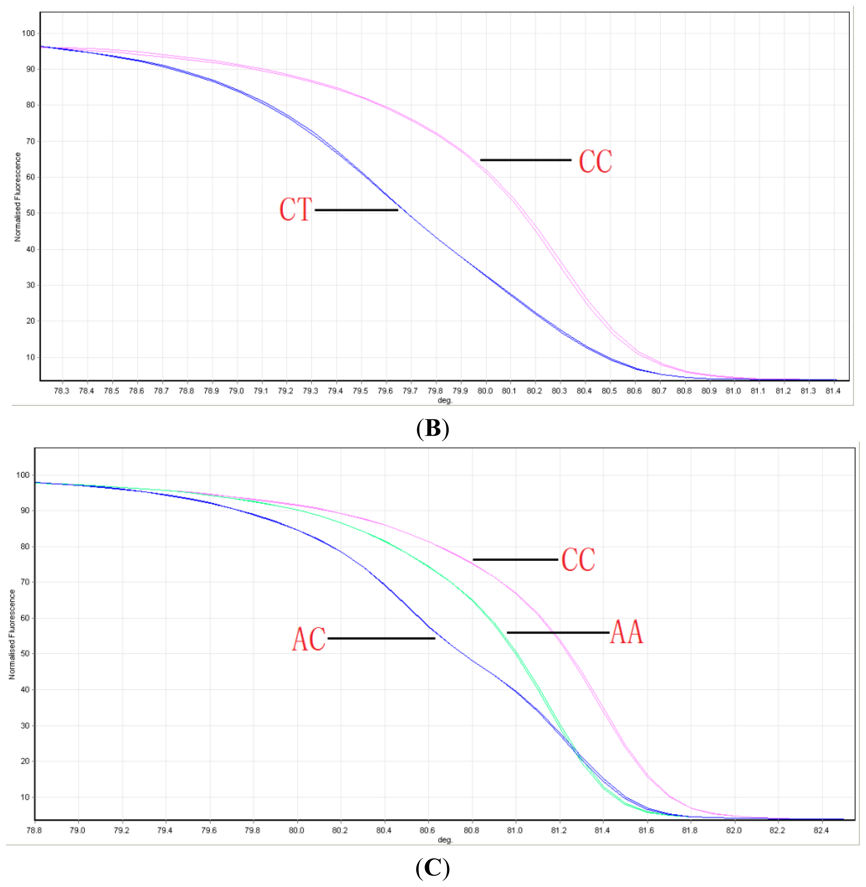
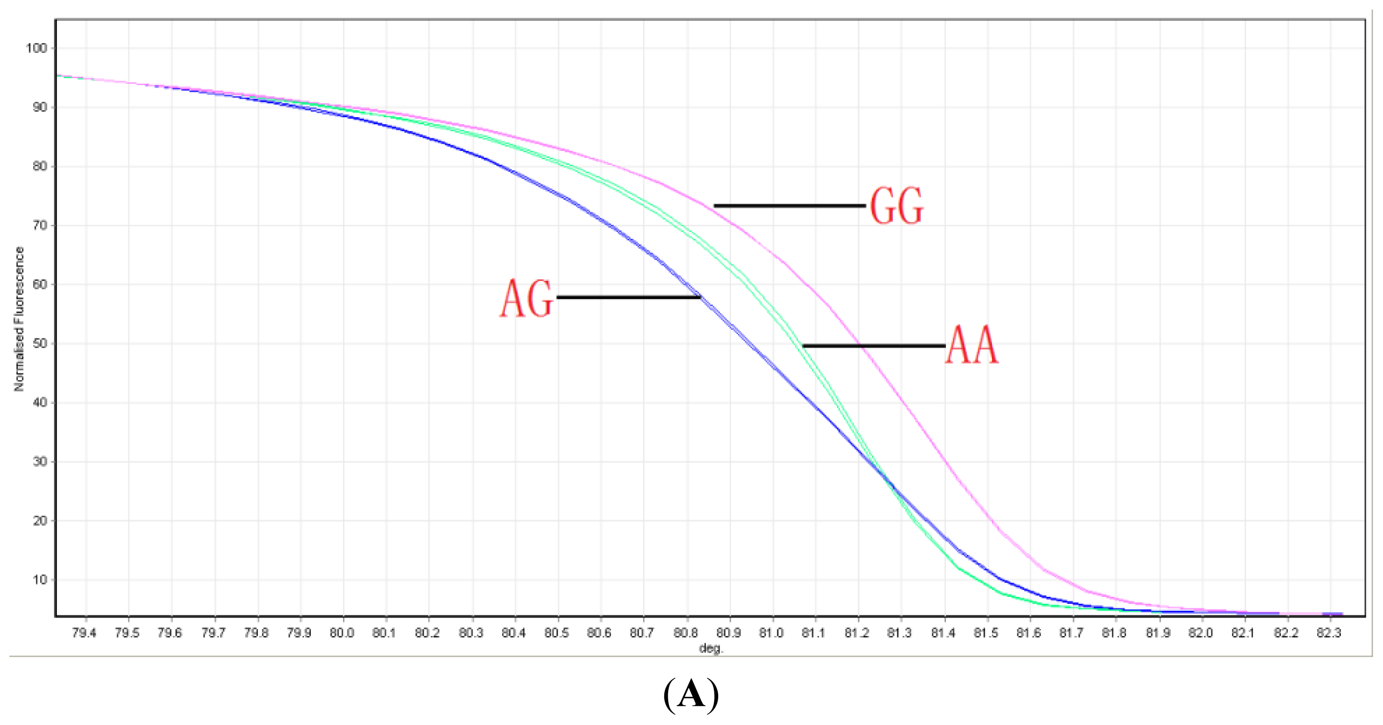
4.2. Genotyping ABCG2 with HRM

Genomic DNA was isolated from the peripheral blood lymphocytes of each patient using a QIAamp DNA Blood Kit (Qiagen, Valencia, CA, USA) according to the manufacturer’s instructions. Primer sequences were designed to amplify a small fragment surrounding the polymorphism and to avoid amplifying other sequence variations. All of the oligonucleotide primers were obtained from Sango Biotech Co. (Shanghai, China). We selected three functional ABCG2 SNPs: V12M, Q126X and Q141K. ABCG2 SNP genotyping was performed via HRM. The primer sequences are shown in Table 5. The reaction mixture consisted of 20 ng of genomic DNA, 1× polymerase chain reaction (PCR) buffer, 2.5 mM MgCl2, 200 nM each primer, 200 μM dNTPs, 5 μM SYTO 9, 0.5 U of HotStarTaq polymerase (Qiagen, Valencia, CA, USA) and PCR grade water in a 20 μL volume. PCR cycling and HRM were performed on a Rotor-Gene 6000™ (Corbett Research, Mortlake, New South Wales, Australia). All of the reactions were performed under the following conditions: one cycle of 95 °C for 15 min; 40 cycles of 95 °C for 30 s, 55 °C for 30 s and 72 °C for 30 s; a melt from 75 to 90 °C was also performed at intervals (ramps) of 0.1 °C/s. An HRM curve analysis was performed using the Rotor-Gene 6000 1.7 software (Corbett Research).

4.3. Sequencing

To confirm the genotyping results, PCR-amplified DNA samples were selected and examined via DNA sequencing. The sequencing primers were the same primers that were used in the HRM assay. The amplicons were gel purified using a QIAquick gel purification kit (Qiagen, Valencia, CA, USA). The DNA sequencing analysis was performed in an ABI PRISM 3130 genetic analyzer (Applied Biosystems, Foster City, CA, USA).

4.4. Statistical Analysis

The chi-squared test was used to analyze categorical data and compare the genotypes and allele frequency distributions. The odds ratios (ORs) and 95% confidence intervals (95% CIs) were calculated. Haplotype frequency analyses were performed using the SHEsis online software ( http://analysis.bio-x.cn/myAnalysis.php). p Values less than 0.05 were considered statistically significant.

5. Conclusions

Because gout leads to a significantly impaired quality of life and imposes high life-long medical costs, early genetic testing of the ABCG2 gene for protein dysfunction will help implement risk-management systems for gout. Developing an understanding of renal transporters will provide interesting targets for the development of future gout therapies. The function of some ABC transporter mutants has reportedly been rescued by pharmacological chaperones [35,36]. The recent findings on the roles of the ABCG2 Q141K and Q126X polymorphism in gout may pave the way for pharmacological chaperones targeting ABCG2 as a potential new therapeutic target for gout. Moreover, risk assessment through genotyping only two SNPs with molecular technologies will provide a cost-effective screening strategy for personalized gout treatment that includes adequate prevention and effective therapy.

Acknowledgments

Funding: This study was funded in part by the Shanghai Committee of Science and Technology Major Program (NO. 11DJ1400101), Shanghai Health Bureau Scientific Research Foundation (20114300, 20134064), and the Baoshan Scientific Bureau Research Foundation (11–E–9).

Conflicts of Interest

The authors declare no conflict of interest.
  • Author ContributionsHejian Zou and Ming Guan designed the experiments and wrote manuscript. Danqiu Zhou, Yunqing Liu, Xinju Zhang, Hua Wang and Xiaoye Gu did the experiments, Xinhua Luo and Jin Zhang analyzed the data.

References

  1. Choi, H.K.; Mount, D.B.; Reginato, A.M. Pathogenesis of gout. Ann. Intern. Med 2005, 143, 499–516. [Google Scholar]
  2. Zhu, Y.; Pandya, B.J.; Choi, H.K. Prevalence of gout and hyperuricemia in the US general population: The National Health and Nutrition Examination Survey 2007–2008. Arthritis Rheumatol 2011, 63, 3136–3141. [Google Scholar]
  3. Krishnan, E.; Lienesch, D.; Kwoh, C.K. Gout in ambulatory care settings in the United States. J. Rheumatol 2008, 35, 498–501. [Google Scholar]
  4. Trieste, L.; Palla, I.; Fusco, F.; Tani, C.; Baldini, C.; Mosca, M.; Turchetti, G. The economic impact of gout: A systematic literature review. Clin. Exp. Rheumatol 2012, 30, S145–S148. [Google Scholar]
  5. Tuttle, K.R.; Short, R.A.; Johnson, R.J. Sex differences in uric acid and risk factors for coronary artery disease. Am. J. Cardiol 2001, 87, 1411–1414. [Google Scholar]
  6. Talaat, K.M.; el-Sheikh, A.R. The effect of mild hyperuricemia on urinary transforming growth factor beta and the progression of chronic kidney disease. Am. J. Nephrol 2007, 27, 435–440. [Google Scholar]
  7. Nath, S.D.; Voruganti, V.S.; Arar, N.H.; Thameem, F.; Lopez-Alvarenga, J.C.; Bauer, R.; Blangero, J.; MacCluer, J.W.; Comuzzie, A.G.; Abboud, H.E. Genome scan for determinants of serum uric acid variability. J. Am. Soc. Nephrol 2007, 18, 3156–3163. [Google Scholar]
  8. Enomoto, A.; Kimura, H.; Chairoungdua, A.; Shigeta, Y.; Jutabha, P.; Cha, S.H.; Hosoyamada, M.; Takeda, M.; Sekine, T.; Igarashi, T.; et al. Molecular identification of a renal urate anion exchanger that regulates blood urate levels. Nature 2002, 417, 447–452. [Google Scholar]
  9. Vitart, V.; Rudan, I.; Hayward, C.; Gray, N.K.; Floyd, J.; Palmer, C.N.; Knott, S.A.; Kolcic, I.; Polasek, O.; Graessler, J.; et al. SLC2A9 is a newly identified urate transporter influencing serum urate concentration, urate excretion and gout. Nat. Genet 2008, 40, 437–442. [Google Scholar]
  10. Kolz, M.; Johnson, T.; Sanna, S.; Teumer, A.; Vitart, V.; Perola, M.; Mangino, M.; Albrecht, E.; Wallace, C.; Farrall, M.; et al. Meta-analysis of 28,141 individuals identifies common variants within five new loci that influence uric acid concentrations. PLoS Genet 2009, 5, e1000504. [Google Scholar]
  11. Cheng, L.S.; Chiang, S.L.; Tu, H.P.; Chang, S.J.; Wang, T.N.; Ko, A.M.; Chakraborty, R.; Ko, Y.C. Genomewide scan for gout in taiwanese aborigines reveals linkage to chromosome 4q25. Am. J. Hum. Genet 2004, 75, 498–503. [Google Scholar]
  12. Woodward, O.M.; Kottgen, A.; Coresh, J.; Boerwinkle, E.; Guggino, W.B.; Kottgen, M. Identification of a urate transporter, ABCG2, with a common functional polymorphism causing gout. Proc. Natl. Acad. Sci. USA 2009, 106, 10338–10342. [Google Scholar]
  13. Ichida, K.; Matsuo, H.; Takada, T.; Nakayama, A.; Murakami, K.; Shimizu, T.; Yamanashi, Y.; Kasuga, H.; Nakashima, H.; Nakamura, T.; et al. Decreased extra-renal urate excretion is a common cause of hyperuricemia. Nat. Commun 2012, 3, 764. [Google Scholar]
  14. Tamura, A.; Wakabayashi, K.; Onishi, Y.; Takeda, M.; Ikegami, Y.; Sawada, S.; Tsuji, M.; Matsuda, Y.; Ishikawa, T. Re-evaluation and functional classification of non-synonymous single nucleotide polymorphisms of the human ATP-binding cassette transporter ABCG2. Cancer Sci 2007, 98, 231–239. [Google Scholar]
  15. Dehghan, A.; Kottgen, A.; Yang, Q.; Hwang, S.J.; Kao, W.L.; Rivadeneira, F.; Boerwinkle, E.; Levy, D.; Hofman, A.; Astor, B.C.; et al. Association of three genetic loci with uric acid concentration and risk of gout: A genome-wide association study. Lancet 2008, 372, 1953–1961. [Google Scholar]
  16. Matsuo, H.; Ichida, K.; Takada, T.; Nakayama, A.; Nakashima, H.; Nakamura, T.; Kawamura, Y.; Takada, Y.; Yamamoto, K.; Inoue, H.; et al. Common dysfunctional variants in ABCG2 are a major cause of early-onset gout. Sci. Rep 3.
  17. Mizuarai, S.; Aozasa, N.; Kotani, H. Single nucleotide polymorphisms result in impaired membrane localization and reduced atpase activity in multidrug transporter ABCG2. Int. J. Cancer 2004, 109, 238–246. [Google Scholar]
  18. Matsuo, H.; Takada, T.; Ichida, K.; Nakamura, T.; Nakayama, A.; Ikebuchi, Y.; Ito, K.; Kusanagi, Y.; Chiba, T.; Tadokoro, S.; et al. Common defects of ABCG2, a high-capacity urate exporter, cause gout: A function-based genetic analysis in a Japanese population. Sci. Transl. Med 2009, 1, 5ra11. [Google Scholar]
  19. Ishikawa, T.; Aw, W.; Kaneko, K. Metabolic Interactions of purine derivatives with human ABC transporter ABCG2: Genetic testing to assess gout risk. Pharmaceuticals (Basel) 2013, 6, 1347–1360. [Google Scholar]
  20. Endou, H.; Anzai, N. Urate transport across the apical membrane of renal proximal tubules. Nucleosides Nucleotides Nucleic Acids 2008, 27, 578–584. [Google Scholar]
  21. Anzai, N.; Jutabha, P.; Amonpatumrat-Takahashi, S.; Sakurai, H. Recent advances in renal urate transport: Characterization of candidate transporters indicated by genome-wide association studies. Clin. Exp. Nephrol 2012, 16, 89–95. [Google Scholar]
  22. George, R.L.; Keenan, R.T. Genetics of hyperuricemia and gout: Implications for the present and future. Curr. Rheumatol. Rep 2013, 15, 309. [Google Scholar]
  23. Guan, M.; Zhou, D.; Ma, W.; Chen, Y.; Zhang, J.; Zou, H. Association of an intronic SNP of SLC2A9 gene with serum uric acid levels in the Chinese male Han population by high-resolution melting method. Clin. Rheumatol 2011, 30, 29–35. [Google Scholar]
  24. Li, C.; Chu, N.; Wang, B.; Wang, J.; Luan, J.; Han, L.; Meng, D.; Wang, Y.; Suo, P.; Cheng, L.; et al. Polymorphisms in the presumptive promoter region of the SLC2A9 gene are associated with gout in a Chinese male population. PLoS One 2012, 7, e24561. [Google Scholar]
  25. Li, C.; Han, L.; Levin, A.M.; Song, H.; Yan, S.; Wang, Y.; Meng, D.; Lv, S.; Ji, Y.; Xu, X.; et al. Multiple single nucleotide polymorphisms in the human urate transporter 1 (hURAT1) gene are associated with hyperuricaemia in Han Chinese. J. Med. Genet 2010, 47, 204–210. [Google Scholar]
  26. Guan, M.; Zhang, J.; Chen, Y.; Liu, W.; Kong, N.; Zou, H. High-resolution melting analysis for the rapid detection of an intronic single nucleotide polymorphism in SLC22A12 in male patients with primary gout in China. Scand. J. Rheumatol 2009, 38, 276–281. [Google Scholar]
  27. Hosomi, A.; Nakanishi, T.; Fujita, T.; Tamai, I. Extra-renal elimination of uric acid via intestinal efflux transporter BCRP/ABCG2. PLoS One 2012, 7, e30456. [Google Scholar]
  28. Woodward, O.M.; Tukaye, D.N.; Cui, J.; Greenwell, P.; Constantoulakis, L.M.; Parker, B.S.; Rao, A.; Kottgen, M.; Maloney, P.C.; Guggino, W.B. Gout-causing Q141K mutation in ABCG2 leads to instability of the nucleotide-binding domain and can be corrected with small molecules. Proc. Natl. Acad. Sci. USA 2013, 110, 5223–5228. [Google Scholar]
  29. Zhang, L.; Spencer, K.L.; Voruganti, V.S.; Jorgensen, N.W.; Fornage, M.; Best, L.G.; Brown-Gentry, K.D.; Cole, S.A.; Crawford, D.C.; Deelman, E.; et al. Association of functional polymorphism rs2231142 (Q141K) in the ABCG2 gene with serum uric acid and gout in 4 US populations: The PAGE study. Am. J. Epidemiol 2013, 177, 923–932. [Google Scholar]
  30. Lepper, E.R.; Nooter, K.; Verweij, J.; Acharya, M.R.; Figg, W.D.; Sparreboom, A. Mechanisms of resistance to anticancer drugs: The role of the polymorphic ABC transporters ABCB1 and ABCG2. Pharmacogenomics 2005, 6, 115–138. [Google Scholar]
  31. Wang, B.; Miao, Z.; Liu, S.; Wang, J.; Zhou, S.; Han, L.; Meng, D.; Wang, Y.; Li, C.; Ma, X. Genetic analysis of ABCG2 gene C421A polymorphism with gout disease in Chinese Han male population. Hum. Genet 2010, 127, 245–246. [Google Scholar]
  32. Kim, H.S.; Sunwoo, Y.E.; Ryu, J.Y.; Kang, H.J.; Jung, H.E.; Song, I.S.; Kim, E.Y.; Shim, J.C.; Shon, J.H.; Shin, J.G. The effect of ABCG2 V12M, Q141K and Q126X, known functional variants in vitro, on the disposition of lamivudine. Br. J. Clin. Pharmacol 2007, 64, 645–654. [Google Scholar]
  33. Itoda, M.; Saito, Y.; Shirao, K.; Minami, H.; Ohtsu, A.; Yoshida, T.; Saijo, N.; Suzuki, H.; Sugiyama, Y.; Ozawa, S.; et al. Eight novel single nucleotide polymorphisms in ABCG2/BCRP in Japanese cancer patients administered irinotacan. Drug Metab. Pharmacokinet 2005, 18, 212–217. [Google Scholar]
  34. Kobayashi, D.; Ieiri, I.; Hirota, T.; Takane, H.; Maegawa, S.; Kigawa, J.; Suzuki, H.; Nanba, E.; Oshimura, M.; Terakawa, N.; et al. Functional assessment of ABCG2 (BCRP) gene polymorphisms to protein expression in human placenta. Drug Metab. Dispos 2005, 33, 94–101. [Google Scholar]
  35. Loo, T.W.; Bartlett, M.C.; Clarke, D.M. Rescue of folding defects in ABC transporters using pharmacological chaperones. J. Bioenerg. Biomembr 2005, 37, 501–507. [Google Scholar]
  36. Chiba, P.; Freissmuth, M.; Stockner, T. Defining the blanks-Pharmacochaperoning of SLC6 transporters and ABC transporters. Pharmacol. Res 2013. [Google Scholar] [CrossRef]
Figure 1. Melting curves of SNP genotypes in the ABCG2 gene. The three groups are well distinguished: (A) V12M; (B) Q126X; and (C) Q141K.
Ijms 15 09149f1aIjms 15 09149f1b
Table 1. Association analysis of ABCG2 variants in gout patients. MAF, minor allele frequency.
SNPGenotype *Allele Frequency Mode


CaseControlp-Valuep-ValueOR95% CI


1/11/22/2MAF1/11/22/2MAF
Q141K84181870.496331501670.3091.18 × 10−118.99 × 10−132.201.77–2.74
Q126X0333190.0470123380.0171.31 × 10−31.57 × 10−32.911.49–5.68
V12M16972390.183351331820.2903.67 × 10−52.55 × 10−60.550.43–0.71
*The minor allele was referred to as allele 1, and the major allele was referred to as allele 2. Allele 1 is A and allele 2 is C in Q141K. Allele 1 is T and allele 2 is C in Q126X. Allele 1 is A and allele 2 is G in V12M.
Table 2. Haplotype frequency analysis of V12M, Q126X and Q141K.
AlleleFrequencyp-ValueOR95% CI


V12MQ126XQ141KGoutControl
GCA0.4810.2891.26 × 10−132.301.84–2.87
GTC0.0440.0172.97 × 10−32.711.37–5.36
GCC0.2920.4048.27 × 10−60.600.48–0.75
ACC0.1650.2711.53 × 10−60.530.41–0.69
Table 3. Participants’ ABCG2 function levels.
Estimated FunctionGenotype CombinationNumber (%)p-ValueOR95% CI


Q141KQ126XGoutControl
≤1/4 functionC/AT/C22 (6.2)8 (2.3)8.47 × 10−65.902.56–13.58
1/2 functionC/CT/C95 (27.0)37 (10.5)1.12 × 10−135.513.46–8.77
A/AC/C
3/4 functionC/AC/C159 (45.2)142 (40.6)1.00 × 10−62.401.69–3.42
Full functionC/CC/C76 (21.6)163 (46.6)
p-Value, OR and 95% CI for each ABCG2 dysfunction were obtained via comparisons with full function.
Table 4. Clinical and biochemical profile of gout patients and controls.
IndexGout PatientsControlsp-Value
Subjects (%)352 (50.1%)350 (49.9%)
Age (year)57.6 ± 14.056.6 ± 16.6NS
BUN (mmol/L)5.4 ± 1.95.5 ± 2.1NS
Creatinine (μmol/L)97.3 ± 15.796.1 ± 16.4NS
Uric Acid (μmol/L)456.4 ± 120.1334.7 ± 88.7<0.01
Data are expressed as the means ± standard deviation (S.D.).
Table 5. Primer sequences.
SNP IDSNP AlleleSequence (5′-3′)Size
V12MA/GATGGTATGGGCCATTCATTG
ATGCCTTCAGGTCATTGGAA
250 bp
Q141KA/CATGTTGTGATGGGCACTCTG
CCACATTACCTTGGAGTCTG
158 bp
Q126XC/TGCTGCAAGGAAAGATCCAAG
CAGCCAAAGCACTTACCCAT
163 bp

