Article

The Canonical Notch Signaling Was Involved in the Regulation of Intestinal Epithelial Cells Apoptosis after Intestinal Ischemia/Reperfusion Injury

Department of General Surgery, Xinqiao Hospital, Third Military Medical University, Chongqing 400037, China
*
Author to whom correspondence should be addressed.
These authors contributed equally to this work.
Int. J. Mol. Sci. 2014, 15(5), 7883-7896; https://doi.org/10.3390/ijms15057883
Submission received: 22 February 2014 / Revised: 21 April 2014 / Accepted: 24 April 2014 / Published: 6 May 2014
(This article belongs to the Section Biochemistry)

Abstract

:
Notch signaling plays a critical role in the maintenance of intestinal homeostasis. The aim of the present study was to investigate the role of Notch signaling in the apoptosis of intestinal epithelial cells after intestinal ischemia reperfusion (I/R) injury. Male C57BL/6 mice were subjected to sham operation or I/R injury. Intestinal tissue samples were collected at 12 h after reperfusion. TUNEL (terminal deoxynucleotidyl transferase-mediated dUTP-biotin nick end labeling) staining showed that intestinal I/R injury induced significantly increased apoptosis of intestinal epithelial cells. Meanwhile, the mRNA expression of Jagged1, DLL1, Notch2, and Hes5, and protein expression of NICD2 and Hes5 were increased significantly after I/R injury in intestinal epithelial cells. In an in vitro IEC-6 culture model, flow cytometry analyses showed that inhibition of Notch signaling by γ-secretase inhibitor DAPT and the suppression of Hes5 expression using siRNA both significantly increased the apoptosis of IEC-6 cells under the condition of hypoxia/reoxygenation (H/R). In conclusion, the Notch2/Hes5 signaling pathway was activated and involved in the regulation of intestinal epithelial cells apoptosis in intestinal I/R injury.

Graphical Abstract

1. Introduction

Intestinal ischemia/reperfusion (I/R) injury may lead to the development of systemic inflammatory response syndrome, sepsis, and multiple organ dysfunction syndrome (MODS). The loss of intestinal barrier function is considered to be the initial triggering event that turns the gut into the “motor” of MODS [1]. It is known that cell apoptosis is a significant contributor to increased intestinal permeability and there is clear evidence of cell apoptosis in intestinal mucosa after intestinal I/R injury [24]. Several signaling pathways, such as PI3K/Akt, ERK1/2, and HIF-1, have been reported to be involved in the apoptosis of intestinal epithelial cells after I/R injury [57]. However, the precise molecular mechanisms of this process are still not fully understood.
Studies have shown that the Notch signaling pathway plays critical roles in intestinal epithelial cell homeostasis [8]. In mammals, there are four transmembrane Notch receptors (Notch1–4), two Jagged-like ligands (Jagged1, Jagged2), and three Delta-like ligands (DLL1, DLL3, DLL4) [9,10]. The interaction of these five ligands with Notch receptors will activate their proteolytic cleavage at two sites. The cleavage releases the Notch intracellular domain (NICD), which translocates to the nucleus and forms an activated transcriptional complex. The complex then activates the transcription of target genes, such as Hes (Hairy/Enhancer of split) and Hey (Hes-related with YRPW motif), which are two families of basic helix-loop-helix genes [11]. Importantly, the second Notch receptor cleavage is mediated by the γ-secretase complex, and inhibition of this proteolytic activity will block the activation of Notch receptors [12].
In hepatic I/R injury, interruption of Notch signaling resulted in increased intracellular ROS and increased apoptosis of hepatocytes. Notch2 signal protected hepatocytes from I/R injury by Hes5 dependent activation of STAT3, leading to the scavenging of ROS [13]. Notch signaling protects myocardial cell, endothelial cell, and lymphocyte from apoptosis after injury [1416]. Notch signaling was also reported to protect neuron and non-parenchymal cells from apoptosis in I/R injury [17,18]. After intestinal I/R injury, intestinal epithelial cells undergo proliferation and apoptosis [24]. In our previous published study, we detected increased expression of Notch signaling components in intestinal epithelium after intestinal I/R injury and showed that Notch signaling was involved in the proliferation of intestinal epithelial cells [19]. However, little is known about the role of Notch signaling in the apoptosis of intestinal epithelial cells after intestinal I/R injury. The purpose of the present study was to investigate the role of Notch signaling pathway in the apoptosis of intestinal epithelial cells after intestinal I/R injury.

2. Results

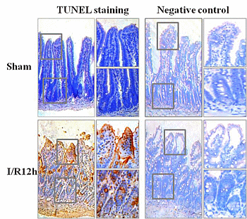
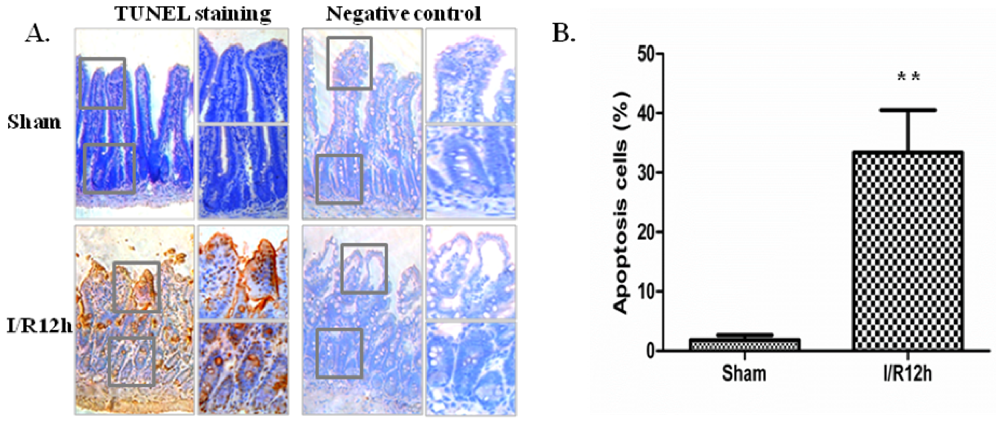
2.1. Intestinal I/R Injury Increased Apoptosis of Intestinal Epithelial Cells

We built the intestinal I/R model through occlusion of superior mesenteric artery (SMA) for 20 min in mice. Jejunum of mice were excised at 12 h after reperfusion. TUNEL examination was carried out to examine the apoptosis of intestinal epithelial cells after intestinal I/R injury. TUNEL staining results showed that the TUNEL-positive cells could be observed in the intestinal epithelial cells of sham operated mice. Importantly, I/R (12 h) significantly increased the number of TUNEL-positive cells compared with sham operation (Figure 1A,B). The TUNEL-positive cells increased 18.6-fold in the I/R (12 h) group compared with the sham group (Figure 1B). However, in the negative control group there was no TUNEL-positive cell (Figure 1A). This shows that all the TUNEL results are credible.

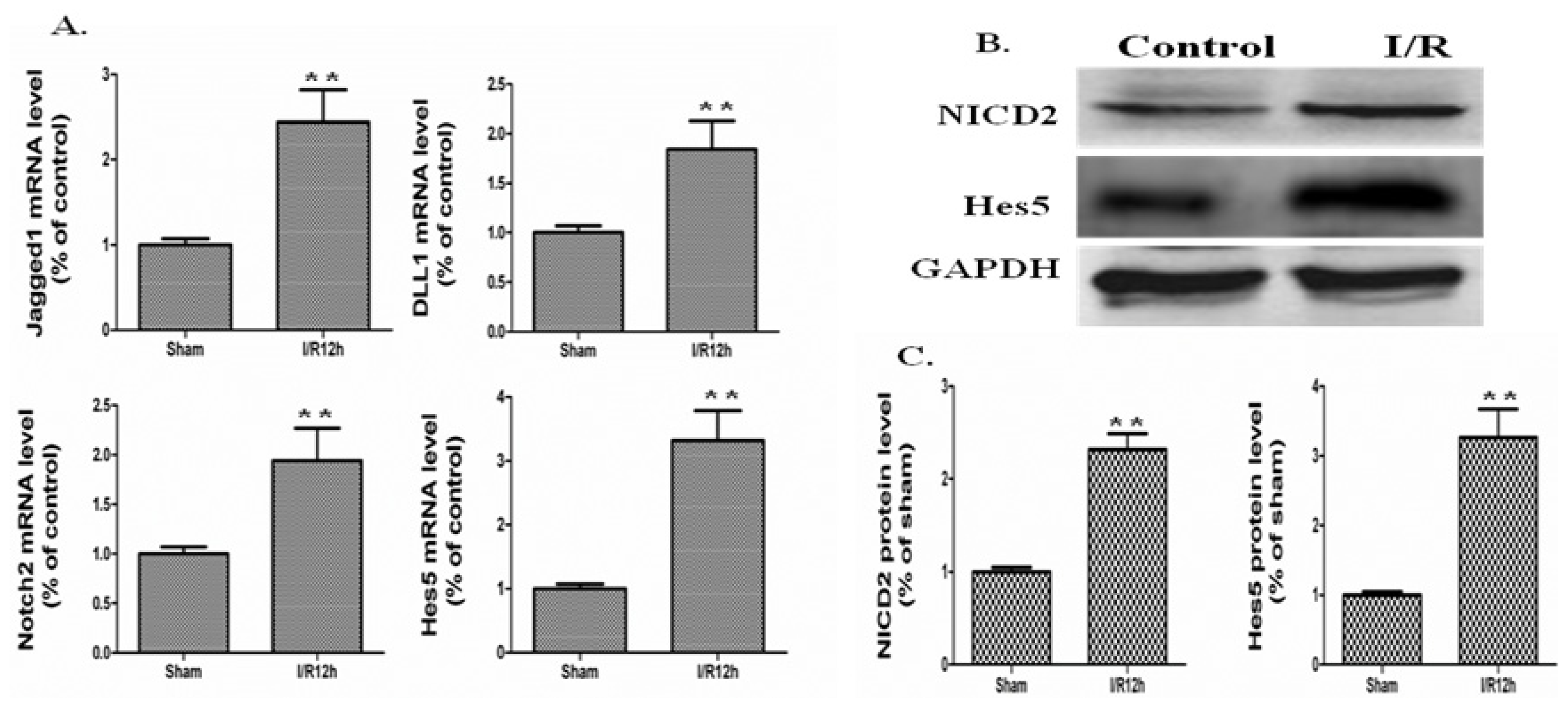
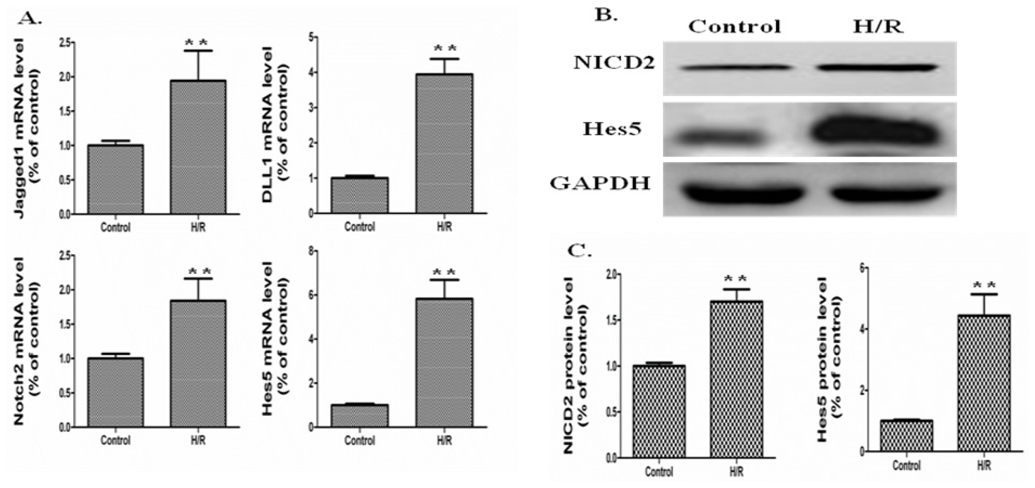
2.2. The Notch Signaling Was Activated in a Mouse Model of Intestinal I/R

To investigate the expression of Notch signaling components in intestinal epithelium after I/R, we generated a mouse model of intestinal I/R by occlusion of the SMA for 20 min. The clamps were removed, and I/R mice were killed at 12 h after reperfusion. According to published studies, Notch signaling components were expressed in intestine of mouse, including four ligands (Jagged1, Jagged2, DLL1, and DLL4), four Notch receptors (Notch1, 2, 3, and 4), and four Hes genes (Hes1, Hes5, Hes6, and Hes7) [20]. In our experiment, real-time PCR analysis was applied to screen the intestinal epithelium genes for ligands, Notch receptors (Notch1, 2, 3, and 4) and target genes. As shown in Figure 2A, the mRNA levels of Jagged1, DLL1, Notch2, and the target gene Hes5 were increased after I/R injury compared to the sham operation group. The mRNA expression of Jagged1, DLL1, Notch2 and Hes5 increased 2.44-, 1.84-, 1.94-, and 3.32-fold, at 12 h after I/R compared to sham operation, respectively (Figure 2A, p < 0.01, I/R 12 h vs. Sham). At this point, the other Notch signaling components, such as Notch1, 3, and 4, exhibited no significant changes statistically (data not shown). Based on the mRNA expression of Notch signaling components, we focused on the protein expression of NICD2 and Hes5 during the following experiments.
The protein level of Hes5 also increased after I/R injury (Figure 2B,C). The protein expression of Hes5 was increased 3.27-fold after I/R injury, respectively (Figure 2C, p < 0.01, I/R 12 h vs. Sham). As cleavage of Notch2 is the indicator of Notch signal activation, then we investigated NICD2 protein expression. Western blot analyses showed a significant increase in the NICD2 level at 12 h after I/R, indicating the activation of Notch signaling (Figure 2B,C). Quantification of the Western blot results revealed a 2.32-fold increase of NICD2 compared with the sham operated mice (Figure 2C, p < 0.01, I/R 12 h vs. Sham).

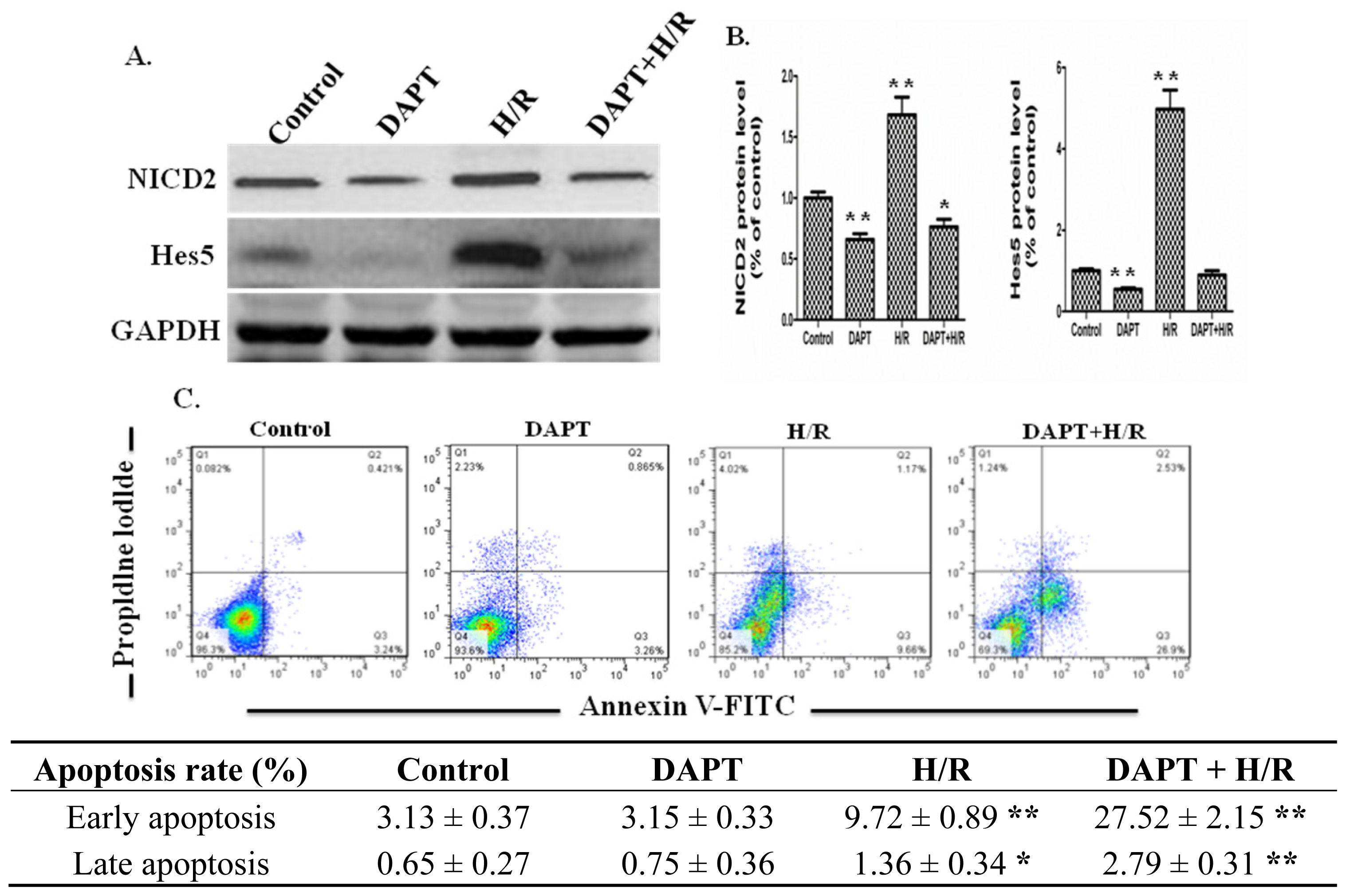
2.3. Effects of DAPT on the Apoptosis of IEC-6 Cells

In order to investigate the functional role of the Notch2 signaling pathway in intestinal epithelial cells, we used a culture system with IEC-6 cells receiving 4 h hypoxia, followed by 4 h reoxygenation (H/R), as reported previously [21].
First, we examined if this culture system mimics the in vivo I/R model, we investigated the mRNA expression of Jagged1, DLL1, Notch2, and Hes5 and the protein expression of NICD2, and Hes5. The real-time PCR results showed that H/R up-regulated the mRNA expression of Jagged1, DLL1, Notch2, and Hes5 by 1.94-, 3.95-, 1.84-, and 5.82-fold compared to the control group, respectively (p < 0.01, H/R vs. Control) (Figure 3A). In accordance with the in vivo real-time PCR results, at this point, the other Notch signaling components of IEC-6 cells, such as Notch1, 3, and 4, also exhibited no significant changes statistically (data not shown). Western blot results showed that H/R up-regulated the protein expression of NICD2 and Hes5 by 1.7- and 4.43-fold compared to the control group, respectively (p < 0.01, H/R vs. Control) (Figure 3B,C). These results indicated that the IEC-6 culture system mimics the in vivo I/R model.
DAPT is one type of γ-secretase inhibitor that inhibits the activation of Notch signaling. DAPT was used to stimulate the IEC-6 cells to investigate the function of Notch signaling in intestinal epithelial cells. After DAPT was added to the culture medium of IEC-6 cells for 12 h, IEC-6 cells received H/R or not. After H/R, cell protein was extracted and western blot was applied to examine the the effect of DAPT on the signal transduction of IEC-6 cells. The Western blot results showed that DAPT down-regulated the protein expressions of NICD2 and Hes5 (Figure 4A,B). Furthermore, we examined the effects of DAPT on the apoptosis of IEC-6 cells receiving H/R or not with flow cytometry. The flow cytometry results showed that H/R increased the early and late apoptosis of IEC-6 cells compared to control group (Figure 4C). Statistically, DAPT had no effect on the apoptosis rate of IEC-6 cells. However, DAPT and H/R together increased the apoptosis rate of IEC-6 cells significantly, especially the early apoptosis of IEC-6 cells (Figure 4C). The results above showed that down-regulation of Notch2 signaling by γ-secretase inhibition contributed to IEC-6 cells apoptosis under an H/R condition (Figure 4A–C). The Notch2-Hes5 signaling was up-regulated in intestinal epithelial cells under an H/R condition and promoted the survival of IEC-6 cells.

2.4 Effects of Silencing RNA for Hes5 on the Apoptosis of IEC-6 Cells

To further investigate the effect of Notch signaling on intestinal epithelial cell apoptosis, IEC-6 cells were transfected with siRNA for Hes5. Western blot results showed that the siRNA for Hes5 significantly down-regulated the protein expression of Hes5 (Figure 5A,B). Then flow cytometry was applied to examine the effect of siRNA for Hes5 on the apoptosis of IEC-6 cells receiving H/R or not. The results showed that suppression of Hes5 expression increased the apoptosis of IEC-6 cells. Importantly, under the H/R condition siRNA for Hes5 further increased the apoptosis of IEC-6 cells significantly, especially the early apoptosis of IEC-6 cells (Figure 5C). The results above confirmed that protein expression of Hes5 could promote the survival of intestinal epithelial cells under the condition of H/R.

3. Discussion

In this study, we found that intestinal I/R stress caused increased apoptosis of intestinal epithelial cells. The mRNA and protein expression of Notch signaling components were significantly increased in intestinal epithelial cells after I/R injury. Our findings also demonstrated that the Notch2/Hes5 signaling pathway was involved in the protection of intestinal epithelial cells from intestinal I/R injury.
Intestinal I/R injury, which is associated with trauma, hemorrhage, and other shock states, is characterized by epithelial apoptosis, necrosis, and mucosal barrier dysfunction. Studies have shown that intestinal epithelial cell apoptosis is a significant contributor to mucosal barrier dysfunction. Immediate early genes, such as c-fos and c-jun are involved in the apoptosis of intestinal epithelial cells [22,23]. Some other factors, such as p38 MAPK, IL-6, HGF, and KGF, were also reported to be involved in the apoptosis of intestinal epithelial cells after I/R injury [2427]. However, the precise molecular mechanisms that accompany I/R within the intestinal epithelial cells need further investigation.
The Notch signaling pathway plays critical roles in the maintenance of intestinal homeostasis [8]. Mutations in the Notch pathway components, such as the Notch DNA binding protein RBP-J or Notch receptors will disturb the normal differentiation of intestinal epithelial cells [2830]. Inhibition of Notch signaling activation by γ-secretase inhibitors also lead to disturbed intestinal homeostasis [2830]. The published studies have shown that the PCNA-positive cells increased at 1 to 6 h after reperfusion and TUNEL-positive cells increased later than PCNA-positive cells in a rat I/R model [22,23,31]. Our previous studies have investigated the role of Notch signaling in the proliferation of intestinal epithelial cells [19,32]. In our previous study by our group, we showed that Notch signaling was activated and increased the proliferation of intestinal epithelial cells through 6 h after reperfusion [19]. However, in our previous study we did not investigate the Notch signaling expression later than 6 h after reperfusion. It is also not clear whether Notch signaling was involved in the apoptosis of intestinal epithelial cells after intestinal I/R injury. Thus, in this study we further investigated the function of Notch signaling in the apoptosis of the intestinal epithelial cells after I/R injury.
In our study, real-time PCR analysis was firstly applied to screen the intestinal epithelium genes for ligands, Notch receptors (Notch1, 2, 3, and 4) and target genes after intestinal I/R injury. Physiologically, Jagged1 and DLL1 were normally localized in the intestinal epithelial cells [20,33]. In this study, we detected increased expression of Jagged1 and DLL1 after I/R injury. To confirm the activation of Notch signaling pathway, we further detected increased expression of Notch2 and Hes5. real-time PCR and Western blot results showed that the expressions of Notch2 and Hes5 increased significantly 12 h after I/R injury. However, the other Notch signaling components, such as Notch1, 3, 4, exhibited no significant changes statistically. Thus, in the following experiments we focused on the expression of Jagged1, DLL1, Notch2, and Hes5. Accompanied with activated Notch2 signaling, the apoptosis of intestinal epithelial cells also increased significantly. From the above, we aimed to investigate whether Notch2 signaling was involved in the regulation of intestinal epithelial cells apoptosis after intestinal I/R injury.
To further investigate the functional role of Notch2 signaling in the regulation of intestinal epithelial cell apoptosis, an IEC-6 cells culture system was applied. In some instances, it may be inappropriate to extend in vitro results to in vivo conditions; The results showed that the mRNA expression of Jagged1, DLL1, Notch2, and Hes5 and the protein expression of NICD2 and Hes5 were up-regulated in IEC-6 cells under the H/R condition. The results above suggested that the in vitro IEC-6 cells culture system could mimic the in vivo I/R model upon activation of Notch2/Hes5 signaling pathway. Next, the γ-secretase inhibitor DAPT, which inhibits γ-secretase activation of Notch receptors, was used to investigate the function of Notch2 signaling on the apoptosis of IEC-6 cells receiving H/R or not. The results showed that the protein expressions of NICD2 and Hes5 were down-regulated significantly. The apoptosis of IEC-6 cells was increased significantly when Notch2 signaling was suppressed by DAPT. Next, siRNA for Hes5 was used to suppress the expression of Hes5 and examine the role of Hes5 in the apoptosis of IEC-6 cells. The results showed that suppression of Hes5 with siRNA significantly increased the apoptosis of IEC-6 cells. All these results suggested that the NICD2/Hes5 signaling was involved in the regulation of intestinal epithelial cells apoptosis after intestinal I/R injury.
In summary, our study showed that the Notch2/Hes5 signaling pathway was activated and involved in the regulation of intestinal epithelial cells apoptosis after intestinal I/R injury. Further studies are needed to gain a more precise understanding of the molecular mechanisms of intestinal epithelial cells apoptosis after intestinal I/R injury.

4. Experimental Section

4.1. Animals

Male, 6–8 week-old, specific pathogen-free, C57BL/6 mice were purchased from the Experiment Animal Center at Daping Hospital of Third Military Medical University, Chongqing, China. All the animal experiments were performed in compliance with the University’s Guidelines for the Care and Use of Laboratory Animals. The protocol was approved by the ethics committee of Xinqiao Hospital, Third Military Medical University, Chongqing, China. Mice were randomly divided into two groups: control group (sham operation, n =7) and experimental group (I/R, n =7). For the I/R group, the superior mesenteric artery (SMA) was occluded using an atraumatic microvascular clamp for 20 min. Then, we removed the clamps and closed the incisions [19]. Mice were sacrificed at 12 h after reperfusion. The jejunums of mice were quickly removed and processed for histological evaluation, RNA extraction, or protein extraction. The control mice received identical operation procedures without occlusion of the SMA.

4.2. Cell Culture

The intestinal epithelial cell line IEC-6, originally purchased from the American type culture collection (Manassas, VA, USA), were grown in Dulbecco’s modified Eagles medium (DMEM, Hyclone, Thermo Fisher, Rockford, IL, USA) supplemented with 10% fetal calf serum (Sigma-Aldrich, St. Louis, MO, USA), 100 μg/mL streptomycin and 100 IU/mL penicillin and cultured overnight for adHesion. Once grown, the IEC-6 cells were cultured at 37 °C in either normoxic (20% O2 and 5% CO2) or hypoxic (1% O2 and 5% CO2 in a hypoxia chamber) conditions (Thermo Fisher, Rockford, IL, USA). The γ-secretase inhibitor DAPT was added to the medium for 12 h, and the apoptosis of IEC-6 cells were investigated with flow cytometric analysis. Total protein was also obtained for Western blot analysis.

4.3. Real-Time PCR Analysis

Total RNA was extracted following a standard isothiocyanate/chloroform extraction method using Trizol (Takara Co., Ltd., Dalian, China), and reverse transcription into first-strand cDNA was performed using the First Strand cDNA Synthesis Kit (FSK-100, TOYOBO CO., Ltd., Osaka, Japan) in the presence of the RNase inhibitor diethylpyrocarbonate (DEPC) (Roche Diagnostics GmbH, Mannheim, Germany) [32]. The amplified cDNA was used as the template DNA for PCR performed with specific primers. In the in vivo experiments, the following primers were used: Jagged1 forward, 5′-CTTGGGTCTGTTGCTTGGTGA-3′ and reverse, 5′-ACATTGTTGGTGGTGTTGTCCTC-3′; DLL1 forward, 5′-CGGCTTCTATGGCAAGGTCTG-3′ and reverse, 5′-TGTAGCCTCCGTCAGGGTTATCT-3′; Notch2 forward, 5′-GCGAGCACCCATACCTGACA-3′ and reverse, 5′-TGGGCTGGTGGTCACATCTG-3′; and Hes5 forward, 5′-AAGCTGGAGAAGGCCGACA-3′ and reverse, 5′-CAGGAGTAGCCCTCGCTGTAGT-3′; GAPDH forward, 5′-AGAAGGTGGTGAAGCAGGCA-3′ and reverse, 5′-AGGTGGAAGAGTGGGAGTTGC-3′. In the in vitro experiments the following primers were used: Jagged1 forward, 5′-CGCCGTGCCCTTTGTGGAG-3′ and reverse, 5′-GGGCCAGACTGCAGGATAAAC-3′; DLL1 forward, 5′-AGAGGGGCCAACGCTACATGTG-3′ and reverse, 5′-GGCGGAGGCTGGTGTTTCTG-3′; Notch2 forward, 5′-GGGGGGACCTGCTCTGACTAC-3′ and reverse, 5′-ACGTGCCGCCATTGAAACAGGAG-3′; and Hes5 forward, 5′-GAAGCCGGTGGTGGAGAAG-3′ and reverse, 5′-CGGCGAAGGCTTTGCTGTG-3′; GAPDH forward, 5′-GGGGCCAAAAGGGTCATCATCTC-3′ and reverse, 5′-AGGGGCCATCCACAGTCTTC-3′; Real-time PCR was performed as previously described [27]. The standard conditions used for real-time PCR were as follows: 94 °C for 10 min, 30 s at 94 °C, 30 s at 60 °C, and 45 s at 72 °C for 45 cycles.

4.4. Western Blot Analysis

Tissues and cells were homogenized in cold RIPA buffer (PBS, 1% NP-40, 0.5% sodium deoxycholate, 0.1% SDS, 1 μg/mL PMSF, 1.0 mM sodium orthovanadate, and 1× mammalian protease inhibitor cocktail; Sigma-Aldrich, Shanghai, China). Protein was quantified by the Bradford method using the BCA assay reagent (Beyotime, Shanghai, China). Equal amounts of protein were loaded into SDS-polyacrylamide gels and transferred onto PVDF-Plus membranes. After blocking in 5% fat-free milk for 1 h at room temperature, the membranes were incubated overnight at 4 °C with the following primary antibodies: NICD2 (ab-52302, Abcam, UK), Hes5 (sc-25395, Santa Cruz, Dallas, CA, USA), and GAPDH (sc-32233, Santa-Cruz, Dallas, CA, USA). The primary antibodies were detected with horseradish peroxidase-conjugated goat anti-rabbit IgG, or goat anti mouse IgG secondary antibody for 1 h at room temperature and detected by the use of a chemiluminescence system (Beyotime, Shanghai, China) and imaging system (Kodak Gel Logic 4000R Imaging System, Carestream, Rochester, NY, USA). A semiquantitative densitometry analysis of the bands were performed using the Kodak Gel Logic 4000R Imaging System (Carestream, Rochester, NY, USA). Protein expression was normalized to the same sample’s expression of GAPDH.

4.5. Silencing Hes5 Using siRNA and in Vitro Transfection

To inhibit the Notch signaling pathway, an in vitro transfection to silence Hes5 expression was performed. siRNA were chemically syntHesized by Ribobio (Guangzhou, China). The sequences of the siRNAs targeting Hes5 were as follows: siRNA1, sense 5′ GCAUUGAGCAGCUGAAACUdTdT 3′ and antisense 3′ dTdTCGUAACUCGUCGACUUUGA 5′; siRNA2, sense 5′ GCAGAUGAAGCUGCUUUACdTdT 3′ and antisense 3′ dTdTCGUCUACUUCGACGAAAUG 5′.
After IEC-6 cells were cultured to 50%–60% confluency in 6-well plates, they were transfected with siRNA at a concentration of 50 nmol for each well using the Lipofectamine 2000 reagent (Invitrogen, Shanghai, China) in antibiotic-free and serum-free Opti-MEM medium, according to the manufacturer’s instructions [19]. An unrelated control siRNA (si-NC) was used in the experiment as the negative control. After 6 h, the medium was replaced with normal IEC-6 cell medium, and the cells were cultured for 48 h. Then, protein was extracted from the cells, and Western blot analysis was performed to detect the protein expression of Hes5. Flow cytometric analysis was applied to investigate the effect of siRNA on the apoptosis of IEC-6 cells.

4.6. Detection of Epithelial Apoptosis

Tissues were fixed with 4% paraformaldehyde, and 5 μm paraffin-embedded sections were prepared. Intestinal epithelial apoptosis was investigated through terminal deoxynucleotidyl transferase mediated dUTP nick-end labeling (TUNEL) staining using an in situ cell death detection POD kit (Roche, Penzberg, Germany) in accordance with the manufacturer’s instructions. As a predealing process, the slides were incubated with 20 μg/mL proteinase K (pH 7.6) for 15 min at room temperature. Then slides were incubated with 0.1% Triton X-100 for 8 min and rinsed with phosphate buffered saline (PBS). Endogenous peroxidase activity was blocked with 3% H2O2 and methanol. Rinse slides with PBS, add 50 μL TUNEL reaction mixture on sample and incubate for 60 min at 37 °C. Rinse slides three times with PBS and examine the samples under a fluorescence microscope. After this pilot evaluation, the slides were stained by DAB coupling. All slides were counterstained with hematoxylin. As a negative control, the terminal transferase was omitted. The TUNEL-positive cells were counted under a light microscope.
In order to detect the apoptosis of IEC-6 cells, flow cytometric analysis was applied with Annexin V-FITC Apoptosis Detection Kit (KeyGEN Biotech, Nanjing, China) according to the manufacturer’s instructions. IEC-6 cells were cultured as above. Double staining for FITC-Annexin V binding and cellular DNA using propidium iodide (PI) was performed as previously described [34]. The acquisition and analysis were performed using MoFlow (Beckman Coulter, Atlanta, GA, USA).

4.7. Statistical Analysis

All the results are presented as the means. The Student’s t test was used for the comparisons of the mean values between two groups. ANOVA was used for comparisons of more than two groups. All statistical analyses were carried out using SPSS13.0 software (SPSS, Chicago, IL, USA). p < 0.05 was considered significant.

5. Conclusions

In this study, we found that intestinal I/R injury caused increased apoptosis of intestinal epithelial cells. The mRNA and protein expressions of Notch signaling components were significantly increased in intestinal epithelial cells after I/R injury. Our findings also demonstrated that the Notch2/Hes5 signaling pathway was involved in the protection of intestinal epithelial cells from intestinal I/R injury.

Acknowledgments

This work was supported through funding from the National Natural Science Foundation of China (NSFC 81330013 and NSFC 81272078 to H.Y., NSFC 81270451 to W.D.X.), and program for Chang jiang scholars and innovative research team in University (IRT 13050 to H.Y.).

Conflicts of Interest

The authors declare no conflict of interest.

References

  1. Deitch, E.A.; Xu, D.; Lu, Q. Gut lymph hypot Hesis of early shock and trauma-induced multiple organ dysfunction: A new look at gut origin of sepsis. J. Org. Dys 2006, 2, 70. [Google Scholar]
  2. An, S.C.; Hishikawa, Y.; Koji, T. Induction of cell death in rat small intestine by ischemia reperfusion: Differential roles of Fas/Fas ligand and Bcl-2/Bax systems depending upon cell types. Histochem. Cell Biol 2005, 123, 249–261. [Google Scholar]
  3. Higa, O.H.; Parra, E.R.; Ab’Saber, A.M.; Farhat, C.; Higa, R.; Capelozzi, V.L. Protective effects of ascorbic acid pretreatment in a rat model of intestinal ischemia-reperfusion injury: A histomorphometric study. Clinics 2007, 62, 315–320. [Google Scholar]
  4. Sukhotnik, I.; Brod, V.; Lurie, M.; Rahat, M.A.; Shnizer, S.; Lahat, N.; Mogilner, J.G.; Bitterman, H. The effect of 100% oxygen on intestinal preservation and recovery following ischemia-reperfusion injury in rats. Crit. Care Med 2009, 37, 1054–1061. [Google Scholar]
  5. Huang, C.Y.; Hsiao, J.K.; Lu, Y.Z.; Lee, T.C.; Yu, L.C.H. Anti-apoptotic PI3K/Akt signaling by sodium/glucose transporter 1 reduces epithelial barrier damage and bacterial translocation in intestinal ischemia. Lab. Investig 2011, 91, 294–309. [Google Scholar]
  6. Ban, K.; Peng, Z.; Kozar, R.A. Inhibition of ERK1/2 worsens intestinal ischemia/reperfusion injury. PLoS One 2013, 8, e76790. [Google Scholar]
  7. Feinman, R.; Deitch, E.A.; Watkins, A.C.; Abungu, B.; Colorado, I.; Kannan, K.B.; Sheth, S.U.; Caputo, F.J.; Lu, Q.; Ramanathan, M.; et al. HIF-1 mediates pathogenic inflammatory responses to intestinal ischemia-reperfusion injury. Am. J. Physiol.-Gastr. L 2010, 299, G833–G843. [Google Scholar]
  8. Robinson, G.W. Using notches to track mammary epithelial cell homeostasis. Cell Stem Cell 2008, 3, 359–360. [Google Scholar]
  9. Ueo, T.; Imayoshi, I.; Kobayashi, T.; Ohtsuka, T.; Seno, H.; Nakase, H.; Chiba, T.; Kageyama, R. The role of Hes genes in intestinal development, homeostasis and tumor formation. Development 2012, 139, 1071–1082. [Google Scholar]
  10. Baron, M. An overview of the Notch signalling pathway. Semin. Cell Dev. Biol 2003, 14, 113–119. [Google Scholar]
  11. Bailey, A.M.; Posakony, J.W. Suppressor of hairless directly activates transcription of enhancer of split complex genes in response to Notch receptor activity. Genes Dev 1995, 9, 2609–2622. [Google Scholar]
  12. Artavanis-Tsakonas, S.; Rand, M.D.; Lake, R.J. Notch signaling: Cell fate control and signal integration in development. Science 1999, 284, 770–776. [Google Scholar]
  13. Yu, H.C.; Qin, H.Y.; He, F.; Wang, L.; Fu, W.; Liu, D.; Guo, F.C.; Liang, L.; Dou, K.F.; Han, H. Canonical Notch pathway protects hepatocytes from ischemia/reperfusion injury in mice by repressing reactive oxygen species production through JAK2/STAT3 signaling. Hepatology 2011, 54, 979–988. [Google Scholar]
  14. Zhou, X.L.; Wan, L.; Xu, Q.R.; Zhao, Y.; Liu, J.C. Notch signaling activation contributes to cardioprotection provided by ischemic preconditioning and postconditioning. J. Transl. Med 2013, 11, 251. [Google Scholar]
  15. Chang, L.D.; Wong, F.; Niessen, K.; Karsan, A. Notch activation promotes endothelial survival through a PI3K-Slug axis. Microvasc. Res 2013, 89, 80–85. [Google Scholar]
  16. Duechler, M.; Shehata, M.; Schwarzmeier, J.D.; Hoelbl, A.; Hilgarth, M.; Hubmann, R. Induction of apoptosis by proteasome inhibitors in B-CLL cells is associated with downregulation of CD23 and inactivation of Notch 2. Leukemia 2005, 19, 260–267. [Google Scholar]
  17. Ma, M.M.; Wang, X.J.; Ding, X.B.; Teng, J.F.; Shao, F.M.; Zhang, J.W. Numb/Notch signaling plays an important role in cerebral ischemia-induced apoptosis. Neurochem. Res 2013, 38, 254–261. [Google Scholar]
  18. Yu, H.C.; Bai, L.; Yue, S.Q.; Wang, D.S.; Wang, L.; Han, H.; Dou, K.F. Notch signal protects non-parenchymal cells from ischemia/reperfusion injury in vitro in vitro by repressing ROS. Ann. Hepatol 2013, 12, 815–821. [Google Scholar]
  19. Chen, G.Q.; Qiu, Y.; Sun, L.H.; Yu, M.; Wang, W.S.; Xiao, W.D.; Yang, Y.; Liu, Y.; Yang, S.; Teitelbaum, D.H.; et al. The Jagged-2/Notch-1/Hes-1 Pathway Is Involved in Intestinal Epithelium Regeneration after Intestinal Ischemia-Reperfusion Injury. PLoS One 2013, 8, e76274. [Google Scholar]
  20. Schroder, N.; Gossler, A. Expression of Notch pathway components in fetal and adult mouse small intestine. Gene Expr. Patterns 2002, 2, 247–250. [Google Scholar]
  21. Ban, K.; Kozar, R.A. Glutamine protects against apoptosis via downregulation of Sp3 in intestinal epithelial cells. Am. J. Physiol. Gastrointest. Liver Physiol 2010, 299, G1344–G1353. [Google Scholar]
  22. Shima, Y.; Tajiri, T.; Taguchi, T.; Suita, S. Increased expression of c-fos and c-jun in the rat small intestinal epithelium after ischemia-reperfusion injury: A possible correlation with the proliferation or apoptosis of intestinal epithelial cells. J. Pediatr. Surg 2006, 41, 830–836. [Google Scholar]
  23. Itoh, H.; Yagi, M.; Hasebe, K.; Fushida, S.; Tani, T.; Hashimoto, T.; Shimizu, K.; Miwa, K. Regeneration of small intestinal mucosa after acute ischemia-reperfusion injury. Dig. Dis. Sci 2002, 47, 2704–2710. [Google Scholar]
  24. Zheng, S.Y.; Fu, X.B.; Xu, J.G.; Zhao, J.Y.; Sun, T.Z.; Chen, W. Inhibition of p38 mitogen-activated protein kinase may decrease intestinal epithelial cell apoptosis and improve intestinal epithelial barrier function after ischemia-reperfusion injury. World J. Gastroenterol 2005, 11, 656–660. [Google Scholar]
  25. Jin, X.L.; Zimmers, T.A.; Zhang, Z.X.; Pierce, R.H.; Koniaris, L.G. Interleukin-6 is an important in vivo in vivo inhibitor of intestinal epithelial cell death in mice. Gut 2010, 59, 186–196. [Google Scholar]
  26. Kuenzler, K.A.; Pearson, P.Y.; Schwartz, M.Z. Hepatoctye growth factor pretreatment reduces apoptosis and mucosal damage after intestinal ischemia-reperfusion. J. Pediatr. Surg 2002, 37, 1093–1097. [Google Scholar]
  27. Cai, Y.J.; Wang, W.S.; Liang, H.Y.; Sun, L.H.; Teitelbaum, D.H.; Yang, H. Keratinocyte growth factor improves epithelial structure and function in a mouse model of intestinal ischemia/reperfusion. PLoS One 2012, 7, e44772. [Google Scholar]
  28. van Es, J.H.; van Gijn, M.E.; Riccio, O.; van den Born, M.; Vooijs, M.; Begthel, H.; Cozijnsen, M.; Robine, S.; Winton, D.J.; Radtke, F.; et al. Notch/gamma-secretase inhibition turns proliferative cells in intestinal crypts and adenomas into goblet cells. Nature 2005, 435, 959–963. [Google Scholar]
  29. Wong, G.T.; Manfra, D.; Poulet, F.M.; Zhang, Q.; Josien, H.; Bara, T.; Engstrom, L.; Pinzon-Ortiz, M.; Fine, J.S. Chronic treatment with the gamma-secretase inhibitor LY-411,575 inhibits beta-amyloid peptide production and alters lymphopoiesis and intestinal cell differentiation. J. Biol. Chem 2004, 279, 12876–12882. [Google Scholar]
  30. Milano, J.; McKay, J.; Dagenais, C.; Foster-Brown, L.; Pognan, F.; Gadient, R.; Jacobs, R.T.; Zacco, A.; Greenberg, B.; Ciaccio, P.J. Modulation of Notch processing by gamma-secretase inhibitors causes intestinal goblet cell metaplasia and induction of genes known to specify gut secretory lineage differentiation. Toxicol. Sci 2004, 82, 341–358. [Google Scholar]
  31. Taguchi, T.; Shima, Y.; Nakao, M.; Fujii, Y.; Tajiri, T.; Ogita, K.; Suita, S. Activation of immediate early genes in relation to proliferation and apoptosis of enterocytes after ischemia-reperfusion injury of small intestine. Transplant. Proc 2002, 34, 983. [Google Scholar]
  32. Chen, G.Q.; Sun, L.H.; Yu, M.; Meng, D.; Wang, W.S.; Yang, Y.; Yang, H. The Jagged-1/Notch-1/Hes-1 Pathway Is Involved in Intestinal Adaptation in a Massive Small Bowel Resection Rat Model. Dig. Dis. Sci 2013, 58, 2478–2486. [Google Scholar]
  33. Sander, G.R.; Powell, B.C. Expression of Notch receptors and ligands in the adult gut. J. Histochem. Cytochem 2004, 52, 509–516. [Google Scholar]
  34. Qiu, Y.; Yu, M.; Yang, Y.; Sheng, H.; Wang, W.; Sun, L.; Chen, G.; Liu, Y.; Xiao, W.; Yang, H. Disturbance of intraepithelial lymphocytes in a murine model of acute intestinal ischemia/reperfusion. J. Mol. Histol 2013, 45, 217–227. [Google Scholar]
Figure 1. TUNEL-positive cells were increased in intestinal epithelial cells after I/R (12 h). (A) TUNEL-positive cells (brown staining with diaminobenzidine) tended to be observed in the intestinal epithelial cells (original magnification ×200). Magnified view of the squared area is shown in the right side of the original picture (original magnification ×400); (B) Graphic representation of relative number of TUNEL-positive cells. Data are given as the means ± SDs (n = 7). ** p < 0.01 vs. sham group.
Ijms 15 07883f1
Figure 2. The transcripts and protein levels of Notch signaling components were increased in intestine after I/R injury. (A) The I/R and sham operated mice were sacrificed at indicated times, mRNA of intestinal mucosa was collected, and real-time PCR was performed to detect mRNA levels of Jagged1, DLL1, Notch2, and Hes5. GAPDH was used to verify equivalent loading. Data are given as the means ± SDs (n = 7). ** p < 0.01 vs. sham group; (B) Western blot was performed to detect protein expression of NICD2 and Hes5. GAPDH was used to verify equivalent loading; (C) Graphic representation of relative expression of NICD2 and Hes5 normalized to GAPDH. Data are given as the means ± SDs (n = 7). ** p < 0.01 vs. sham group.
Ijms 15 07883f2
Figure 3. The mRNA and protein expressions of Notch signaling components for IEC-6 cells under a hypoxia condition followed by reoxygenation (H/R). (A) mRNA expression of Jagged1, DLL1, Notch2, and Hes5 detected by real-time PCR. The mRNA expression was increased in the H/R group compared to the control group. Data are shown as the means ± SDs (n = 5). ** p < 0.01 vs. control group; (B) Protein was extracted from IEC-6 cells. Western blot analyses was performed to detect the protein expressions of NICD2 and Hes5. GAPDH was used as the loading control; (C) Quantitative analyses of Western blot results were performed for NICD2 and Hes5. Data are shown as the means ± SDs (n = 5). ** p < 0.01 vs. control group.
Ijms 15 07883f3
Figure 4. Inhibition of Notch signaling by DAPT increased cell apoptosis of IEC-6 cells receiving H/R. (A) IEC-6 cells were plated onto a 6-well culture plate and DAPT (20 μM) was added. Protein was extracted from IEC-6 cells and protein expression of NICD2 and Hes5 was examined by Western blot. GAPDH was used as the loading control; (B) Graphic representation of relative expression of NICD2 and Hes5 normalized to GAPDH. Data are given as the means ± SDs (n = 5). ** p < 0.01 vs. control group; * p < 0.05 vs. control group; (C) Flow cytometry was applied to examine the apoptosis of IEC-6 cells with apoptosis markers (FITC-Annexin V and PI). In the apoptosis map, FITC-Annexin V+/PI+ indicates late apoptosis, FITC-Annexin V+/PI− indicates early apoptosis, and FITC-Annexin V−/PI− indicates normal live cells. H/R and DAPT + H/R increased the early apoptosis and late apoptosis of IEC-6 cells compared to control group. Data are given as the means ± SDs (n = 5). ** p < 0.01 vs. control group; * p < 0.05 vs. control group.
Ijms 15 07883f4
Figure 5. siRNA for Hes5 increased apoptosis of IEC-6 cells receiving H/R. (A) Cells were plated onto 6-well plates. Inhibition of Hes5 with siRNA was carried out as mentioned in the materials and methods part. After 48 h of culture, protein was extracted from IEC-6 hcells plated on 6-well plates. Western blot analysis showed that siRNA for Hes5 down-regulated protein expression of Hes5 compared to the si-NC or control group. GAPDH was used as loading control; (B) Graphic representation of relative expression of Hes5 normalized to GAPDH. Data are given as the means ± SDs (n = 5). ** p < 0.01 vs. control group; (C) Flow cytometry was applied to examine the apoptosis of IEC-6 cells with apoptosis markers (FITC-Annexin V and PI). In the apoptosis map, FITC-Annexin V+/PI+ indicates late apoptosis, FITC-Annexin V+/PI− indicate early apoptosis, and FITC-Annexin V−/PI− indicate normal live cells. SiRNA1 and siRNA2 all increased the apoptosis rate of IEC-6 cells receiving H/R significantly. ** p < 0.01 vs. control group.
Ijms 15 07883f5

