Article

Substance P Activates the Wnt Signal Transduction Pathway and Enhances the Differentiation of Mouse Preosteoblastic MC3T3-E1 Cells

1
Department of Orthopaedics and Traumatology, Nanfang Hospital, Southern Medical University, 1838 North Guangzhou Avenue, Guangzhou 510515, Guangdong, China
2
Department of Orthopaedic, Wuzhou Red Cross Hospital, Wuzhou 543002, Guangxi, China
3
School of Engineering and Materials Science, Queen Mary University of London, Mile End, London E1 4NS, UK
*
Author to whom correspondence should be addressed.
Int. J. Mol. Sci. 2014, 15(4), 6224-6240; https://doi.org/10.3390/ijms15046224
Submission received: 16 February 2014 / Revised: 7 March 2014 / Accepted: 24 March 2014 / Published: 11 April 2014
(This article belongs to the Special Issue Signalling Molecules and Signal Transduction in Cells 2014)

Abstract

:
Recent experiments have explored the impact of Wnt/β-catenin signaling and Substance P (SP) on the regulation of osteogenesis. However, the molecular regulatory mechanisms of SP on the formation of osteoblasts is still unknown. In this study, we investigated the impact of SP on the differentiation of MC3T3-E1 cells. The osteogenic effect of SP was observed at different SP concentrations (ranging from 10−10 to 10−8 M). To unravel the underlying mechanism, the MC3T3-E1 cells were treated with SP after the pretreatment by neurokinin-1 (NK1) antagonists and Dickkopf-1 (DKK1) and gene expression levels of Wnt/β-catenin signaling pathway components, as well as osteoblast differentiation markers (collagen type I, alkaline phosphatase, osteocalcin, and Runx2), were measured using quantitative polymerase chain reaction (PCR). Furthermore, protein levels of Wnt/β-catenin signaling pathway were detected using Western blotting and the effects of SP, NK1 antagonist, and DKK1 on β-catenin activation were investigated by immunofluorescence staining. Our data indicated that SP (10−9 to 10−8 M) significantly up-regulated the expressions of osteoblastic genes. SP (10−8 M) also elevated the mRNA level of c-myc, cyclin D1, and lymphocyte enhancer factor-1 (Lef1), as well as c-myc and β-catenin protein levels, but decreased the expression of Tcf7 mRNA. Moreover, SP (10−8 M) promoted the transfer of β-catenin into nucleus. The effects of SP treatment were inhibited by the NK1 antagonist and DKK1. These findings suggest that SP may enhance differentiation of MC3T3-E1 cells via regulation of the Wnt/β-catenin signaling pathway.

1. Introduction

Neurons not only innervate skeletal muscles, but also function as a network innervating cancellous bone [1]. They contain a variety of peripherally released neurotransmitters, whose functional receptors have been found in bone cells [2,3]. Substance P (SP) is a neuropeptide present in primary sensory neurons and the central, as well as peripheral nervous systems [47]. SP is a member of the tachykinin family. This group of peptides consists of five subtypes: neuropeptide K, neurokinin A, neurokinin B, neuropeptide C, and SP. Thus far, three receptor subtypes have been identified for that group of peptides (NK-1, -2, and -3 receptor) [8]. SP is a mediator in many physiological and pathological processes. Recent studies have revealed function of SP in neurokinin-1 receptor (NK1-Rs) dependent modulation of osteoblastic bone formation [9]. Furthermore, SP is associated with bone metabolism, including osteoblastic bone formation and osteoclastic bone absorption. SP stimulates the proliferation and differentiation of bone marrow stromal stem cells [4,1012].
Stability and localization of β-catenin is crucial for signal transduction in the Wnt/β-catenin pathway [13,14]. Canonical Wnts bind to the 7-transmembrane domain-spanned frizzled (Fz) receptor and low-density lipoprotein 5 and 6 (LRP5/6) co-receptors. This complex recruits the scaffolding protein Dishevelled (Dvl) and initiates a cascade of events. Dvl interacts with the destruction complex consisting of the scaffold protein Axin, adenomatous polyposis coli (APC) and glycogen synthase kinase-3 (GSK-3) [15,16]. Disruption of this complex leads to phosphorylation of GSK-3β and stabilization of β-catenin through dephosphorylation. Subsequent translocation of β-catenin into the nucleus leads to interaction with Tcf/Lef transcription factors and up-regulation of Wnt-responsive genes, including cyclin D1, c-jun, c-myc, and fra-1 [1719]. Wnt signaling is tightly regulated by secreted regulatory proteins, such as Dickkopf (Dkk), which antagonizes LRP5/6 [20]. Over the past few years the Wnt/β-catenin-signaling pathway has been shown to be an important component of bone mass accrual, regulation, and maintenance [2123]. However, the link between SP and the canonical Wnt/β-catenin signaling pathway in the case of osteogenic process has not been clarified.
Recent reports showed that calcitonin gene-related peptide, another neurotransmitter released by peripheral nerve system, promoted the differentiation of MC3T3-E1 osteoblast-like cells [24]. However, the effect of SP on the differentiation of pre-osteoblastic cell line and the related mechanism are still unknown. We hypothesized that SP may stimulate the differentiation of MC3T3-E1 cells via regulation of Wnt/β-catenin signaling pathway. In this study, the mice pre-osteoblastic cell line MC3T3-E1 was used as an in vitro model with osteogenic properties and fast growth. Our findings revealed that SP enhanced the differentiation of MC3T3-E1 cells and activated the Wnt/β-catenin signaling pathway.

2. Results and Discussion

2.1. Results

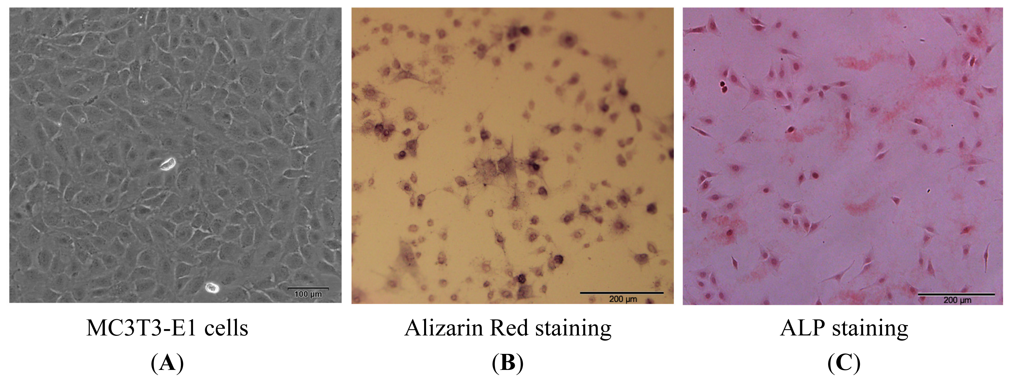
2.1.1. Isolation, Screening, Alkaline Phosphatase (ALP) and Alizarin Red Staining of MC3T3-E1 Cells

To show the osteoblastic features of the MC3T3-E1 line, cells were used for alkaline phosphatase (ALP) and Alizarin red staining before exposure to the differentiation medium. ALP activity and presence of Ca deposits in extracellular matrix indicate that the MC3T3-E1 cells possess osteogenic properties (Figure 1).

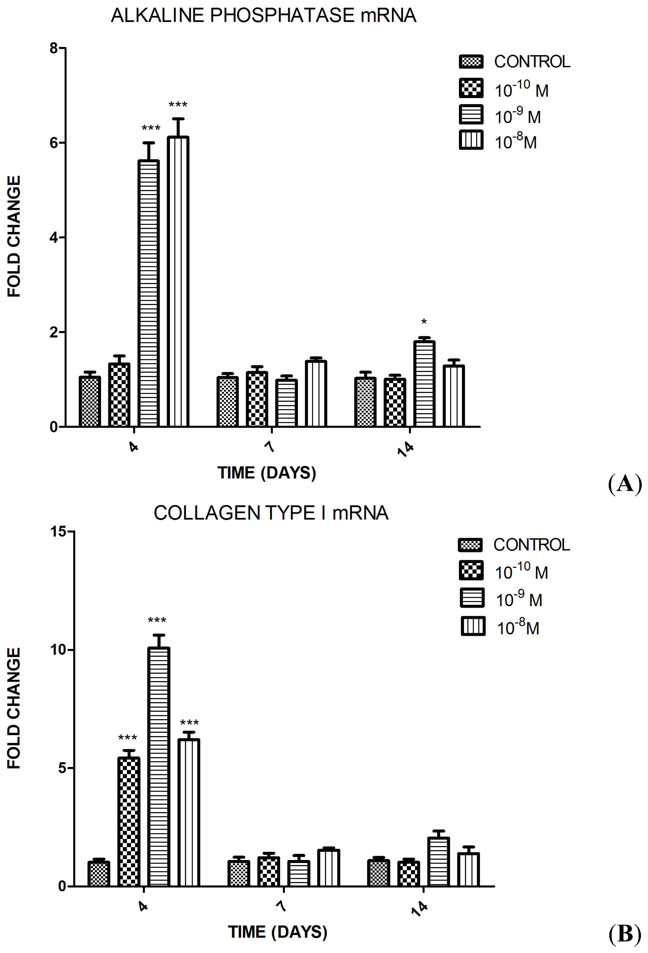
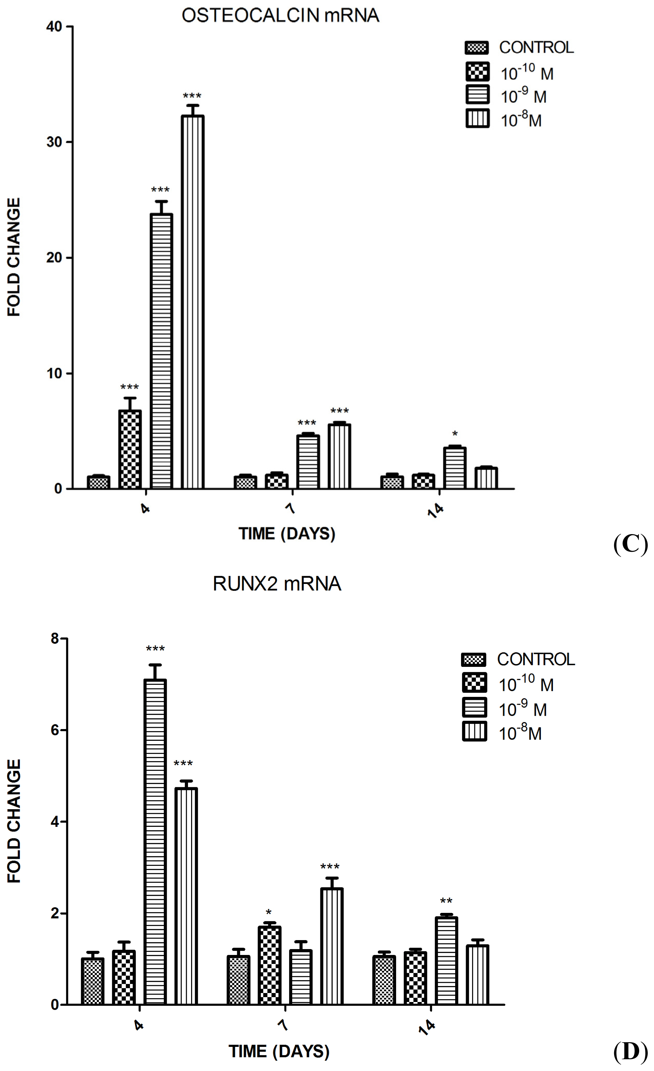
2.1.2. Influence of Substance P (SP) at Different Concentrations on Differentiation of the MC3T3-E1 Cells into Osteoblasts

The impact of SP at different concentrations on the differentiation of MC3T3-E1 cells into osteoblasts was assessed by testing the osteoblastic gene expressions (collagen type I, alkaline phosphatase, osteocalcin, and Runx2) using quantitative polymerase chain reaction (qPCR). Collagen type I and alkaline phosphatase were selected as early markers of osteoblastic differentiation. Osteocalcin was selected as a late marker of that process. Furthermore, the expression of Runx2, a transcription factor crucial in osteoblast differentiation, was examined. Three different concentrations of SP (10−8, 10−9, and 10−10 M) were tested in MC3T3-E1 cells at days 4, 7, and 14 in the osteoinductive medium. SP (10−8 M) significantly increased the level of alkaline phosphatase and collagen type I mRNA at day 4, significantly increased the levels of osteocalcin and Runx2 mRNA at days 4 and 7. SP at concentration of 10−9 M, significantly increased the levels of alkaline phosphatase and Runx2 mRNA at days 4 and 14, significantly increased the level of osteocalcin mRNA at days 4, 7, and 14 and collagen type I mRNA at day 4. Treatment with 10−10 M SP resulted only in elevated collagen type I and osteocalcin mRNA level at day 4 compared with the control group (group 4) (Figure 2).

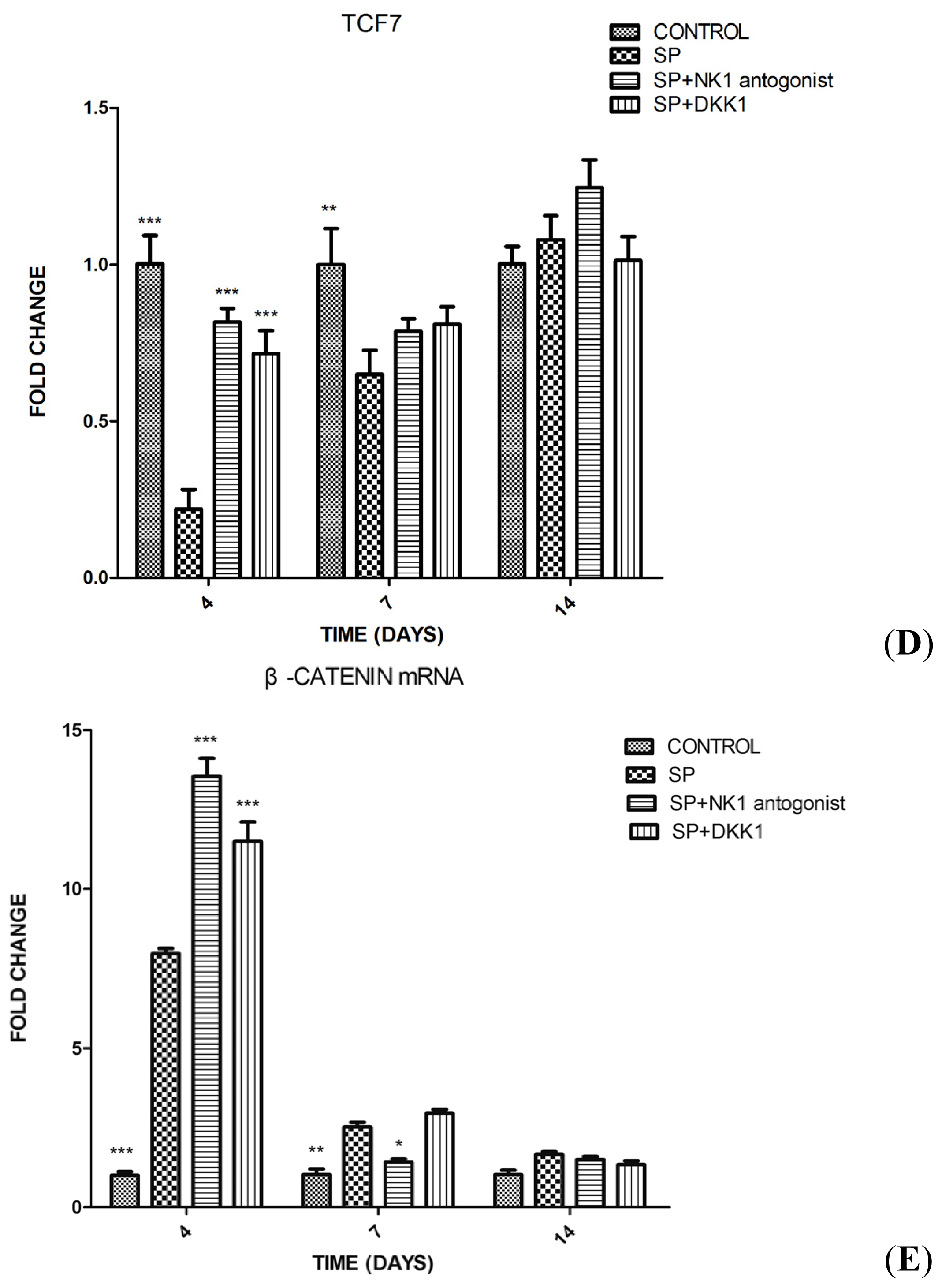
2.1.3. Impact of SP, Neurokinin-1 (NK1) Antagonist and Dickkopf-1 (DKK1) on the Differentiation of MC3T3-E1 Cells into Osteoblasts

qPCR revealed that SP (10−8 M) significantly increased the expression of alkaline phosphatase and collagen type I mRNA at day 4 compared with the control group (group D) and also significantly increased the levels of osteocalcin and Runx2 mRNA on days 4 and 7 (Figure 3). Pretreatment of the cells with NK1 antagonist and DKK1 significantly inhibited the elevated expression of those genes upon SP treatment (Figure 3).

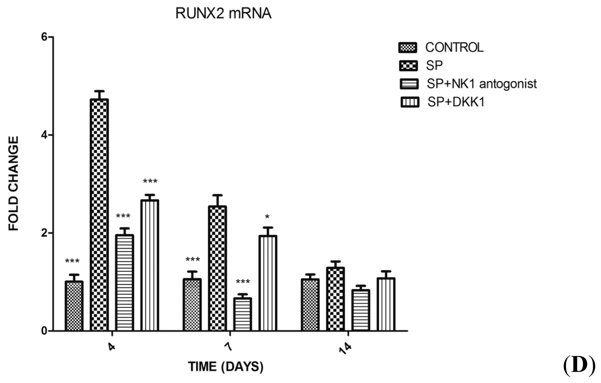
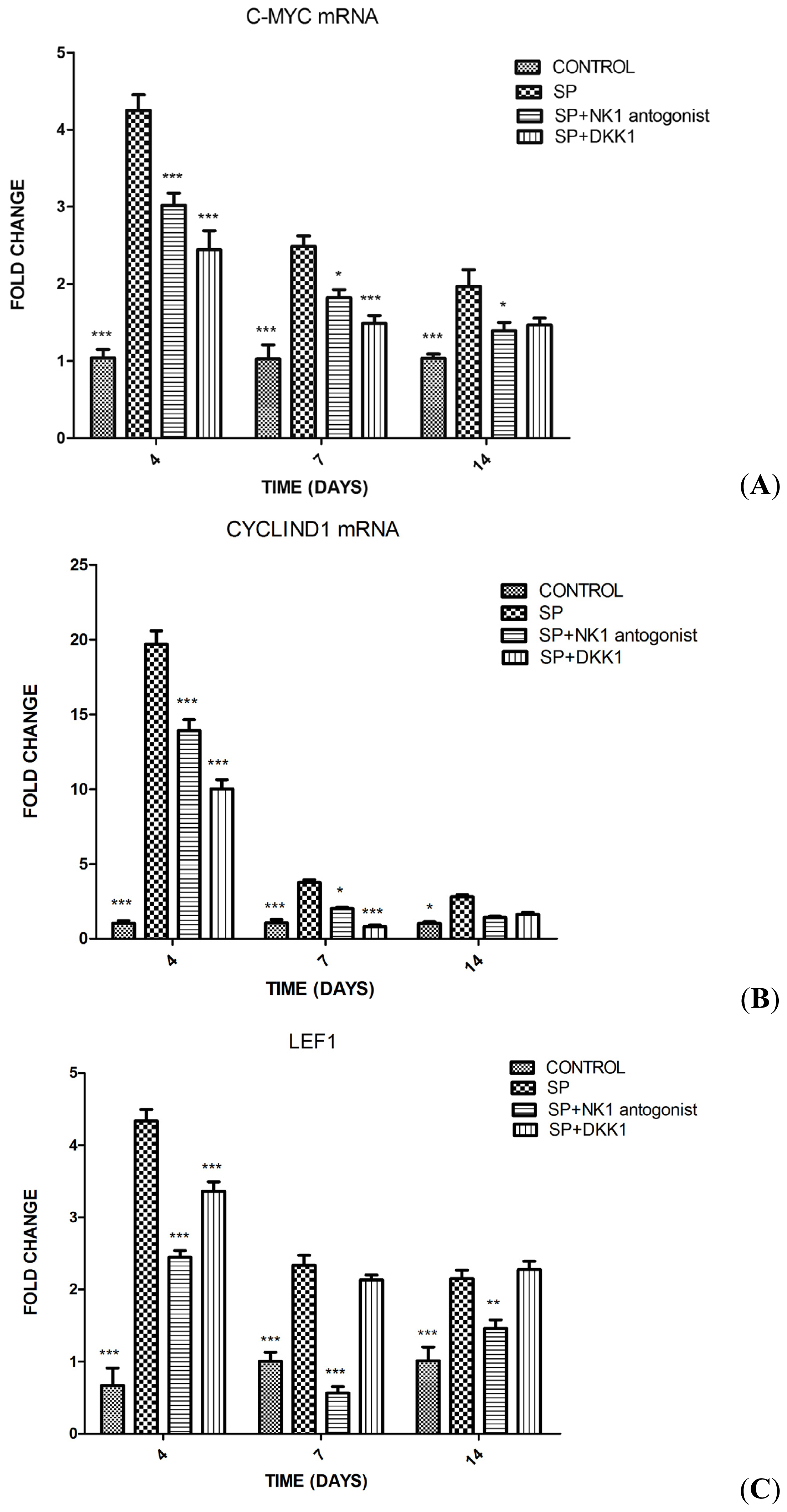
2.1.4. Impact of SP, NK1 Antagonist and DKK1 on the Expression of Genes of the Wnt/β-Catenin Signaling Pathway

To examine the potential mechanism of SP-induced osteoblast differentiation in MC3T3-E1 cells, gene expression of Wnt/β-catenin signaling pathway (c-myc, cyclin D1, Lef1, Tcf7 and β-catenin) was evaluated by qPCR. The addition of SP (10−8 M) to the osteoinductive medium significantly increased the expression of c-myc and Lef1 on days 4, 7, and 14 compared with the control group (group D). Moreover, it significantly increased the expression of cyclin D1 on days 4 and 7 but decreased the expression of Tcf7 on days 4 and 7. These effects of SP were significantly inhibited by the pretreatment with NK1 antagonist and DKK1. SP also significantly increased the expression of β-catenin mRNA on day 4 but, instead of down-regulating the expression of β-catenin mRNA, the pretreatment with NK1 antagonist and DKK1 increased the expression of β-catenin mRNA compared with the SP group (group A) (Figure 4).

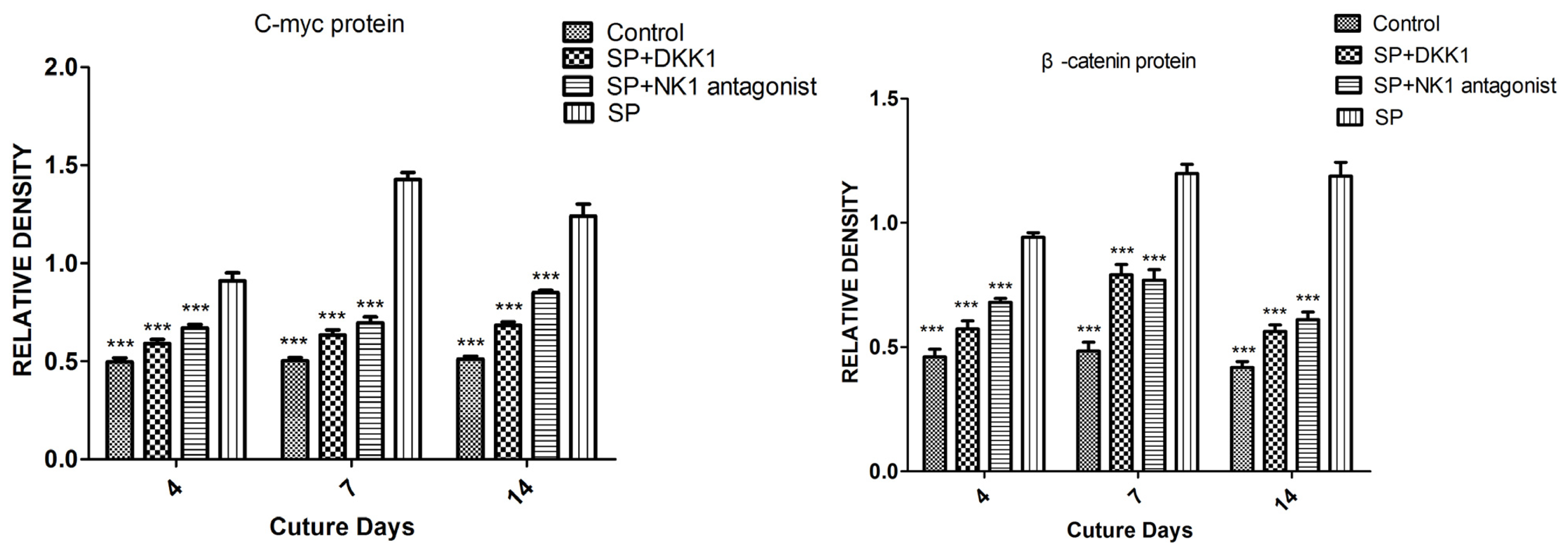
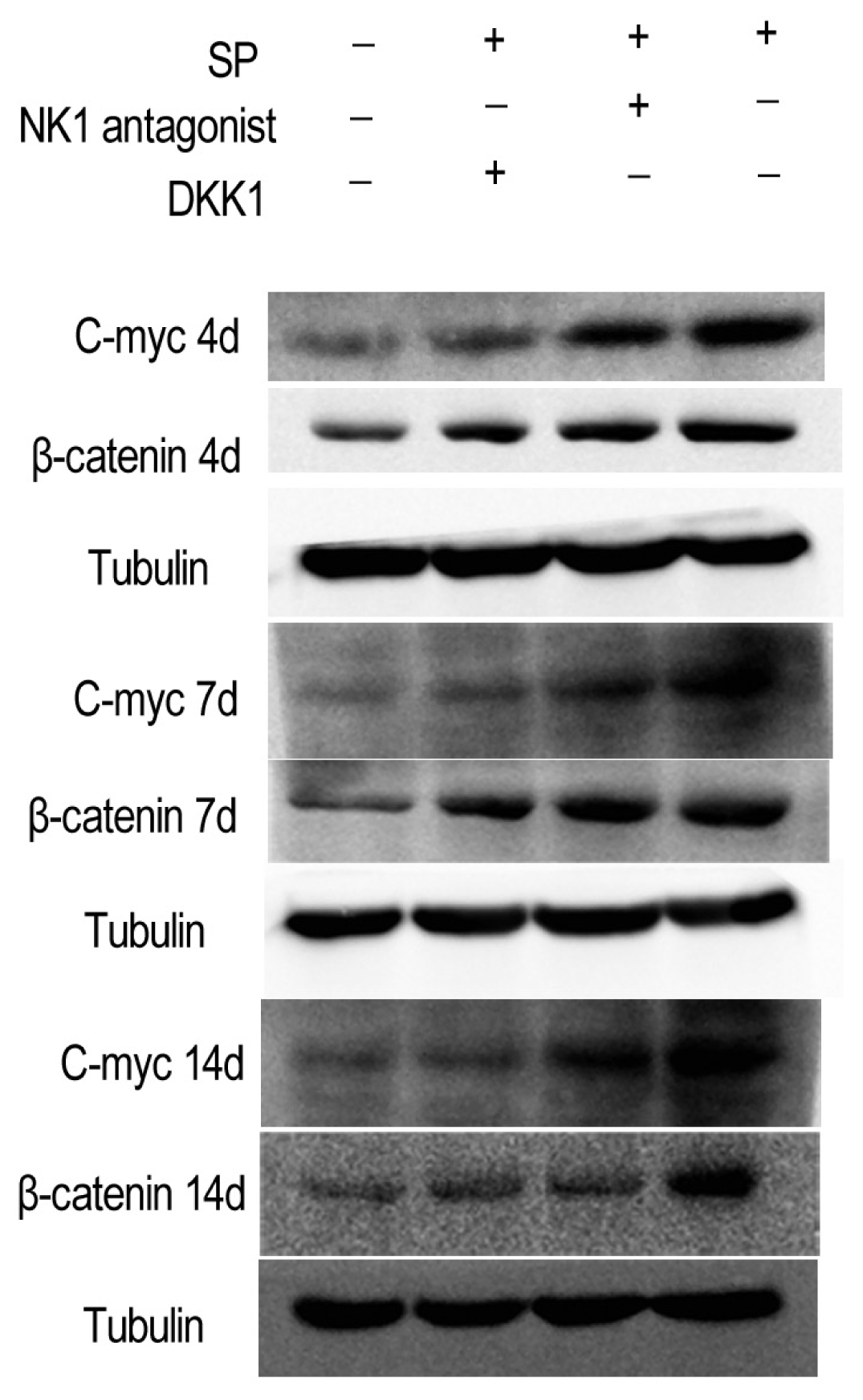
2.1.5. Impact of SP, NK1 Antagonist and DKK1 on the Protein Levels of the Wnt/β-Catenin Signaling Pathway

The addition of SP (10−8 M) to the osteoinductive medium increased the level of c-myc and β-catenin protein on days 4, 7, and 14 compared with the control group (group D). Pretreatment with NK1 antagonist and DKK1 inhibited the SP induced c-myc and β-catenin protein level increase on days 4, 7, and 14 (Figure 5).

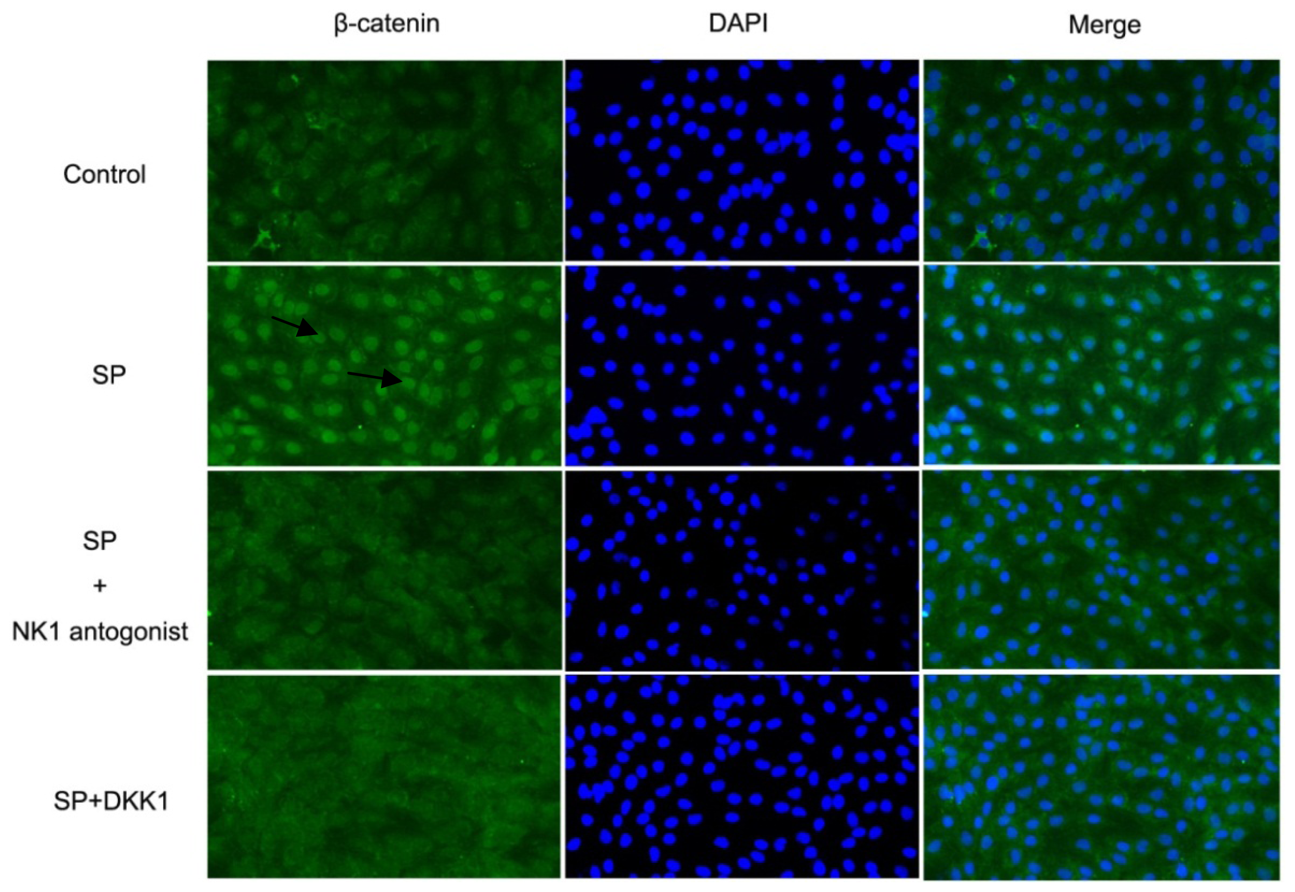
2.1.6. Impact of SP, NK1 Antagonist and DKK1 on β-Catenin Activation in MC3T3-E1 Cells

We next examined whether the effect of SP on the activation of Wnt/β-catenin signaling was associated with an increase in β-catenin translocation to the nucleus. Immunolabeling and fluorescence microscopy of β-catenin in revealed low level of β-catenin in the nucleus of the MC3T3E1 cells from the control group (group D). Addition of SP (10−8 M) to the osteoinductive medium resulted in transfer of β-catenin (green) into the nucleus (blue). Pretreatment with NK1 antagonist and DKK1 prevented the SP induced transfer of β-catenin into the nucleus (Figure 6).

2.2. Discussion

In this study we explored the osteogenic effects of SP treatment on MC3T3-E1 osteoblast-like cells. We found that SP significantly promoted the differentiation of MC3T3-E1 cells. The osteoblastic differentiation in vitro is marked by expressions of osteoblastic genes, including collagen type I, alkaline phosphatase, osteocalcin, and Runx2. The role of SP in osteoblastic differentiation was investigated by detection of those four genes expression. Treatment of MC3T3-E1 cells with SP (10−9–10−8 M) led to an increase in osteoblastic gene expression including collagen type I, alkaline phosphatase, Runx2, and osteocalcin on day 4 and to a less extent on day 7. The role of SP was in turn inhibited by the NK1 receptor antagonist and DKK1, the antagonist of Wnt/β-catenin signaling pathway. SP treatment significantly increased the expressions of genes and levels of proteins associated with activation of the Wnt/β-catenin signaling pathway, but down-regulated the genes associated with inhibition of the Wnt/β-catenin signaling pathway on days 4, 7, and 14. The NK1 receptor antagonist and DKK1 inhibited these effects of SP. These findings indicate that SP may stimulate the differentiation of MC3T3-E1 cells via the Wnt/β-catenin signaling, whereas the NK1 antagonist and DKK1 may weaken the role of SP inducing the differentiation of MC3T3-E1 cells. Our data suggests a potential mechanism of nerves involvement in bone repair.
Previous studies have established the role of β-catenin as a key element of Wnt/β-catenin signaling pathway in control of developmental gene expression programs. Upon activation of the destruction complex, phosphorylation of β-catenin by GSK-3 triggers subsequent ubiquitination and degradation of β-catenin by the proteasome. This mechanism allows maintaining low level of β-catenin in the cytoplasm [15,19]. When the Wnt/β-catenin signaling pathway is activated, the destruction complex is inhibited, β-catenin protein accumulates in the cytoplasm and enters the nucleus where it interacts with Tcf/Lef transcription factors and contributes to activation of the expression of target genes [2528]. In this study we examined the gene and protein expressions of β-catenin after the pretreatment of MC3T3-E1 cells with SP, NK1 antagonist + SP, and DKK1 + SP. We found that SP significantly increased the gene and protein expressions of β-catenin while NK1 receptor antagonist and DKK1 inhibited the expression of β-catenin protein, but increased the expression of β-catenin mRNA, so we speculate that the amount of β-catenin protein in the cytoplasm may depend mainly on the state of the destruction complex, but not mainly on the expression of β-catenin mRNA. As transferring of β-catenin into the nuclei is one of the most important steps in the activation of Wnt/β-catenin signaling pathway, we investigated the role of SP in promoting the transferring of β-catenin into the nuclei. We observed that SP promoted the accumulation and travelling of β-catenin to the nuclei. This may allow subsequent complex formation with Tcf/Lef and activation of Wnt target gene expression.
The role of Tcf7/Lef1 in the Wnt/β-catenin signaling is still controversial. Tcf7 may act as both a repressor and an activator while Lef1 might be usually an activator but sometimes a repressor [2931]. In the dorsal midbrain, Lef1 and Tcf7 were shown to cooperatively activate the expression of the Wnt/β-catenin signaling target genes and promote cell proliferation [32]. In line with previous studies, we found that treatment of MC3T3-E1 cells with SP (10−8 M) for 4, 7, and 14 days led to increased expression of Lef1 and decreased expression of Tcf7. Therefore, we speculate that the cooperation of Lef1 and Tcf7 may be required in the process of activation of Wnt/β-catenin signaling induced by SP.
As Wnt/β-catenin signaling plays an important role in proliferation and differentiation in numerous developmental stages as well as adult tissue homeostasis, Wnt target genes have to be context specific [33,34]. In this study we investigated the expressions of two Wnt target genes and one corresponding protein (c-myc mRNA, cyclin D1 mRNA, and c-myc protein). Our results showed that SP remarkably increased the expressions of the two Wnt target genes and the one protein in MC3T3-E1 cells, indicating that the targeted genes, c-myc and cyclin D1, may be involved in the process of differentiation of MC3T3-E1 cells, which is promoted by the activation of the Wnt/β-catenin signaling pathway [35,36].

3. Experimental Section

3.1. Cell Culture

Murine osteoblastic MC3T3-E1 cells (ATCC, Manassas, VA, USA) were cultured in 5% CO2 atmosphere at 37 °C in α-MEM (minimum essential medium) containing 10% heat-inactivated fetal bovine serum (FBS). The medium was supplemented with 100 U/mL penicillin and 100 μg/mL streptomycin. After reaching 80% confluence, cells were passaged with 0.02% trypsin (Gibco, Grand Island, NY, USA) and transferred to new culture flasks in a ratio of 1:3. To induce osteogenic differentiation culture medium was changed to differentiation medium (α-MEM containing 10 mM β-glycerophosphate and 50 μg/mL ascorbic acid). The cell culture was performed in two steps. To find optimal concentration for promotion of cellular differentiation, the cells were divided into 4 groups according to different concentrations of SP (Sigma, S6883, St. Louis, MO, USA): Group 1, incubated with SP (10−8 M); Group 2, incubated with SP (10−9 M); Group 3 incubated with SP (10−10 M); and group 4, the control group, treated with the same volume of PBS. To determine the effects of SP, NK1 antagonist, and DKK1 on the differentiation of MC3T3-E1 cells, cells were divided into 4 groups: Group A, incubated with SP (10−8 M); Group B, incubated with a mixture of SP and NK1 antagonist (1 μM, Sigma, CP-96345) [37,38]; Group C, incubated with a mixture of SP and 0.2 μg/mL DKK1 (recombinant Human DKK-1, Peprotech, Rocky Hill, CT, USA) [39]; and Group D, the control group, added with the same amount of PBS.

3.2. Alizarin Red Staining and Alkaline Phosphatase Staining

Medium from the cultures before the shifting to differentiation medium was discarded and the cells were fixed in 4% paraformaldehyde for 30 min. After washing three times with ice-cold PBS, cells were stained for 5 min with alizarin red (Sigma) and analyzed under a light microscope.
The alkaline phosphatase (ALP) activity staining was performed with the cells before exposure to the differentiation medium. Cells were rinsed once with PBS, fixed in 2% formaldehyde and subsequently stained at 37 °C in solution containing naphthol AS-MX phosphate disodium salt, fast red salt (Sigma) and N,N-dimethyl formamide for 30 min or until a yellow color appeared. Cells were washed with PBS and photographed using a phase-contrast microscope. The alkaline phosphatase activity was assessed as red stains indicating the products of enzyme activity [40].

3.3. Quantitative Polymerase Chain Reaction (qPCR)

Total RNA of the MC3T3-E1 cells from the eight groups (groups 1–4 and groups A–D) at the 3 time points (4, 7, and 14 days) was extracted using a RNeasy Mini Kit (Qiagen, Valencia, CA, USA) according to the manufacturers protocol. After subsequent DNase digestion, 1 μg of RNA was used to synthetize 20 μL of cDNA using an iScript cDNA Synthesis Kit (Bio-Rad Laboratories, Hercules, CA, USA). Quantitative real time PCR reactions were performed in the ABI prism 7900 real time PCR system using the SYBR Green PCR master mix (Applied Biosystems, Foster City, CA, USA). Expression in each sample was evaluated in three technical replicates. Sequences of the oligonucleotides used in this study are listed in Table 1. Specificity of primer pairs and absence of primer dimmers was validated by analysis of the dissociation curves and the agarose gel electrophoresis. Comparative CT method was used for data analysis. Expression in each sample was normalized to the amount of glyceraldehyde 3-phosphate dehydrogenase (GAPDH) mRNA. Data represent mean expression from 3 experiments.

3.4. Protein Extraction and Western Blot

The cells from groups A–D at indicated time points (4, 7, and 14 days) were used for protein extraction. Cells were treated with the lysis buffer (Cell Signaling Technology, Danvers, MA, USA) and protein extracts were dissolved in sample buffer containing 50 mM Tris-HCl, 2% SDS, 10% glycerol, 100 mM dithiothreitol (pH = 6.80). Proteins were separated by SDS-PAGE in 10% polyacrylamide gel and transferred to a nitrocellulose membrane. Blots were decorated with anti-β-catenin antibodies and anti-c-myc antibodies (Santa Cruz Biotechnology, Santa Cruz, CA, USA). Bands were captured and documented through a CCD system (Imagestation 2000 MM, Kodak, Rochester, NY, USA). Blots were stripped and re-probed with anti-tubulin antibodies to demonstrate equal loading and to allow normalization of the protein content. Densitometry of the bands was performed using Molecular Imaging Software Version 4.0 (Kodak).

3.5. Immunofluorescence Staining

The cells from groups A–D at day 4 were fixed (4% paraformaldeyde in PBS, 15 min, room temperature), permeabilized (0.25% TritonX100 in PBS, 15 min) and blocked in 1% bovine serum albumin (BSA) in poly butylene succinate-co-butylene terephthalate (PBST) for 30 min. After washing three times in PBS, cells were incubated overnight with rabbit anti-mouse anti-β-catenin antibodies (Santa Cruz Biotechnology) diluted 1:100 in PBST. Cells were washed 3 times with PBS and incubated for 1 h with fluorescein isothiocyanate (FITC)-linked rabbit anti-mouse IgG antibody diluted 1:100 (USCN) and again washed three times in PBS. 4′-6-Diamidino-2-phenylindole (DAPI) was added to mark the nuclei. Slides were examined in Fluoview 300 (Olympus, Tokyo, Japan) fluorescent microscope and images were acquired [4,41].

3.6. Statistical Analysis

Statistical analyses were performed in SPSS (version 13.0, IBM, Armonk, NY, USA). One-way ANOVA test followed by Dunnett (2-tailed t) post hoc test was used to evaluate the statistical significance of the differences. Significance was achieved when p < 0.05. Presented error bars indicate standard error of the mean (SEM). * is indicative of p < 0.05, ** p < 0.01 and *** p < 0.001.

4. Conclusions

In conclusion, this study demonstrates that SP may stimulate the osteogenic differentiation of MC3T3-E1 cells and the canonical Wnt signaling may contribute to this process. The role of SP and Wnt/β-catenin signaling in osteogenesis should be further and depth studied by detecting more genes and proteins, such as GSK-3β, Axin, and APC, to elucidate the role of SP and Wnt/β-catenin signaling in bone development and remodeling to improve our understanding of the role of nerves in bone repair.

Acknowledgments

This work was supported by National Natural Science of Foundation of China (81171723, 30872638). We are grateful to Allen P. Liang for revising and editing this manuscript. We also appreciate the technical assistance from Guangdong Province Key Laboratory for Regenerative Medicine of Bone and Cartilage and Research Center of Clinical Medicine at Nanfang Hospital.

Conflicts of Interest

The authors declare no conflict of interest.
  • Author ContributionsGang Mei and Dan Jin conceived the project and wrote the first draft. All authors contributed to the analysis and interpretations of date and to the writing of subsequent drafts. All authors have seen and approved the final version of the manuscript. All authors read and approved the final manuscript.

References

  1. Bjurholm, A.; Kreicbergs, A.; Brodin, E.; Schultzberg, M. Substance P- and CGRP-immunoreactive nerves in bone. Peptides 1988, 9, 165–171. [Google Scholar]
  2. Lundberg, P.; Lundgren, I.; Mukohyama, H.; Lehenkari, P.P.; Horton, M.A.; Lerner, U.H. Vasoactive intestinal peptide (VIP)/pituitary adenylate cyclase-activating peptide receptor subtypes in mouse calvarial osteoblasts: Presence of VIP-2 receptors and differentiation-induced expression of VIP-1 receptors. Endocrinology 2001, 142, 339–347. [Google Scholar]
  3. Tuo, Y.; Guo, X.; Zhang, X.; Wang, Z.; Zhou, J.; Xia, L.; Zhang, Y.; Wen, J.; Jin, D. The biological effects and mechanisms of calcitonin gene-related peptide on human endothelial cell. J. Recept. Signal Transduct. Res 2013, 33, 114–123. [Google Scholar]
  4. An, Y.S.; Lee, E.; Kang, M.H.; Hong, H.S.; Kim, M.R.; Jang, W.S.; Son, Y.; Yi, J.Y. Substance P stimulates the recovery of bone marrow after the irradiation. J. Cell. Physiol 2011, 226, 1204–1213. [Google Scholar]
  5. Malkesman, O.; Braw, Y.; Weller, A. Assessment of antidepressant and anxiolytic properties of NK1 antagonists and substance P in Wistar Kyoto rats. Physiol. Behav 2007, 90, 619–625. [Google Scholar]
  6. Delgado, A.V.; McManus, A.T.; Chambers, J.P. Production of tumor necrosis factor-α, interleukin 1-β, interleukin 2, and interleukin 6 by rat leukocyte subpopulations after exposure to substance P. Neuropeptides 2003, 37, 355–361. [Google Scholar]
  7. Marolda, R.; Ciotti, M.T.; Matrone, C.; Possenti, R.; Calissano, P.; Cavallaro, S.; Severini, C. Substance P activates ADAM9 mRNA expression and induces α-secretase-mediated amyloid precursor protein cleavage. Neuropharmacology 2012, 62, 1954–1963. [Google Scholar]
  8. Goto, T.; Tanaka, T. Tachykinins and tachykinin receptors in bone. Microsc. Res. Tech 2002, 58, 91–97. [Google Scholar]
  9. Goto, T.; Nakao, K.; Gunjigake, K.K.; Kido, M.A.; Kobayashi, S.; Tanaka, T. Substance P stimulates late-stage rat osteoblastic bone formation through neurokinin-1 receptors. Neuropeptides 2007, 41, 25–31. [Google Scholar]
  10. Shih, C.; Bernard, G.W. Neurogenic substance P stimulates osteogenesisin vitro. Peptides 1997, 18, 323–326. [Google Scholar]
  11. Sohn, S.J. Substance P upregulates osteoclastogenesis by activating nuclear factor κB in osteoclast precursors. Acta Otolaryngol 2005, 125, 130–133. [Google Scholar]
  12. Wang, L.; Zhao, R.; Shi, X.; Wei, T.; Halloran, B.P.; Clark, D.J.; Jacobs, C.R.; Kingery, W.S. Substance P stimulates bone marrow stromal cell osteogenic activity, osteoclast differentiation, and resorption activityin vitro. Bone 2009, 45, 309–320. [Google Scholar]
  13. Kim, S.Y.; Kim, S.; Yun-Choi, H.S.; Jho, E.H. Wnt5a potentiates U46619-induced platelet aggregation via the PI3K/Akt pathway. Mol. Cells 2011, 32, 333–336. [Google Scholar]
  14. Zheng, Q.; Chen, P.; Xu, Z.; Li, F.; Yi, X.P. Expression and redistribution of β-catenin in the cardiac myocytes of left ventricle of spontaneously hypertensive rat. J. Mol. Histol 2013, 44, 565–573. [Google Scholar]
  15. Peterson-Nedry, W.; Erdeniz, N.; Kremer, S.; Yu, J.; Baig-Lewis, S.; Wehrli, M. Unexpectedly robust assembly of the Axin destruction complex regulates Wnt/Wg signaling in Drosophila as revealed by analysis in vivo. Dev. Biol. 2008, 320, 226–241. [Google Scholar]
  16. Li, J.; Li, J.; Chen, B. Oct4 was a novel target of Wnt signaling pathway. Mol. Cell. Biochem 2012, 362, 233–240. [Google Scholar]
  17. Sun, Y.C. Examination of effects of GSK3β phosphorylation, β-catenin phosphorylation, and β-catenin degradation on kinetics of Wnt signaling pathway using computational method. Theor. Biol. Med. Model 2009. [Google Scholar] [CrossRef]
  18. Lee, J.H.; Kim, B.G.; Ahn, J.M.; Park, H.J.; Park, S.K.; Yoo, J.S.; Yates, J.R.; Cho, J.Y. Role of PI3K on the regulation of BMP2-induced β-catenin activation in human bone marrow stem cells. Bone 2010, 46, 1522–1532. [Google Scholar]
  19. MacDonald, B.T.; Tamai, K.; He, X. Wnt/β-catenin signaling: Components, mechanisms, and diseases. Dev. Cell 2009, 17, 9–26. [Google Scholar]
  20. Fleury, D.; Vayssiere, B.; Touitou, R.; Gillard, C.; Lebhar, H.; Rawadi, G.; Mollat, P. Expression, purification and functional characterization of Wnt signaling co-receptors LRP5 and LRP6. Protein Expr. Purif 2010, 70, 39–47. [Google Scholar]
  21. Georgiou, K.R.; King, T.J.; Scherer, M.A.; Zhou, H.; Foster, B.K.; Xian, C.J. Attenuated Wnt/β-catenin signalling mediates methotrexate chemotherapy-induced bone loss and marrow adiposity in rats. Bone 2012, 50, 1223–1233. [Google Scholar]
  22. Tamura, M.; Sato, M.M.; Nashimoto, M. Regulation of CXCL12 expression by canonical Wnt signaling in bone marrow stromal cells. Int. J. Biochem. Cell Biol 2011, 43, 760–767. [Google Scholar]
  23. Guo, J.; Liu, M.; Yang, D.; Bouxsein, M.L.; Saito, H.; Galvin, R.J.; Kuhstoss, S.A.; Thomas, C.C.; Schipani, E.; Baron, R.; et al. Suppression of Wnt signaling by Dkk1 attenuates PTH-mediated stromal cell response and new bone formation. Cell Metab 2010, 11, 161–171. [Google Scholar]
  24. Wang, L.; Shi, X.; Zhao, R.; Halloran, B.P.; Clark, D.J.; Jacobs, C.R.; Kingery, W.S. Calcitonin-gene-related peptide stimulates stromal cell osteogenic differentiation and inhibits RANKL induced NF-κB activation, osteoclastogenesis and bone resorption. Bone 2010, 46, 1369–1379. [Google Scholar]
  25. Zhang, M.; Yan, Y.; Lim, Y.B.; Tang, D.; Xie, R.; Chen, A.; Tai, P.; Harris, S.E.; Xing, L.; Qin, Y.X.; et al. BMP-2 modulates β-catenin signaling through stimulation of Lrp5 expression and inhibition of β-TrCP expression in osteoblasts. J. Cell. Biochem 2009, 108, 896–905. [Google Scholar]
  26. Lee, E.; Salic, A.; Kruger, R.; Heinrich, R.; Kirschner, M.W. The roles of APC and Axin derived from experimental and theoretical analysis of the Wnt pathway. PLoS Biol 2003, 1, e10. [Google Scholar]
  27. Kestler, H.A.; Kuhl, M. From individual Wnt pathways towards a Wnt signalling network. Philos. Trans. R. Soc 2008, 363, 1333–1347. [Google Scholar]
  28. Cho, K.H.; Baek, S.; Sung, M.H. Wnt pathway mutations selected by optimal β-catenin signaling for tumorigenesis. FEBS Lett 2006, 580, 3665–3670. [Google Scholar]
  29. Hoppler, S.; Kavanagh, C.L. Wnt signalling: Variety at the core. J. Cell Sci 2007, 120, 385–393. [Google Scholar]
  30. Lee, J.E.; Wu, S.F.; Goering, L.M.; Dorsky, R.I. Canonical Wnt signaling through Lef1 is required for hypothalamic neurogenesis. Development 2006, 133, 4451–4461. [Google Scholar]
  31. Barolo, S. Transgenic Wnt/TCF pathway reporters: All you need is Lef? Oncogene 2006, 25, 7505–7511. [Google Scholar]
  32. Shimizu, N.; Kawakami, K.; Ishitani, T. Visualization and exploration of Tcf/Lef function using a highly responsive Wnt/β-catenin signaling-reporter transgenic zebrafish. Dev. Biol 2012, 370, 71–85. [Google Scholar]
  33. Vlad, A.; Rohrs, S.; Klein-Hitpass, L.; Muller, O. The first five years of the Wnt targetome. Cell Signal 2008, 20, 795–802. [Google Scholar]
  34. Logan, C.Y.; Nusse, R. The Wnt signaling pathway in development and disease. Annu. Rev. Cell Dev. Biol 2004, 20, 781–810. [Google Scholar]
  35. Juan, J.; Muraguchi, T.; Iezza, G.; Sears, R.C.; McMahon, M. Diminished WNT → β-catenin → c-myc signaling is a barrier for malignant progression of BRAFV600E-induced lung tumors. Genes Dev 2014, 28, 561–575. [Google Scholar]
  36. Sukhotnik, I.; Roitburt, A.; Pollak, Y.; Dorfman, T.; Matter, I.; Mogilner, J.G.; Bejar, J.; Coran, A.G. Wnt/β-catenin signaling cascade down-regulation following massive small bowel resection in a rat. Pediatr. Surg. Int 2014, 30, 173–180. [Google Scholar]
  37. Ramnath, R.D.; Sun, J.; Adhikari, S.; Bhatia, M. Effect of mitogen-activated protein kinases on chemokine synthesis induced by substance P in mouse pancreatic acinar cells. J. Cell. Mol. Med 2007, 11, 1326–1341. [Google Scholar]
  38. Schmidt, P.T.; Rasmussen, T.N.; Holst, J.J. Tachykinins may mediate capsaicin-induced, but not vagally induced motility in porcine antrum. Peptides 1997, 18, 1511–1516. [Google Scholar]
  39. Sharon, N.; Mor, I.; Golan-lev, T.; Fainsod, A.; Benvenisty, N. Molecular and functional characterizations of gastrula organizer cells derived from human embryonic stem cells. Stem Cells 2011, 29, 600–608. [Google Scholar]
  40. Alcantara, E.H.; Shin, M.Y.; Sohn, H.Y.; Park, Y.M.; Kim, T.; Lim, J.H.; Jeong, H.J.; Kwon, S.T.; Kwun, I.S. Diosgenin stimulates osteogenic activity by increasing bone matrix protein synthesis and bone-specific transcription factor Runx2 in osteoblastic MC3T3-E1 cells. J. Nutr. Biochem 2011, 22, 1055–1063. [Google Scholar]
  41. Dong, J.; He, Y.; Zhang, X.; Wang, L.; Sun, T.; Zhang, M.; Liang, Y.; Qi, M. Calcitonin gene-related peptide regulates the growth of epidermal stem cellsin vitro. Peptides 2010, 31, 1860–1865. [Google Scholar]
Figure 1. Growth and morphologic characteristics of MC3T3-E1 cells. (A) Cells grew in triangular and polygonal shapes; (B) Alizarin red staining for Ca deposits, extracellular matrix Ca deposits for matrix mineralization was measured using Alizarin red dye which bound with Ca; and (C) staining for alkaline phosphatase (ALP), cells were cultured in 6-well plates and incubated in AS-MX phosphate solution as substrate with Fast Red salt as stain, with matrix ALP activity shown as red stain.
Ijms 15 06224f1
Figure 2. Effects of substance P (SP) treatment at days 4, 7, and 14 (10−8, 10−9 and 10−10 M) on gene expressions in MC3T3-E1 cells measured by qPCR for (A) alkaline phosphatase; (B) collagen type I; (C) osteocalcin; and (D) Runx2 mRNA levels. The values reported are the mean ± SEM of three independent experiments. * p < 0.05 versus Control; ** p < 0.01 versus Control; and *** p < 0.001 versus Control.
Ijms 15 06224f2aIjms 15 06224f2b
Figure 3. Effects of SP, SP + NK1 (neurokinin-1) antagonist, SP + DKK1 (Dickkopf-1) and the same amount of phosphate-buffered saline (PBS) on days 4, 7, 14 on gene expressions in MC3T3-E1 cells measured by quantitative polymerase chain reaction (qPCR). (A) alkaline phosphatase; (B) collagen type I; (C) osteocalcin; and (D) Runx2 mRNA. Results are representative of 3 independent experiments. * p < 0.05 versus SP; ** p < 0.01 versus SP; and *** p < 0.001 versus SP, respectively.
Ijms 15 06224f3aIjms 15 06224f3b
Figure 4. Effects of SP, SP + NK1 antagonist, SP + DKK1 and the same amount of PBS on days 4, 7, 14 on gene expressions in MC3T3-E1 cells measured by qPCR. (A) c-myc mRNA; (B) cyclin D1 mRNA; (C) Lef1; (D) Tcf7; and (E) β-catenin mRNA. Results are representative of 3 independent experiments. * p < 0.05 versus SP; **p < 0.01 versus SP; and *** p < 0.001 versus SP, respectively.
Ijms 15 06224f4aIjms 15 06224f4b
Figure 5. Effects of SP, SP + NK1 antagonist, SP + DKK1 and the same amount of PBS on days 4, 7, and 14 on β-catenin protein and c-myc protein expressions in MC3T3-E1 cells measured by Western blot analysis. *** p < 0.001 versus SP.
Ijms 15 06224f5aIjms 15 06224f5b
Figure 6. Effects of SP, SP + NK1 antagonist, and SP +DKK1 on β-catenin activation in MC3T3-E1 cells. Immunofluorescence staining with antibodies of β-catenin. Cells were incubated in the presence of SP, SP + NK1 antagonist, SP + DKK1, and the same amount of PBS for 4 days, then the β-catenin was detected by the immunostaining method as shown in green fluorescence (arrowheads). The nuclei were stained with 4′-6-diamidino-2-phenylindole (DAPI) as shown in blue.
Ijms 15 06224f6
Table 1. Sequences of the primers used in this study.
GeneSourceSequencePredicted length (bp)
ALPNM_0074315′-AACCCAGACACAAGCATTCC-3′200
5′-GCCTTTGAGGTTTTTGGTCA-3′
COL1NM_0077425′-GCCAAGAAGACATCCCTGAA-3′107
5′-GCCATTGTGGCAGATACAGA-3′
OCNNM_0075415′-TGACAAAGCCTTCATGTCCA-3′175
5′-TTTGTAGGCGGTCTTCAAGC-3′
RUNX2NM_0098205′-AAGTGCGGTGCAAACTTTCT-3′175
5′-ACGCCATAGTCCCTCCTTTT-3′
C-MYCNM_0011773535′-TCCTGTACCTCGTCCGATTC-3′195
5′-GGTTTGCCTCTTCTCCACAG-3′
CCND1NM_0076315′-GCGTACCCTGACACCAATCT-3′183
5′-CTCCTCTTCGCACTTCTGCT-3′
LEF1NM_0107035′-TATGAACAGCGACCCGTACA-3′132
5′-TCGTCGCTGTAGGTGATGAG-3′
TCF7NM_0093315′-ATCCTTGATGCTGGGATCTG-3′139
5′-CTTCTCTGCCTTGGGTTCTG-3′
β-CATENINNM_0076145′-ATGGCTTGGAATGAGACTGC-3′150
5′-ATGCTCCATCATAGGGTCCA-3′
GAPDHNM_0080845′-ATTGTCAGCAATGCATCCTG-3′102
5′-ATGGACTGTGGTCATGAGCC-3′

