Review

Mitochondria in the Center of Human Eosinophil Apoptosis and Survival

1
The Immunopharmacology Research Group, University of Tampere School of Medicine and Tampere University Hospital, Tampere FI-33014, Finland
2
Department of Respiratory Medicine, Seinäjoki Central Hospital, Seinäjoki, Finland and University of Tampere, Tampere FI-60220, Finland
*
Author to whom correspondence should be addressed.
Int. J. Mol. Sci. 2014, 15(3), 3952-3969; https://doi.org/10.3390/ijms15033952
Submission received: 17 January 2014 / Revised: 20 February 2014 / Accepted: 26 February 2014 / Published: 5 March 2014
(This article belongs to the Collection Programmed Cell Death and Apoptosis)

Abstract

:
Eosinophils are abundantly present in most phenotypes of asthma and they contribute to the maintenance and exacerbations of the disease. Regulators of eosinophil longevity play critical roles in determining whether eosinophils accumulate into the airways of asthmatics. Several cytokines enhance eosinophil survival promoting eosinophilic airway inflammation while for example glucocorticoids, the most important anti-inflammatory drugs used to treat asthma, promote the intrinsic pathway of eosinophil apoptosis and by this mechanism contribute to the resolution of eosinophilic airway inflammation. Mitochondria seem to play central roles in both intrinsic mitochondrion-centered and extrinsic receptor-mediated pathways of apoptosis in eosinophils. Mitochondria may also be important for survival signalling. In addition to glucocorticoids, another important agent that regulates human eosinophil longevity via mitochondrial route is nitric oxide, which is present in increased amounts in the airways of asthmatics. Nitric oxide seems to be able to trigger both survival and apoptosis in eosinophils. This review discusses the current evidence of the mechanisms of induced eosinophil apoptosis and survival focusing on the role of mitochondria and clinically relevant stimulants, such as glucocorticoids and nitric oxide.

1. Introduction

Eosinophils are cells of the innate immune system involved in the pathogenesis of allergic, gastrointestinal and hypereosinophilic disorders, in anti-parasitic defence and in tumor immunity [15]. Eosinophils account only for approximately 3% of blood leukocytes in healthy individuals but their number is elevated in subjects with eosinophilic conditions such as allergic asthma. However, shortages of cells often hamper studies on eosinophils. In asthmatic airways, eosinophils are driven into an activated state e.g., by pro-inflammatory cytokines such as IL-5. When activated, they release toxic and pro-inflammatory mediators able to induce bronchoconstriction, mucus hypersecretion, and damage to airway epithelium as well as contribute to T helper 2 cell polarization and airway thickening [68]. Recent data from clinical studies with anti-IL-5 antibody and eosinophil-deficient mice suggest that eosinophils are important for asthma exacerbations and airway remodelling. Anti-IL-5 treatment led to decreased exacerbation rate in patients with severe eosinophilic asthma and these patients were able to reduce their glucocorticoid dose in response to anti-IL-5 treatment [9,10]. Thereby, eosinophils are important for maintenance and exacerbations of asthma [11].
In healthy individuals eosinophils are short-living cells. In in vitro conditions, blood eosinophils undergo spontaneous apoptosis in a few days but in a physiological situation they tend to migrate and accumulate into liver and spleen, where they are likely to live longer than a few days [1215]. Apoptosis of eosinophils can be delayed or accelerated by various agents [14,15]. Eosinophil longevity may be enhanced up to 1–2 weeks by pro-inflammatory cytokines such as IL-5, IL-3 and GM-CSF present in inflamed airways [16]. Indeed, blood and tissue eosinophils from patients with asthma have been shown to live longer when compared to eosinophils from healthy individuals [17,18]. Eosinophil removal from the airways is beneficial to reduce eosinophilic inflammation and alleviate symptoms of asthma [19]. Apoptosis is a non-inflammatory way of cell death comprising a beneficial means for cell removal. Membrane integrity is retained throughout the process and the harmful cell content maintained inside the cell. The immunological silence of apoptosis is ensured by formation of smaller apoptotic bodies that are rapidly ingested by phagocytes. Apoptosis may be executed via two different main routes, extrinsic (receptor-mediated) or intrinsic (mitochondrion-centered) pathway [20]. Extrinsic pathway is activated e.g., by ligation of the death receptor Fas/CD95. This leads to formation of a multiprotein complex called death-inducing signalling complex (DISC) that regulates activation of initiator caspase-8. Initiator caspase-8 may either directly activate effector caspases that execute apoptosis or cleave BH3-interacting-domain death agonist (Bid) resulting in activation of an additional mitochondrial loop. Intrinsic pathway can be initiated by several intracellular stress conditions such as DNA damage, oxidative stress and cytosolic Ca2+ overload. Members of the Bcl-2 family are critical in monitoring intracellular damage and aim to mediate activation of pore-forming Bax and the following mitochondrial membrane permeabilization (MMP), a central event in apoptosis [20,21]. Mitochondrial membrane permeabilization can also be mediated via mitochondrial permeability transition (mPT) [21,22]. MMP results in loss of mitochondrial membrane potential (ΔΨm), halted mitochondrial ATP synthesis and release of pro-apoptotic proteins such as cytochrome c to the cytosol. Cytochrome c stimulates formation of the apoptosome, a platform that activates initiator caspase-9 [20,21]. Initiator caspase 9 activates effector caspases 3, 6 and 7 resulting in degradation of cellular components and apoptosis.
Eosinophil apoptosis can be accelerated by physiological factors such as Fas activation [23]. Fas ligand is a significant pro-apoptotic agent for eosinophils in vivo because its neutralization enhanced airway eosinophilia in a mouse model of allergic asthma [24]. NO is produced in high amounts in the lungs of asthmatics and has been shown to regulate eosinophil apoptosis in a complex manner. NO has shown both anti- and pro-apoptotic effects on eosinophils [2527] and both enhancing and reducing properties regarding lung eosinophilia [2830]. Thereby, the net effect in response to NO can be different in different pathophysiological situations and is not known at the moment. Also many anti-asthmatic agents such as glucocorticoids, theophylline and cysteinyl leukotriene receptor antagonists enhance eosinophil apoptosis in the absence and presence of eosinophil survival-prolonging cytokines [3136] and the pro-apoptotic effects of these drugs may contribute to their clinical efficacy [3742]. Anti-inflammatory glucocorticoid medication is the corner stone in the treatment of asthma and understanding its actions is of critical importance. Glucocorticoids modulate longevity of many immune cell types and the sensitivity to glucocorticoid-induced cell death depends on the cell type. For example, CD4+ T cells but not CD8+ T cells are sensitive to glucocorticoid-induced apoptosis [4345]. In contrast, glucocorticoids inhibited neutrophil apoptosis [31,4648], even though not in an environment with severe hypoxia [49]. In eosinophils, glucocorticoids accelerate apoptosis [3133].
Understanding the signalling related to eosinophil survival and apoptosis is extremely important for understanding the pathogenesis of eosinophilic inflammation and for the development of novel drugs to treat diseases associated with eosinophilia. Studies using primary human eosinophils are hampered by several issues. Low numbers of cells available for the studies restricts carrying out experiments with long-time series and different treatments. Additionally, the short lifespan of eosinophils excludes use of most modern molecular biology methods such as transfection and RNA interference. Because of the restrictions, knowledge of primary human eosinophil functions is based mainly on the use of pharmacological inhibitors and methods available for direct measurements of cellular functions and intracellular mediators.
In eosinophils, mitochondria play a central role in apoptosis and survival [50]. Mitochondrial events have been shown to be critical for spontaneous, glucocorticoid-, nitric oxide- and anti-Fas-induced apoptosis of eosinophils [5154] and thereby mediate both extrinsic and intrinsic forms of eosinophil apoptosis. Mitochondria are forums where pro- and anti-apoptotic signals merge and the fate of the cell is determined. Because of the evident importance of this cell organ for eosinophil survival and death, this review concentrates on discussing the mechanisms of eosinophil apoptosis and survival focusing on the role of mitochondria and the clinically relevant pro-apoptotic stimulant glucocorticoid.

2. Mitochondria and Bcl-2 Family Members

In most cells, mitochondria function as “energy factories” producing ATP via function of the electron transport chain maintaining mitochondrial membrane potential (ΔΨm). Eosinophils have been suggested to contain only a low number of mitochondria, 24–36 per cells [50] but this result needs to be confirmed by current techniques. It has also been reported that eosinophils maintain mitochondrial membrane potential rather by hydrolysis of ATP than via respiratory chain [50], even though evidence also exists of functional respiratory chains in eosinophils [55]. It is clear that eosinophil mitochondria are able to release cytochrome c from the intermembrane space and by this mechanism activate caspases and induce apoptosis [50].

2.1. Bcl-2 Members and Pore-Forming Activity of Bax and Bid

Members of the Bcl-2 family are critical in monitoring intracellular damage and the balance between anti-apoptotic and pro-apoptotic Bcl-2 members (ratio of pro-apoptotic Bax to anti-apoptotic Bcl-2) determines the susceptibility of cells to apoptosis. Short-living granulocytes have high ratios of Bax/Bcl-2 while the corresponding ratios in monocytes and lymphocytes are relatively low. This ratio determined the susceptibility of leukocytes to anti-Fas-induced apoptosis, granulocytes being the most susceptible and lymphocytes the least susceptible [56]. Eosinophils express high levels of pro-apoptotic Bid and Bax, which are proteins capable to oligomerize and form pores to the mitochondrial outer membrane [53,54,57,58]. Bid is processed during spontaneous apoptosis and in enhanced manner during glucocorticoid- and anti-Fas-induced apoptosis but it is the most critical mediator for Fas-induced apoptosis [53,54]. Also Bax was demonstrated to spontaneously translocate into mitochondria in untreated eosinophils and in an accelerated manner in response to glucocorticoid treatment [51,52]. Peptidyl-prolyl isomerase Pin1 may be the key regulator of Bax translocation. Bax translocation into mitochondria was prevented by GM-CSF, which activated ERK1/2 to phosphorylate the threonine residue of Bax. This phosphorylation enabled interaction of Pin1 with Bax preventing its mitochondrial targeting [58]. Pin1 was also shown to be a key regulator of apoptosis induced by anti-Fas via the Fas-associated death domain (FADD) in activated eosinophils. In the presence of survival-prolonging IL-5, Pin1 quenched phosphorylation of FADD at Ser194 and prevented apoptosis [59]. These mechanisms may be highly important in determining whether allergic inflammation is continued by IL-5/GM-CSF or diminished by Fas receptor signalling.
Anti-apoptotic Mcl-1 has been found in eosinophils and was degraded during spontaneous apoptosis and in an accelerated manner during apoptosis induced by glucocorticoids or by an inhibitor of cyclin-dependent kinase [57,6063]. In HeLa cells, Mcl-1 was demonstrated to inhibit Bax downstream to its mitochondrial translocation. Mcl-1 prevented formation of Bax oligomers at mitochondria, required for pore formation, but this inhibition required no direct interaction [64]. Thereby, degradation of Mcl-1 in apoptotic eosinophils probably enables pore-forming activity of Bax and mitochondrial outer membrane permeabilization. The mechanism of Mcl-1 degradation accelerated by glucocorticoids remains unclear but may for example involve transcription of proteins involved in the degradation [63]. In neutrophils, glucocorticoids, in contrast to eosinophils, induced Mcl-1 expression, which may play an important role in the mechanism of prolongation of neutrophil survival [65]. Contradictions exist concerning the expression of anti-apoptotic Bcl-2 in eosinophils; expression seems to depend on the origin of the eosinophils [39,57,60,66].

2.2. Mitochondrial Permeability Transition

Mitochondrial permeability transition (mPT) is one mechanism for the mitochondrial membrane permeabilization. During mPT, permeability of the inner mitochondrial membrane is increased for solutes and molecules up to 1.5 kDa. A channel sensitive to Ca2+, oxidants and pro-apoptotic Bcl-2 family members is responsible for this phenomenon [21,22]. Mitochondrial permeability transition results in mitochondrial matrix swelling, most likely due to the influx of ions that are accompanied by water. The mitochondrial outer membrane is ruptured due to matrix swelling and apoptosis-inducing proteins are released to the cytosol [67]. The mPT channel is thought to be a multiprotein complex but its molecular structure is still unknown. Glucocorticoids and nitric oxide induced apoptosis in eosinophils that was mediated by mPT [68,69]. However, mPT had no critical role in mediating spontaneous or anti-Fas-induced apoptosis [23]. What defines the mechanism of mitochondrial membrane permeabilization in response to different pro-apoptotic stimulants in eosinophils remains unclear. Oxidants are generally important mediators of eosinophil apoptosis and known inducers of mPT but do not, however, always mediate mPT. It seems that ROS have several mechanisms to mediate apoptosis, one of which is stimulation of mPT. For example, Bid has been shown to engage a ROS-dependent but mPT-independent mechanism for mitochondrial membrane permeabilization and cytochrome c release [70].
Mitochondrial permeability transition may function in two different modes. In addition to the irreversible sustained opening of the mPT channel occurring during cell death, the channel may also fluctuate between open and closed states (flicker) [71,72]. In NO-treated eosinophils, early flickering mPT preceded permanent mPT and mPT-dependent eosinophil apoptosis [68]. The early flickering mPT was not necessary for apoptosis to proceed and may actually represent a cell survival mechanism [73,74]. It has been demonstrated that flickering mPT may act as a mechanism to release ROS or calcium [7577] and by these mechanisms, flickering mPT may participate in cell signalling. For example, in NO-treated eosinophils flickering mPT mediated activation of JNK [68].

3. Reactive Oxygen Species (ROS) and Pro-Apoptotic Signalling Pathways

Reactive oxygen species such as superoxide O2•− and hydrogen peroxide H2O2 were demonstrated to mediate induced eosinophil apoptosis as well as spontaneous eosinophil apoptosis [52,68,7881]. In most tissues, the mitochondrial electron transport chain and especially the complexes I and III serve as the primary source of superoxide (O2•−) even thought O2•− can also be generated by function of NADPH oxidase or xanthine oxidase in certain immune cells such as eosinophils, neutrophils and macrophages following their activation [82]. Approximately five- to ten-fold higher steady state concentrations of O2•− exist in the mitochondrial matrix when compared to the cytosol, according to one estimation [83]. Mitochondria have a diverse antioxidant defence system including superoxide dismutases (SODs such as Manganese (Mn)-containing SOD), glutathione, glutathione peroxidase, catalase, peroxiredoxins etc. In the presence of superoxide dismutase (SOD), O2•− is converted into a more stable non-radical oxidant, hydrogen peroxide (H2O2) that may also function as a signalling molecule [84]. Catalase functions by decomposing H2O2 to oxygen and water.

3.1. ROS

H2O2 has been shown to induce eosinophil apoptosis and catalase has been demonstrated to decrease spontaneous eosinophil apoptosis [55,85]. Eosinophil apoptosis induced by excretory-secretory products from helminth was associated with increased levels of H2O2 but not superoxide and reversed by catalase but not by mimetic of superoxide dismutase (SOD). Increased H2O2 preceded mitochondrial injury [86]. It is possible that H2O2 often acts as the actual mediator of eosinophil apoptosis instead of O2•−. H2O2-induced apoptosis also required products of the mitochondrial respiratory chain because inhibition of mitochondrial respiration by rotenone decreased H2O2-induced eosinophil apoptosis [55]. In thymocytes, glucocorticoids induced production of H2O2 and overexpression of catalase in these cells resulted in their resistance to glucocorticoid-induced apoptosis supporting a mediator role of H2O2 in the process [87,88]. In eosinophils, evidence exists also for the important role of superoxide itself as the mediator of apoptosis. Glucocorticoids increased levels of superoxide in eosinophils after 24 h of treatment [52]. Spontaneous apoptosis as well as glucocorticoid-induced apoptosis were associated with decreased level of the mitochondrial antioxidant MnSOD but not that of the cytosolic antioxidant CuZnSOD at 24 h [52]. Decreased level of MnSOD would lead to increased levels of superoxide radical and decreased formation of H2O2. Also, nitric oxide-induced eosinophil apoptosis was reduced by a SOD mimetic, suggesting that superoxide is an important mediator [68]. Why are the levels of ROS elevated when eosinophils are on their way towards apoptosis? Generally, it is thought that most often increased levels of ROS result from compromised antioxidant capacity rather than increased production of superoxide [82,89]. Indeed, as discussed above, glucocorticoids were shown to decrease levels of MnSOD in eosinophils [52]. The mechanism of glucocorticoid-induced enhancement of ROS has also been studied in many cell types other than eosinophils. In thymocytes, glucocorticoid-induced production of H2O2 was dependent on complex III of the mitochondrial respiratory chain and these events mediated glucocorticoid-induced apoptosis. In isolated mitochondria, glucocorticoids have also been shown to inhibit members of the mitochondrial respiratory chain [90,91]. In neural stem cells, treatment with dexamethasone resulted in down-regulation of 72% of the investigated genes involved in the mitochondrial respiratory chain, as well as 29% of the genes encoding for antioxidant enzymes [92]. Altogether, the source of ROS production may vary depending on the pro-apoptotic stimulant. Glucocorticoids may elevate ROS by directly modulating function of the enzymes in the mitochondrial electron transport chain as well as by transcriptional regulation of antioxidant or respiratory chain enzymes.
ROS are significant mediators of eosinophil apoptosis induced by many pro-apoptotic agents but how do they actually mediate eosinophil cell death? At some point, excessive mitochondrial ROS may reach a threshold that leads to mPT pore opening resulting in ROS release to the cytosol [76]. In the cytosol, ROS may activate several protein kinases as discussed below. According to the hypothesis of Zorov et al. the released ROS might trigger a similar phenomenon in neighbouring mitochondria and lead to amplified oxidative stress signals, mitochondrial injury and possibly cell death [76]. Alternatively, a mPT-independent mechanism was demonstrated where ROS was required for mitochondrial membrane permeabilization and cytochrome c release induced by Bid [70].

3.2. Kinases Activated by ROS

Mitogen-activated protein kinases (MAPKs) are serine/threonine kinases mainly activated by bacterial products, proinflammatory cytokines, growth factors and environmental stress. MAPK family consists of c-jun N-terminal kinase (JNK) 1–3, extracellular regulated kinase (ERK) 1/2, 3, 5 and 7, and p38 kinases. A phosphorylation cascade conducted by MAPK kinase kinases (MAP3K) and MAPK kinases (MAP2K) leads to activation of MAPK. MAPKs phosphorylate transcription factors resulting in transcription of genes involved in apoptosis, survival, proliferation and differentiation. Additionally, MAPKs affect the function of numerous other proteins via phosphorylation. MAPKs are inactivated by phosphoprotein phosphatases (MAPK phosphatases (MKPs)) [9395]. Germinal center kinases (GSK) such as mammalian sterile 20-like kinase (Mst) 1 are also activators of MAPK pathways and at least some family members function by acting as MAPK kinase kinase kinases (MAP4K) [96].

3.2.1. JNK

ROS are known activators of MAPKs JNK, p38 and ERK 1/2, known to regulate cell survival and death pathways [97]. Of these MAPKs, JNK has been shown to mediate glucocorticoid-induced eosinophil apoptosis as well as spontaneous apoptosis, apoptosis induced by nitric oxide and several drugs [27,52,68,98101]. Activation of JNK by ROS seems to be indirect. Even though not shown in eosinophils, the actual targets of oxidants such as MAP3K (MEKK1) and apoptosis signal-regulating kinase 1 (ASK1 or MAP3K5) reside upstream of JNK, and lead to JNK activation via function of MAP2K [97]. In addition, peroxynitrite led to activation of JNK in eosinophils but was found to require Fas. Fas was demonstrated to be a direct oxidation target of reactive nitrogen species (RNS) but the mechanism of JNK activation via Fas remains unclear [102].
Kinetics of JNK activation seems to be an important determinant in whether the activation leads to survival or apoptosis. Early JNK activation has been described to represent a stress response resulting in cell survival signalling while delayed and sustained JNK activation has been typically related to apoptosis [97,103,104]. Indeed, early and strong JNK activation was demonstrated as a feature preceding glucocorticoid-induced eosinophil apoptosis, as well as apoptosis induced by several other factors in eosinophils [52,68,101]. Eosinophil apoptosis induced by glucocorticoids and nitric oxide exhibit many similarities such as early and late JNK activation, mPT and caspase activation [52,68]. Early JNK activation induced by nitric oxide, was however, not critical for NO-induced apoptosis and might initiate a stress response aiming to cell survival [68]. In the study of Gardai et al., early JNK phosphorylation stimulated by glucocorticoids was prevented by antioxidant treatment suggesting involvement of ROS [52]. Furthermore, in NO-treated eosinophils, early JNK activation was dependent on partial mitochondrial permeability transition (mPT) [68]. By combining these results with findings of Zorov et al. it can be suggested that early JNK activation in response to glucocorticoids could be mediated by mPT stimulated by ROS and the following release of ROS to the cytosol [76]. Activation of JunD by JNK might provide a link to cell survival [105].
Instead, an additional later and sustained activation phase of JNK seems to take place in eosinophils undergoing glucocorticoid- and NO-induced apoptosis [52,68] and evidence exists that the late phase mediates apoptosis [68,103,104]. Interestingly, it was shown, that ROS oxidizes the inactivators of MAPKs, MAP kinase phosphatases (MKPs), thereby inhibiting their action and enabling prolonged JNK activation [106]. It is possible that only high levels of ROS lead to oxidation and inactivation of MKPs and prolonged JNK activation while low levels of ROS may not influence MKP activity resulting in rapid JNK inactivation (Figure 1). This could explain the kinetics of JNK activation in eosinophils. In eosinophils, JNK may be mainly involved in regulating DNA fragmentation, because its inhibition prevented DNA fragmentation but not e.g., phosphatidylserine exposure or morphological signs in induced apoptosis [99,101].

3.2.2. ERK

There remains some controversy whether ERK has a role in mediating cytokine-afforded eosinophil survival [57,107,108]. However, evidence exists for its role in mediating eosinophil cell death even though it was not involved in eosinophil apoptosis induced by dexamethasone [109]. Activation of ERK1/2 (but not ERK5) preceded H2O2-induced caspase activation and eosinophil apoptosis [55]. Additionally, Siglec-8 induced ROS-dependent cell death in IL-5-treated eosinophils that was mediated by enhanced activation of ERK1/2 [81]. However, siglec-8-induced cell death tended to be more necrotic than apoptotic. Recently, paired immunoglobulin-like receptor A (PIR-A) was demonstrated to drive eosinophils into apoptosis in the absence of its suppressor PIR-B. The pro-apoptotic activity of PIR-A was found to involve Grb2 association and ERK1/2 phosphorylation [110]. Furthermore, anti-CD30 antibody induced eosinophil apoptosis that was partially prevented by inhibitors of MAP/extracellular signal-regulated kinase kinase (MEK) 1 and MEK1/2 that lie upstream of ERK1/2 [111].

3.2.3. p38

In eosinophils, MAPK p38 has been mainly shown to mediate survival rather than apoptosis [108,112,113]. However, eosinophil apoptosis induced by anti-CD30 antibody was partially prevented by SB203580, an inhibitor of p38 [111], suggesting that similarly to JNK and ERK, p38 may also act as mediator of apoptosis in certain circumstances. Whether ROS were involved in the activation of p38, remains to be determined.

3.2.4. Mst 1/2

Mammalian sterile 20-like kinase (Mst) 1 belonging to the group of germinal center kinases (GSKs), is involved in many functions of immune cells including apoptosis [96]. Release of 36 kDa fragment of Mst1 correlated with eosinophil apoptosis and was inhibited by catalase and inhibitor of caspases [114], suggesting that Mst1 activation was dependent on H2O2 and caspases. In embryonic stem cells, Mst1 was demonstrated to be involved in activation of JNK and chromatin condensation during apoptosis. This effect was dependent on upstream activators of JNK, MAP2K4 and MAP2K7, because when these kinases were suppressed, Mst1 was not able to mediate chromatin condensation [115].

4. Summary and Conclusions

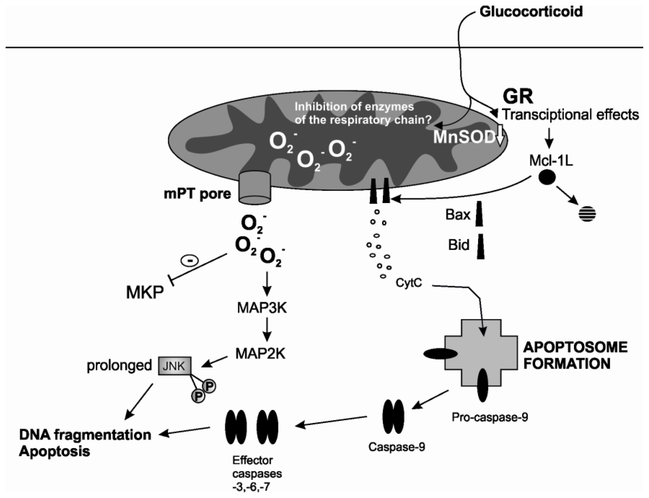
Mitochondria are extremely central in mediating induced eosinophil apoptosis and are involved in many steps from the early stress response to the decision of the cell to cope or undergo apoptosis as well as to the final loss of mitochondrial membrane potential. Many clinically relevant inducers of eosinophil apoptosis utilize the intrinsic pathway of apoptosis and even the extrinsic pathway stimulated by Fas activation involves a critical mitochondrial loop. Glucocorticoids and nitric oxide stimulate an intrinsic pathway with many similar features involving ROS, early and late JNK activation and mPT (Figure 2). Additionally, glucocorticoids accelerate degradation of anti-apoptotic Mcl-1 which might enable oligomerization and pore-forming activity of Bax. Processing of pro-apoptotic Bid into its truncated, pore-forming fragment is also enhanced by glucocorticoids. Studies support that ROS may have a central role in mediating many of these events: JNK activation, mPT induction and Bid-mediated cytochrome c release. Glucocorticoids decrease levels of mitochondrial antioxidants in eosinophils, which most likely enhance their pro-apoptotic effect. Understanding of these pathways in eosinophil apoptosis is critical to support development of new agents to treat eosinophilic disorders such as asthma. Furthermore, these pathways may also occur in other immune cells in response to glucocorticoids, and increase our understanding of the mechanisms behind the divergent effects of glucocorticoids on the longevity of different cell types.

Acknowledgments

This study was financially supported by the Competitive State Research Financing of the Expert Responsibility area of Tampere University Hospital (Tampere, Finland), grant numbers VTR 15 and VTR224, Competitive Research Funding of Seinäjoki Central Hospital (Seinäjoki, Finland), Tampere Tuberculosis Foundation (Tampere, Finland) and Finnish Anti-Tuberculosis Association Foundation (Helsinki, Finland), which are gratefully acknowledged.

Conflicts of Interest

The authors declare no conflict of interest.

References

  1. Trivedi, S.G.; Lloyd, C.M. Eosinophils in the pathogenesis of allergic airways disease. Cell. Mol. Life Sci. 2007, 64, 1269–1289. [Google Scholar]
  2. Zuo, L.; Rothenberg, M.E. Gastrointestinal eosinophilia. Immunol. Allergy Clin. N. Am. 2007, 27, 443–455. [Google Scholar]
  3. Gleich, G.J.; Leiferman, K.M. The hypereosinophilic syndromes: Current concepts and treatments. Br. J. Haematol. 2009, 145, 271–285. [Google Scholar]
  4. Ellyard, J.I.; Simson, L.; Parish, C.R. Th2-mediated anti-tumour immunity: Friend or foe? Tissue Antigens 2007, 70, 1–11. [Google Scholar]
  5. Klion, A.D.; Nutman, T.B. The role of eosinophils in host defense against helminth parasites. J. Allergy Clin. Immunol. 2004, 113, 30–37. [Google Scholar]
  6. Hogan, S.P.; Rosenberg, H.F.; Moqbel, R.; Phipps, S.; Foster, P.S.; Lacy, P.; Kay, A.B.; Rothenberg, M.E. Eosinophils: Biological properties and role in health and disease. Clin. Exp. Allergy 2008, 38, 709–750. [Google Scholar]
  7. Jacobsen, E.A.; Ochkur, S.I.; Pero, R.S.; Taranova, A.G.; Protheroe, C.A.; Colbert, D.C.; Lee, N.A.; Lee, J.J. Allergic pulmonary inflammation in mice is dependent on eosinophil-induced recruitment of effector T cells. J. Exp. Med. 2008, 205, 699–710. [Google Scholar]
  8. Humbles, A.A.; Lloyd, C.M.; McMillan, S.J.; Friend, D.S.; Xanthou, G.; McKenna, E.E.; Ghiran, S.; Gerard, N.P.; Yu, C.; Orkin, S.H.; et al. A critical role for eosinophils in allergic airways remodeling. Science 2004, 305, 1776–1779. [Google Scholar]
  9. Nair, P.; Pizzichini, M.M.; Kjarsgaard, M.; Inman, M.D.; Efthimiadis, A.; Pizzichini, E.; Hargreave, F.E.; O’Byrne, P.M. Mepolizumab for prednisone-dependent asthma with sputum eosinophilia. N. Engl. J. Med. 2009, 360, 985–993. [Google Scholar]
  10. Haldar, P.; Brightling, C.E.; Hargadon, B.; Gupta, S.; Monteiro, W.; Sousa, A.; Marshall, R.P.; Bradding, P.; Green, R.H.; Wardlaw, A.J.; et al. Mepolizumab and exacerbations of refractory eosinophilic asthma. N. Engl. J. Med. 2009, 360, 973–984. [Google Scholar]
  11. Wenzel, S.E. Eosinophils in asthma—Closing the loop or opening the door? N. Engl. J. Med. 2009, 360, 1026–1028. [Google Scholar]
  12. Lukawska, J.J.; Livieratos, L.; Sawyer, B.M.; Lee, T.; O’Doherty, M.; Blower, P.J.; Kofi, M.; Ballinger, J.R.; Corrigan, C.J.; Gnanasegaran, G.; et al. Real-time differential tracking of human neutrophil and eosinophil migration in vivo. J. Allergy Clin. Immunol. 2014, 133, 233–239. [Google Scholar]
  13. Farahi, N.; Singh, N.R.; Heard, S.; Loutsios, C.; Summers, C.; Solanki, C.K.; Solanki, K.; Balan, K.K.; Ruparelia, P.; Peters, A.M.; et al. Use of 111-Indium-labeled autologous eosinophils to establish the in vivo kinetics of human eosinophils in healthy subjects. Blood 2012, 120, 4068–4070. [Google Scholar]
  14. Walsh, G. Eosinophil apoptosis and clearance in asthma. J. Cell Death 2013, 6, 17–25. [Google Scholar]
  15. Kankaanranta, H.; Moilanen, E.; Zhang, X. Pharmacological regulation of human eosinophil apoptosis. Curr. Drug Targets Inflamm. Allergy 2005, 4, 433–445. [Google Scholar]
  16. Rothenberg, M.E.; Owen, W.F., Jr; Silberstein, D.S.; Soberman, R.J.; Austen, K.F.; Stevens, R.L. Eosinophils cocultured with endothelial cells have increased survival and functional properties. Science 1987, 237, 645–647. [Google Scholar]
  17. Kankaanranta, H.; Lindsay, M.A.; Giembycz, M.A.; Zhang, X.; Moilanen, E.; Barnes, P.J. Delayed eosinophil apoptosis in asthma. J. Allergy Clin. Immunol. 2000, 106, 77–83. [Google Scholar]
  18. Simon, H.U.; Yousefi, S.; Schranz, C.; Schapowal, A.; Bachert, C.; Blaser, K. Direct demonstration of delayed eosinophil apoptosis as a mechanism causing tissue eosinophilia. J. Immunol. 1997, 158, 3902–3908. [Google Scholar]
  19. Duffin, R.; Leitch, A.E.; Fox, S.; Haslett, C.; Rossi, A.G. Targeting granulocyte apoptosis: Mechanisms models and therapies. Immunol. Rev. 2010, 236, 28–40. [Google Scholar]
  20. Galluzzi, L.; Vitale, I.; Abrams, J.M.; Alnemri, E.S.; Baehrecke, E.H.; Blagosklonny, M.V.; Dawson, T.M.; Dawson, V.L.; El-Deiry, W.S.; Fulda, S.; et al. Molecular definitions of cell death subroutines: Recommendations of the nomenclature committee on cell death 2012. Cell Death Differ. 2012, 19, 107–120. [Google Scholar]
  21. Kroemer, G.; Galluzzi, L.; Brenner, C. Mitochondrial membrane permeabilization in cell death. Physiol. Rev. 2007, 87, 99–163. [Google Scholar]
  22. Rasola, A.; Bernardi, P. Mitochondrial permeability transition in Ca2+-dependent apoptosis and necrosis. Cell Calcium 2011, 50, 222–233. [Google Scholar]
  23. Letuve, S.; Druilhe, A.; Grandsaigne, M.; Aubier, M.; Pretolani, M. Involvement of caspases and of mitochondria in Fas ligation-induced eosinophil apoptosis: Modulation by interleukin-5 and interferon-gamma. J. Leukoc. Biol. 2001, 70, 767–775. [Google Scholar]
  24. Sharma, S.K.; Almeida, F.A.; Kierstein, S.; Hortobagyi, L.; Lin, T.; Larkin, A.; Peterson, J.; Yagita, H.; Zangrilli, J.G.; Haczku, A. Systemic FasL neutralization increases eosinophilic inflammation in a mouse model of asthma. Allergy 2012, 67, 328–335. [Google Scholar]
  25. Beauvais, F.; Joly, F. Effects of nitric oxide on the eosinophil survival in vitro A role for nitrosyl-heme. FEBS Lett. 1999, 443, 37–40. [Google Scholar]
  26. Zhang, X.; Msc Moilanen, E.; Lahti, A.; Hamalainen, M.; Giembycz, M.A.; Barnes, P.J.; Lindsay, M.A.; Kankaanranta, H. Regulation of eosinophil apoptosis by nitric oxide: Role of c-Jun-N-terminal kinase and signal transducer and activator of transcription 5. J. Allergy Clin. Immunol. 2003, 112, 93–101. [Google Scholar]
  27. Hebestreit, H.; Dibbert, B.; Balatti, I.; Braun, D.; Schapowal, A.; Blaser, K.; Simon, H.U. Disruption of fas receptor signaling by nitric oxide in eosinophils. J. Exp. Med. 1998, 187, 415–425. [Google Scholar]
  28. Oliveira, M.S.; de O Barreto, E.; Zamuner, S.; Pires, A.L.; Ferreira, T.P.; Cordeiro, R.S.; Lagente, V.; Martins, M.A.; Wallace, J.L.; e Silva, P.M. Suppressive effects of nitric oxide-releasing prednisolone NCX-1015 on the allergic pleural eosinophil recruitment in rats. Clin. Exp. Allergy 2008, 38, 1830–1837. [Google Scholar]
  29. Feder, L.S.; Stelts, D.; Chapman, R.W.; Manfra, D.; Crawley, Y.; Jones, H.; Minnicozzi, M.; Fernandez, X.; Paster, T.; Egan, R.W.; et al. Role of nitric oxide on eosinophilic lung inflammation in allergic mice. Am. J. Respir. Cell Mol. Biol. 1997, 17, 436–442. [Google Scholar]
  30. Lehtimaki, L.; Kankaanranta, H.; Saarelainen, S.; Hahtola, P.; Jarvenpaa, R.; Koivula, T.; Turjanmaa, V.; Moilanen, E. Extended exhaled NO measurement differentiates between alveolar and bronchial inflammation. Am. J. Respir. Crit. Care Med. 2001, 163, 1557–1561. [Google Scholar]
  31. Zhang, X.; Moilanen, E.; Kankaanranta, H. Enhancement of human eosinophil apoptosis by fluticasone propionate budesonide and beclomethasone. Eur. J. Pharmacol. 2000, 406, 325–332. [Google Scholar]
  32. Lamas, A.M.; Leon, O.G.; Schleimer, R.P. Glucocorticoids inhibit eosinophil responses to granulocyte-macrophage colony-stimulating factor. J. Immunol. 1991, 147, 254–259. [Google Scholar]
  33. Wallen, N.; Kita, H.; Weiler, D.; Gleich, G.J. Glucocorticoids inhibit cytokine-mediated eosinophil survival. J. Immunol. 1991, 147, 3490–3495. [Google Scholar]
  34. Lee, E.; Robertson, T.; Smith, J.; Kilfeather, S. Leukotriene receptor antagonists and synthesis inhibitors reverse survival in eosinophils of asthmatic individuals. Am. J. Respir. Crit. Care Med. 2000, 161, 1881–1886. [Google Scholar]
  35. Yasui, K.; Hu, B.; Nakazawa, T.; Agematsu, K.; Komiyama, A. Theophylline accelerates human granulocyte apoptosis not via phosphodiesterase inhibition. J. Clin. Investig. 1997, 100, 1677–1684. [Google Scholar]
  36. Ilmarinen, P.; Kankaanranta, H. Eosinophil apoptosis as a therapeutic target in allergic asthma. Basic Clin. Pharmacol. Toxicol. 2013, 114, 109–117. [Google Scholar]
  37. Woolley, K.L.; Gibson, P.G.; Carty, K.; Wilson, A.J.; Twaddell, S.H.; Woolley, M.J. Eosinophil apoptosis and the resolution of airway inflammation in asthma. Am. J. Respir. Crit. Care Med. 1996, 154, 237–243. [Google Scholar]
  38. Druilhe, A.; Wallaert, B.; Tsicopoulos, A.; Lapa e Silva, J.R.; Tillie-Leblond, I.; Tonnel, A.B.; Pretolani, M. Apoptosis proliferation and expression of Bcl-2 Fas and Fas ligand in bronchial biopsies from asthmatics. Am. J. Respir. Cell Mol. Biol. 1998, 19, 747–757. [Google Scholar]
  39. Vignola, A.M.; Chanez, P.; Chiappara, G.; Siena, L.; Merendino, A.; Reina, C.; Gagliardo, R.; Profita, M.; Bousquet, J.; Bonsignore, G. Evaluation of apoptosis of eosinophils macrophages and T lymphocytes in mucosal biopsy specimens of patients with asthma and chronic bronchitis. J. Allergy Clin. Immunol. 1999, 103, 563–573. [Google Scholar]
  40. Duncan, C.J.; Lawrie, A.; Blaylock, M.G.; Douglas, J.G.; Walsh, G.M. Reduced eosinophil apoptosis in induced sputum correlates with asthma severity. Eur. Respir. J. 2003, 22, 484–490. [Google Scholar]
  41. Montuschi, P.; Peters-Golden, M.L. Leukotriene modifiers for asthma treatment. Clin. Exp. Allergy 2010, 40, 1732–1741. [Google Scholar]
  42. Lim, S.; Tomita, K.; Caramori, G.; Jatakanon, A.; Oliver, B.; Keller, A.; Adcock, I.; Chung, K.F.; Barnes, P.J. Low-dose theophylline reduces eosinophilic inflammation but not exhaled nitric oxide in mild asthma. Am. J. Respir. Crit. Care Med. 2001, 164, 273–276. [Google Scholar]
  43. Jetzek-Zader, M.; Gudowius, S.; Feyen, O.; Stevens, M.; Lipfert, P.; Niehues, T. A single intravenous dose of prednisolone induces phosphatidylserine externalization loss of surface marker expression and a 24-h net increase in human peripheral blood lymphocytes ex vivo. Rheumatol. Int. 2007, 27, 667–673. [Google Scholar]
  44. Wiegers, G.J.; Knoflach, M.; Bock, G.; Niederegger, H.; Dietrich, H.; Falus, A.; Boyd, R.; Wick, G. CD4(+)CD8(+)TCR(low) thymocytes express low levels of glucocorticoid receptors while being sensitive to glucocorticoid-induced apoptosis. Eur. J. Immunol. 2001, 31, 2293–2301. [Google Scholar]
  45. Leussink, V.I.; Jung, S.; Merschdorf, U.; Toyka, K.V.; Gold, R. High-dose methylprednisolone therapy in multiple sclerosis induces apoptosis in peripheral blood leukocytes. Arch. Neurol. 2001, 58, 91–97. [Google Scholar]
  46. Meagher, L.C.; Cousin, J.M.; Seckl, J.R.; Haslett, C. Opposing effects of glucocorticoids on the rate of apoptosis in neutrophilic and eosinophilic granulocytes. J. Immunol. 1996, 156, 4422–4428. [Google Scholar]
  47. Zhang, X.; Moilanen, E.; Kankaanranta, H. Beclomethasone budesonide and fluticasone propionate inhibit human neutrophil apoptosis. Eur. J. Pharmacol. 2001, 431, 365–371. [Google Scholar]
  48. Zhang, X.; Moilanen, E.; Adcock, I.M.; Lindsay, M.A.; Kankaanranta, H. Divergent effect of mometasone on human eosinophil and neutrophil apoptosis. Life Sci. 2002, 71, 1523–1534. [Google Scholar]
  49. Marwick, J.A.; Dorward, D.A.; Lucas, C.D.; Jones, K.O.; Sheldrake, T.A.; Fox, S.; Ward, C.; Murray, J.; Brittan, M.; Hirani, N.; et al. Oxygen levels determine the ability of glucocorticoids to influence neutrophil survival in inflammatory environments. J. Leukoc. Biol. 2013, 94, 1285–1292. [Google Scholar]
  50. Peachman, K.K.; Lyles, D.S.; Bass, D.A. Mitochondria in eosinophils: Functional role in apoptosis but not respiration. Proc. Natl. Acad. Sci. USA 2001, 98, 1717–1722. [Google Scholar]
  51. Dewson, G.; Cohen, G.M.; Wardlaw, A.J. Interleukin-5 inhibits translocation of Bax to the mitochondria cytochrome c release and activation of caspases in human eosinophils. Blood 2001, 98, 2239–2247. [Google Scholar]
  52. Gardai, S.J.; Hoontrakoon, R.; Goddard, C.D.; Day, B.J.; Chang, L.Y.; Henson, P.M.; Bratton, D.L. Oxidant-mediated mitochondrial injury in eosinophil apoptosis: Enhancement by glucocorticoids and inhibition by granulocyte-macrophage colony-stimulating factor. J. Immunol. 2003, 170, 556–566. [Google Scholar]
  53. Segal, M.; Niazi, S.; Simons, M.P.; Galati, S.A.; Zangrilli, J.G. Bid activation during induction of extrinsic and intrinsic apoptosis in eosinophils. Immunol. Cell Biol. 2007, 85, 518–524. [Google Scholar]
  54. Maret, M.; Ruffie, C.; Letuve, S.; Phelep, A.; Thibaudeau, O.; Marchal, J.; Pretolani, M.; Druilhe, A. A role for Bid in eosinophil apoptosis and in allergic airway reaction. J. Immunol. 2009, 182, 5740–5747. [Google Scholar]
  55. Lee, Y.A.; Shin, M.H. Mitochondrial respiration is required for activation of ERK1/2 and caspase-3 in human eosinophils stimulated with hydrogen peroxide. J. Investig. Allergol. Clin. Immunol. 2009, 19, 188–194. [Google Scholar]
  56. Van Der Vliet, H.J.; Wever, P.C.; van Diepen, F.N.; Yong, S.L.; Ten Berge, I.J. Quantification of Bax/Bcl-2 ratios in peripheral blood lymphocytes monocytes and granulocytes and their relation to susceptibility to anti-Fas (anti-CD95)-induced apoptosis. Clin. Exp. Immunol. 1997, 110, 324–328. [Google Scholar]
  57. Druilhe, A.; Arock, M.; le Goff, L.; Pretolani, M. Human eosinophils express bcl-2 family proteins: Modulation of Mcl-1 expression by IFN-gamma. Am. J. Respir. Cell Mol. Biol. 1998, 18, 315–322. [Google Scholar]
  58. Shen, Z.J.; Esnault, S.; Schinzel, A.; Borner, C.; Malter, J.S. The peptidyl-prolyl isomerase Pin1 facilitates cytokine-induced survival of eosinophils by suppressing Bax activation. Nat. Immunol. 2009, 10, 257–265. [Google Scholar]
  59. Oh, J.; Malter, J.S. Pin1-FADD interactions regulate Fas-mediated apoptosis in activated eosinophils. J. Immunol. 2013, 190, 4937–4945. [Google Scholar]
  60. Zangrilli, J.; Robertson, N.; Shetty, A.; Wu, J.; Hastie, A.; Fish, J.E.; Litwack, G.; Peters, S.P. Effect of IL-5 glucocorticoid and Fas ligation on Bcl-2 homologue expression and caspase activation in circulating human eosinophils. Clin. Exp. Immunol. 2000, 120, 12–21. [Google Scholar]
  61. Duffin, R.; Leitch, A.E.; Sheldrake, T.A.; Hallett, J.M.; Meyer, C.; Fox, S.; Alessandri, A.L.; Martin, M.C.; Brady, H.J.; Teixeira, M.M.; et al. The CDK inhibitor R-roscovitine promotes eosinophil apoptosis by down-regulation of Mcl-1. FEBS Lett. 2009, 583, 2540–2546. [Google Scholar]
  62. Farahi, N.; Uller, L.; Juss, J.K.; Langton, A.J.; Cowburn, A.S.; Gibson, A.; Foster, M.R.; Farrow, S.N.; Marco-Casanova, P.; Sobolewski, A.; et al. Effects of the cyclin-dependent kinase inhibitor R-roscovitine on eosinophil survival and clearance. Clin. Exp. Allergy 2011, 41, 673–687. [Google Scholar]
  63. Sivertson, K.L.; Seeds, M.C.; Long, D.L.; Peachman, K.K.; Bass, D.A. The differential effect of dexamethasone on granulocyte apoptosis involves stabilization of Mcl-1L in neutrophils but not in eosinophils. Cell. Immunol. 2007, 246, 34–45. [Google Scholar]
  64. Germain, M.; Milburn, J.; Duronio, V. MCL-1 inhibits BAX in the absence of MCL-1/BAX interaction. J. Biol. Chem. 2008, 283, 6384–6392. [Google Scholar]
  65. Saffar, A.S.; Dragon, S.; Ezzati, P.; Shan, L.; Gounni, A.S. Phosphatidylinositol 3-kinase and p38 mitogen-activated protein kinase regulate induction of Mcl-1 and survival in glucocorticoid-treated human neutrophils. J. Allergy Clin. Immunol. 2008, 121, 492–498. [Google Scholar]
  66. Dewson, G.; Walsh, G.M.; Wardlaw, A.J. Expression of Bcl-2 and its homologues in human eosinophils Modulation by interleukin-5. Am. J. Respir. Cell Mol. Biol. 1999, 20, 720–728. [Google Scholar]
  67. Kaasik, A.; Safiulina, D.; Zharkovsky, A.; Veksler, V. Regulation of mitochondrial matrix volume. Am. J. Physiol. Cell Physiol. 2007, 292, C157–C163. [Google Scholar]
  68. Ilmarinen-Salo, P.; Moilanen, E.; Kinnula, V.L.; Kankaanranta, H. Nitric oxide-induced eosinophil apoptosis is dependent on mitochondrial permeability transition (mPT), JNK and oxidative stress: Apoptosis is preceded but not mediated by early mPT-dependent JNK activation. Respir. Res. 2012, 13. [Google Scholar] [CrossRef]
  69. Letuve, S.; Druilhe, A.; Grandsaigne, M.; Aubier, M.; Pretolani, M. Critical role of mitochondria but not caspases during glucocorticosteroid-induced human eosinophil apoptosis. Am. J. Respir. Cell Mol. Biol. 2002, 26, 565–571. [Google Scholar]
  70. Garcia-Perez, C.; Roy, S.S.; Naghdi, S.; Lin, X.; Davies, E.; Hajnoczky, G. Bid-induced mitochondrial membrane permeabilization waves propagated by local reactive oxygen species (ROS) signaling. Proc. Natl. Acad. Sci. USA 2012, 109, 4497–4502. [Google Scholar]
  71. Petronilli, V.; Penzo, D.; Scorrano, L.; Bernardi, P.; di Lisa, F. The mitochondrial permeability transition release of cytochrome c and cell death Correlation with the duration of pore openings in situ. J. Biol. Chem. 2001, 276, 12030–12034. [Google Scholar]
  72. Ichas, F.; Jouaville, L.S.; Mazat, J.P. Mitochondria are excitable organelles capable of generating and conveying electrical and calcium signals. Cell 1997, 89, 1145–1153. [Google Scholar]
  73. Bernardi, P.; Petronilli, V. The permeability transition pore as a mitochondrial calcium release channel: A critical appraisal. J. Bioenerg. Biomembr. 1996, 28, 131–138. [Google Scholar]
  74. Barsukova, A.; Komarov, A.; Hajnoczky, G.; Bernardi, P.; Bourdette, D.; Forte, M. Activation of the mitochondrial permeability transition pore modulates Ca2+ responses to physiological stimuli in adult neurons. Eur. J. Neurosci. 2011, 33, 831–842. [Google Scholar]
  75. Bernardi, P.; von Stockum, S. The permeability transition pore as a Ca2+ release channel: New answers to an old question. Cell Calcium 2012, 52, 22–27. [Google Scholar]
  76. Zorov, D.B.; Juhaszova, M.; Sollott, S.J. Mitochondrial ROS-induced ROS release: An update and review. Biochim. Biophys. Acta 2006, 1757, 509–517. [Google Scholar]
  77. Ma, Q.; Fang, H.; Shang, W.; Liu, L.; Xu, Z.; Ye, T.; Wang, X.; Zheng, M.; Chen, Q.; Cheng, H. Superoxide flashes: Early mitochondrial signals for oxidative stress-induced apoptosis. J. Biol. Chem. 2011, 286, 27573–27581. [Google Scholar]
  78. Wedi, B.; Straede, J.; Wieland, B.; Kapp, A. Eosinophil apoptosis is mediated by stimulators of cellular oxidative metabolisms and inhibited by antioxidants: Involvement of a thiol-sensitive redox regulation in eosinophil cell death. Blood 1999, 94, 2365–2373. [Google Scholar]
  79. Martinez-Losa, M.; Cortijo, J.; Juan, G.; Ramon, M.; Sanz, M.J.; Morcillo, E.J. Modulatory effects of N-acetyl-l-cysteine on human eosinophil apoptosis. Eur. Respir. J. 2007, 30, 436–442. [Google Scholar]
  80. Nissim Ben Efraim, A.H.; Eliashar, R.; Levi-Schaffer, F. Hypoxia modulates human eosinophil function. Clin. Mol. Allergy 2010, 8. [Google Scholar] [CrossRef]
  81. Kano, G.; Almanan, M.; Bochner, B.S.; Zimmermann, N. Mechanism of siglec-8-mediated cell death in IL-5-activated eosinophils: Role for reactive oxygen species-enhanced MEK/ERK activation. J. Allergy Clin. Immunol. 2013, 132, 437–445. [Google Scholar]
  82. Orrenius, S.; Gogvadze, V.; Zhivotovsky, B. Mitochondrial oxidative stress: Implications for cell death. Annu. Rev. Pharmacol. Toxicol. 2007, 47, 143–183. [Google Scholar]
  83. Cadenas, E.; Davies, K.J. Mitochondrial free radical generation oxidative stress and aging. Free Radic. Biol. Med. 2000, 29, 222–230. [Google Scholar]
  84. Gough, D.R.; Cotter, T.G. Hydrogen peroxide: A Jekyll and Hyde signalling molecule. Cell Death Dis. 2011, 2, e213. [Google Scholar]
  85. Kankaanranta, H.; Giembycz, M.A.; Barnes, P.J.; Haddad, E.l.-B.; Saarelainen, S.; Zhang, X.; Moilanen, E.; Lindsay, M.A. Hydrogen peroxide reverses IL-5 afforded eosinophil survival and promotes constitutive human eosinophil apoptosis. Int. Arch. Allergy Immunol. 2002, 127, 73–78. [Google Scholar]
  86. Serradell, M.C.; Guasconi, L.; Masih, D.T. Involvement of a mitochondrial pathway and key role of hydrogen peroxide during eosinophil apoptosis induced by excretory-secretory products from Fasciola hepatica. Mol. Biochem. Parasitol. 2009, 163, 95–106. [Google Scholar]
  87. Tonomura, N.; McLaughlin, K.; Grimm, L.; Goldsby, R.A.; Osborne, B.A. Glucocorticoid-induced apoptosis of thymocytes: requirement of proteasome-dependent mitochondrial activity. J. Immunol. 2003, 170, 2469–2478. [Google Scholar]
  88. Tome, M.E.; Baker, A.F.; Powis, G.; Payne, C.M.; Briehl, M.M. Catalase-overexpressing thymocytes are resistant to glucocorticoid-induced apoptosis and exhibit increased net tumor growth. Cancer Res. 2001, 61, 2766–2773. [Google Scholar]
  89. Andreyev, A.Y.; Kushnareva, Y.E.; Starkov, A.A. Mitochondrial metabolism of reactive oxygen species. Biochemistry 2005, 70, 200–214. [Google Scholar]
  90. Simon, N.; Jolliet, P.; Morin, C.; Zini, R.; Urien, S.; Tillement, J.P. Glucocorticoids decrease cytochrome c oxidase activity of isolated rat kidney mitochondria. FEBS Lett. 1998, 435, 25–28. [Google Scholar]
  91. Morin, C.; Zini, R.; Simon, N.; Charbonnier, P.; Tillement, J.P.; le Louet, H. Low glucocorticoid concentrations decrease oxidative phosphorylation of isolated rat brain mitochondria: An additional effect of dexamethasone. Fundam. Clin. Pharmacol. 2000, 14, 493–500. [Google Scholar]
  92. Mutsaers, H.A.; Tofighi, R. Dexamethasone enhances oxidative stress-induced cell death in murine neural stem cells. Neurotox. Res. 2012, 22, 127–137. [Google Scholar]
  93. Korhonen, R.; Moilanen, E. MAP kinase phosphatase-1 as an inflammatory factor and drug target. Basic Clin. Pharmacol. Toxicol. 2014, 114, 24–36. [Google Scholar]
  94. Raman, M.; Chen, W.; Cobb, M.H. Differential regulation and properties of MAPKs. Oncogene 2007, 26, 3100–3112. [Google Scholar]
  95. Wancket, L.M.; Frazier, W.J.; Liu, Y. Mitogen-activated protein kinase phosphatase (MKP)-1 in immunology physiology and disease. Life Sci. 2012, 90, 237–248. [Google Scholar]
  96. Yin, H.; Shi, Z.; Jiao, S.; Chen, C.; Wang, W.; Greene, M.I.; Zhou, Z. Germinal center kinases in immune regulation. Cell. Mol. Immunol. 2012, 9, 439–445. [Google Scholar]
  97. McCubrey, J.A.; Lahair, M.M.; Franklin, R.A. Reactive oxygen species-induced activation of the MAP kinase signaling pathways. Antioxid. Redox Signal. 2006, 8, 1775–1789. [Google Scholar]
  98. Hasala, H.; Zhang, X.; Saarelainen, S.; Moilanen, E.; Kankaanranta, H. c-Jun N-terminal kinase mediates constitutive human eosinophil apoptosis. Pulm. Pharmacol. Ther. 2007, 20, 580–587. [Google Scholar]
  99. Kankaanranta, H.; Zhang, X.; Tumelius, R.; Ruotsalainen, M.; Haikala, H.; Nissinen, E.; Moilanen, E. Antieosinophilic activity of simendans. J. Pharmacol. Exp. Ther. 2007, 323, 31–38. [Google Scholar]
  100. Hasala, H.; Moilanen, E.; Janka-Junttila, M.; Giembycz, M.A.; Kankaanranta, H. First-generation antihistamines diphenhydramine and chlorpheniramine reverse cytokine-afforded eosinophil survival by enhancing apoptosis. Allergy Asthma Proc. 2007, 28, 79–86. [Google Scholar]
  101. Kankaanranta, H.; Ilmarinen, P.; Zhang, X.; Nissinen, E.; Moilanen, E. Antieosinophilic activity of orazipone. Mol. Pharmacol. 2006, 69, 1861–1870. [Google Scholar]
  102. Shrivastava, P.; Pantano, C.; Watkin, R.; McElhinney, B.; Guala, A.; Poynter, M.L.; Persinger, R.L.; Budd, R.; Janssen-Heininger, Y. Reactive nitrogen species-induced cell death requires Fas-dependent activation of c-Jun N-terminal kinase. Mol. Cell. Biol. 2004, 24, 6763–6772. [Google Scholar]
  103. Sanchez-Perez, I.; Murguia, J.R.; Perona, R. Cisplatin induces a persistent activation of JNK that is related to cell death. Oncogene 1998, 16, 533–540. [Google Scholar]
  104. Ventura, J.J.; Hubner, A.; Zhang, C.; Flavell, R.A.; Shokat, K.M.; Davis, R.J. Chemical genetic analysis of the time course of signal transduction by JNK. Mol. Cell 2006, 21, 701–710. [Google Scholar]
  105. Lamb, J.A.; Ventura, J.J.; Hess, P.; Flavell, R.A.; Davis, R.J. JunD mediates survival signaling by the JNK signal transduction pathway. Mol. Cell 2003, 11, 1479–1489. [Google Scholar]
  106. Kamata, H.; Honda, S.; Maeda, S.; Chang, L.; Hirata, H.; Karin, M. Reactive oxygen species promote TNFalpha-induced death and sustained JNK activation by inhibiting MAP kinase phosphatases. Cell 2005, 120, 649–661. [Google Scholar]
  107. Miike, S.; Nakao, A.; Hiraguri, M.; Kurasawa, K.; Saito, Y.; Iwamoto, I. Involvement of JAK2 but not PI 3-kinase/Akt and MAP kinase pathways in anti-apoptotic signals of GM-CSF in human eosinophils. J. Leukoc. Biol. 1999, 65, 700–706. [Google Scholar]
  108. Kankaanranta, H.; de Souza, P.M.; Barnes, P.J.; Salmon, M.; Giembycz, M.A.; Lindsay, M.A. SB 203580 an inhibitor of p38 mitogen-activated protein kinase enhances constitutive apoptosis of cytokine-deprived human eosinophils. J. Pharmacol. Exp. Ther. 1999, 290, 621–628. [Google Scholar]
  109. Zhang, J.P.; Wong, C.K.; Lam, C.W. Role of caspases in dexamethasone-induced apoptosis and activation of c-Jun NH2-terminal kinase and p38 mitogen-activated protein kinase in human eosinophils. Clin. Exp. Immunol. 2000, 122, 20–27. [Google Scholar]
  110. Baruch-Morgenstern, N.B.; Shik, D.; Moshkovits, I.; Itan, M.; Karo-Atar, D.; Bouffi, C.; Fulkerson, P.C.; Rashkovan, D.; Jung, S.; Rothenberg, M.E.; Munitz, A. Paired immunoglobulin-like receptor A is an intrinsic self-limiting suppressor of IL-5-induced eosinophil development. Nat. Immunol. 2013, 15. [Google Scholar] [CrossRef]
  111. Matsumoto, K.; Terakawa, M.; Miura, K.; Fukuda, S.; Nakajima, T.; Saito, H. Extremely rapid and intense induction of apoptosis in human eosinophils by anti-CD30 antibody treatment in vitro. J. Immunol. 2004, 172, 2186–2193. [Google Scholar]
  112. Tsukahara, K.; Nakao, A.; Hiraguri, M.; Miike, S.; Mamura, M.; Saito, Y.; Iwamoto, I. Tumor necrosis factor-alpha mediates antiapoptotic signals partially via p38 MAP kinase activation in human eosinophils. Int. Arch. Allergy Immunol. 1999, 120, 54–59. [Google Scholar]
  113. Wong, C.K.; Hu, S.; Cheung, P.F.; Lam, C.W. Thymic stromal lymphopoietin induces chemotactic and prosurvival effects in eosinophils: Implications in allergic inflammation. Am. J. Respir. Cell Mol. Biol. 2010, 43, 305–315. [Google Scholar]
  114. De Souza, P.M.; Kankaanranta, H.; Michael, A.; Barnes, P.J.; Giembycz, M.A.; Lindsay, M.A. Caspase-catalyzed cleavage and activation of Mst1 correlates with eosinophil but not neutrophil apoptosis. Blood 2002, 99, 3432–3438. [Google Scholar]
  115. Ura, S.; Nishina, H.; Gotoh, Y.; Katada, T. Activation of the c-Jun N-terminal kinase pathway by MST1 is essential and sufficient for the induction of chromatin condensation during apoptosis. Mol. Cell. Biol. 2007, 27, 5514–5522. [Google Scholar]
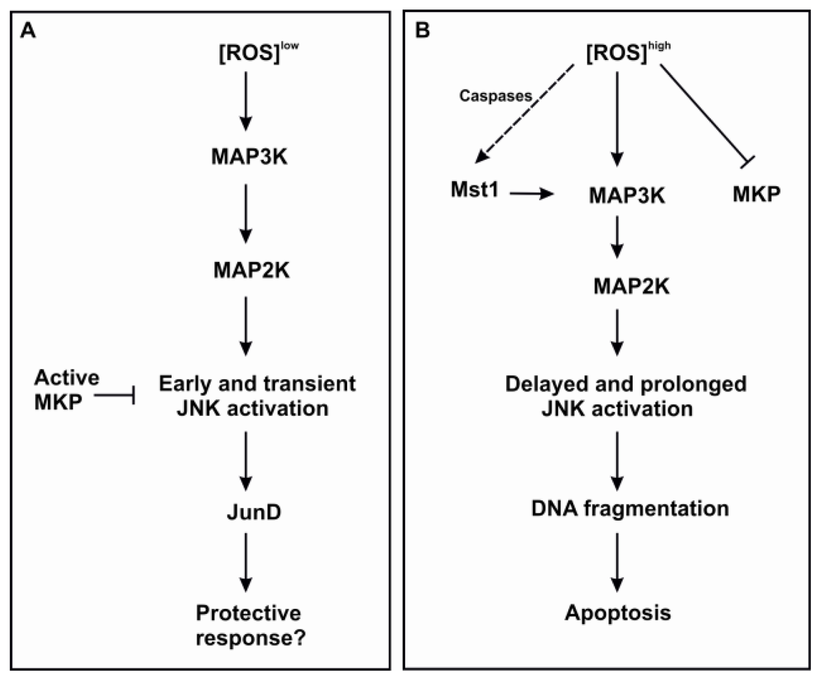
Figure 1. Hypothetical model to describe the mechanisms and outcomes of MAPK activation induced by different levels of ROS. If a pro-apoptotic stimulant induces release of ROS that is high enough to quench MKP, this may lead to prolonged JNK activation and apoptosis (B); Low levels of ROS may lead to transient activation of JNK due to presence of active MKP and end up in a protective response (A). The specific pathway mediating activation of Mst1 is unclear. ROS, reactive oxygen species; MAP3K, mitogen-activated protein kinase kinase kinase; MAP2K, mitogen-activated protein kinase kinase; JNK, c-Jun N-terminal kinase; MKP, MAP kinase phosphatase; Mst1, mammalian sterile 20-like kinase 1.
Ijms 15 03952f1
Figure 2. A proposed model of the pro-apoptotic mechanism of glucocorticoids in eosinophils. Glucocorticoid-induced eosinophil apoptosis is partially dependent on glucocorticoid receptor and stimulate an intrinsic pathway of apoptosis. Transcriptional effects may lead to reduced level of mitochondrial antioxidant MnSOD and elevated ROS. It is also possible that glucocorticoids directly inhibit mitochondrial respiratory chain enzymes. Elevated ROS may lead to mPT and release of ROS to the cytosol. ROS inhibits MKP and stimulates activation of the MAPK pathway. Glucocorticoids may also enhance degradation of anti-apoptotic Mcl-1L by a transcriptional route. Degradation of Mcl-1L enables oligomerization and pore-forming activity of Bax leading to mitochondrial outer membrane permeabilization. Processing of pro-apoptotic Bid into its truncated, pore-forming fragment is also enhanced by glucocorticoids. Abbreviations: GR, glucocorticoid receptor; Mcl-1L, myeloid cell leukemia 1 (long); MnSOD, manganese superoxide dismutase; mPT, mitochondrial permeability transition; CytC, cytochrome c; MKP, map kinase phosphatase; JNK, c-Jun N-terminal kinase; MAP3K, mitogen-activated protein kinase kinase kinase; MAP2K, mitogen-activated protein kinase kinase.
Ijms 15 03952f2