Review

Intercellular Signaling Pathway among Endothelia, Astrocytes and Neurons in Excitatory Neuronal Damage

1
Medical Research Institute, Tokyo Women's Medical University, Shinjuku, Tokyo 162-8666, Japan
2
Neural Plasticity Project, Tokyo Metropolitan Institute of Medical Science, Tokyo 156-8506, Japan
*
Author to whom correspondence should be addressed.
Int. J. Mol. Sci. 2013, 14(4), 8345-8357; https://doi.org/10.3390/ijms14048345
Submission received: 1 February 2013 / Revised: 20 March 2013 / Accepted: 3 April 2013 / Published: 16 April 2013
(This article belongs to the Special Issue Signalling Molecules and Signal Transduction in Cells)

Abstract

:
Neurons interact closely with astrocytes via glutamate; this neuron-glia circuit may play a pivotal role in synaptic transmission. On the other hand, astrocytes contact vascular endothelial cells with their end-feet. It is becoming obvious that non-neuronal cells play a critical role in regulating the neuronal activity in the brain. We find that kainic acid (KA) administration induces the expression of microsomal prostaglandin E synthase-1 (mPGES-1) in venous endothelial cells and the prostaglandin E2 (PGE2) receptor prostaglandin E receptor (EP)-3 on astrocytes. Endothelial mPGES-1 exacerbates KA-induced neuronal damage in in vivo experiments. In in vitro experiments, mPGES-1 produces PGE2, which enhances astrocytic Ca2+ levels via the EP3 receptor and increases Ca2+-dependent glutamate release, thus aggravating neuronal injury. This novel endothelium-astrocyte-neuron signaling pathway may be crucial for driving neuronal damage after repetitive seizures and could be a new therapeutic target for epilepsy and other brain disorders.

1. Introduction

Prostaglandin E2 (PGE2) is one of the most important modulators in inflammation. In the brain, PGE2 is also involved in pathological processes, such as fever, seizure and cerebral ischemia [13], suggesting that these processes might be associated with inflammation. PGE2 is sequentially synthesized from arachidonic acid by cyclooxygenase (COX) and PGE2 synthase (PGES) in various cells and tissues. COX catalyzes the first step in the synthesis of prostaglandins (PGs) and exists in two homologous isoforms. One isoform is the constitutively active COX-1, which is widely distributed in various cell types and is thought to mediate physiological responses. The other isoform is the inducible COX-2, which is expressed in several cell types in response to various stimuli, such as neuronal activity, cytokines and pro-inflammatory molecules [48]. Inducible COX-2 expression in the brain is associated with acute neurotoxicity, such as seizures and ischemia [2,3,9,10]. COX-2 is also involved in delayed pro-inflammatory activities, which aggravate the neuronal damage found in neurodegenerative diseases, such as amyotrophic lateral sclerosis (ALS), Parkinson’s disease (PD), multiple sclerosis (MS) and Alzheimer’s disease (AD) [9,11]. We find that COX-2 is induced in non-neuronal cells late after seizure and facilitates neuronal loss in the hippocampus [12].
In this review, we first focus on the co-induction of COX-2 and microsomal prostaglandin E synthase-1 (mPGES-1), an enzyme downstream of COX-2, in brain endothelial cells after seizures. Next, we provide a view on the role of endothelial mPGES-1 in neuronal loss in the hippocampus. Finally, we will present a novel mechanism for exacerbation of neuronal damage by PGE2 derived from endothelial mPGES-1 and discuss the intercellular signaling pathway among endothelia, astrocytes and neurons in this process.

2. Role of Endothelial mPGES-1 in KA-Induced Neuronal Damage

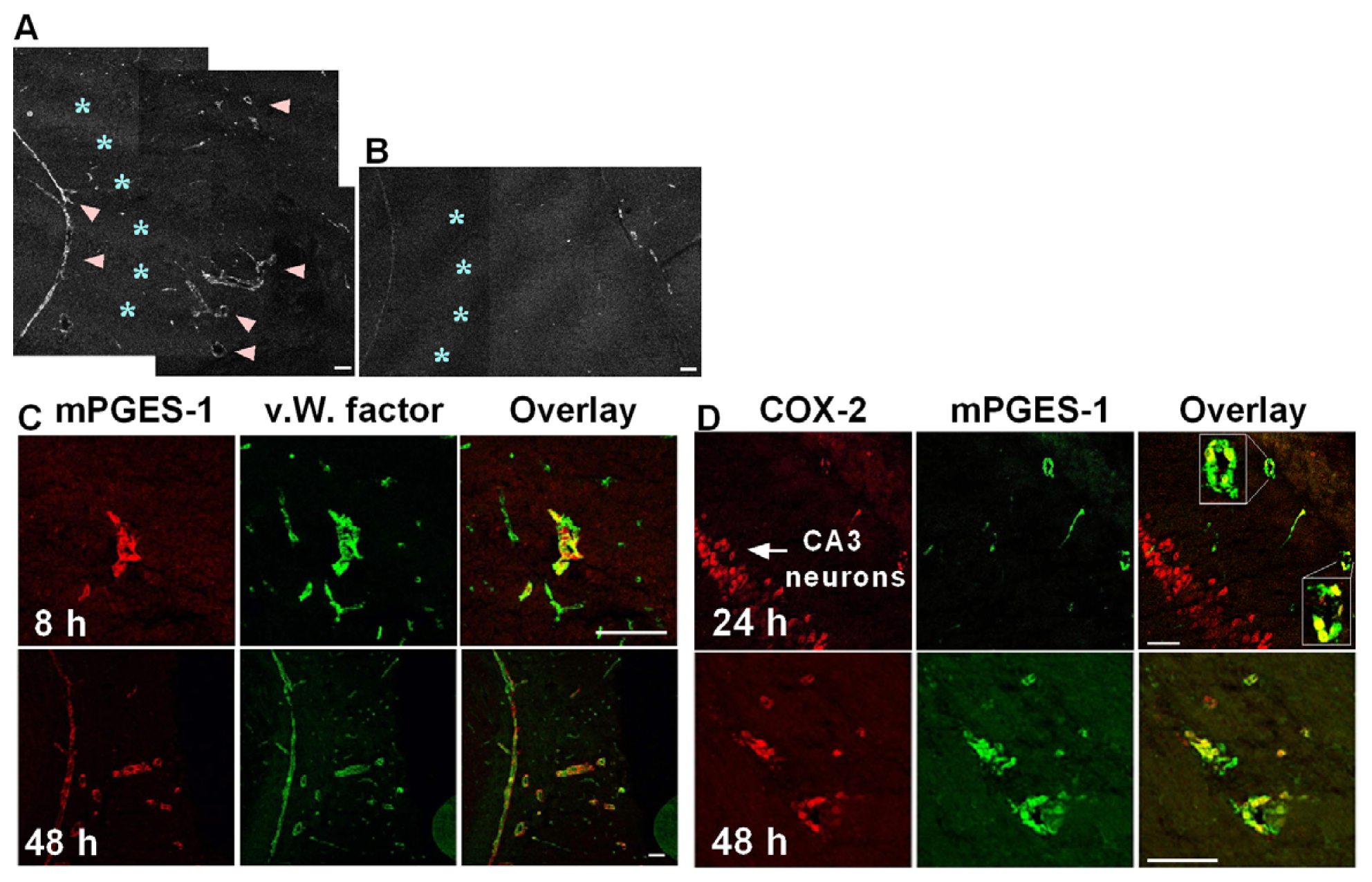
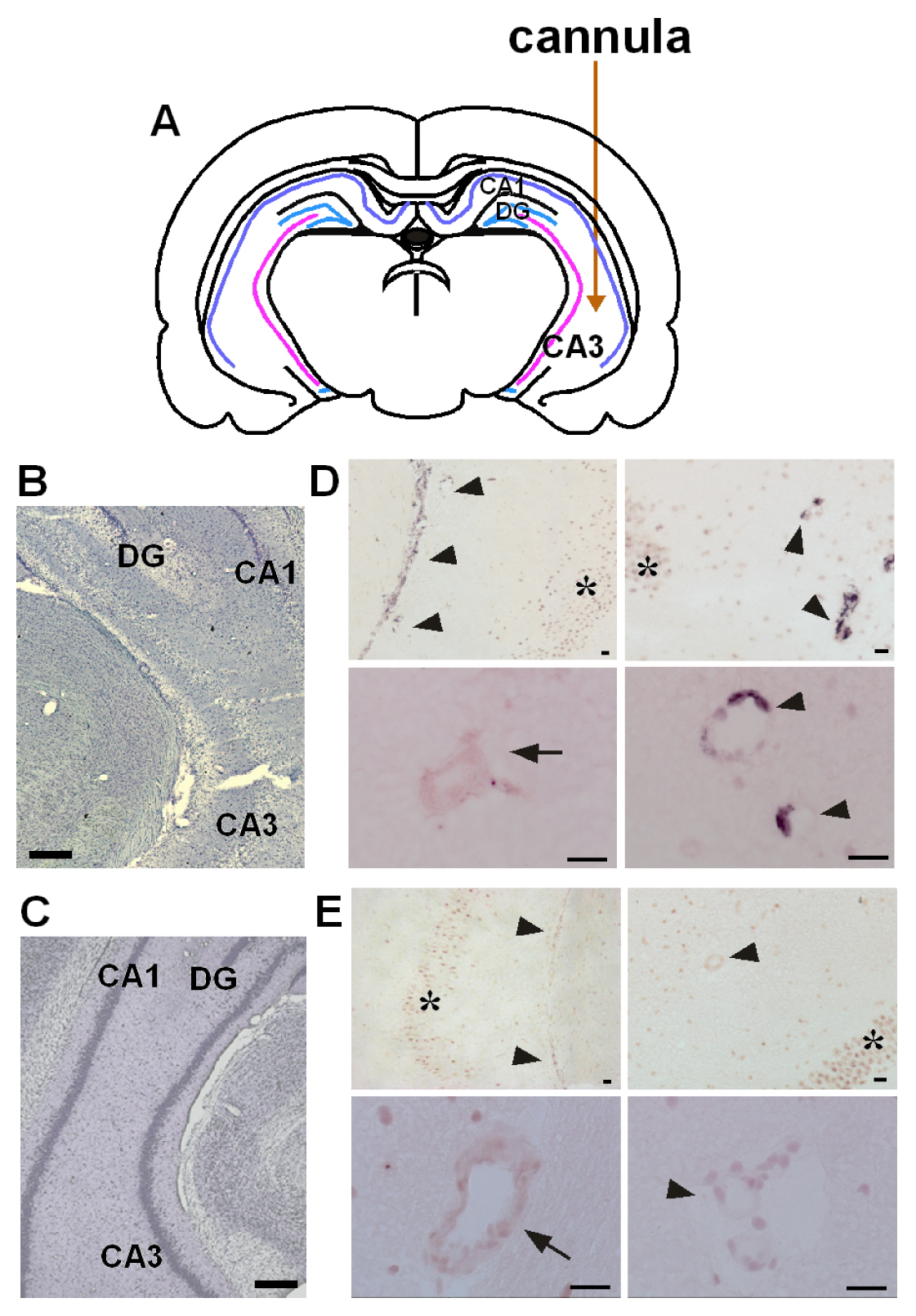
2.1. Co-Induction of COX-2 and mPGES-1 in Brain Endothelial Cells

We were the first to demonstrate that PGE2 is synthesized by mPGES-1 coupling with COX-2 in brain endothelial cells in lipopolysaccharide (LPS)-induced fever [13], and subsequent publications have confirmed that mPGES-1 is co-induced with COX-2 during fever or inflammation [1417]. Furthermore, mPGES-1 is induced in the hippocampus after epileptic seizures caused by kainic acid (KA) microinjection. KA is an analogue of the excitatory amino acid glutamate and is suited for research to investigate the mechanisms for hippocampal neuronal loss after seizures, because KA induces generalized convulsion and causes neuronal damage in the hippocampus several days after seizures [18]. Unilateral KA microinjection causes neuronal loss in the injection side, but not in the contralateral side (Figure 1A–C) [12]. Kainate induces mPGES-1 mRNA in the veins, but not in the arteries, neurons or other cells on the ipsilateral side (Figure 1D) or in any cells on the contralateral side (Figure 1E), at 8h after KA microinjection [19]. The mPGES-1 protein is localized in the structure of blood vessel, but not in other cells, such as neurons or glial cells, 24 h after KA injection (Figure 2A,B) [19]. Double-immunostaining for both mPGES-1 and von Willebrand (v.W.) factor (an endothelial cell marker) shows that mPGES-1 is induced in endothelial cells (Figure 2C). In addition, the co-induction of mPGES-1 and COX-2 in the endothelial cells was found to continue up to 48 h after the microinjection (Figure 2D) [19].

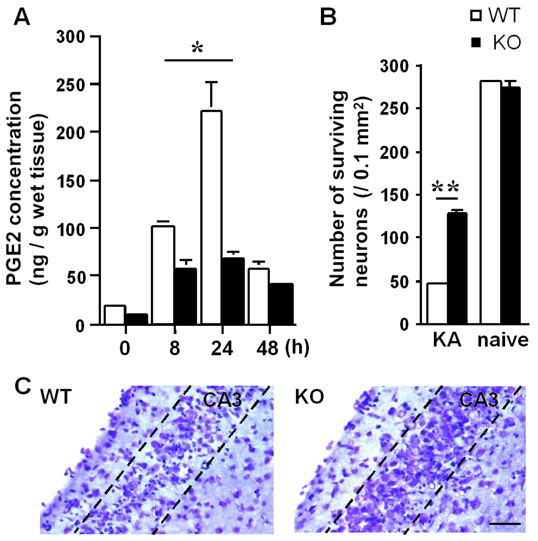
2.2. Role of Endothelial mPGES-1 in Hippocampal Neuronal Loss

We also addressed whether mPGES-1 has an effect on neuronal death after KA-induced seizure by using mPGES-1−/− mice [20]. Our results revealed that endothelial mPGES-1 is a key enzyme for the production of PGE2 (Figure 3A), which stimulates neuronal cell death (Figure 3B,C) [19]. Although it is widely thought that brain PGE2 is synthesized in and derived from astrocytes [2123], microglia [24] and neurons [25], we found that most brain PGE2, which affects neuronal death, is supplied by endothelial mPGES-1. These findings raise the question of how endothelial PGE2 regulates neuronal damage. Next, we propose a mechanism for the stimulating role of endothelial PGE2 in neuronal death and then verify our hypothesis step by step.

3. Mechanism for Exacerbation of Neuronal Damage by Endothelial mPGES-1

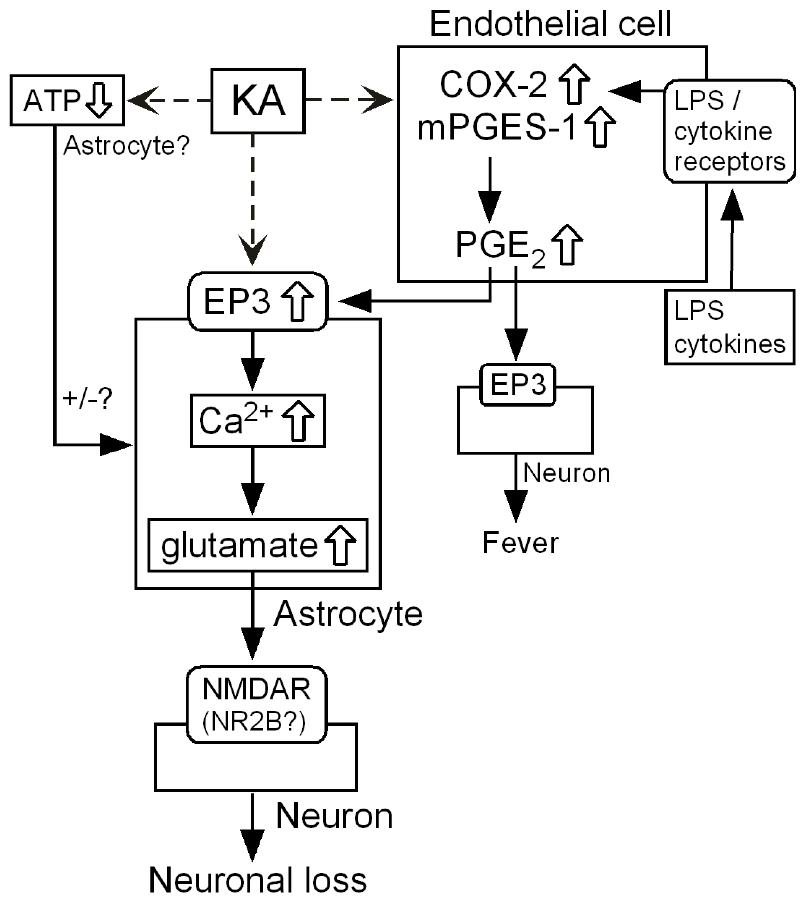
3.1. Hypothetical Mechanism for Exacerbation by mPGES-1

Brain endothelial cells are surrounded by astrocytic end-feet [26], suggesting that the PGE2 produced in endothelial cells may have a direct effect on the adjacent astrocytes. Several lines of evidence indicate that prostaglandin E receptor (EP) receptors are present on cultured astrocytes. In addition, exogenous PGE2 immediately evokes Ca2+-dependent glutamate release from astrocytes [27]; therefore, astrocytes may be activated by endogenous PGE2 to elevate the intracellular Ca2+ ([Ca2+]i) levels directly through the PGE2 receptor. Moreover, astrocytes can modulate synaptic transmission through the release of glutamate [2830], which may play a crucial role in delayed neuronal injury after seizures [31]. Taken together, we hypothesize that the endothelial PGE2 produced by mPGES-1 directly activates EP receptors on astrocytes, elevating the astrocytic [Ca2+]i levels and, subsequently, evoking sustained glutamate release, ultimately leading to neuronal damage. To investigate the mechanisms of the neuronal damage while maintaining the intercellular associations among endothelial cells, astrocytes and neurons, we used hippocampal slice culture prepared from wild-type (WT) and mPGES-1−/− mice.

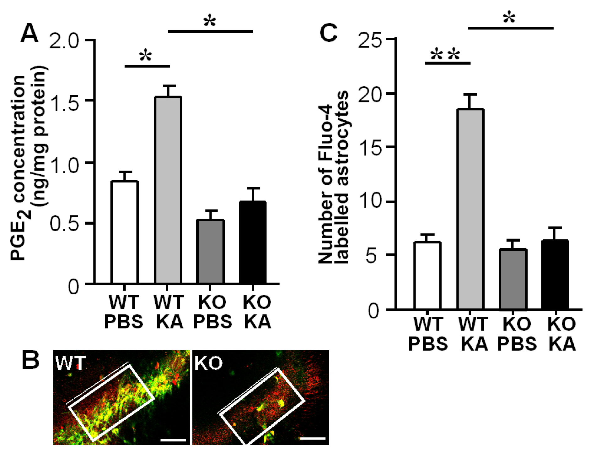
3.2. Increases in Hippocampal PGE2 Concentration and Astrocytic Ca2+ Levels after KA Treatment

First, we treated the WT hippocampal slices with KA, which significantly elevated the PGE2 concentration in the slices. This increase in PGE2 was not observed in the mPGES-1−/− slices (Figure 4A). Next, we labeled [Ca2+]i and astrocytes in the slices with Fluo-4 (Ca2+ indicator) and sulforhodamine101 (SR101; astrocyte marker), respectively (Figure 4B), and counted the number of astrocytes with elevated [Ca2+]i levels within a set rectangular area of the hippocampal CA3 region (Figure 4C). The number of these cells within the rectangular area was significantly smaller in the mPGES-1−/− slices (Figure 4D, right panel) than in the WT slices (Figure 4D, left panel and Figure 4E). These results suggest that the PGE2 derived from mPGES-1 upregulates the astrocytic [Ca2+]i levels in the hippocampus, especially in the CA3 region.

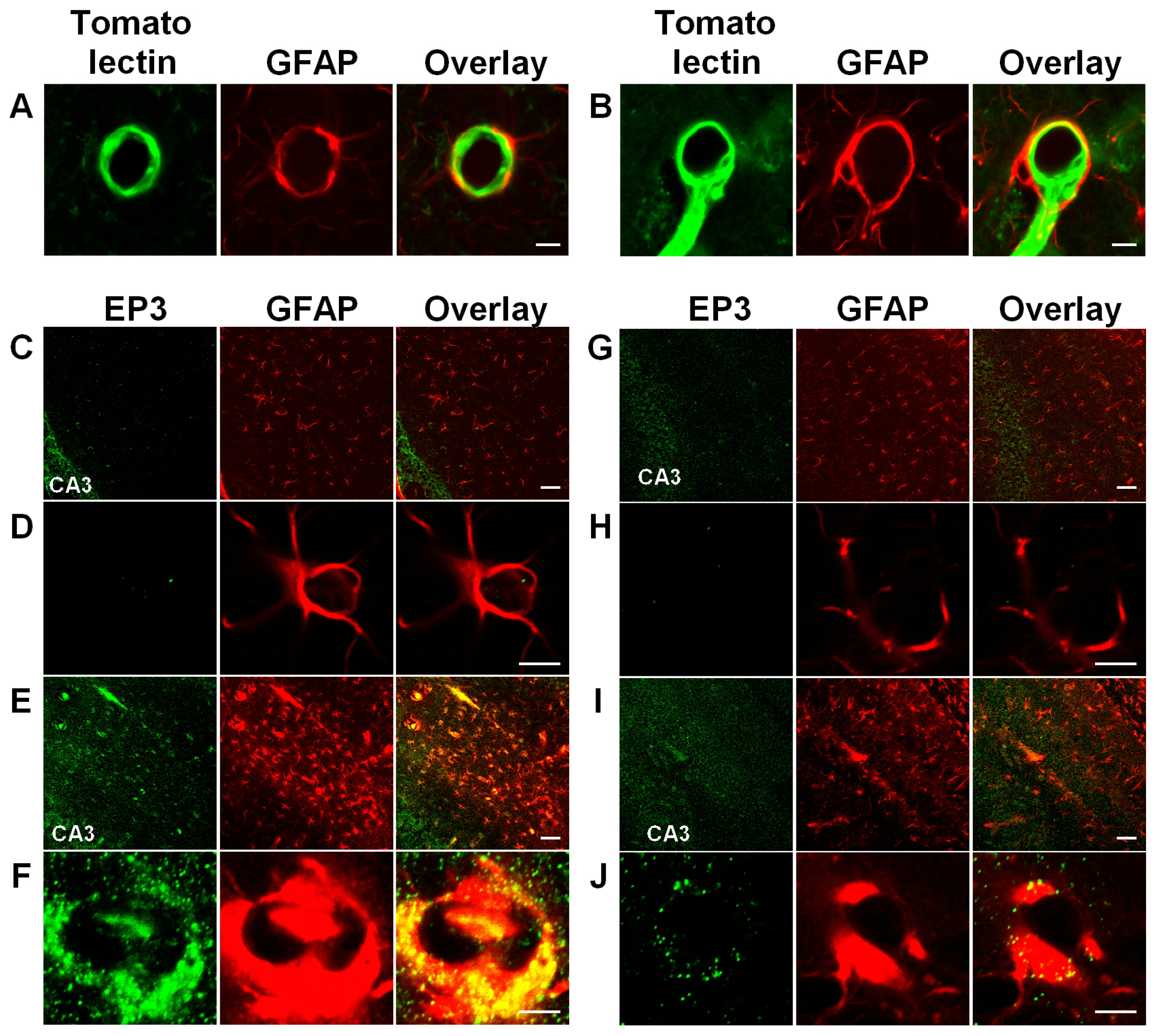
3.3. Activation of Astrocytic EP3 Receptors by KA

Astrocytic end-feet, shown by the GFAP-stained ring-like structures surround the blood vessels (Figure 5A,B), indicating that endothelial PGE2 directly binds to the PGE2 receptor on astrocytic end-feet. We identified the PGE2 receptor subtype responsible for the endothelial regulation of astrocytes. We examined the effects of each EP receptor antagonist and agonist on the number of Fluo-4-labeled astrocytes in the KA-treated WT and mPGES-1−/− slice cultures. ONO-AE3-240 (an EP3 receptor antagonist) [33] decreased the number of Fluo-4 labeled astrocytes in the KA-treated WT slices [32]. Conversely, ONO-AE-248 (an EP3 receptor agonist) [33] increased the number of Fluo-4 labeled astrocytes in the KA-treated mPGES-1−/− slices, suggesting that the EP3 receptor has a crucial role in astrocytic Ca2+ elevation [32].
EP3 immunoreactivity is rarely detected in the end-feet in naive mice (Figure 5C,D,G,H); however, it is enhanced in the WT end-feet with swelling after KA injection (Figure 5E,F). In the mPGES-1−/− mice, the end-feet also show swelling, but the EP3 immunoreactivity is not increased as much as in the WT mice (Figure 5I,J). These results indicate that the EP3 receptor is locally induced by KA in hippocampal astrocytes, which may receive PGE2 from endothelial cells. Preceding publications have already shown that EP3 mRNA is expressed in cultured astrocytes [34], and EP3 protein is induced in astrocytomas by interleukin-1β [35]. These findings indicate that astrocytic EP3 receptors may be upregulated under pathological conditions, and endothelial PGE2 may directly activate EP3 receptors on astrocytic end-feet, not distant neuronal EP receptors in neurotoxic brain diseases, such as epileptic seizures.

3.4. Enhanced Glutamate Release and Neuronal Damage by Endothelial PGE2

In addition to astrocytic [Ca2+]i elevation, we observed that the level of glutamate release was drastically enhanced in the WT slices by KA for 17 h, but not in the mPGES-1−/− slices [32]. To verify whether mPGES-1 regulates hippocampal neuronal death via glutamate release, we stained the cells with propidium iodide (PI) and then calculated the fluorescence ratio of the PI uptake (Fp/Fpi) by dividing the fluorescence within the region of interest by that of the unstained region. The results show a greater degree of PI incorporation into the CA3 region of the WT slices than into that of the mPGES-1−/− slices [32]. This significant increase in Fp/Fpi in the WT slices suggests that neuronal injury may be enhanced by mPGES-1, which controls the Ca2+-dependent glutamate release from astrocytes.
To validate the above findings on the endogenous PGE2, we added exogenous PGE2 to the mPGES-1−/− slices. The application of 5 μM PGE2 enhanced the astrocytic [Ca2+]i levels, particularly around the CA3 stratum radiatum, and also increased the number of Fluo-4 labeled astrocytes [32]. Moreover, PGE2 caused an increase in the glutamate concentration and exacerbated the neuronal damage in the CA3 region, but not in the CA1 region [32]. These results indicate that the PGE2 derived from mPGES-1 modulates KA-induced neuronal injury by elevating the astrocytic [Ca2+]i levels. Moreover, we confirmed whether exogenous PGE2 increases the [Ca2+]i levels in cultured neurons, because a [Ca2+]i increase in neurons might cause neuronal damage. We found that PGE2 could raise the [Ca2+]i levels in neurons co-cultured with astrocytes, but not without astrocytes (unpublished data). In addition, the [Ca2+]i increase in neurons was found to follow the [Ca2+]i increase in astrocytes (unpublished data). These results suggest that PGE2 indirectly increases the neuronal [Ca2+]i levels via the astrocytic [Ca2+]i increase and subsequent glutamate release. Finally, we tested whether this PGE2-evoked glutamate release from astrocytes occurs in a Ca2+-dependent manner.

3.5. Ca2+-Dependent Glutamate Release

A membrane-permeable Ca2+ chelator, BAPTA-AM, was applied to the slices. BAPTA-AM diminished the increase in the [Ca2+]i levels in the astrocytes in the WT slice and abolished the increase in glutamate concentration [32]. Moreover, BAPTA-AM partially ameliorated the neuronal damage in the CA3 region, but not in the CA1 region, suggesting that CA3 neuronal damage is locally regulated by Ca2+-dependent glutamate release from neighboring astrocytes [32].
Taken together, these results suggest that the PGE2 produced by endothelial mPGES-1 activates the astrocytic EP3 receptor to elevate the [Ca2+]i levels in astrocytes, causing Ca2+-dependent glutamate release and leading to neuronal injury (Figure 6) [32].

3.6. Intercellular Signaling among Endothelia, Astrocytes and Neurons

Accumulating evidence suggests that neuron-to-astrocyte signaling regulates arterial blood flow in the brain [22,36,37]. Conversely, there is also mounting evidence for dynamic astrocyte-to-neuron interactions, for example, astrocytes modulate synaptic transmission [2830]. These interactions are also involved in neuronal synchrony [38] and epileptic discharges [39,40], which contribute to a delayed neuronal loss after seizures [31]. In this review, we propose an advanced mechanism for excitotoxicity via vascular endothelial cell that controls astrocyte-to-neuron signaling.
We demonstrate that endothelial mPGES-1 regulates Ca2+ signaling in astrocytes and Ca2+-dependent glutamate release from astrocytes, consistent with the finding that application of exogenous PGE2 propagates astrocytic Ca2+ waves and evokes Ca2+-dependent glutamate release from astrocytes [27]. However, as the application of PGE2 alone did not increase astrocytic [Ca2+]i levels in our experimental system (data not shown), PGE2 may require another factor, such as a concomitant activation of astrocytic EP3, to elevate [Ca2+]i levels in astrocytes after KA treatment. Furthermore, it has been shown that PGE2 barely increases [Ca2+]i levels in neurons (unpublished data) and cannot evoke neuronal current directly [41], suggesting that endothelial PGE2 may specifically regulate astrocytes.
In addition to PGE2, ATP and related purine derivatives are known transmitters that are released from astrocytes, and they have been shown to depress astrocytes and neurons [42]. There is still controversy over the ATP function, because other groups claim that ATP stimulates Ca2+-dependent glutamate release from astrocytes via the P2Y1 receptor and propagates Ca2+ oscillations to neighboring astrocytes [43,44], leading to neuronal excitation. We find a reduction in the ATP concentration in the slices treated with KA for 17 h. Previous studies also showed that the systemic administration of KA causes an ATP decrease in the rat hippocampus [45,46]. Further detailed investigation is needed to infer the role of ATP in the KA-induced neurotoxicity.
Hippocampal neurons are vulnerable to glutamate, and it is thought to be mediated by N-methyl-d-aspartate (NMDA) receptors (NMDARs) [47]. In particular, glutamate release from astrocytes activates extrasynaptic NMDAR NR2B, which induces neuronal currents [31] or triggers neuronal cell death [31,48][49], suggesting that extrasynaptic NR2B receptors have important roles in the neurotoxicity caused by the glutamate released from astrocytes. Conversely, neuronal glutamate activates astrocytic mGluR5 to cause an increase in [Ca2+]i levels in astrocytes, which may in turn release glutamate and feedback to extrasynaptic NMDAR NR2B [31]. Thus, the neuron-astrocyte circuit may amplify the glutamate signaling, which aggravates neuronal excitotoxicity following seizures.
The results described here indicate that brain endothelial cells are not merely a physiological barrier between the blood and brain, but may also act as a signal transducer or amplifier. In particular, endothelial cells may be active under pathological conditions, such as in epileptic seizure. In response to such insults, endothelial cells would continuously supply a large amount of PGE2 to astrocytes, which would in turn affect neurons. The interaction among neurons, astrocytes and endothelial cells may be a key for investigating the processes of neuropathological disorders.

4. Conclusions

We find that PGE2 is synthesized by inducible mPGES-1 in cooperation with COX-2 in vascular endothelial cells in response to KA treatment. Moreover, endothelial PGE2 activates astrocytic the EP3 receptor to elevate [Ca2+]i levels in astrocytes, causing Ca2+-dependent glutamate release and, subsequently, stimulating neuronal damage. This is a first clarification of a mechanism for neuronal damage regulated by endothelial cells; therefore, this review emphasizes that brain endothelial cells act as a signal transducer or amplifier, especially, under pathological conditions, such as epileptic seizure. The analysis of the interactions among neurons, astrocytes and endothelial cells provides a better understanding of the processes of neuropathological disorders and will facilitate the development of new treatments.

Acknowledgments

This work was supported by KAKENHI (22591139) from the Japan Society for the Promotion of Science. We acknowledge Neuroscience Research (Elsevier Ltd.), the Journal of Neuroscience Research (Wiley-Liss, Inc.) and Neurochemistry International (Elsevier Ltd.) for their permission to reproduce figures from our published manuscripts.

Conflict of Interest

The authors declare no conflict of interest.

References

  1. Matsumura, K.; Watanabe, Y.; Onoe, H.; Hayaishi, O. High density of prostaglandin E2 binding sites in the anterior wall of the 3rd ventricle: A possible site of its hyperthermic action. Brain Res 1990, 533, 147–151. [Google Scholar]
  2. Takemiya, T.; Suzuki, K.; Sugiura, H.; Yasuda, S.; Yamagata, K.; Kawakami, Y.; Maru, E. Inducible brain COX-2 facilitates the recurrence of hippocampal seizures in mouse rapid kindling. Prostaglandins Other Lipid Mediat 2003, 71, 205–216. [Google Scholar]
  3. Sasaki, T.; Kitagawa, K.; Yamagata, K.; Takemiya, T.; Tanaka, S.; Omura-Matsuoka, E.; Sugiura, S.; Matsumoto, M.; Hori, M. Amelioration of hippocampal neuronal damage after transient forebrain ischemia in cyclooxygenase-2-deficient mice. J. Cereb. Blood Flow Metab 2004, 24, 107–113. [Google Scholar]
  4. Cao, C.; Matsumura, K.; Shirakawa, N.; Maeda, M.; Jikihara, I.; Kobayashi, S.; Watanabe, Y. Pyrogenic cytokines injected into the rat cerebral ventricle induce cyclooxygenase-2 in brain endothelial cells and also upregulate their receptors. Eur. J. Neurosci 2001, 13, 1781–1790. [Google Scholar]
  5. Cao, C.; Matsumura, K.; Yamagata, K.; Watanabe, Y. Endothelial cells of the rat brain vasculature express cyclooxygenase-2 mRNA in response to systemic interleukin-1 beta: A possible site of prostaglandin synthesis responsible for fever. Brain Res 1996, 733, 263–272. [Google Scholar]
  6. Cao, C.; Matsumura, K.; Yamagata, K.; Watanabe, Y. Induction by lipopolysaccharide of cyclooxygenase-2 mRNA in rat brain; its possible role in the febrile response. Brain Res 1995, 697, 187–196. [Google Scholar]
  7. Matsumura, K.; Cao, C.; Ozaki, M.; Morii, H.; Nakadate, K.; Watanabe, Y. Brain endothelial cells express cyclooxygenase-2 during lipopolysaccharide-induced fever: Light and electron microscopic immunocytochemical studies. J. Neurosci 1998, 18, 6279–6289. [Google Scholar]
  8. Yamagata, K.; Andreasson, K.I.; Kaufmann, W.E.; Barnes, C.A.; Worley, P.F. Expression of a mitogen-inducible cyclooxygenase in brain neurons: Regulation by synaptic activity and glucocorticoids. Neuron 1993, 11, 371–386. [Google Scholar]
  9. Takemiya, T.; Yamagata, K. Chapter 4. Effects of COX-2 Inhibitors on Brain Diseases. In Trends in COX-2 Inhibitor Research; Howardell, M., Ed.; Nova Science Publishers: New York, NY, USA, 2007; pp. 47–75. [Google Scholar]
  10. Takemiya, T.; Matsumura, K.; Yamagata, K. Roles of prostaglandin synthesis in excitotoxic brain diseases. Neurochem. Int 2007, 51, 112–120. [Google Scholar]
  11. Takemiya, T.; Yamagata, K. Chapter 3. The Modulatory Role of COX-2 and Prostaglandins in Brain Diseases. In Prostaglandins: New Research; Bronson, C., Ed.; Nova Science Publishers: New York, NY, USA, 2006; pp. 63–89. [Google Scholar]
  12. Takemiya, T.; Maehara, M.; Matsumura, K.; Yasuda, S.; Sugiura, H.; Yamagata, K. Prostaglandin E2 produced by late induced COX-2 stimulates hippocampal neuron loss after seizure in the CA3 region. Neurosci. Res 2006, 56, 103–110. [Google Scholar]
  13. Yamagata, K.; Matsumura, K.; Inoue, W.; Shiraki, T.; Suzuki, K.; Yasuda, S.; Sugiura, H.; Cao, C.; Watanabe, Y.; Kobayashi, S. Coexpression of microsomal-type prostaglandin E synthase with cyclooxygenase-2 in brain endothelial cells of rats during endotoxin-induced fever. J. Neurosci 2001, 21, 2669–2677. [Google Scholar]
  14. Engblom, D.; Saha, S.; Engstrom, L.; Westman, M.; Audoly, L.P.; Jakobsson, P.J.; Blomqvist, A. Microsomal prostaglandin E synthase-1 is the central switch during immune-induced pyresis. Nat. Neurosci 2003, 6, 1137–1138. [Google Scholar]
  15. Trebino, C.E.; Stock, J.L.; Gibbons, C.P.; Naiman, B.M.; Wachtmann, T.S.; Umland, J.P.; Pandher, K.; Lapointe, J.M.; Saha, S.; Roach, M.L.; et al. Impaired inflammatory and pain responses in mice lacking an inducible prostaglandin E synthase. Proc. Natl. Acad. Sci. USA 2003, 100, 9044–9049. [Google Scholar]
  16. Inoue, W.; Matsumura, K.; Yamagata, K.; Takemiya, T.; Shiraki, T.; Kobayashi, S. Brain-specific endothelial induction of prostaglandin E(2) synthesis enzymes and its temporal relation to fever. Neurosci. Res 2002, 44, 51–61. [Google Scholar]
  17. Ek, M.; Engblom, D.; Saha, S.; Blomqvist, A.; Jakobsson, P.J.; Ericsson-Dahlstrand, A. Inflammatory response: Pathway across the blood-brain barrier. Nature 2001, 410, 430–431. [Google Scholar]
  18. Cavalheiro, E.A.; Riche, D.A.; Le Gal La Salle, G. Long-term effects of intrahippocampal kainic acid injection in rats: A method for inducing spontaneous recurrent seizures. Electroencephalogr. Clin. Neurophysiol 1982, 53, 581–589. [Google Scholar]
  19. Takemiya, T.; Matsumura, K.; Sugiura, H.; Maehara, M.; Yasuda, S.; Uematsu, S.; Akira, S.; Yamagata, K. Endothelial microsomal prostaglandin E synthase-1 exacerbates neuronal loss induced by kainate. J. Neurosci. Res 2010, 88, 381–390. [Google Scholar]
  20. Uematsu, S.; Matsumoto, M.; Takeda, K.; Akira, S. Lipopolysaccharide-dependent prostaglandin E(2) production is regulated by the glutathione-dependent prostaglandin E(2) synthase gene induced by the Toll-like receptor 4/MyD88/NF-IL6 pathway. J. Immunol 2002, 168, 5811–5816. [Google Scholar]
  21. Parri, R.; Crunelli, V. An astrocyte bridge from synapse to blood flow. Nat. Neurosci 2003, 6, 5–6. [Google Scholar]
  22. Zonta, M.; Angulo, M.C.; Gobbo, S.; Rosengarten, B.; Hossmann, K.A.; Pozzan, T.; Carmignoto, G. Neuron-to-astrocyte signaling is central to the dynamic control of brain microcirculation. Nat. Neurosci 2003, 6, 43–50. [Google Scholar]
  23. Mulligan, S.J.; MacVicar, B.A. Calcium transients in astrocyte endfeet cause cerebrovascular constrictions. Nature 2004, 431, 195–199. [Google Scholar]
  24. Turrin, N.P.; Rivest, S. Innate immune reaction in response to seizures: Implications for the neuropathology associated with epilepsy. Neurobiol. Dis 2004, 16, 321–334. [Google Scholar]
  25. Ciceri, P.; Zhang, Y.; Shaffer, A.F.; Leahy, K.M.; Woerner, M.B.; Smith, W.G.; Seibert, K.; Isakson, P.C. Pharmacology of celecoxib in rat brain after kainate administration. J. Pharmacol. Exp. Ther 2002, 302, 846–852. [Google Scholar]
  26. Janzer, R.C.; Raff, M.C. Astrocytes induce blood-brain barrier properties in endothelial cells. Nature 1987, 325, 253–257. [Google Scholar]
  27. Bezzi, P.; Carmignoto, G.; Pasti, L.; Vesce, S.; Rossi, D.; Rizzini, B.L.; Pozzan, T.; Volterra, A. Prostaglandins stimulate calcium-dependent glutamate release in astrocytes. Nature 1998, 391, 281–285. [Google Scholar]
  28. Volterra, A.; Steinhauser, C. Glial modulation of synaptic transmission in the hippocampus. Glia 2004, 47, 249–257. [Google Scholar]
  29. Haydon, P.G.; Carmignoto, G. Astrocyte control of synaptic transmission and neurovascular coupling. Physiol. Rev 2006, 86, 1009–1031. [Google Scholar]
  30. Perea, G.; Araque, A. Astrocytes potentiate transmitter release at single hippocampal synapses. Science 2007, 317, 1083–1086. [Google Scholar]
  31. Ding, S.; Fellin, T.; Zhu, Y.; Lee, S.Y.; Auberson, Y.P.; Meaney, D.F.; Coulter, D.A.; Carmignoto, G.; Haydon, P.G. Enhanced astrocytic Ca2+ signals contribute to neuronal excitotoxicity after status epilepticus. J. Neurosci 2007, 27, 10674–10684. [Google Scholar]
  32. Takemiya, T.; Matsumura, K.; Sugiura, H.; Yasuda, S.; Uematsu, S.; Akira, S.; Yamagata, K. Endothelial microsomal prostaglandin E synthase-1 facilitates neurotoxicity by elevating astrocytic Ca2+ levels. Neurochem. Int 2011, 58, 489–496. [Google Scholar]
  33. Amano, H.; Hayashi, I.; Endo, H.; Kitasato, H.; Yamashina, S.; Maruyama, T.; Kobayashi, M.; Satoh, K.; Narita, M.; Sugimoto, Y.; et al. Host prostaglandin E(2)-EP3 signaling regulates tumor-associated angiogenesis and tumor growth. J. Exp. Med 2003, 197, 221–232. [Google Scholar]
  34. Kitanaka, J.; Hashimoto, H.; Gotoh, M.; Kondo, K.; Sakata, K.; Hirasawa, Y.; Sawada, M.; Suzumura, A.; Marunouchi, T.; Matsuda, T.; et al. Expression pattern of messenger RNAs for prostanoid receptors in glial cell cultures. Brain Res 1996, 707, 282–287. [Google Scholar]
  35. Waschbisch, A.; Fiebich, B.L.; Akundi, R.S.; Schmitz, M.L.; Hoozemans, J.J.; Candelario-Jalil, E.; Virtainen, N.; Veerhuis, R.; Slawik, H.; Yrjanheikki, J.; et al. Interleukin-1 beta-induced expression of the prostaglandin E-receptor subtype EP3 in U373 astrocytoma cells depends on protein kinase C and nuclear factor-kappaB. J. Neurochem 2006, 96, 680–693. [Google Scholar]
  36. Takano, T.; Tian, G.F.; Peng, W.; Lou, N.; Libionka, W.; Han, X.; Nedergaard, M. Astrocyte-mediated control of cerebral blood flow. Nat Neurosci 2006, 9, 260–267. [Google Scholar]
  37. Iadecola, C.; Nedergaard, M. Glial regulation of the cerebral microvasculature. Nat. Neurosci 2007, 10, 1369–76. [Google Scholar]
  38. Fellin, T.; Pascual, O.; Gobbo, S.; Pozzan, T.; Haydon, P.G.; Carmignoto, G. Neuronal synchrony mediated by astrocytic glutamate through activation of extrasynaptic NMDA receptors. Neuron 2004, 43, 729–743. [Google Scholar]
  39. Kang, N.; Xu, J.; Xu, Q.; Nedergaard, M.; Kang, J. Astrocytic glutamate release-induced transient depolarization and epileptiform discharges in hippocampal CA1 pyramidal neurons. J. Neurophysiol 2005, 94, 4121–4130. [Google Scholar]
  40. Tian, G.F.; Azmi, H.; Takano, T.; Xu, Q.; Peng, W.; Lin, J.; Oberheim, N.; Lou, N.; Wang, X.; Zielke, H.R.; et al. An astrocytic basis of epilepsy. Nat. Med 2005, 11, 973–981. [Google Scholar]
  41. Sanzgiri, R.P.; Araque, A.; Haydon, P.G. Prostaglandin E(2) stimulates glutamate receptor-dependent astrocyte neuromodulation in cultured hippocampal cells. J. Neurobiol 1999, 41, 221–229. [Google Scholar]
  42. Ortinau, S.; Laube, B.; Zimmermann, H. ATP inhibits NMDA receptors after heterologous expression and in cultured hippocampal neurons and attenuates NMDA-mediated neurotoxicity. J. Neurosci 2003, 23, 4996–5003. [Google Scholar]
  43. Jeremic, A.; Jeftinija, K.; Stevanovic, J.; Glavaski, A.; Jeftinija, S. ATP stimulates calcium-dependent glutamate release from cultured astrocytes. J. Neurochem 2001, 77, 664–675. [Google Scholar]
  44. Domercq, M.; Brambilla, L.; Pilati, E.; Marchaland, J.; Volterra, A.; Bezzi, P. P2Y1 receptor-evoked glutamate exocytosis from astrocytes: Control by tumor necrosis factor-alpha and prostaglandins. J. Biol. Chem 2006, 281, 30684–30696. [Google Scholar]
  45. Vezzani, A.; Sangalli, L.; Wu, H.Q.; Schwarcz, R. ATP as a marker of excitotoxin-induced nerve cell death in vivo. J. Neural. Transm 1987, 70, 349–56. [Google Scholar]
  46. Kobayashi, S.; Kikuchi, H.; Ishikawa, M.; Hashimoto, K. Regional changes of tissue pH and ATP content in rat brain following systemic administration of kainic acid. Brain Res 1990, 514, 352–354. [Google Scholar]
  47. Kambe, Y.; Nakamichi, N.; Georgiev, D.D.; Nakamura, N.; Taniura, H.; Yoneda, Y. Insensitivity to glutamate neurotoxicity mediated by NMDA receptors in association with delayed mitochondrial membrane potential disruption in cultured rat cortical neurons. J. Neurochem 2008, 105, 1886–1900. [Google Scholar]
  48. Hardingham, G.E.; Fukunaga, Y.; Bading, H. Extrasynaptic NMDARs oppose synaptic NMDARs by triggering CREB shut-off and cell death pathways. Nat. Neurosci 2002, 5, 405–414. [Google Scholar]
  49. Vizi, E.S.; Fekete, A.; Karoly, R.; Mike, A. Non-synaptic receptors and transporters involved in brain functions and targets of drug treatment. Br. J. Pharmacol 2010, 160, 785–809. [Google Scholar]
Figure 1. Neuronal injury elicited by kainic acid (KA) and microsomal prostaglandin E synthase-1 (mPGES-1) mRNA induction in the hippocampus following KA microinjection (summarized from ref. [12] and [19]). (A) Placement of cannula tips for microinjection into the hippocampal CA3 region. (B and C) KA-injected hippocampus showing marked cell loss in the ipsilateral side of the CA3 region (B), but little cell loss in the contralateral side (C). Scale bars: 400 μm. (D and E) mPGES-1 mRNA levels were increased in the veins (arrowheads), but not in the arteries (arrows), neurons (asterisks) or other cells on the ipsilateral side (D) or in any cells on the contralateral side (E) at 8 h after KA injection. Scale bars: 20 μm.
Ijms 14 08345f1
Figure 2. Induction of the mPGES-1 protein in the hippocampus following KA microinjection (summarized from ref. [19]). (A and B) Immunostaining of the mPGES-1 protein in the hippocampal CA3 region at 24 h after KA (A) or PBS (B) injection. mPGES-1 appeared in the veins (arrowheads), but not in the CA3 neurons (asterisks). (C) Double immunostaining of mPGES-1 and von Willebrand (v.W.) factor at 8 h and 48 h after KA treatment; (D) Double immunostaining for COX-2 and mPGES-1 at 24 h and 48 h after KA injection. Scale bars: 100μm.
Ijms 14 08345f2
Figure 3. Inducible mPGES-1 produces prostaglandin E2 (PGE2) and stimulates neuronal damage (summarized from ref. [19]). (A) PGE2 concentration in the hippocampi of wild-type (WT) mice (n = 6–8) and mPGES-1−/− mice (n = 6–8) following KA injection; (B) Under naive conditions, there was no significant difference in neuronal density between the WT (n = 7) and mPGES-1−/− mice (n = 6), whereas a significant difference was observed in the number of neurons between the WT (n = 7) and mPGES-1−/− mice (n = 7) at 48 h after KA microinjection; (C) Nissl staining of the hippocampal CA3 region of the WT (left) and mPGES-1−/− mice (right) at 48 h after KA microinjection. *p < 0.01, **p < 0.005, Scale bars: 100 μm.
Ijms 14 08345f3
Figure 4. KA-induced mPGES-1 produces PGE2 and increases the astrocytic Ca2+ levels, promoting glutamate release from astrocytes and causing neuronal damage (summarized from ref. [32]). (A) PGE2 concentrations in cultured slices from WT and mPGES-1−/− mice after the addition of KA or PBS for 17 h (n = 5–6); (B) Ca2+ imaging in the WT (left) and mPGES-1−/− slices (right); (C) The number of Fluo-4 labeled astrocytes within the rectangular areas in WT and mPGES-1−/− slices treated with KA or PBS (n = 5). *p < 0.001, **p < 0.0001.
Ijms 14 08345f4
Figure 5. Astrocytic prostaglandin E receptor (EP)-3 is increased after KA microinjection (reprinted from ref. [19]). (A and B) Double staining with tomato lectin and an anti-GFAP antibody in the hippocampi of naive WT (A) and mPGES-1−/− mice (B). (CJ) Double staining with anti-EP3 and anti-GFAP antibodies in the hippocampal CA3 region of naive WT mice (C,D), WT mice at 24 h after KA microinjection (E,F), naive mPGES-1−/− mice (G,H) and mPGES-1−/− mice at 24 h after KA microinjection (I, J). Scale bars: 5 μm in A,B,D,F,H,J; 50 μm in C,E,G,I.
Ijms 14 08345f5
Figure 6. Schematic diagram of the endothelial regulation of neural damage after treatment with KA.
Ijms 14 08345f6

