Review

Phospholipids in Milk Fat: Composition, Biological and Technological Significance, and Analytical Strategies

Consiglio per la Ricerca e la Sperimentazione in Agricoltura–Centro di Ricerca per le Produzioni Foraggere e Lattiero-Casearie, Via A. Lombardo, 11-26900 Lodi, Italy
*
Author to whom correspondence should be addressed.
Int. J. Mol. Sci. 2013, 14(2), 2808-2831; https://doi.org/10.3390/ijms14022808
Submission received: 20 December 2012 / Revised: 24 January 2013 / Accepted: 25 January 2013 / Published: 29 January 2013
(This article belongs to the Special Issue Phospholipids: Molecular Sciences 2012)

Abstract

:
Glycerophospholipids and sphingolipids are quantitatively the most important phospholipids (PLs) in milk. They are located on the milk fat globule membrane (MFGM) and in other membranous material of the skim milk phase. They include principally phosphatidylcholine, phosphatidylethanolamine, phosphatidylinositol and phosphatidylserine, while sphingomyelin is the dominant species of sphingolipids There is considerable evidence that PLs have beneficial health effects, such as regulation of the inflammatory reactions, chemopreventive and chemotherapeutic activity on some types of cancer, and inhibition of the cholesterol absorption. PLs show good emulsifying properties and can be used as a delivery system for liposoluble constituents. Due to the amphiphilic characteristics of these molecules, their extraction, separation and detection are critical points in the analytical approach. The extraction by using chloroform and methanol, followed by the determination by high pressure liquid chromatography (HPLC), coupled with evaporative light scattering (ELSD) or mass detector (MS), are the most applied procedures for the PL evaluation. More recently, nuclear magnetic resonance spectrometry (NMR) was also used, but despite it demonstrating high sensitivity, it requires more studies to obtain accurate results. This review is focused on milk fat phospholipids; their composition, biological activity, technological properties, and significance in the structure of milk fat. Different analytical methodologies are also discussed.

Graphical Abstract

1. Introduction

Phospholipids (PLs) are basic constituents of natural membranes; their amphiphilic properties derive from the presence of both a hydrophobic tail and a hydrophilic head. This characteristic affects their role, behavior and function. They belong to the class of polar lipids and, literally, are defined as “lipids containing phosphorus”. Polar lipids are fundamental in milk for the emulsification of fat in water, because together with proteins, they are the main constituents of the milk fat globule membrane (MFGM), which encircles the lipid droplets secreted by the mammary gland cells.
Like other biological membranes, MFGM includes, together with PLs, (glyco)proteins, glycolipids (i.e., cerebrosides and gangliosides), total and partial glycerides, free fatty acids and cholesterol [1].
The most representative MFGM proteins are Mucin 1, Mucin 15, CD36, Butyrophilin, Lactadherin, Xanthine Oxidoreductase, Adipophilin, and FABP, the last three unglycosylated.
Literature data on the composition of the MFGM are highly variable due to the different procedures applied for isolation and purification.
Studies applying laser confocal scanning microscopy provide interesting information about the structure and the lateral organization of MFGM.
MFGM is trilaminar, with a first surface-active layer, mainly consisting of proteins, surrounding the intracellular neutral lipids. This inner part is covered by a bilayer membrane deriving from the secretory cell apical plasma membrane [2]. PLs are mainly located in this outer leaflet and are organized as a liquid-disordered phase coexisting with a liquid-ordered phase (also called a lipid raft), the latter rich in sphingomyelin and cholesterol [3,4].
The interest in these molecules is high due to both the possible positive effects on human health [57] and their technological properties in the food industry as emulsifiers or emulsion stabilizers [8]. Milk and some by-products of dairy production are an interesting natural source of PLs.
In this paper, the state of the art on milk fat PLs is reviewed with respect to their structure, composition, biological activities, technological properties and analytical techniques.

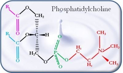
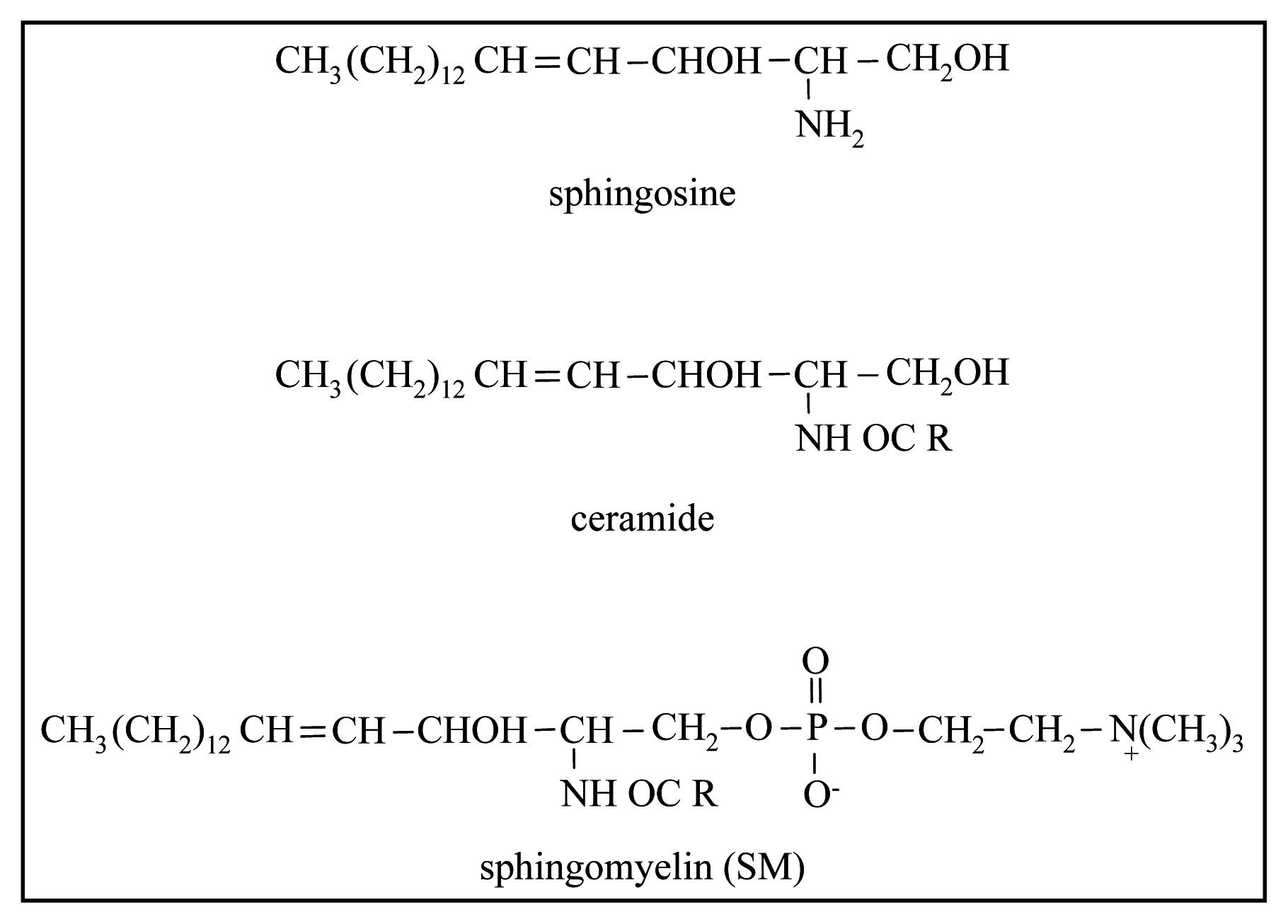
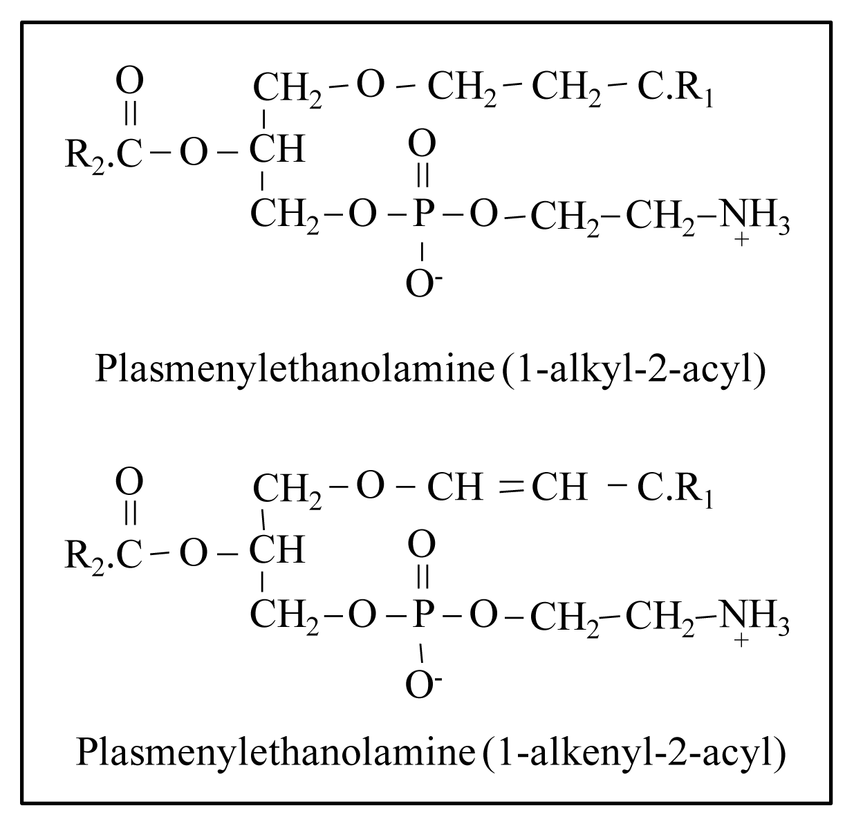
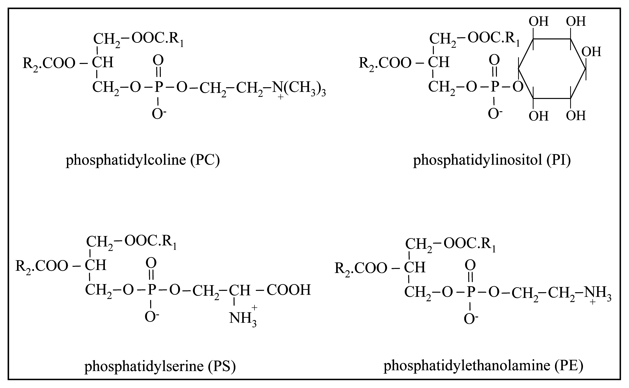
2. Nature and Characteristics of Phospholipids in Dairy Products

Glycerophospholipids and sphingolipids are the two main groups belonging to the class of PLs. Glycerophospholipids are formed by glycerol, phosphoric acid, fatty acids and a hydroxy compound (e.g., choline, ethanolamine, serine, inositol). Figure 1 shows the structure of the most important PLs of milk fat: phosphatidylcholine (PC), phosphatidylethanolamine (PE), phosphatidylinositol (PI) and phosphatidylserine (PS). The two fatty acids, mainly represented by unsaturated FAs, are esterified at the sn-1 and sn-2 positions of the glycerol backbone.
Sphingolipids are compounds containing a long chain base, the so called sphingoid base (e.g., sphingosine or phytosphingosine), fatty acids and sugars or phosphoric acid or alcohols [9]. Sphingosine is the principal sphingoid base in mammalian sphingolipids, forming a ceramide when its amino group is linked (amide bond), generally, with a saturated fatty acid. Sphingomyelin (SM) is the dominant species and it is composed of a phosphorylcholine head group linked to the ceramide (Figure 2).
Among minor PLs in milk, some authors detected lysophosphatidylethanolammine (LPE), lysophosphatidylcholine (LPC) and plasmalogens (Figure 3) [1013]. LPE and LPC derive from partial hydrolysis of PC and PE. Their origin is still uncertain though, because they could be formed as a consequence of a hydrolysis process taking place during the handling of milk (pumping and keeping it at a refrigerating temperature for a few hours). Plasmalogens are glycerophospholipids with ether-linked alkyl chain at the sn-1 position instead of the ester linked fatty acid. Some of the ether-linked phospholipids also display a cis double bond on the alkyl chain, forming a “vinyl-ether linkage” [14]. These molecules are widely distributed in many animal tissues. In particular, the interest in these compounds is growing as they are abundant in brain and human heart tissue and seem to be linked with some pathologies and with human genetic disorders [14]. In animal tissues the principal plasmalogens are usually in the PE class, less in the PC class and little or absent in the other groups. As for the role of these compounds, they are constituents of the cell membranes but the absence of a carbonyl oxygen in the sn-1 position makes their structure much more lipophilic and leads to changes to the arrangement of lipids within membranes. As a consequence, plasmalogen-containing cell membranes are less fluid than those deficient in plasmalogens [15].
Glycerophospholipids and sphingolipids are quantitatively the most important PLs in milk. They represent about 0.5%–1% of milk fat and about 60%–70% of the PL in milk are in the MFGM, placed mainly in the external bilayer membrane [3]. The origin of PLs, as most of the MFGM, is the apical plasma membrane of the mammary gland secretory cell [6]. It is hypothesized that most PLs containing choline (PC) and SM are located on the outside of the membrane, whereas PE, PS and PI are mainly placed on the inner surface of the membrane [16].
In the PLs of milk fat the short and medium chain length FAs are nearly absent and, in particular, PE is highly unsaturated, whereas PC contains more saturated FA in comparison with the other PLs [6]. Fong et al. [17] detected only a small amount of very long chain FAs (carbon atoms >20) esterified in the glycerophospholipids PE, PC, PI and PS. Completely different is the FA composition of dairy SM: the main FAs are C16:0, C18:0, C18:1n9, C22:0, C23:0 and C24:0, thus it is a highly saturated PL [12,17,18]. The particular FA composition of SM provides this molecule with the ability to form in the cellular membranes, together with cholesterol, rigid domains, called “lipid rafts”, involved in different important cellular processes. This behavior is possible because the structure has a higher melting point and degree of packing in comparison with the domains where the glycerophospholipids are present [6].
An overview of the total polar lipid content of milk, together with the detailed PL class composition, obtained by several authors, is reported in Table 1. In order to render the different papers more uniform, the total PL amount was calculated, where necessary and possible, with respect to the fat content by using the total lipid value reported in each paper. Moreover the percentage of the five main PLs was recalculated excluding the other phosphorous-based molecules, if detected. The total amount of polar lipid content ranges from 0.25 to 0.96 g/100 g of fat. This variability can be explained by differences in the extraction and analysis methods as well as by other factors, such as the diet of the animal [19] and the period of lactation [20]. The most abundant PLs in milk fat, expressed as percentage of total PLs, are PE (26.4%–72.3% of total PL), PC (8.0%–45.5% of total PL) and SM (4.1%–29.2% of total PL) followed by PI (1.4%–14.1% of total PL) and PS (2.0%–16.1% of total PL). Graves et al. [18] observed a relation with the breed regarding SM, the most interesting PL under the biological point of view. A higher content of SM was detected in milk fat from Holstein cows than Jersey, suggesting that the greater fat globule size in Jersey milk is responsible for this difference. Graves et al. [18] concluded that its content increased with the stage of lactation and during summer.
Less numerous are the studies aiming to determine the PL composition of milk of different mammalian species (Table 2). Rodriguez-Alcalà et al. [28] detected statistically significant differences among species and in particular between cow and ewe’s milk. Both Benoit et al. [32] and Garcia et al. [13] analyzed human milk and found a lower percentage of PE and a higher percentage of SM with respect to the milk of the other species. Moreover Garcia et al. [13] observed that the human milk was richer in PLs classes and, in particular, in phosphatidylethanolamine plasmalogens, with respect to the other species. The composition of PLs of donkey milk, reported by Donato et al. [30], appeared extremely different from that of milk of all the other species, particularly for the high values of PE and the low values of SM. However it is worth noting that even the values of cow milk (Table 1) were very far from those obtained by the other authors, suggesting possible problems related to the analytical method applied.
Not only are the characteristics of the raw material, but also the technological process applied, responsible for the PL content of dairy products. Being PLs mainly present in the MFGM, any treatment that produces a disruption of the membrane and/or a fractionation or separation of fat globules (e.g., centrifugation) and/or of polar and neutral components of fat, can affect the distribution and composition of PLs in the final matrix [12,24]. Thus, cream has a polar lipid content (expressed on the total lipids) lower than skimmed milk, just as butter and cheese have a lower polar lipid content with respect to buttermilk and whey (Table 3).

3. Biological Activity and Health Effects

The beneficial effects of dietary PLs concerning heart diseases, inflammation and cancer would seem to have been known since the early 1900s. Sphingolipids are abundant in the apical membrane in the absorptive epithelium in the gut [36], and their digestion products (ceramides and sphingosine) are considered as the most bioactive compounds, having important effects on cell regulation. These compounds are critical for the maintenance of membrane structure, modulating the behavior of growth factor receptors and serving as binding sites for some microorganisms, microbial toxins and viruses [37]. Ceramide is a major lipid messenger that inhibits cell proliferation and induces apoptosis, whereas sphingosine-1-phosphate is a second messenger inside the cell and there is evidence that indicates the important role of the latter molecule in regulation of cell growth, angiogenesis, immune function and lymphocyte traffic [38].
It has been estimated that the average human dietary intake of PLs is 2–8 g/day, introduced with different types of food, such as eggs, cereal grains, oilseeds, fish, beef and cow’s milk [39]. Dairy products are most likely to be the major source of sphingolipids. In the US diet, 116 g (calculated as SM), corresponding to 0.01%–0.02% w/w of the diet and 0.3–0.4 g/day, is the yearly intake per capita [37].
The oral application of dietary PLs with a specific FA composition seems able to modify the FA composition of the membrane of a certain cell type, modulating thus the cellular functions, as well as the activity of membrane bound enzymes [7].
Researches on the positive effects of PLs on membranes have been carried out in vitro, since it is very difficult, especially in humans, to perform these studies in vivo. The following is a review of the main diseases where the beneficial effects of milk PLs have been seen.

3.1. Cardiovascular Diseases

Recent studies showed that the increased consumption of milk and dairy products is associated with a reduced incidence of obesity, insulin resistance, dyslipidemia and type 2 diabetes, that are cardiovascular risk factors [40,41]. Wat et al. [42] investigated the effect of milk-derived PLs on the lipid metabolism of plasma and liver in mice, and observed that the supplementation of the high-fat diet with a phospholipid-rich dairy milk extract caused a significant decrease of the liver weight, total liver lipid, liver triglycerides and total cholesterol and serum lipids. Similar results on mice were obtained by Watanabe et al. [43]. Studies carried out by Ohlsson et al. [44,45] on humans, consisting of the supplementation of a sphingolipid-enriched dairy formulation, partially supported the findings that these molecules may affect cholesterol concentrations in TG-rich lipoproteins, but they did not find any effect on the level of plasma lipids or lipoproteins. Keller et al. [46] concluded that milk PL supplementations influenced the plasma cholesterol amount, but did not change the LDL/HDL ratio.
Nevertheless, PL human metabolism, through the group of phospholipases A2 (PLA2s) enzymes, can produce molecules that are associated with cardiovascular diseases onset. These enzymes are esterases that catalyze the hydrolysis of glycerophospholipids at the sn-2 position, producing non-esterified fatty acids and lysophospholipids. PLA2s can be cytosolic or extracellular (associated with lipoproteins: Lp-PLA2; or secreted: sPLA2) enzymes [47]. What their role is on human health can be very different, depending on the enzyme. One of the sPLA2 enzymes has a function in the digestion of dietary PLs, another in host defense against bacterial infections, but there is some research that suggests the involvement of other enzymes in promoting atherosclerosis and cancer [4749].

3.2. Inflammation and Gastrointestinal Infections

Inflammation is a response to harmful stimulus, in order to remove it and start a healing process. The mucosal surface of the digestive tract represents a barrier between a wide spectrum of potentially harmful factors [50], and polar lipids were effective in protecting cellular membranes. In a recent research by Veereman-Wauters et al. [51], the consumption of a MFGM-enriched milk by young children has seen to have a protective effect against gastrointestinal infections, producing a significant decrease in the number of short febrile episodes.
Studies have suggested an activity of PLs in regulating the inflammatory reaction. Dial et al. [52] attributed the antiulcer action of milk to the protective role of dipalmitoyl lecithin. A further study, conducted on human subjects, demonstrated that the acetylsalicylic acid (ASA) induced injury to the gastric mucosa, was markedly reduced or completely abolished if ASA was chemically associated with the PC [53].
In addition, several researches of Sprong et al. [5456] highlighted the positive effect, in vitro, of both C10:0, C12:0, unsaturated C18 fatty acids and sphingolipids against gastrointestinal infections. In particular, 100 mmol L−1 of lysophingomyelin was highly bactericidal against Campylobacter jejuni, Listeria monocitogenes and Clostridium perfringens, and moderately lowered viable counts of E. coli and Salmonella enteritidis. In addition, 100 mmol L−1 of sphingosine decreased viable counts of all pathogens tested.
PC was even found to be active in reducing the development of arthritis. Hartman et al. [57] demonstrated that exogenous PC ameliorates leukocyte-mediated signs of acute arthritis, and the outcome is comparable to that of non-steroidal anti-inflammatory drug treatment in the short term. The analgesic properties of PC were associated with reduced joint swelling, and decreased leukocyte adhesion and infiltration at the level of the synovial microcirculation. The study of Eros et al. [58] provided evidence that an increased dietary PC uptake prior to collagen-induced arthritis was associated with significantly enhanced anti-inflammatory protection. The same authors then suggested that the use of phosphatidylcholine-enriched food as a pretreatment, but not as a therapy, could exert beneficial effects on the morphological, functional and microcirculatory characteristics of chronic arthritis.

3.3. Stress Conditions

One central stress-regulating system is the hypothalamic-pituitary-adrenal axis (HPAA), which allows adaptation to stressful challenges by releasing the glucocorticoid cortisol via corticotrophin-releasing hormone and adrenocorticotropic hormone (ACTH). Stress-buffering effects of PLs have been related to dampened plasma cortisol responses to short-term exercise and mental stress [59].
Milk-based PLs have shown positive effects on individuals under chronic stress, improving the ability of the organism to adapt to the condition, increasing the cortisol availability and attenuating stress-induced memory impairments. A dose of 1% PL, administered as concentrated milk PL, protected people who were continually exposed to chronic stress with respect to both physical and mental health [60].
Hellhammer et al. [61] investigated whether the daily intake of PL concentrate rich in PS and SM had similar beneficial effects on working memory and the acute stress response. A better response with a tendency for shorter reaction times in the working memory was observed in PL-treated subjects, compared to placebo-exposed individuals. The two treatment groups did not significantly differ in their endocrine stress response. However, PL-treated subjects with a higher stress load showed a blunted psychological stress response.

3.4. Cancer

Ceramides and sphingosine, the digestion products of sphingolipids, affect cell growth, differentiation and apoptosis, suggesting that their release may have an effect on the behavior of normal or transformed cells, especially of the intestine [38].
Cancer cell membranes acquire particular properties, which vary from those found in the differentiated progenitor cells. The membrane of breast and prostate cancer cells was shown to have a higher concentration of “lipid rafts” than their normal counterpart cells, which was associated with higher apoptotic sensitivity. As above-mentioned, SM, due to its content of long chain saturated fatty acids, highly contributes to form, together with cholesterol, the rigid domains, called “lipid rafts”. Consequently, the regulation of the composition and density of lipid rafts could potentially alter cancer cell viability and metastatic behavior [7].
Non-pharmacological amounts of SM in the diet showed chemopreventive and chemotherapeutic effects on chemically induced colon cancer in mice [62].
Studies on other cell types indicate that sphingolipids may have a protective activity even against damage from γ-irradiation and chemical agents [37].
Finally, the research of Russell et al. [63] suggests that milk PLs, and in particular SM, act upon skin cells protecting them against the effect of ultraviolet radiation.
The impact of lipid components of the MFGM on human health, particularly in relation to colorectal cancer has recently been reviewed by Kuchta et al. [50].

3.5. Cholesterol Absorption

PLs also seem to have an effect on cholesterol intestinal uptake [64]. Kamili et al. [65] in a study with mice fed a high-fat diet observed that milk PLs reduce the hepatic accumulation of intestinal cholesterol and increase the fecal cholesterol excretion. In particular, the associations between SM and cholesterol may be responsible for this behavior, since the two molecules in the cellular membranes are localized in the same domain [66]. SM affects different aspects of cholesterol transport and metabolism suggesting that it may influence atherosclerosis [37]. Noh et al. [67] observed that milk SM is a more potent inhibitor of the intestinal absorption of cholesterol than egg SM and this behavior can be explained by the higher degree of saturation and longer chain length of the fatty acyl groups.

3.6. Nervous System Myelination and Neurological Development

SM and sphingolipid metabolites are fundamental components in the central nervous system of myelin sheath that surrounds the axons of some neurons. The importance of the myelination process in the development of the human brain is confirmed by the fact that it begins at 12–14 weeks of gestation and continues up to the second postnatal year [68]. A research of Oshida et al. [69] on developing rats demonstrated that dietary SM can contribute to the myelination of the central nervous system; furthermore the study of Tanaka et al. [70], carried out on premature infants, showed that the administration of SM-fortified milk to the babies had a positive association with the neurobehavioral development.
PLs are carriers of essential polyunsaturated FAs, very important molecules for the fluidity of the membrane. This function is crucial during aging because in this period the lipid composition of brain cells changes and the content of polyunsaturated n3-FAs decreases. PC has also a role as antioxidative agent in the treatment of alcohol induced brain changes and choline affects brain development and lifelong memory characteristics [7]. Finally, there are studies supporting the hypothesis that dietary PLs could contribute in the therapeutic approach to Alzheimer’s disease, even if this is still a controversial issue. Neurodegenerative diseases, such as Alzheimer’s and Parkinson’s, are caused by endoplasmic reticulum stress [71]. Nagai [72] demonstrated the protective function of milk PLs on endoplasmic reticulum stress induced neuronal cell death, and concluded that consumption of milk PLs or milk products may reduce the risk of some neurodegenerative diseases.

4. Technological Properties

The emulsifying properties of PLs are due to the simultaneous presence in the molecule of a water-loving hydrophilic head and an oil-loving hydrophobic tail. As above reported, milk PLs are mainly located in MFGM in association with protein and other polar lipids. Because of its original function in emulsifying the fat globules in whole milk, MFGM material isolated from buttermilk or cream is considered to be an efficient natural surface-active material, highly effective in lowering the interfacial tension [1].
The studies aiming to exploit the technological properties of milk PLs are thus based on the purification of MFGM from dairy by-products showing the highest concentration of MFGM fragments, particularly buttermilk and butter serum.
Buttermilk is the aqueous phase released during the churning of cream in the manufacturing of butter. This definition includes a wide range of milk fat by-products, according to the raw material used, the pre-treatment conditions and the butter making process [73]. The most common type of buttermilk is sweet buttermilk deriving from churning sweet cream into butter. However, other types of buttermilk can be produced from milk fat, such as sour buttermilk, produced from churning cultured cream, in the manufacture of European-style butter; or whey buttermilk from churning whey cream, obtained by centrifugation of whey deriving from cheese making [74].
Butter serum is a by-product of anhydrous milk fat production. The anhydrous milk fat is produced by melting and centrifuging butter (or a 75% cream can be homogenized, forcing phase inversion), resulting in pure butterfat and butter serum. The composition of butter serum is comparable to that of buttermilk, except for the fat content (2.6%–3.7%), which is much higher than buttermilk (0.5%–1.5%). The phospho- and sphingolipid contents of butter serum (0.9–1.2) are much higher than those of buttermilk (0.1–0.2) [75].
Whole buttermilk contains, together with MFGM fragments, skim milk-derived proteins (casein and whey proteins), lactose and minerals. Therefore, purification processes, mainly based on membrane filtration, are needed to separate the MFGM material. Corredig et al. [76] carried out several ultrafiltration, microfiltration and diafiltration experiments and evaluated the ratio of concentration of MFGM constituents together with the degree of purification. The authors concluded that microfiltration, through a 0.1 μm filter, of commercial buttermilk, in which casein micelles were disrupted by the addition of sodium citrate, was the most effective procedure to obtain an isolate containing a high ratio of MFGM material.
The emulsifying properties of the reconstituted microfiltered buttermilk (MF-BM: 9.3% PLs on dry matter), in comparison with those of buttermilk powder (BMP: 3.3% PLs on dry matter), skim milk powder (SM: 0.2% PLs on dry matter) and sodium caseinate (SC: PLs not determined), were evaluated by Phan et al. [77]. Mixtures prepared with of 35% soybean (Oil/Water), were homogenized at different pressures. The emulsions prepared with MF-BM and SC showed a narrower particle size distribution compared with that of emulsions prepared with BMP and SMP, and those containing MF-BM were the most stable, among the four types of emulsions.
In addition, MF-BM emulsions had very low viscosity and Newtonian-like flow characteristics. The authors confirmed that the highest concentration of PLs in MF-BM was responsible for its higher emulsifying/stabilizing activity, in comparison with the other materials.
MFGM fragments have also gained considerable attention for their ability to improve the heat stability of recombined evaporated milk emulsions [78]. Two powdered dairy by-products, sweet buttermilk (SBP) and the cream residue after the production of butter oil (CRP), were added, in different concentrations, to 16.5% (w/w) skim milk powder, 6.5% (w/w) sunflower oil and 77% of an aqueous solution with 0.02% NaN3, to obtain recombined evaporated milk emulsion samples.
The recombined samples were then heated at 121 °C for 15 min. Viscosity and particle size distribution measurements revealed that MFGM-enriched and hence PL enriched dairy by-products were able to avoid the whey protein aggregation as well as the whey protein-casein interaction. This usually occurs during the sterilization of the recombined evaporated milk. These results lead to the hypothesis that PL-enriched dairy by-products can replace exogenous ingredients, such as soybean lecithin or alternative surfactants, in recombined milk emulsions.
MFGM components can also play a role in the transport and delivery of liposoluble ingredients in the gastrointestinal tract. Bezelgues et al. [79] observed, by an in vitro digestion study with tocopherol and lycopene, that MFGM-purified fraction had a higher ability in the transfer of liposoluble molecules into bile salts micelles than other conventional emulsifying milk proteins.
Milk PLs have been successfully tested for the preparation of liposomes, PL bilayer vesicles used both for the encapsulation and controlled release of bioactive compounds and for enhancing their stability and bioavailability [80,81]. Gülseren et al. [80] applied milk PLs for the encapsulation of polyphenols with the aim to optimize the delivery of these potential bioactive molecules. Farhang et al. [81] used these molecules for the encapsulation of ascorbic acid, a very labile compound that can be used as both a vitamin supplement and an antioxidant.

5. Analytical Strategies for PL Determination

A significant effort has been made in the past decades to develop quali-quantitative methods for determining phospholipid composition of milk and dairy products.

5.1. Extraction of Fat from Milk and Dairy Products

Compared to other food, milk is a very complex matrix with a high amount of water and similar content of fat, protein and lactose. PLs interact with both lipids and proteins, due to their amphiphilic properties, and therefore particular care should be taken during fat extraction to be sure to recover the whole lipidic fraction.
The Rose Gottlieb principle, which includes a digestion with ammonia and an extraction with a mixture of diethyl and petroleum ether, and the Schmid-Bondzynski-Ratzlaff principle, which applies the same solvent mixture, but a digestion with hydrochloric acid, are the ISO official methods adopted for the quantitative extraction of fat from milk [82] and cheese [83]. This solvent mixture was not really reliable for the PL extraction [84], particularly regarding PS and PI [22]. The most applied procedure to obtain a lipidic fraction suitable for the PL determination, is based on the extraction with a solvent mixture including chloroform and methanol.
The use of this mixture arises from the studies aiming to extract PLs and SPLs from biological materials, particularly animal tissues [85,86]. Both procedures start with the addition of a chloroform/methanol mixture. The initial solvent system is binary; during the extraction process, it becomes a ternary system consisting of chloroform, methanol, and water in various proportions, depending on the moisture content of the sample. The Folch procedure [85] uses a solvent-to-sample ratio of 2:1 (v/w) with a mixture of chloroform and methanol (2:1 v/v) in a two-step extraction. The extract is then diluted with a salt solution until the phases separate and the lower phase containing lipids is collected. Bligh and Dyer [86] use a 1:1 (v/v) chloroform–methanol mixture for the first step extraction, and the ratio is then adjusted to 2:1 (v/v) in the alternate steps of extraction and washing. Moreover, in this procedure, the amount of water naturally present in the sample is specifically taken into account, and the amount of sample should be adjusted to obtain, in the first extraction step, a ternary system chloroform/methanol/water with a ratio of 1:1:0.9 (v/v/v).
Both procedures use large amounts of sample and solvents; therefore, some authors provided modifications aiming to reduce the amount of solvents and sample [87], to improve the extraction of lysophosphatides and plasmalogens [88] and to adapt the system to the dairy products [21,29,89]. Recently, the accelerated solvent technique has been applied to cheese samples to enhance the capabilities of conventional solvent extraction [90]. The extraction was carried out with ethyl acetate/cyclohexane (54:46, v/v) and methanol/ethyl acetate (1:1, v/v) and the recovery, calculated by spiking the samples with PC standards, ranged between 87.8% and 105%.

5.2. PL Separation from Lipid Matrix

Due to the low concentration of PLs in milk and dairy products (0.3–1 g/100 g of fat), except for dried butter serum and buttermilk, a second extraction step is often applied to separate PLs from the other fat constituents, mainly triglycerides.
Lipid extracts are usually fractionated by column chromatography on a preparative scale before subjecting them to detailed analysis. The solid–liquid chromatography, in which the elution of the desired lipid class is achieved by varying the polarity and strength of the mobile phase, is the most applied technique for PL separation. The traditional glass columns of diameter of few cm and heights of not more than 40 cm, are nowadays substituted by prepacked commercial columns, named SPE (solid phase extraction), which require less time, solvent, and packing material.
Table 4 reports the different types of SPE sorbents applied for the PLs purification. Both normal phase (silica and NH2) and reversed phase (C8 and C18) chromatography were used by the different authors. When different sorbents were compared [12,22,91], normal phase SPE with silica sorbent provided the most accurate results, except for Caboni et al. [91] who obtained the highest yield by using the C8 SPE.
Normal phase SPE procedure typically involves a polar analyte, a mid-to non-polar sample solvent and a polar stationary phase. Retention of PLs, under normal phase conditions, is primarily due to interactions between polar functional groups of PL and polar groups on the sorbent surface. Non polar compounds are eluted in the early step of the procedure, whereas PLs interact with the polar groups on the sorbent surface. The disruption of this binding mechanism, i.e., the elution of PLs, is obtained by applying a solvent that is more polar than that in which sample is dissolved. Exactly the opposite behavior occurs when reverse phase SPE is applied.
Thin layer chromatography (TLC) is one of the other chromatographic methods frequently used for the fractionation of complex lipid mixtures. It is based on the difference in the affinity of a component toward a stationary, generally silica gel, and a mobile phase. In comparison with SPE and HPLC, TLC is more time consuming and less quantitatively precise. Nowadays, this technique is applied principally for testing the effectiveness of other methods of separation, e.g., SPE [12], for qualitative [34] and for preparative purposes [18,26,27,32].
It can be used to separate both PLs from the whole lipidic matrix, and the single PL species.
The separation of polar lipids from neutral lipids was obtained applying a mobile phase composed of hexane/ether/acetic acid [32] or acetone [18]; the detailed separation of the single PLs was performed by using: chloroform/methanol/methylamine [26,32], or chloroform/methanol/water [12,18].
A two-dimension TLC, using chloroform/methanol/ammonia/water for the first dimension and chloroform/methanol/acetic acid/water for the second one, was adopted by Sanchez-Juanes et al. [27] to obtain pure PL constituents.

5.3. Quantification and Identification of Single PLs

5.3.1. HPLC Coupled with ELSD or MS

High performance liquid chromatography (HPLC) with evaporative light scattering detection (ELSD) is the most widely chromatographic system used for PL analysis in the dairy sector (Table 5).
ELSD is a mass flow sensitive detection method suitable for non-volatile sample components, without significant chromophore groups, in a volatile eluent. The eluent stream passes through a nebulizer into an evaporation chamber, where the solvent is evaporated to leave a mist of tiny sample particles. These scatter a light beam, and the extent of the light scattering is proportional to the amount of sample present.
To maximize the response and the linear range, particular care should be taken to adjust the flow rate of the nebulizer gas and the temperature of the evaporator chamber. Moreover, good quantitative results could be obtained if the calibration conditions were rigidly set to be the same as in the analysis of real samples.
HPLC coupled with ELSD was applied both on the PL purified fraction [22,30,91] and directly on the whole fat extracted from milk and dairy products [25,28,29].
The HPLC separation of PLs was always performed in normal phase by using a silica column, except Donato et al. [30] who used a hydrophilic interaction liquid chromatography (HILIC). It is a particular version of normal phase liquid chromatography that uses a hydrophilic stationary phase and can be performed with partially aqueous mobile phases. With respect to the traditional normal-phase chromatography, HILIC has the double advantage of using water-miscible solvents, which are compatible with electrospray ionisation detection (ESI), making on-line hyphenation to mass spectrometry (MS) detection straightforward.
An alternative HPLC detector, charge aerosol detector (CAD), based upon aerosol charging, has been recently applied by Kielbowicz et al. [31] to milk PL analysis. Its principle of operation is based on charging aerosol particles by corona discharge and subsequent measurement of the charged particles using an electrometer. CAD adopts the same mobile phase nebulization principle as ELSD but uses a charge transfer for solute detection that makes this detector more sensitive and precise than ELSD [92].
Several mobile phases were adopted, all in gradient mode. Except for Rombaut et al. [25], the other analytical procedures included more than one solvent mixture, and Rodríguez-Alcalá [28] even a quaternary gradient.
The identification by mass spectrometry detector (ESI-MS/MS) was also adopted by Gallier et al. [12] to assess the accuracy of different extraction/purification methods of PLs and to measure the effect that processing had on PL composition. In that paper the extracts from the three SPE conditions tested (Table 4), were directly introduced by continuous infusion into the ESI source on a triple-quadrupole MS/MS. This technique allowed the detection, in dairy products, of a large number of PL molecules (LPC, PC, SM, ePC, LPE, PE, PE-cer, ePE, PI, PS, PA).

5.3.2. NMR Technique

Phosphorus occurs predominantly as the isotope 31P, which has a nuclear spin value of 1/2 and is therefore visible in nuclear magnetic resonance (NMR) spectrometry. The 31P NMR method is widely used, in such diverse areas as the characterization of organic and inorganic molecular structures and the analysis of biological fluids, including milk and derivatives.
The large range of chemical shifts (about 700 ppm) reported for the 31P nucleus, its 100% natural abundance, and its high sensitivity, which is only about 15 times less than that of the proton nucleus, makes 31P NMR a reliable analytical tool to determine very low concentrations of molecules containing phosphorous [93]. Nevertheless, NMR is still an expensive instrument and requires very experienced analysts.
Several authors [11,13,23,34] applied 31P NMR to dairy products, mainly milk, to evaluate the PL constituents (Table 6).
Murgia et al. [11] identified and quantified PLs in both ewe and cow milk cream, obtained from milk centrifugation. The polar lipid fraction was then separated by applying the Folch extraction method. They compared the traditional NMR solvent mixture including chloroform/methanol/water-EDTA with a monophasic mixture composed of dimethylformamide/triethylamine/guanidinium hydrochloride. The monophasic mixture seemed to solve some of the partition problems related to the traditional biphasic system, and slightly improved the PL resolution, but caused the formation of some adducts leading to an underestimation of PE. Together with the PLs reported in Table 6, the analytical procedure applied by Murgia et al. [11] provided the qualitative detection of LPC, MMPE, and LPE.
Andreotti et al. [23] applied 31P NMR to recognize not only the PL composition of buffalo milk in comparison with cow milk, but also the composition of the other phosphorilated compounds. They applied the same monophasic mixture as Murgia et al. [11] and, together with the PL composition of the lipidic fraction extracted by the Folch method [85], analyzed both the whole milk and the defatted milk, ten fold concentrated by ultrafiltration. No significant differences were observed in the PL composition of fat fraction, between the two types of milk. Total inorganic P, glycerophosphorylethanolamine and glycerophosphorylcoline were detected by the 31P NMR analysis of whole milk; in addition, N-acetylglucosamine-1-phosphate, galactose-1-phosphate, phosphorylcholine, phosphorylethanolamine, glycerol-1-phosphate, and glucose-6-phosphate, were identified in ultrafiltered milk.
A comparison between the quantitative two-dimensional thin-layer-chromatography (2D-TLC) and 31P NMR was performed by MacKenzie et al. [34], on different types of dairy products: cream, fresh beta serum, and buttermilk protein concentrate powders. TLC method appeared more sensitive and detected a number of compounds not seen by NMR. Nevertheless, when the pH of the detergent (sodium cholate/EDTA) was raised 7.1 to 9.5, major phospholipids PC, PE, SM, DHSM, PI and PS were correctly measured by NMR. Comparable NMR results were also obtained by analyzing both the lipid fraction and the whole sample of some concentrated dairy products, providing that they have a high PL content and a good solubility in the NMR detergent.
Results on the PL composition of cow, camel, mare and human milk, analyzed by 31P NMR, were recently reported by Garcia et al. [13]. Among the parameters checked to optimize the NMR analysis, the temperature significantly affected the spectral resolution. The temperature appeared specifically correlated with the milk species, even though the authors did not provide any explanation for this behavior. The optimized method proposed by Garcia et al. [13], was suitable for determining the largest number of bioactive phospholipids, including plasmalogens.
Despite its high sensitivity and the simple pulse sequences that are used, the 31P NMR analytical technique needs more detailed studies to be accurately applied for the determination of PL composition of dairy products. Sample preparation, solvent mixtures, pH and temperature were the most critical parameters investigated by the different authors and for which new data are certainly advisable.

5.4. Determination of the FAs Bonded to PL Molecules

The FA composition of the PLs is carried out by two approaches: by GC and GC/MS of methyl esters of PLs previously purified by different techniques [26,27,32,90], and directly by LC/MS [12,17,30]. As for the application of LC/MS technique, the main results were related only to the qualitative occurrence of FAs in the PL molecules. Gallier et al. [12] discriminated the FAs only on the basis of the degree of unsaturation, whereas Donato et al. [30] and Fong et al. [17] provided more detailed indications about the type of fatty acid esterified in the PL molecules.
As far as the derivatization process is concerned, different methods can be applied [94]. Base-catalyzed methods, e.g., with sodium or potassium methoxide in methanol, can be adopted, but they do not esterify the amide-bound fatty acids as in sphingolipids. One of the most used approaches is the acid-catalyzed transmethylation by boron trifluoride in methanol. This method has the advantage that it esterifies even the free fatty acids, if present, but it has the disadvantage of being able to produce many side effects and for this reason it should be avoided. Methanolic hydrogen chloride and methanolic-sulfuric acid are considered the best general purpose esterifying agents [94].

6. Conclusions

The studies carried out on milk PLs, glycerophospholipids and sphingolipids, clearly showed that these molecules play a key role in both the nutritional and technological field.
The low amount of PLs in milk together with their amphipilic chemical structure, are responsible for the numerous efforts made by different researchers to improve the methodologies for the analytical determination of these compounds. However, milk fat extraction, PLs separation from lipidic matrix, detailed identification and precise quantification, still remain a not completely solved problem. It is not surprising that a standardized method for milk PLs determination is not yet available and it is certainly one of the main causes of both the variability of the results and the difficulties of comparison among the different researches. A reliable analytical procedure would be essential for supporting the evidence of the beneficial effects of milk PLs on the human health.
Finally, it would be advisable to enhance the development and the application of some dairy by-products, e.g., buttermilk, which, due to the high content of PLs and reduced amount of triglycerides, have potential health properties.

Abbreviations

DHSM
dihydrosphingomyelin
ELSD
evaporative light scattering detector
ePC
ether phosphatidylcholine
ePE
ether phosphatidylethanolamine
EPLAS
phosphatidylethanolamine plasmalogen
FA
fatty acid
GluCer
glucosylceramide
LacCer
lactosylceramide
LDL
low density lipoproteins: HDL, high density lipoproteins
HPLC
high performance liquid chromatography
LPA
lysophosphatidic acid
LPC
lysophosphatidylcholine
LPE
lysophosphatidylethanolamine
LPS
lysophosphatidylserine
MFGM
milk fat globule membrane
MMPE
monomethylphosphatidylethanolamine
MS
mass spectrometer
NMR
nuclear magnetic resonance
PA
phosphatidic acid
PC
phosphatidylcholine
PE
phosphatidylethanolamine
PE-cer
phosphoethanolamine-ceramide
PG
phosphatidylglycerol
PI
phosphatidylinositol
PL
phospholipid
PLA2
phospholipase A2
PS
phosphatidylserine
SM
sphingomyelin

Conflict of Interest

The authors declare no conflict of interest.

References

  1. Singh, H. The milk fat globule membrane—A biophysical system for food applications. Curr. Opin. Colloid Interface Sci 2006, 11, 154–163. [Google Scholar]
  2. Evers, J.M.; Haverkamp, R.G.; Holroyd, S.E.; Jameson, G.B.; Mackenzie, D.D.S.; McCarthy, O.J. Heterogeneity of milk fat globule membrane structure and composition as observed using fluorescence microscopy techniques. Int. Dairy J 2008, 18, 1081–1089. [Google Scholar]
  3. Gallier, S.; Gragson, D.; Cabral, C.; Jimenez-Flores, R.; Everett, D.W. Using confocal laser scanning microscopy to probe the milk fat globule membrane and associated proteins. J. Agric. Food Chem 2010, 58, 4250–4257. [Google Scholar]
  4. Lopez, C.; Madec, M.N.; Jimenez-Flores, R. Lipid rafts in the bovine milk fat globule membrane revealed by the lateral segregation of phospholipids and heterogeneous distribution of glycoproteins. Food Chem 2010, 120, 22–33. [Google Scholar]
  5. Spitsberg, V.L. Bovine milk fat globule membrane as a potential nutraceutical. J. Dairy Sci 2005, 88, 2289–2294. [Google Scholar]
  6. Dewettinck, K.; Rombaut, R.; Thienpont, N.; Le, T.T.; Messens, K.; van Camp, J. Nutritional and technological aspects of milk fat globule membrane material. Int. Dairy J 2008, 18, 436–457. [Google Scholar]
  7. Küllenberg, D.; Taylor, L.A.; Schneider, M.; Massing, U. Health effects of dietary phospholipids. Lipids Health Dis 2012, 11, 1–16. [Google Scholar]
  8. Kanno, C. Emulsifying properties of bovine milk fat globule membrane in milk fat emulsion: Conditions for the reconstitution of milk fat globules. J. Food Sci 1989, 54, 1534–1539. [Google Scholar]
  9. Beare-Rogers, J.; Dieffenbacher, A.; Holm, J.V. Lexicon of lipid nutrition. (IUPAC Technical Report). Pure Appl. Chem 2001, 73, 685–744. [Google Scholar]
  10. Hay, J.D.; Morrison, W.R. Polar lipids in bovine milk. III. Isomeric cis and trans monoenoic and dienoic fatty acids, and alkyl and alkenyl ethers in phopshatidylcholine and phopshatidylethanolammine. Biochim. Biophys. Acta 1971, 248, 71–79. [Google Scholar]
  11. Murgia, S.; Mele, S.; Monduzzi, M. Quantitative characterization of phospholipids in milk fat via 31P NMR using a monophasic solvent mixture. Lipids 2003, 38, 585–591. [Google Scholar]
  12. Gallier, S.; Gragson, D.; Cabral, C.; Jimenez-Flores, R.; Everett, D.W. Composition and fatty acid distribution of bovine milk phospholipids from processed milk products. J. Agric. Food Chem 2010, 58, 10503–10511. [Google Scholar]
  13. Garcia, C.; Lutz, N.W.; Confort-Gouny, S.; Cozzone, P.J.; Armand, M.; Bernard, M. Phospholipid fingerprints of milk from different mammalians determined by 31P NMR: Towards specific interest in human health. Food Chem 2012, 135, 1777–1783. [Google Scholar]
  14. Nagan, N.; Zoeller, R.A. Plasmalogens: Biosynthesis and functions. Prog. Lipid Res 2001, 40, 199–229. [Google Scholar]
  15. Christie, W.W. Lipid library. Available online: http://www.lipidlibrary.aocs.org accessed on 8 December 2012.
  16. Deeth, H.C. The role of phospholipids in the stability of milk fat globules. Aust. J. Dairy Technol 1997, 52, 44–46. [Google Scholar]
  17. Fong, B.Y.; Norris, C.S.; MacGibbon, A.K.H. Protein and lipid composition of bovine milk-fat globule membrane. Int. Dairy J 2007, 17, 275–288. [Google Scholar]
  18. Graves, E.L.F.; Beaulieu, A.D.; Drackley, J.K. Factors affecting the concentration of sphingomyelin in bovine milk. J. Dairy Sci 2007, 90, 706–715. [Google Scholar]
  19. Lopez, C.; Briard-Bion, V.; Menard, O.; Rousseau, F.; Pradel, P.; Besle, J.-M. Phospholipid, sphingolipid, and fatty acid composition of the milk fat globule membrane are modified by diet. J. Agric. Food Chem 2008, 56, 5226–5236. [Google Scholar]
  20. Bitman, J.; Wood, D.L. Changes in milk fat phospholipids during lactation. J. Dairy Sci 1990, 73, 1208–1216. [Google Scholar]
  21. Fagan, P.; Wijesundera, C. Liquid chromatographic analysis of milk phospholipids with on-line pre-concentration. J. Chromatogr. A 2004, 1054, 241–249. [Google Scholar]
  22. Avalli, A.; Contarini, G. Determination of phospholipids in dairy products by SPE/HPLC/ELSD. J. Chromatogr. A 2005, 1071, 185–190. [Google Scholar]
  23. Andreotti, G.; Trivellone, E.; Motta, A. Characterization of buffalo milk by 31P-nuclear magnetic resonance spectroscopy. J. Food Compos. Anal 2006, 19, 843–849. [Google Scholar]
  24. Rombaut, R.; van Camp, J.; Dewettinck, K. Phospho- and sphingolipid distribution during processing of milk, butter and whey. Int. J. Food Sci. Technol 2006, 41, 435–443. [Google Scholar]
  25. Rombaut, R.; Dewettinck, K.; van Camp, J. Phospho- and sphingolipid content of selected dairy products as determined by HPLC coupled to an evaporative light scattering detector (HPLC–ELSD). J. Food Compos. Anal 2007, 20, 308–312. [Google Scholar]
  26. Fauquant, C.; Briard-Bion, V.; Leconte, N.; Guichardant, M.; Michalski, M.C. Membrane phospholipids and sterols in microfiltered milk fat globules. Eur. J. Lipid Sci. Technol 2007, 109, 1167–1173. [Google Scholar]
  27. Sanchez-Juanes, F.; Alonso, J.M.; Zancada, L.; Hueso, P. Distribution and fatty acid content of phospholipids from bovine milk and bovine milk membranes. Int. Dairy J 2009, 19, 273–278. [Google Scholar]
  28. Rodríguez-Alcalá, L.M.; Fontecha, J. Major lipid classes separation of buttermilk, and cows, goats and ewes milk by high performance liquid chromatography with an evaporative light scattering detector focused on the phospholipid fraction. J. Chromatogr. A 2010, 1217, 3063–3066. [Google Scholar]
  29. Le, T.T.; Miocinovic, J.; Nguyen, T.M.; Rombaut, R.; van Camp, J.; Dewettinck, K. Improved solvent extraction procedure and high-performance liquid chromatography evaporative light-scattering detector method for analysis of polar lipids from dairy materials. J. Agric. Food Chem 2011, 59, 10407–10413. [Google Scholar]
  30. Donato, P.; Cacciola, F.; Cichello, F.; Russo, M.; Dugo, P.; Mondello, L. Determination of phospholipids in milk samples by means of hydrophilic interaction liquid chromatography coupled to evaporative light scattering and mass spectrometry detection. J. Chromatogr. A 2011, 1218, 6476–6482. [Google Scholar]
  31. Kielbowicz, G.; Micek, P.; Wawrzenczyk, C. A new liquid chromatography method with charge aerosol detector (CAD) for the determination of phospholipid classes. Application to milk phospholipids. Talanta 2013, 105, 28–33. [Google Scholar]
  32. Benoit, B.; Fauquant, C.; Daira, P.; Peretti, N.; Guichardant, M.; Michalski, M.-C. Phospholipid species and minor sterols in French human milks. Food Chem 2010, 120, 684–691. [Google Scholar]
  33. Lamothe, S.; Robitaille, G.; St-Gelais, D.; Britten, M. Butter making from caprine creams: Effect of washing treatment on phospholipids and milk fat globule membrane proteins distribution. J. Dairy Res 2008, 75, 439–443. [Google Scholar]
  34. MacKenzie, A.; Vyssotski, M.; Nekrasov, E. Quantitative analysis of dairy phospholipids by 31P NMR. J. Am. Oil Chem. Soc 2009, 86, 757–763. [Google Scholar]
  35. Costa, M.R.; Elias-Argote, X.E.; Jiménez-Flores, R.; Gigante, M.L. Use of ultrafiltration and supercritical fluid extraction to obtain a whey buttermilk powder enriched in milk fat globule membrane phospholipids. Int. Dairy J 2010, 20, 598–602. [Google Scholar]
  36. Danielsen, E.M.; Hansen, G.H. Lipid raft organization and function in brush borders of epithelial cells. Mol. Membr. Biol 2006, 23, 71–79. [Google Scholar]
  37. Vesper, H.; Schmelz, E.-M.; Nikolova-Karakashian, M.N.; Dillehay, D.L.; Lynch, D.V.; Merrill, A.H. Sphingolipids in food and the emerging importance of sphingolipids to nutrition. J. Nutr 1999, 129, 1239–1250. [Google Scholar]
  38. Duan, R.-D.; Nilsson, A. Metabolism of sphingolipids in the gut and its relation to inflammation and cancer development. Prog. Lipid Res 2009, 48, 62–72. [Google Scholar]
  39. Cohn, J.S.; Kamili, A.; Wat, E.; Chung, R.W.S.; Tandy, S. Dietary phospholipids and intestinal cholesterol absorption. Nutrients 2010, 2, 116–127. [Google Scholar]
  40. Pereira, M.A.; Jacobs, D.R.; van Horn, L.; Slattery, M.L.; Kartashov, A.I.; Ludwig, D.S. Dairy consumption, obesity, and the insulin resistance syndrome in young adults: The CARDIA study. JAMA 2002, 287, 2081–2089. [Google Scholar]
  41. Choi, H.K.; Willett, W.C.; Stampfer, M.J.; Rimm, E.; Hu, F.B. Dairy consumption and risk of type 2 diabetes mellitus in men: A prospective study. Arch. Intern. Med 2005, 165, 997–1003. [Google Scholar]
  42. Wat, E.; Tandy, S.; Kaper, E.; Kamili, A.; Chung, R.W.S.; Brown, A.; Rowney, M.; Cohn, J.S. Dietary phospholipid-rich dairy milk extract reduces hepatomegaly, hepatic steatosis and hyperlipidemia in mice fed a high-fat diet. Atherosclerosis 2009, 205, 144–150. [Google Scholar]
  43. Watanabe, S.; Takahashi, T.; Tanaka, L.; Haruta, Y.; Shiota, M.; Hosokawa, M.; Miyashita, K. The effect of milk polar lipids separated from butter serum on the lipid levels in the liver and the plasma of obese-model mouse (KK-Ay). J. Funct. Foods 2011, 3, 313–320. [Google Scholar]
  44. Ohlsson, L.; Burling, H.; Duan, R.-D.; Nilsson, A. Effects of a sphingolipid-enriched dairy formulation on postprandial lipid concentrations. Eur. J. Clin. Nutr 2010, 64, 1344–1349. [Google Scholar]
  45. Ohlsson, L.; Burling, H.; Nilsson, A. Long term effects on human plasma lipoproteins of a formulation entiched in butter milk polar lipid. Lipids Health Dis 2009, 8, 44. [Google Scholar]
  46. Keller, S.; Malarski, A.; Reuther, C.; Kertscher, R.; Kiehntopf, M.; Jahreis, G. Milk phospholipid and plant sterol-dependent modulation of plasma lipids in healthy volunteers. Eur. J. Nutr. 2012. [Google Scholar] [CrossRef]
  47. Mallat, Z.; Lambeau, G.; Tedgui, A. Lipoprotein-associated and secreted phospholipases a2 in cardiovascular disease: Roles as biological effectors and biomarkers. Circulation 2010, 122, 2183–2200. [Google Scholar]
  48. Lambeau, G.; Gelb, M.H. Biochemistry and physiology of mammalian secreted phospholipases A2. Annu. Rev. Biochem 2008, 77, 495–520. [Google Scholar]
  49. Hui, D.Y. Phospholipase A2 enzymes in metabolic and cardiovascular diseases. Curr. Opin. Lipidol 2012, 23, 235–240. [Google Scholar]
  50. Kuchta, A.M.; Kelly, P.M.; Stanton, C.; Devery, R.A. Milk fat globule membrane—A source of polar lipids for colon health? A review. Int. J. Dairy Technol 2012, 65, 315–333. [Google Scholar]
  51. Veereman-Wauters, G.; Staelens, S.; Rombaut, R.; Dewettinck, K.; Deboutte, D.; Brummer, R.-J.; Boone, M.; Le Ruyet, P. Milk fat globule membrane (INPULSE) enriched formula milk decreases febrile episodes and may improve behavioral regulation in young children. Nutrition 2012, 28, 749–752. [Google Scholar]
  52. Dial, E.J.; Lichtenberg, L.M. A role for milk phospholipids in protection against gastric acid. Studies in adult and suckling rats. Gastroenterology 1984, 87, 379–385. [Google Scholar]
  53. Anand, B.S.; Romero, J.J.; Sanduja, S.K.; Lichtenberger, L.M. Phospholipid association reduces the gastric mucosal toxicity of aspirin in human subjects. Am. J. Gastroenterol 1999, 94, 1818–1822. [Google Scholar]
  54. Sprong, C.; Hulstein, M.; van der Meer, R. Phospholipid rich butter milk decreases the gastro-intestinal survival and translocation of listeria in rats. Gastroenterology 1998, 114, A1090. [Google Scholar]
  55. Sprong, R.C.; Hulstein, M.F.E.; van der Meer, R. Bactericidal activities of milk lipids. Antimicrob. Agents Ch 2001, 45, 1298–1301. [Google Scholar]
  56. Sprong, R.C.; Hulstein, M.F.E.; van der Meer, R. Bovine milk fat components inhibit food-borne pathogens. Int. Dairy J 2002, 12, 209–215. [Google Scholar]
  57. Hartmann, P.; Szabó, A.; Erős, G.; Gurabi, D.; Horváth, G.; Németh, I.; Ghyczy, M.; Boros, M. Anti-inflammatory effects of phosphatidylcholine in neutrophil leukocyte-dependent acute arthritis in rats. Eur. J. Pharmacol 2009, 622, 58–64. [Google Scholar]
  58. Erős, G.; Ibrahim, S.; Siebert, N.; Boros, M.; Vollmar, B. Oral phosphatidylcholine pretreatment alleviates the signs of experimental rheumatoid arthritis. Arthritis Res. Ther 2009, 11, R43. [Google Scholar]
  59. Hellhammer, J.; Fries, E.; Buss, C.; Engert, V.; Tuch, A.; Rutenberg, D.; Hellhammer, D. Effects of soy lecithin phosphatidic acid and phosphatidylserine complex (PAS) on the endocrine and psychological responses to mental stress. Stress 2004, 7, 119–126. [Google Scholar]
  60. Schubert, M.; Contreras, C.; Franz, N.; Hellhammer, J. Milk-based phospholipids increase morning cortisol availability and improve memory in chronically stressed men. Nutr. Res 2011, 31, 413–420. [Google Scholar]
  61. Hellhammer, J.; Waladkhani, A.R.; Hero, T.; Buss, C. Effects of milk phospholipid on memory and psychological stress response. Br. Food J 2010, 112, 1124–1137. [Google Scholar]
  62. Lemonnier, L.A.; Dillehay, D.L.; Vespremi, M.J.; Abrams, J.; Brody, E.; Schmelz, E.M. Sphingomyelin in the suppression of colon tumors: Prevention versus intervention. Arch. Biochem. Biophys 2003, 419, 129–138. [Google Scholar]
  63. Russell, A.; Laubscher, A.; Jimenez-Flores, R.; Laiho, L.H. Investigating the protective properties of milk phospholipids against ultraviolet light exposure in a skin equivalent model. Proc. SPIE 2010, 7569, 75692Z:1–75692Z:9. [Google Scholar]
  64. Nilsson, A.; Duan, R.-D. Absorption and lipoprotein transport of sphingomielin. J. Lipid Res 2006, 47, 154–171. [Google Scholar]
  65. Kamili, A.; Wat, E.; Chung, R.W.S.; Tandy, S.; Weir, J.M.; Meikle, P.J.; Cohn, J.S. Hepatic accumulation of intestinal cholesterol is decreased and fecal cholesterol excretion is increased in mice fed a high-fat diet supplemented with milk phospholipids. Nutr. Metab 2010, 7, 90–101. [Google Scholar]
  66. Harder, T.; Simons, K. Caveolae, DIGs, and the dynamics of sphingolipid cholesterol microdomains. Curr. Opin. Cell Biol 1997, 9, 534–542. [Google Scholar]
  67. Noh, S.K.; Koo, S.I. Milk sphingomyelin is more effective than egg sphingomyelin in inhibiting intestinal absorption of cholesterol and fat in rats. J. Nutr 2004, 134, 2611–2616. [Google Scholar]
  68. Kinney, H.C.; Brody, B.A.; Kloman, A.S.; Gilles, F.H. Sequence of central nervous system myelination in human infancy. II. Patterns of myelination in autopsied infants. J. Neuropath. Exp. Neur 1988, 47, 217–234. [Google Scholar]
  69. Oshida, K.; Shimizu, T.; Takase, M.; Tamura, Y.; Shimizu, T.; Yamashiro, Y. Effects of dietary sphingomyelin on central nervous system myelination in developing rats. Pediatr. Res 2003, 53, 589–593. [Google Scholar]
  70. Tanaka, K.; Hosozawa, M.; Kudo, N.; Yoshikawa, N.; Hisata, K.; Shoji, H.; Shinohara, K.; Shimizu, T. The pilot study: Sphingomyelin-fortified milk has a positive association with the neurobehavioural development of very low birth weight infants during infancy, randomized control trial. Brain Dev 2012, 35, 45–52. [Google Scholar]
  71. Lindholm, D.; Wootz, H.; Korhonen, L. ER stress and neurodegenerative diseases. Cell Death Differ 2006, 13, 385–392. [Google Scholar]
  72. Nagai, K. Bovine milk phospholipid fraction protects Neuro2a cells from endoplasmic reticulum stress via PKC activation and autophagy. J. Biosci. Bioeng 2012, 114, 466–471. [Google Scholar]
  73. Vanderghem, C.; Bodson, P.; Danthine, S.; Paquot, M.; Deroanne, C.; Blecker, C. Milk fat globule membrane and buttermilks: From composition to valorization. Biotechnol. Agron. Soc 2010, 14, 485–500. [Google Scholar]
  74. Sodini, I.; Morin, P.; Olabi, A.; Jimenez-Flores, R. Compositional and functional properties of buttermilk: A comparison between sweet, sour, and whey buttermilk. J. Dairy Sci 2006, 89, 525–536. [Google Scholar]
  75. Rombaut, R.; Dejonckheere, V.; Dewettinck, K. Microfiltration of butter serum upon casein micelle destabilization. J. Dairy Sci 2006, 89, 1915–1925. [Google Scholar]
  76. Corredig, M.; Roesch, R.R.; Dalgleish, D.G. Production of a novel ingredient from buttermilk. J. Dairy Sci 2003, 86, 2744–2750. [Google Scholar]
  77. Phan, T.T.Q.; Asaduzzaman, M.; Le, T.T.; Fredrick, E.; van der Meeren, P.; Dewettinck, K. Composition and emulsifying properties of a milk fat globule membrane enriched material. Int. Dairy J 2013, 29, 99–106. [Google Scholar]
  78. Kasinos, M.; Le, T.T.; van der Meeren, P. Improved heat stability of recombined evaporated milk emulsions upon addition of phospholipid enriched dairy by-products. Food Hydrocolloids.
  79. Bezelgues, J.-B.; Morgan, F.; Palomo, G.; Crosset-Perrotin, L.; Ducret, P. Milk fat globule membrane as a potential delivery system for liposoluble nutrients. J. Dairy Sci 2009, 92, 2524–2528. [Google Scholar]
  80. Gülseren, I.; Guri, A.; Corredig, M. Encapsulation of tea polyphenols in nanoliposomes prepared with milk phospholipids and their effect on the viability of HT-29 human carcinoma cells. Food Dig 2012, 3, 36–45. [Google Scholar]
  81. Farhang, B.; Kakuda, Y.; Corredig, M. Encapsulation of ascorbic acid in liposomes prepared with milk fat globule membrane-derived phospholipids. Dairy Sci. Technol 2012, 92, 353–366. [Google Scholar]
  82. ISO 1211, Milk: Determination of Fat Content—Gravimetric Method (Reference method); International Standard ISO: Geneva, Switzerland, 2010.
  83. ISO 1735, Cheese and processed cheese products: Determination of Fat Content—Gravimetric Method (Reference method); International Standard ISO: Geneva, Switzerland, 2004.
  84. Duthie, A.H.; Patton, S. New method for extraction of milk phospholipids. J. Dairy Sci 1964, 48, 170–174. [Google Scholar]
  85. Folch, J.; Lees, M.; Sloane Stanley, G.H. A simple method for the isolation and purification of total lipids from animal tissues. J. Biol. Chem 1957, 226, 497–509. [Google Scholar]
  86. Bligh, E.G.; Dyer, W.J. A rapid method of total lipid extraction and purification. Can. J. Biochem. Phys 1959, 37, 911–917. [Google Scholar]
  87. Shahidi, F.; Wanasundara, P.K.J.P.D. Extraction and Analysis of Lipids. In Food Lipids: Chemistry, Nutrition, and Biotechnology, 3rd ed; Akho, C.C., Min, D.B., Eds.; Francis & Taylor: New York, NY, USA, 2008; pp. 125–156. [Google Scholar]
  88. Shaikh, N.A. Assessment of various techniques for the quantitative extraction of lysophospholipids from myocardial tissues. Anal. Biochem 1994, 216, 313–321. [Google Scholar]
  89. Rombaut, R.; Camp, J.V.; Dewettinck, K. Analysis of phospho-and sphingolipids in dairy products by a new hplc method. J. Dairy Sci 2005, 88, 482–488. [Google Scholar]
  90. Hauff, S.; Vetter, W. Quantification of fatty acids as methyl esters and phospholipids in cheese samples after separation of triacylglycerides and phospholipids. Anal. Chim. Acta 2009, 636, 229–235. [Google Scholar]
  91. Caboni, M.F.; Menotta, S.; Lercker, G. Separation and analysis of phospholipids in different foods with a light-scattering detector. J. Am. Oil Chem. Soc 1996, 73, 1561–1566. [Google Scholar]
  92. Godoy Ramos, R.; Libong, D.; Rakotomanga, M.; Gaudin, K.; Loiseau, P.M.; Chaminade, P. Comparison between charged aerosol detection and light scattering detection for the analysis of Leishmania membrane phospholipids. J. Chromatogr. A 2008, 1209, 88–94. [Google Scholar]
  93. Spyros, A.; Dais, P. Application of 31P NMR spectroscopy in food analysis. Quantitative determination of the mono- and diglyceride composition of olive oils. J. Agric. Food Chem 2000, 48, 802–805. [Google Scholar]
  94. Christie, W.W. Preparation of Derivatives of Fatty Acids. In Lipid Analysis, 3rd ed; The Oily Press, PJ Barnes & Associates: Bridgewater, UK, 2003; pp. 205–224. [Google Scholar]
Figure 1. Structures of the principal glycerophospholipids in milk fat.
Ijms 14 02808f1
Figure 2. Structures of the sphingomyelin and its derivatives.
Ijms 14 02808f2
Figure 3. Structures of plasmalogens subclasses of phosphatidylethanolamine (PE).
Ijms 14 02808f3
Table 1. Polar lipid content (g/100 g of fat) and phospholipids composition (percentage of total phospholipids (PLs)) of liquid milk.
ReferencePolar lipidsPEPIPSPCSMNote
[21]0.6938.6--32.229.21
[22]0.3632.39.310.527.320.5
[23]26.913.74.127.527.7
[24]0.9633.25.29.327.425.1
[25]0.746.45.37.421.119.8
[17]32.67.65.333.221.3
[26]36.47.66.532.117.3
[19]0.25–0.3026.813.616.12221.6
[27]0.4828.514.1-32.7231,2
[28]0.3638.56.57.725.921.4
[12]26.43.4242.825.5
[29]0.6936.96.16.32723.7
[30]72.31.411.587.9
[13]33.83.910.630.521.2
[31]0.65–0.8934.27.78.645.54.11
1the total amount of polar lipids was calculated considering an average fat content of cow milk of 3.5%.
2The percentage of PI includes also PS.
Table 2. Content of main PLs in milk fat of different mammalian species. Values are expressed as percentage of total PLs, applying the same calculation reported for Table 1.
ReferenceSpeciesPEPIPSPCSMNote
[23]buffalo24,519,76,624,324,9
[28]goat31.76.38.328.525.2
ewe34.44.45.228.627.4
[32]human21.316.41943.31
[30]donkey60.22.411.217.38.8
[13]mare24.38.510.627.828.9
human21.74.59.62935.2
camel34.34.910.522.128.1
1The percentage of PI includes also PS.
Table 3. Polar lipid content (g/100 g of fat) and phospholipids composition (percentage of total PLs) in milk fat of dairy products and by-products of butter-making process.
ReferenceMatrixPolar lipidsPEPIPSPCSM
[22]cream0.8642.76.87.214.628.6
butter0.23111.915.324.717.1
buttermilk4.4933.52.410.335.518.3
[33]cow cream0.17
cow buttermilk0.1738.79.39.123.918.9
cow butter serum0.8827.210.87.229.824.9
goat cream0.2
goat buttermilk0.1935.29.89.924.820.3
goat butter serum1.0127.111.78.226.226.8
[34]cream26.77.511.726.520.8
[35]cream5.6517.715.411.333.721.8
butter5.3117.715.811.533.321.8
buttermilk12.4177.18.146.121.7
[12]buttermilk8.48.24.651.227.6
Table 4. SPE conditions applied to purify PLs (the asterisk indicates the procedure that provided the most accurate results).
ReferenceMatrixSPE phaseSolvents for non polar compound elution (v/v)Solvents for PL elution (v/v)
[91]Egg powder, chicken meat, cheese, salamiSilicahexane/diethyl-ether (8:2) and (1:1)methanol and metanol/acetic acid (1% to 5%)
Aminopropyl (NH2)chloroform/isopropanol (2:1) and diethyl-ether/acetic acid (98:2)methanol
Octadecyl (C18)methanol and methanol/chloroform (4:1)methanol/water (4:1)
Octyl (C8) (*)chloroform/methanol (3:2) and chloroformmethanol
[22]milk, cream, butter, fresh buttermilkSilicahexane/diethyl-ether (8:2) and (1:1)methanol
Silica (*)hexane/diethyl-ether (8:2) and (1:1)methanol and chloroform/methanol/water (3:5:2)
Octyl (C8)chloroform/methanol (3:2) and chloroformmethanol
[90]cheeseSilica with 20% watercyclohexane/ethyl acetate (1:1)ethyl acetate/methanol (1:1), methanol and methanol/water (98:2).
[12]milk, cream, powdered buttermilkAminopropyl (NH2-bonded)chloroform/isopropanol (2:1) and diethyl-ether/acetic acid (98:2)methanol
Silica (*)hexane/diethyl-ether (1:1)methanol and chloroform/methanol/water (3:5:2)
Silicahexane/diethyl-ether (8:2) and (1:1)methanol and chloroform/methanol/water (3:5:2)
[30]milk of different speciesSilicahexane/diethyl-ether (8:2 and 1:1)methanol and chloroform/methanol/water (3:5:2)
[13]milk of different speciesSilicahexane/diethyl-ether (8:2 and 1:1)methanol and chloroform/methanol/water (3:5:2)
[31]milkSilicachloroform/methanol (95:5)methanol and chloroform/methanol/water (3:5:2)
Table 5. Analytical conditions applied to quantify and identify PLs (* HILIC = Hydrophilic interaction liquid chromatography).
ReferenceMatrixHPLC Column phaseHPLC mobile phaseELSD: temperature/pressure or flowPL identificationMolecules identified in samples
[91]Egg powder, chicken meat, cheese, salamiSilicaSolvent A: chloroform/metanol/NH4OH (80:19.5:0.5) Solvent B: chloroform/methanol/water/NH4OH (60:34:5.5:0.5)60 °C/2 atmauthentic standardsPE, PC, PI, PG, SM, LPC
[22]milk, cream, butter, fresh buttermilkSilicaSolvent A: chloroform/metanol/NH4OH (80:19.5:0.5) Solvent B: chloroform/methanol/water/NH4OH (60:34:5.5:0.5)50 °C/2.2 barauthentic standardsPE, PC, PI, PS, SM
[25]milk, cream, butter, cheese, whey, yoghurt, fermented buttermilkSilicachloroform/methanol/buffer (1M formic acid, neutralized to pH 3 with triethylamine) (87.5:12:0.5)85 °C/1.4 L/minauthentic standardsPE, PC, PI, PS, SM, GluCer, LacCer
[28]milk of different species, powdered buttermilkSilicaSolvent A: chloroform/methanol/water (1M formic acid; triethylamine) (87.5:12:0.5). Solvent B: chloroform/methanol/water (1M formic acid; triethylamine) (28:60:12). Solvent C: isooctane/tetrahydrofurane (99:1). Solvent D: 2-Propanol60 °C/3.5 barauthentic standardsPE, PC, PI, PS, SM, LacCer
[29]milk, cheese, butterSilicaSolvent A: dichloromethane Solvent B: methanol/buffer (7.2 mL acetic acid, 8.0 ml triethylamine and 118 mL HPLC water) (500:21)65 °C/2.1 L/minauthentic standardsPE, PC, PI, PS, SM, GluCer, LacCer
[30]milk of different speciesHILIC *Solvent A: acetonitrile Solvent B: acetonitrile/water (2:1)50 °C/180 KPaESI/TOFPE, PC, PI, PS, SM
[31]milkSilicaSolvent A:13% formic acid Solvent B: hexane Solvent C: 2-propanolCAD detector (see text)authentic standardsPE, PC, PI, PS, SM
Table 6. PL composition, expressed as percent on the total PLs, obtained by 31P NMR analysis.
ReferenceMatrixPCPEPIPSSMDHSMEPLASLPCLPEPA
[11]cow milk26.825.814.01.526.84.60.5
[23]cow milk24.023.512.03.624.2
[13]cow milk28.731.43.611.219.94.5
[34]cow cream26.526.77.511.720.83.91.11.8
[11]ewe milk23.927.59.43.928.36.50.5
[23]buffalo milk21.621.817.55.922.1
[13]camel milk19.330.04.39.224.66.40.9
[13]mare milk21.318.66.58.122.23.41.08.33.1
[13]human milk24.518.33.88.129.711.42.5
[34]buttermilk (lipid fraction)27.025.75.89.720.44.60.71.0
[34]buttermilk (direct analysis)26.425.87.611.516.94.60.71.0

