Review

The Quiescent Cellular State is Arf/p53-Dependent and Associated with H2AX Downregulation and Genome Stability

1
Division of Genome Stability Research, National Cancer Center Research Institute, 5-1-1 Tsukiji, Chuo-ku, Tokyo 104-0045, Japan
2
Department of Biosciences, School of Science, Kitasato University, 1-15-1 Kitasato, Minami-ku, Sagamihara 228-8555, Japan
3
Division of Cancer Development System, National Cancer Center Research Institute, 5-1-1 Tsukiji, Chuo-ku, Tokyo 104-0045, Japan
4
Department of Pathological Biochemistry, Medical Research Institute, Tokyo Medical and Dental University, 2-3-10 Kandasurugadai, Chiyoda-ku, Tokyo 101-0062, Japan
*
Author to whom correspondence should be addressed.
Int. J. Mol. Sci. 2012, 13(5), 6492-6506; https://doi.org/10.3390/ijms13056492
Submission received: 11 April 2012 / Revised: 21 May 2012 / Accepted: 22 May 2012 / Published: 24 May 2012

Abstract

:
Cancer is a disease associated with genomic instability and mutations. Excluding some tumors with specific chromosomal translocations, most cancers that develop at an advanced age are characterized by either chromosomal or microsatellite instability. However, it is still unclear how genomic instability and mutations are generated during the process of cellular transformation and how the development of genomic instability contributes to cellular transformation. Recent studies of cellular regulation and tetraploidy development have provided insights into the factors triggering cellular transformation and the regulatory mechanisms that protect chromosomes from genomic instability.

Graphical Abstract

1. Introduction

During cancer development, cells acquire immortality in association with the development of genomic instability [13] and mutations in certain genes including those of the Arf/p53 pathway [47]. Except for certain tumors associated with specific chromosomal translocations [8], such as infant leukemia and sarcoma [913], most cancers that develop at an advanced age [1422] are characterized by an unstable genome, with either chromosomal instability (CIN) or microsatellite instability (MIN) [23], and specific mutations [24] (Figure 1). Although MIN usually develops on a mismatch repair (MMR)-deficient background [2529], CIN frequently develops in the presence of a functional MMR system [23]. While cancer cells with MIN are rarely associated with aberrant chromosomes, cancer cells with CIN are characterized by a diversity of chromosomal abnormalities such as aneuploidy; chromosome-loss, -translocation, and -gain; gene amplification; and loss of heterozygosity [30,31]. Similar to the process of in vivo carcinogenesis, cells immortalized in vitro show genomic instability with either CIN or MIN and mutations in the Arf/p53 module [32]. Furthermore, CIN is inducible on a normal genetic background [33]. These in vitro findings illustrate the critical role of genomic instability and loss of Arf/p53 function in the acquisition of immortality, and raise the following critical questions: how is genomic instability induced and how does it contribute to cellular transformation? What is the role of the Arf/p53 module in cancer suppression? This review examines recent evidence regarding the oncogenic stress-induced development of tetraploidy and the role of the Arf/p53 module in suppressing cellular transformation.

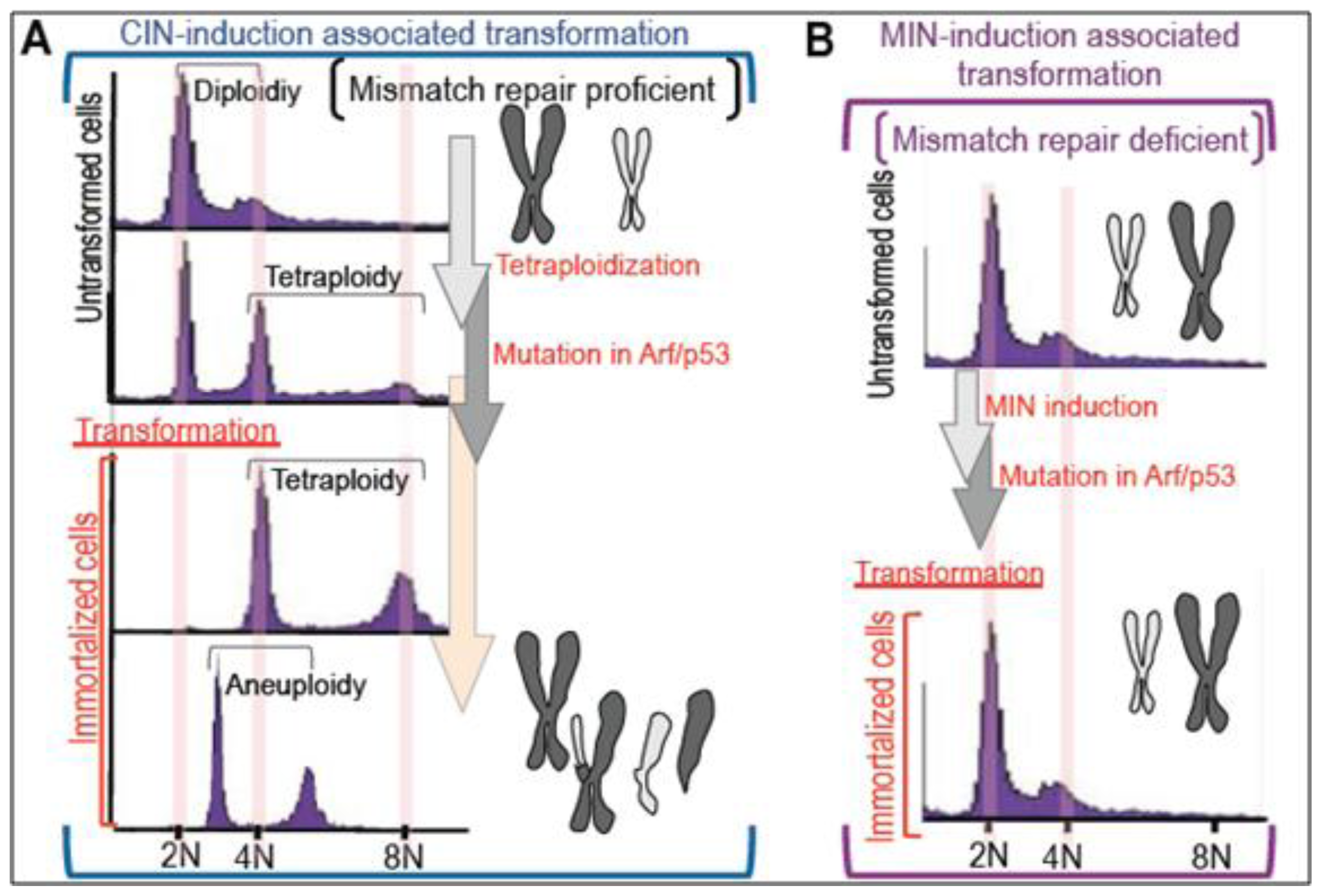
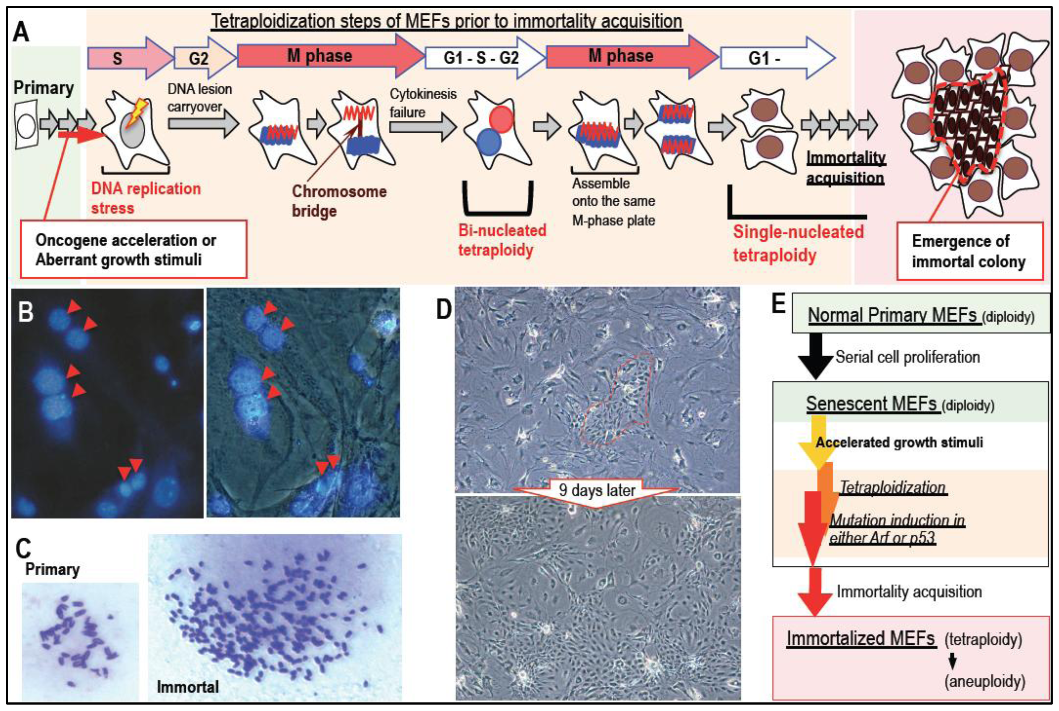
2. Massive Genomic Rearrangements during Cellular Transformation are Associated with Tetraploidization

Recent advances in DNA sequencing have helped to identify genomic rearrangements associated with tumorigenesis and have revealed the diversity of cancer cell genomes [30,3437]. Although genomic rearrangements and mutations in cancer have traditionally been thought to accumulate gradually over time, a recent report analyzed cancer cells with complex rearrangements and showed that massive genomic rearrangements can occur during a single catastrophic event [3740]. Although the exact mechanisms underlying such catastrophic events are still unclear, the accumulated findings suggest that one such event could be associated with tetraploidization. In fact, tetraploid cells have been documented in the early stages of colorectal, breast, and cervical cancer [41,42] and also precancerous lesions [43]. By contrast, the genomes of most malignant cancer cells with CIN are characterized by aneuploidy. Therefore, after massive genomic rearrangement in association with tetraploidization, transformed cells might continuously change the chromosomal status to become aneuploid [44].
Oxidative stress-induced senescence in normal mouse embryonic fibroblast cells (MEFs) [45] can lead to the acquisition of immortality [46] and mutations in the Arf/p53 module [32] as well as CIN [33], in a process analogous to that observed during cancer development. In agreement with the hypothesis that tetraploidization is one of the catastrophic events that triggers massive genomic rearrangements, MEF immortality occurs with the acquisition of tetraploidy and mutation of the Arf/p53 module [33] (Figure 1A). In addition, immortalized tetraploid MEFs eventually become aneuploid during serial cultivation [33,47], similar to the changes observed in cancer cells [44]. Identical tetraploidization and the subsequent aneuploidization were also observed in some other models [48,49]. Thus, “tetraploidy development” is likely involved in the events triggering cellular transformation and the ensuing genomic instability. Importantly, as tetraploidy is observed in the in vivo precancerous states [50], in vitro tetraploidization occurs prior to the acquisition of immortality, during a period in which MEFs rarely proliferate and inevitably exhibit accumulated γH2AX foci and a senescent appearance, i.e., a flattened and enlarged morphology [33].
Although multiple mechanisms of tetraploidy development have been reported [44], the main tetraploidization process leading to cancerous transformation is most likely a failure of chromosome-bridge-mediated cytokinesis [5153], which primarily results in bi-nucleated tetraploidy [33,54]. This is because (1) chromosome-bridge formation is associated with DNA lesions induced by oncogene acceleration and aberrant growth activation during pre-cancerous stages [55,56]; and (2) other tetraploidization processes, such as cell-to-cell fusion and mitotic slippage-mediated tetraploidization [5760] do not induce mutations and massive genomic rearrangements in a single catastrophic event (chromothripsis) [3740]; however, chromosome-bridge mediated tetraploidization does. In fact, the process of chromosome-bridge mediated tetraploidization is associated with DNA damage under a repair defective background, directly inducing aberrations in the genome. Oncogenic stress can be reproduced in vitro by oncogene activation and exogenous growth stimulation due to accelerated S phase entry and the resulting DNA replication stress [55,56]. Importantly, cells subjected to oncogenic stress develop tetraploidy [33] despite being under the opposing influences of cancer progression, reflected by senescence and apoptosis induction [61]. In agreement with the argument supporting aging-associated cancer development with CIN, many senescent cells and aging organs show persistent DNA damage [62,63].
In response to oncogenic DNA replication stress-associated lesions, cells activate damage checkpoint responses and downstream barrier reactions, such as senescence and apoptosis induction [55,56,64]. However, induced DNA lesions are not efficiently repaired and, thus, are often carried over into M phase without completion of the repair process (Figure 2A). This causes chromosome-bridge formation upon missegregation of chromosomes during mitosis, which leads to cytokinesis failure and tetraploidy development [33]. Although the induced tetraploidy is initially bi-nucleated, this is only transient because the chromosomes of two nuclei assemble on the same M phase plate and then segregate to each side, leading to the formation of two single-nucleus tetraploids at the following G1 phase [33] (Figure 2A). In fact, prior to the acquisition of immortality, senescent MEFs cultured using the 3T3 protocol [46] often show chromosome-bridge formation and accumulation of bi-nucleated tetraploid cells [33,54] (Figure 2B). Immortalized MEFs are subsequently generated, which have a mutated Arf/p53 module (either in Arf or p53) [32] in association with tetraploidy [33] (Figures 1A and 2C). In these immortalized MEFs, the loss of senescent morphology and the acquisition of primary-like phenotypes in terms of morphology and growth activity (Figure 2D) become predominant (Figure 2D). During these processes, tetraploidization occurs in rarely-growing senescent cells. In contrast, the emergence of immortality is associated with the loss of senescent characteristics; the resulting immortalized cells, therefore, gain growth activity and an altered morphology.

3. Mutation Induction during the Development of Tetraploidy

In addition to developing genomic instability, cancer cells accumulate a number of mutations [24], although only a few may be required for carcinogenesis. The mutations essential for carcinogenesis include at least the following two types: (a) tissue-specific mutations, such as mutations in the APC regulation module in the colon [65]; and (b) mutations in the Arf/p53 module, which are likely to be common mutations in malignant cancer cells [32]. Importantly, unlike the tissue specific mutations observed in cancer, the Arf/p53 module is also mutated in cells immortalized/transformed in vitro [32]. This indicates that the Arf/p53 module is involved in cellular regulatory pathways that are common to various tissues.
Although the mechanisms underlying the induction of mutations in the Arf/p53 module are still unclear, an in vitro model suggests that they are the direct consequence of tetraploidization [47] (Figure 2E). In fact, whereas immortality acquisition in normal MEFs occurs only with tetraploidy and mutations in the Arf/p53 module, p53-knockout MEFs immortalize while diploid [47]. This suggests that tetraploidization is necessary for immortalization of wild-type MEFs with mutations in the Arf/p53 module, but not for the development of immortality (as long as p53 function is lost). In addition, wild-type MEFs cannot develop immortality with genome stability [47] under conditions in which the Arf/p53 module is continuously functional. These lines of evidence indicate that tetraploidy development directly contributes to mutation induction in the Arf/p53 module, and tetraploidy develops prior to the acquisition of immortality in barely-proliferating normal MEFs.

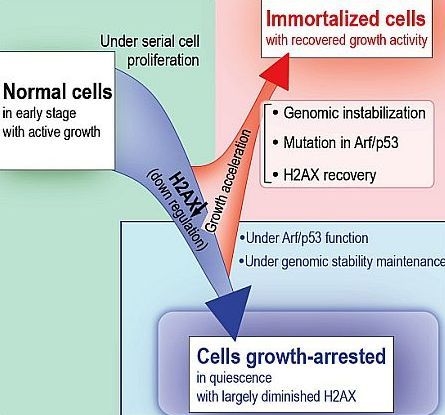
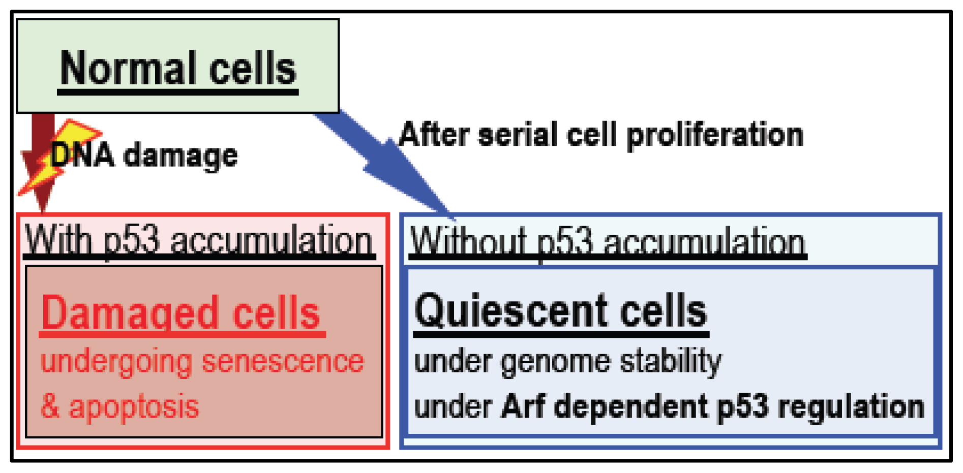
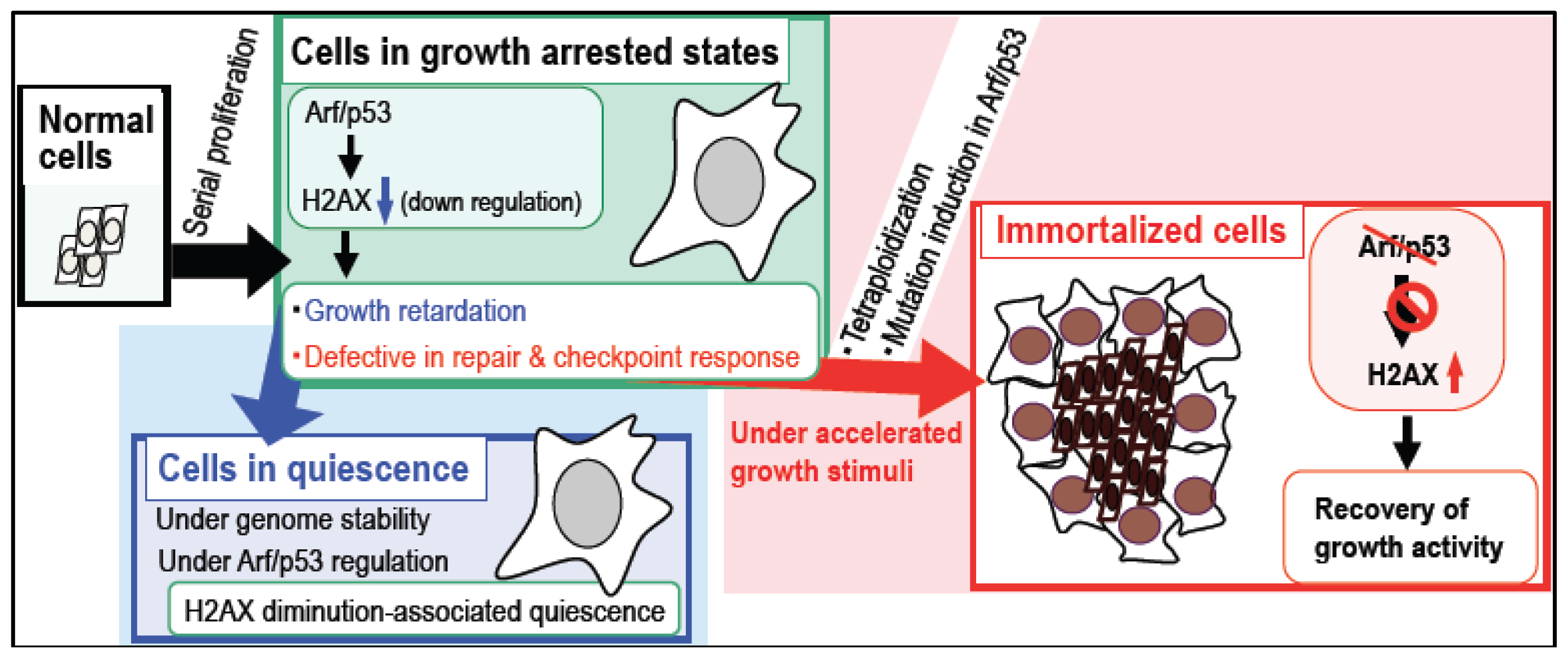
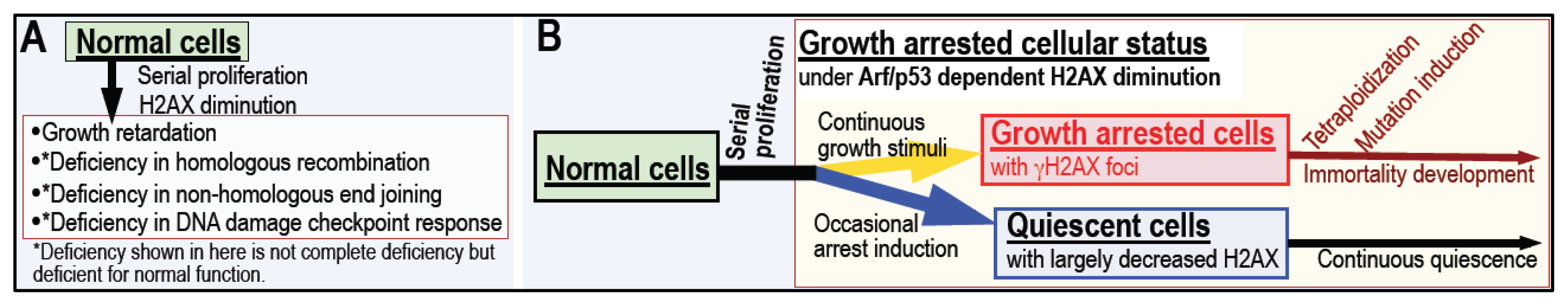
4. Arf/p53 Module-Dependent Quiescent Cellular Status

Similar to the process of development of malignant cancers, mutations in the Arf/p53 module are widely induced during immortality acquisition in vitro [32,33,47]. However, the exact role of Arf/p53 in the suppression of cellular transformation is still unclear. A recent study shows that most of the direct transcription targets of p53 are associated with the acute DNA damage response, but are not required for tumor suppression (Figure 3), suggesting two separate functions for p53 [66]. In addition, Arf and p53, the two most frequently mutated genes in cancer, are part of the same MDM2-mediated regulatory module and are mutated in a mutually exclusive manner [4]. This strongly suggests that the essential role of p53 in cancer suppression is dependent on Arf regulation, and that cells acquire immortality only in the presence of mutations in Arf and p53. By contrast, p53-dependent acute damage responses are even observed in cancer cells, indicating that cancer cells without p53 mutations are more sensitive to DNA damaging agents than p53-mutated cancer cells. Thus, unlike Arf-independent p53 activation (e.g., through checkpoint responses), the role of p53 in cancer suppression is likely to be regulated by Arf.
Because Arf and p53 are critical tumor suppressors, Arf- and p53-knockout (KO) mice show a significantly increased predisposition to cancer [67,68]. By contrast, transgenic mice with additional single gene copies of Arf and p53 are characterized by cancer suppression and an extended lifespan [32], indicating normal regulation of the Arf/p53 genes. However, unlike mice that show functional Arf and p53 regulation, transgenic mice with hyper-active p53 with no MDM2-binding site show a reduced lifespan and premature aging [6971]. In these mice, p53 is not regulated by Arf because the normal MDM2-mediated inhibition of Arf is absent. Thus, unlike its stress response-associated function, under normal conditions the Arf/p53 module functions simultaneously to extend lifespan and suppress cancer. Importantly, the two functions of p53 are distinguished by differences in p53 levels; i.e., (1) hardly-detectable levels of p53 under normal conditions are associated with extended lifespan and cancer suppression; and (2) accumulated p53 is associated with premature aging and with senescent and/or apoptotic cells [55,56], which are also characterized by p53 overexpression [72].
Arf is coded on the same gene locus as another cancer suppressor, INK4a. Therefore, mutations in Arf may affect INK4a expression. However, unlike INK4a-KO mice, Arf-KO mice show spontaneous tumor development, as do p53-KO mice [73]. Furthermore, unlike INK4a-KO MEFs, which senesce in a similar manner to wild-type MEFs, primary Arf-KO MEFs directly acquire immortality in a manner similar to p53-KO MEFs [73]. Thus, unlike INK4a, Arf (along with p53) is involved in essential cellular regulatory functions that induce growth-arrest.

5. Cellular Quiescence Is Produced with Arf/p53-Dependent H2AX Diminution

The exact contribution of the Arf/p53 module to growth arrest is unclear. We recently determined that normal cells show decreased H2AX levels after serial proliferation under the regulation of the Arf/p53 module, which contributes to growth arrest [47]. In agreement with this, cells in which H2AX was either knocked-down or knocked-out show severe growth retardation [7481]. Importantly, decreased H2AX levels are also observed in adult mouse organs, such as liver, spleen, and pancreas, in which cells rarely proliferate [47]. On the other hand, the mechanisms underlying H2AX downregulation are absent in cancer cells due to mutations in the Arf/p53 module.
Intriguingly, certain characteristics of senescent cells and growth-arrested cells may be the result of Arf/p53-dependent H2AX downregulation, because cells without H2AX show the same characteristics, including growth retardation and defects in DNA damage repair and checkpoint responses (Figure 4A) [7481]. Therefore, cells showing Arf/p53-dependent H2AX downregulation are sensitive to accelerated growth stimulation by exogenous stresses, which leads to the development of tetraploidy (Figure 2A). This includes the effects of preserving the quiescent state and the risk of developing genomic instability [47] (Figure 4B). During cell maintenance (with occasional growth-arrest), cells enter a quiescent state, in which H2AX is largely downregulated. This quiescent cellular state is preserved under the functional regulation of the Arf/p53 module and the maintenance of genome stability [47]. On the other hand, cells under continuous growth stimulation accumulate DNA replication stress-associated lesions and exhibit γH2AX because they undergo accelerated entry into S phase [47]. In addition, cells showing a reduced level of H2AX are defective in DNA damage repair and DNA damage checkpoint responses [47]. These unrepairable DNA lesions are not efficiently removed and are carried over into M phase, causing tetraploidization and, subsequently, inducing mutations in the Arf/p53 module (Figure 2A), which lead to recovery of H2AX expression and growth activity and the acquisition of immortality. Taken together, growth arrest in normal cells can be separated into two states [47]: (1) a continuously quiescent state with largely downregulated H2AX under genome stability maintenance; and (2) a state at risk of developing tetraploidy with γH2AX accumulation. Although the regulatory mechanisms that lead to the different cellular states are still unclear, our recent results demonstrate that growth stimulation is involved [47].
After serial cell proliferation, cells enter a growth-arrested state associated with Arf/p53-dependent downregulation of H2AX (Figure 5). By contrast, cells subjected to stress by oncogenes and growth stimuli are characterized by persistent exhibition of γH2AX [55,56], which is also a characteristic of senescent cells and aging organs where it is induced by a variety of stresses [1422]. However, Arf/p53-dependent downregulation of H2AX is often abrogated during the development of cancer, as well as during in vitro cellular transformation associated with mutations in the Arf/p53 module [7481].
To suppress cellular transformation, normal cells generally enter a growth-arrested state and downregulate H2AX in an Arf/p53-dependent manner; immortality is, therefore, inevitably associated with Arf/p53 mutations, which are triggered by genomic instability. The mechanism(s) underlying the involvement of the Arf/p53 module in H2AX downregulation is still unclear. H2AX is probably not the direct target of p53 because the promoter region of H2AX does not contain a p53-binding site. In addition, Arf/p53 might not be the only mechanism by which H2AX is downregulated because miR24, which reduces H2AX in terminally-differentiated blood cells [82], is unlikely to be the direct target of p53 [83]. These are some of the issues that need to be addressed in future studies.

6. Conclusions

After serial cell proliferation, normal cells eventually undergo growth arrest (Figure 5). The cells may then become quiescent; a state in which cells show largely diminished levels of H2AX under the regulation of the Arf/p53 module. However, this state is abrogated by exogenous growth stimuli, which cause accelerated entry into S phase and DNA replication stress. Because cells with reduced levels of H2AX are defective in DNA damage repair and checkpoint responses, the unrepairable DNA lesions are often carried over into M phase and induce tetraploidy. Although most of these tetraploid cells are still growth arrested and show a senescent morphology, immortalized cells will appear with the Arf/p53 module-mutation, which develops in association with tetraploidization. Thus, normal cells generally undergo quiescence when H2AX is downregulated by the Arf/p53 module under conditions of genome stability, in which cells maintain quiescence. However, in the presence of exogenous growth stimulation, the development of genomic instability (tetraploidy) and mutations in the Arf/p53 module lead to the loss of the quiescence and ultimately result in cellular transformation.
These recent findings illustrate the critical role of H2AX downregulation in the establishment of a growth-arrested cellular state. The phenotypes of the cells in this state are often expressed in association with Arf/p53-dependent downregulation of H2AX; these include deficiencies in DNA repair and checkpoint responses, and an increased risk of genomic instability. Therefore, to avoid cellular transformation, genome stability must be maintained in cells after they reach the growth-arrested state characterized by H2AX downregulation.

Acknowledgments

We are grateful to N. Takamatsu for critical discussion of the study. K.Y. is partly supported by the National Cancer Center Research and Development Fund (23-C-10).

References

  1. Lengauer, C.; Kinzler, K.W.; Vogelstein, B. Genetic instability in colorectal cancers. Nature 1997, 386, 632–637. [Google Scholar]
  2. Lengauer, C.; Kinzler, K.W.; Vogelstein, B. Genetic instabilities in human cancers. Nature 1998, 396, 643–649. [Google Scholar]
  3. Negrini, S.; Gorgoulis, V.G.; Halazonetis, T.D. Genomic instability—An evolving hallmark of cancer. Nat. Rev. Mol. Cell Biol 2010, 11, 220–228. [Google Scholar]
  4. Matheu, A.; Maraver, A.; Serrano, M. The Arf/p53 pathway in cancer and aging. Cancer Res 2008, 68, 6031–6034. [Google Scholar]
  5. Debies, M.T.; Gestl, S.A.; Mathers, J.L.; Mikse, O.R.; Leonard, T.L.; Moody, S.E.; Chodosh, L.A.; Cardiff, R.D.; Gunther, E.J. Tumor escape in a Wnt1-dependent mouse breast cancer model is enabled by p19Arf/p53 pathway lesions but not p16 Ink4a loss. J. Clin. Invest 2008, 118, 51–63. [Google Scholar]
  6. Mallakin, A.; Sugiyama, T.; Taneja, P.; Matise, L.A.; Frazier, D.P.; Choudhary, M.; Hawkins, G.A.; D’Agostino, R.B., Jr; Willingham, M.C.; Inoue, K. Mutually exclusive inactivation of DMP1 and ARF/p53 in lung cancer. Cancer Cell 2007, 12, 381–394. [Google Scholar]
  7. Peng, C.Y.; Chen, T.C.; Hung, S.P.; Chen, M.F.; Yeh, C.T.; Tsai, S.L.; Chu, C.M.; Liaw, Y.F. Genetic alterations of INK4alpha/ARF locus and p53 in human hepatocellular carcinoma. Anticancer Res 2002, 22, 1265–1271. [Google Scholar]
  8. Gale, K.B.; Ford, A.M.; Repp, R.; Borkhardt, A.; Keller, C.; Eden, O.B.; Greaves, M.F. Backtracking leukemia to birth: Identification of clonotypic gene fusion sequences in neonatal blood spots. Proc. Natl. Acad. Sci. USA 1997, 94, 13950–13954. [Google Scholar]
  9. von Levetzow, C.; Jiang, X.; Gwye, Y.; von Levetzow, G.; Hung, L.; Cooper, A.; Hsu, J.H.; Lawlor, E.R. Modeling initiation of Ewing sarcoma in human neural crest cells. PLoS One 2011, 6, e19305. [Google Scholar]
  10. Hu-Lieskovan, S.; Zhang, J.; Wu, L.; Shimada, H.; Schofield, D.E.; Triche, T.J. EWS-FLI1 fusion protein up-regulates critical genes in neural crest development and is responsible for the observed phenotype of Ewing’s family of tumors. Cancer Res 2005, 65, 4633–4644. [Google Scholar]
  11. Gmidene, A.; Frikha, R.; Sennana, H.; Elghezal, H.; Elloumi, M.; Saad, A. T(1;21;8)(p34;q22;q22): A novel variant of t(8;21) in acute myeloblastic leukemia with maturation. Med. Oncol 2011, 28, S509–S512. [Google Scholar]
  12. Nanri, T.; Matsuno, N.; Kawakita, T.; Suzushima, H.; Kawano, F.; Mitsuya, H.; Asou, N. Mutations in the receptor tyrosine kinase pathway are associated with clinical outcome in patients with acute myeloblastic leukemia harboring t(8;21)(q22;q22). Leukemia 2005, 19, 1361–1366. [Google Scholar]
  13. Kozu, T.; Fukuyama, T.; Yamami, T.; Akagi, K.; Kaneko, Y. MYND-less splice variants of AML1-MTG8 (RUNX1-CBFA2T1) are expressed in leukemia with t(8;21). Genes Chrom. Cancer 2005, 43, 45–53. [Google Scholar]
  14. Campisi, J. Senescent cells, tumor suppression, and organismal aging: Good citizens, bad neighbors. Cell 2005, 120, 513–522. [Google Scholar]
  15. Rodier, F.; Campisi, J. Four faces of cellular senescence. J. Cell Biol 2011, 192, 547–556. [Google Scholar]
  16. Davalos, A.R.; Coppe, J.P.; Campisi, J.; Desprez, P.Y. Senescent cells as a source of inflammatory factors for tumor progression. Cancer Metastasis Rev 2010, 29, 273–283. [Google Scholar]
  17. Maslov, A.Y.; Vijg, J. Genome instability, cancer and aging. Biochim. Biophys. Acta 2009, 1790, 963–969. [Google Scholar]
  18. Vijg, J.; Dolle, M.E. Genome instability: Cancer or aging? Mech. Ageing Dev 2007, 128, 466–468. [Google Scholar]
  19. Hoeijmakers, J.H. Genome maintenance mechanisms are critical for preventing cancer as well as other aging-associated diseases. Mech. Ageing Dev 2007, 128, 460–462. [Google Scholar]
  20. Chaturvedi, S.; Hass, R. Extracellular signals in young and aging breast epithelial cells and possible connections to age-associated breast cancer development. Mech. Ageing Dev 2011, 132, 213–219. [Google Scholar]
  21. Keyes, M.K.; Jang, H.; Mason, J.B.; Liu, Z.; Crott, J.W.; Smith, D.E.; Friso, S.; Choi, S.W. Older age and dietary folate are determinants of genomic and p16-specific DNA methylation in mouse colon. J. Nutr 2007, 137, 1713–1717. [Google Scholar]
  22. Pal, S.K.; Hurria, A. Impact of age, sex, and comorbidity on cancer therapy and disease progression. J. Clin. Oncol 2010, 28, 4086–4093. [Google Scholar]
  23. Lengauer, C.; Kinzler, K.W.; Vogelstein, B. Genetic instability in colorectal cancers. Nature 1997, 386, 623–627. [Google Scholar]
  24. Loeb, L.A.; Loeb, K.R.; Anderson, J.P. Multiple mutations and cancer. Proc. Natl. Acad. Sci. USA 2003, 100, 776–781. [Google Scholar]
  25. Laurent-Puig, P.; Blons, H.; Cugnenc, P.H. Sequence of molecular genetic events in colorectal tumorigenesis. Eur. J. Cancer Prev 1999, 1, S39–S47. [Google Scholar]
  26. Strauss, B.S. Frameshift mutation, microsatellites and mismatch repair. Mutat. Res 1999, 437, 195–203. [Google Scholar]
  27. Shibata, D. When does MMR loss occur during HNPCC progression? Cancer Biomark 2006, 2, 29–35. [Google Scholar]
  28. Pal, T.; Permuth-Wey, J.; Kumar, A.; Sellers, T.A. Systematic review and meta-analysis of ovarian cancers: Estimation of microsatellite-high frequency and characterization of mismatch repair deficient tumor histology. Clin. Cancer Res 2008, 14, 6847–6854. [Google Scholar]
  29. Shah, S.N.; Hile, S.E.; Eckert, K.A. Defective mismatch repair, microsatellite mutation bias, and variability in clinical cancer phenotypes. Cancer Res 2010, 70, 431–435. [Google Scholar]
  30. Stephens, P.J.; McBride, D.J.; Lin, M.L.; Varela, I.; Pleasance, E.D.; Simpson, J.T.; Stebbings, L.A.; Leroy, C.; Edkins, S.; Mudie, L.J.; et al. Complex landscapes of somatic rearrangement in human breast cancer genomes. Nature 2009, 462, 1005–1010. [Google Scholar]
  31. Weigman, V.J.; Chao, H.H.; Shabalin, A.A.; He, X.; Parker, J.S.; Nordgard, S.H.; Grushko, T.; Huo, D.; Nwachukwu, C.; Nobel, A.; et al. Basal-like Breast cancer DNA copy number losses identify genes involved in genomic instability, response to therapy, and patient survival. Breast Cancer Res.Treat 2011. [Google Scholar] [CrossRef]
  32. Matheu, A.; Maraver, A.; Klatt, P.; Flores, I.; Garcia-Cao, I.; Borras, C.; Flores, J.M.; Vina, J.; Blasco, M.A.; Serrano, M. Delayed ageing through damage protection by the Arf/p53 pathway. Nature 2007, 448, 375–379. [Google Scholar]
  33. Ichijima, Y.; Yoshioka, K.; Yoshioka, Y.; Shinohe, K.; Fujimori, H.; Unno, J.; Takagi, M.; Goto, H.; Inagaki, M.; Mizutani, S.; et al. DNA lesions induced by replication stress trigger mitotic aberration and tetraploidy development. PLoS One 2010, 5. [Google Scholar] [CrossRef]
  34. Kong, A.; Steinthorsdottir, V.; Masson, G.; Thorleifsson, G.; Sulem, P.; Besenbacher, S.; Jonasdottir, A.; Sigurdsson, A.; Kristinsson, K.T.; Jonasdottir, A.; et al. Parental origin of sequence variants associated with complex diseases. Nature 2009, 462, 868–874. [Google Scholar] [Green Version]
  35. Ding, L.; Ellis, M.J.; Li, S.Q.; Larson, D.E.; Chen, K.; Wallis, J.; Harris, C.C.; McLellan, M.D.; Fulton, R.S.; Fulton, L.L.; et al. Genome remodelling in a basal-like breast cancer metastasis and xenograft. Nature 2010, 464, 999–1005. [Google Scholar]
  36. Kan, Z.Y.; Jaiswal, B.S.; Stinson, J.; Janakiraman, V.; Bhatt, D.; Stern, H.M.; Yue, P.; Haverty, P.M.; Bourgon, R.; Zheng, J.B.; et al. Diverse somatic mutation patterns and pathway alterations in human cancers. Nature 2010, 466, 869–873. [Google Scholar]
  37. Stephens, P.J.; Greenman, C.D.; Fu, B.Y.; Yang, F.T.; Bignell, G.R.; Mudie, L.J.; Pleasance, E.D.; Lau, K.W.; Beare, D.; Stebbings, L.A.; et al. Massive Genomic Rearrangement Acquired in a Single Catastrophic Event during Cancer Development. Cell 2011, 144, 27–40. [Google Scholar]
  38. Kloosterman, W.P.; Guryev, V.; van Roosmalen, M.; Duran, K.J.; de Bruijn, E.; Bakker, S.C.; Letteboer, T.; van Nesselrooij, B.; Hochstenbach, R.; Poot, M.; et al. Chromothripsis as a mechanism driving complex de novo structural rearrangements in the germline. Hum. Mol. Genet 2011, 20, 1916–1924. [Google Scholar]
  39. Kloosterman, W.P.; Hoogstraat, M.; Paling, O.; Tavakoli-Yaraki, M.; Renkens, I.; Vermaat, J.S.; van Roosmalen, M.J.; van Lieshout, S.; Nijman, I.J.; Roessingh, W.; et al. Chromothripsis is a common mechanism driving genomic rearrangements in primary and metastatic colorectal cancer. Genome Biol 2011, 12, R103:1–R103-11. [Google Scholar]
  40. Colnaghi, R.; Carpenter, G.; Volker, M.; O’Driscoll, M. The consequences of structural genomic alterations in humans: Genomic disorders, genomic instability and cancer. Semin. Cell Dev. Biol 2011, 22, 875–885. [Google Scholar]
  41. Gisselsson, D.; Jin, Y.; Lindgren, D.; Persson, J.; Gisselsson, L.; Hanks, S.; Sehic, D.; Mengelbier, L.H.; Ora, I.; Rahman, N.; et al. Generation of trisomies in cancer cells by multipolar mitosis and incomplete cytokinesis. Proc. Natl. Acad. Sci. USA 2010, 107, 20489–20493. [Google Scholar]
  42. Storchova, Z.; Kuffer, C. The consequences of tetraploidy and aneuploidy. J. Cell Sci 2008, 121, 3859–3866. [Google Scholar]
  43. Maley, C.C.; Galipeau, P.C.; Li, X.H.; Sanchez, C.A.; Paulson, T.G.; Blount, P.L.; Reid, B.J. The combination of genetic instability and clonal expansion predicts progression to esophageal adenocarcinoma. Cancer Res 2004, 64, 7629–7633. [Google Scholar]
  44. Vitale, I.; Galluzzi, L.; Senovilla, L.; Criollo, A.; Jemaa, M.; Castedo, M.; Kroemer, G. Illicit survival of cancer cells during polyploidization and depolyploidization. Cell Death Differ 2011, 18, 1403–1413. [Google Scholar]
  45. Parrinello, S.; Samper, E.; Krtolica, A.; Goldstein, J.; Melov, S.; Campisi, J. Oxygen sensitivity severely limits the replicative lifespan of murine fibroblasts. Nat. Cell Biol 2003, 5, 741–747. [Google Scholar]
  46. Todaro, G.J.; Green, H. Quantitative studies of the growth of mouse embryo cells in culture and their development into established lines. J. Cell Biol 1963, 17, 299–313. [Google Scholar]
  47. Atsumi, Y.; Fujimori, H.; Fukuda, H.; Inase, A.; Shinohe, K.; Yoshioka, Y.; Shikanai, M.; Ichijima, Y.; Unno, J.; Mizutani, S.; et al. Onset of quiescence following p53 mediated down-regulation of H2AX in normal cells. PLoS One 2011, 6. [Google Scholar] [CrossRef]
  48. Fujiwara, T.; Bandi, M.; Nitta, M.; Ivanova, E.V.; Bronson, R.T.; Pellman, D. Cytokinesis failure generating tetraploids promotes tumorigenesis in p53-null cells. Nature 2005, 437, 1043–1047. [Google Scholar]
  49. Shi, Q.; King, R.W. Chromosome nondisjunction yields tetraploid rather than aneuploid cells in human cell lines. Nature 2005, 437, 1038–1042. [Google Scholar]
  50. Heselmeyer, K.; Schrock, E.; duManoir, S.; Blegen, H.; Shah, K.; Steinbeck, R.; Auer, G.; Ried, T. Gain of chromosome 3q defines the transition from severe dysplasia to invasive carcinoma of the uterine cervix. Proc. Natl. Acad. Sci. USA 1996, 93, 479–484. [Google Scholar]
  51. Mullins, J.M.; Biesele, J.J. Terminal phase of cytokinesis in D-98s cells. J. Cell Biol 1977, 73, 672–684. [Google Scholar]
  52. Stewenius, Y.; Gorunova, L.; Jonson, T.; Larsson, N.; Hoglund, M.; Mandahl, N.; Mertens, F.; Mitelman, F.; Gisselsson, D. Structural and numerical chromosome changes in colon cancer develop through telomere-mediated anaphase bridges, not through mitotic multipolarity. Proc. Natl. Acad. Sci. USA 2005, 102, 5541–5546. [Google Scholar]
  53. Weaver, B.A.; Silk, A.D.; Cleveland, D.W. Cell biology: Nondisjunction, aneuploidy and tetraploidy. Nature 2006, 442, E9–E10, discussion E10. [Google Scholar]
  54. Steigemann, P.; Wurzenberger, C.; Schmitz, M.H.A.; Held, M.; Guizetti, J.; Maar, S.; Gerlich, D.W. Aurora B-mediated abscission checkpoint protects against tetraploidization. Cell 2009, 136, 473–484. [Google Scholar]
  55. Gorgoulis, V.G.; Vassiliou, L.V.F.; Karakaidos, P.; Zacharatos, P.; Kotsinas, A.; Liloglou, T.; Venere, M.; DiTullio, R.A.; Kastrinakis, N.G.; Levy, B.; Kletsas, D.; et al. Activation of the DNA damage checkpoint and genomic instability in human precancerous lesions. Nature 2005, 434, 907–913. [Google Scholar]
  56. Bartkova, J.; Horejsi, Z.; Koed, K.; Kramer, A.; Tort, F.; Zieger, K.; Guldberg, P.; Sehested, M.; Nesland, J.M.; Lukas, C.; et al. DNA damage response as a candidate anti-cancer barrier in early human tumorigenesis. Nature 2005, 434, 864–870. [Google Scholar]
  57. White, J.M.; Blobel, C.P. Cell-to-cell fusion. Curr. Opin. Cell Biol 1989, 1, 934–939. [Google Scholar]
  58. Iida, S.; Hirota, T.; Morisaki, T.; Marumoto, T.; Hara, T.; Kuninaka, S.; Honda, S.; Kosai, K.; Kawasuji, M.; Pallas, D.C.; et al. Tumor suppressor WARTS ensures genomic integrity by regulating both mitotic progression and G(1) tetraploidy checkpoint function. Oncogene 2004, 23, 5266–5274. [Google Scholar]
  59. Elhajouji, A.; Cunha, M.; Kirsch-Volders, M. Spindle poisons can induce polyploidy by mitotic slippage and micronucleate mononucleates in the cytokinesis-block assay. Mutagenesis 1998, 13, 193–198. [Google Scholar]
  60. Dai, W.; Wang, Q.; Liu, T.Y.; Swamy, M.; Fang, Y.Q.; Xie, S.Q.; Mahmood, R.; Yang, Y.M.; Xu, M.; Ra, C.V. Slippage of mitotic arrest and enhanced tumor development in mice with BubR1 haploinsufficiency. Cancer Res 2004, 64, 440–445. [Google Scholar]
  61. Michaloglou, C.; Vredeveld, L.C.; Soengas, M.S.; Denoyelle, C.; Kuilman, T.; van der Horst, C.M.; Majoor, D.M.; Shay, J.W.; Mooi, W.J.; Peeper, D.S. BRAFE600-associated senescence-like cell cycle arrest of human naevi. Nature 2005, 436, 720–724. [Google Scholar]
  62. Sedelnikova, O.A.; Horikawa, I.; Zimonjic, D.B.; Popescu, N.C.; Bonner, W.M.; Barrett, J.C. Senescing human cells and ageing mice accumulate DNA lesions with unrepairable double-strand breaks. Nat. Cell Biol 2004, 6, 168–170. [Google Scholar]
  63. Nakamura, A.J.; Chiang, Y.J.; Hathcock, K.S.; Horikawa, I.; Sedelnikova, O.A.; Hodes, R.J.; Bonner, W.M. Both telomeric and non-telomeric DNA damage are determinants of mammalian cellular senescence. Epigenet. Chromatin 2008, 1. [Google Scholar] [CrossRef]
  64. Bartkova, J.; Rezaei, N.; Liontos, M.; Karakaidos, P.; Kletsas, D.; Issaeva, N.; Vassiliou, L.V.F.; Kolettas, E.; Niforou, K.; Zoumpourlis, V.C.; et al. Oncogene-induced senescence is part of the tumorigenesis barrier imposed by DNA damage checkpoints. Nature 2006, 444, 633–637. [Google Scholar]
  65. Humphries, A.; Wright, N.A. Colonic crypt organization and tumorigenesis. Nat. Rev. Cancer 2008, 8, 415–424. [Google Scholar]
  66. Brady, C.A.; Jiang, D.; Mello, S.S.; Johnson, T.M.; Jarvis, L.A.; Kozak, M.M.; Broz, D.K.; Basak, S.; Park, E.J.; McLaughlin, M.E.; et al. Distinct p53 Transcriptional Programs Dictate Acute DNA-Damage Responses and Tumor Suppression. Cell 2011, 145, 571–583. [Google Scholar]
  67. Donehower, L.A.; Harvey, M.; Slagle, B.L.; McArthur, M.J.; Montgomery, C.A., Jr; Butel, J.S.; Bradley, A. Mice deficient for p53 are developmentally normal but susceptible to spontaneous tumours. Nature 1992, 356, 215–221. [Google Scholar]
  68. Kamijo, T.; Zindym, F.; Roussel, M.F.; Quelle, D.E.; Downing, J.R.; Ashmun, R.A.; Grosveld, G.; Sherr, C.J. Tumor suppression at the mouse INK4a locus mediated by the alternative reading frame product p19(ARF). Cell 1997, 91, 649–659. [Google Scholar]
  69. Tyner, S.D.; Venkatachalam, S.; Choi, J.; Jones, S.; Ghebranious, N.; Igelmann, H.; Lu, X.; Soron, G.; Cooper, B.; Brayton, C.; et al. p53 mutant mice that display early ageing-associated phenotypes. Nature 2002, 415, 45–53. [Google Scholar]
  70. Maier, B.; Gluba, W.; Bernier, B.; Turner, T.; Mohammad, K.; Guise, T.; Sutherland, A.; Thorner, M.; Scrable, H. Modulation of mammalian life span by the short isoform of p53. Genes Dev 2004, 18, 306–319. [Google Scholar]
  71. Hinkal, G.W.; Gatza, C.E.; Parikh, N.; Donehower, L.A. Altered senescence, apoptosis, and DNA damage response in a mutant p53 model of accelerated aging. Mech. Ageing Dev 2009, 130, 262–271. [Google Scholar]
  72. Polyak, K.; Waldman, T.; He, T.C.; Kinzler, K.W.; Vogelstein, B. Genetic determinants of p53-induced apoptosis and growth arrest. Genes Dev 1996, 10, 1945–1952. [Google Scholar]
  73. Sherr, C.J. Parsing Ink4a/Arf: “pure” p16-null mice. Cell 2001, 106, 531–534. [Google Scholar]
  74. Bassing, C.H.; Alt, F.W. H2AX may function as an anchor to hold broken chromosomal DNA ends in close proximity. Cell Cycle 2004, 3, 149–153. [Google Scholar]
  75. Bassing, C.H.; Suh, H.; Ferguson, D.O.; Chua, K.F.; Manis, J.; Eckersdorff, M.; Gleason, M.; Bronson, R.; Lee, C.; Alt, F.W. Histone H2AX: A dosage-dependent suppressor of oncogenic translocations and tumors. Cell 2003, 114, 359–370. [Google Scholar]
  76. Bonner, W.M.; Redon, C.E.; Dickey, J.S.; Nakamura, A.J.; Sedelnikova, O.A.; Solier, S.; Pommier, Y. γH2ax and Cancer. Nat. Rev. Cancer 2008, 8, 957–967. [Google Scholar]
  77. Sedelnikova, O.A.; Pilch, D.R.; Redon, C.; Bonner, W.M. Histone H2AX in DNA damage and repair. Cancer Biol. Ther 2003, 2, 233–235. [Google Scholar]
  78. Sokolov, M.V.; Dickey, J.S.; Bonner, W.M.; Sedelnikova, O.A. γ-H2AX in bystander cells: Not just a radiation-triggered event, a cellular response to stress mediated by intercellular communication. Cell Cycle 2007, 6, 2210–2212. [Google Scholar]
  79. Pilch, D.R.; Sedelnikova, O.A.; Redon, C.; Celeste, A.; Nussenzweig, A.; Bonner, W.M. Characteristics of γ-H2AX foci at DNA double-strand breaks sites. Biochem. Cell Biol 2003, 81, 123–129. [Google Scholar]
  80. Fernandez-Capetillo, O.; Lee, A.; Nussenzweig, M.; Nussenzweig, A. H2AX: The histone guardian of the genome. DNA Repair (Amst. ) 2004, 3, 959–967. [Google Scholar]
  81. Dickey, J.S.; Redon, C.E.; Nakamura, A.J.; Baird, B.J.; Sedelnikova, O.A.; Bonner, W.M. H2AX: Functional roles and potential applications. Chromosoma 2009, 118, 683–692. [Google Scholar]
  82. Lal, A.; Pan, Y.; Navarro, F.; Dykxhoorn, D.M.; Moreau, L.; Meire, E.; Bentwich, Z.; Lieberman, J.; Chowdhury, D. miR-24-mediated downregulation of H2AX suppresses DNA repair in terminally differentiated blood cells. Nat. Struct. Mol. Biol 2009, 16, 492–498. [Google Scholar]
  83. Suzuki, H.I.; Yamagata, K.; Sugimoto, K.; Iwamoto, T.; Kato, S.; Miyazono, K. Modulation of microRNA processing by p53. Nature 2009, 460, 529–533. [Google Scholar]
Figure 1. Development of genomic instability prior to cellular transformation. Most cancers that arise in aging organs and cells transformed in vitro develop due to mutations, such as those in the Arf/p53 module, and genomic instability, with either chromosomal instability (CIN) (A) or microsatellite instability (MIN) (B). Whereas MIN is associated with a mismatch repair-deficient background, CIN can also develop on a normal genetic background. In normal mouse embryonic fibroblast cells (MEFs), tetraploidy develops prior to the acquisition of immortality and causes mutations in the Arf/p53 module. Immortalization of MEFs is initially associated with tetraploidy and eventually changes to aneuploidy. By contrast, mismatch repair-deficient cells maintain chromosomal stability during transformation, but MIN develops in the transformed cells.
Ijms 13 06492f1
Figure 2. Process of tetraploidization and immortality acquisition. (A) Model of the tetraploidization process with the subsequent emergence of immortality. Oncogenic stress induced by oncogene activation or aberrant growth stimulation leads to the accumulation of DNA replication stress-associated lesions in cells and accelerated entry into S phase. These DNA lesions are not efficiently repaired, and are thereby carried over into M phase and mediate chromosomal-bridge formation, which leads to tetraploidy development after the failure of cytokinesis. The resulting tetraploidy is initially associated with two nuclei (bi-nucleated tetraploidy) until the next M phase, in which chromosomes assemble on the same M phase plate to develop single-nucleated tetraploidy in the subsequent G1 phase; (B) MEFs in the senescent stage show accumulated bi-nucleated tetraploidy. Nuclei are stained with DAPI. Red double arrowheads indicate bi-nucleated tetraploid cells; (C) Images showing the induction of chromosomal instability after immortality acquisition; (D) The emergence of immortality is usually observed in morphologically senescent MEFs. Colonies of immortalized MEFs are generated from morphologically senescent MEFs (flattened and enlarged morphology). Such immortalized MEFs eventually become predominant; (E) Steps leading to senescence and immortalization of normal MEFs are schematically described along with the chromosomal status at each step.
Ijms 13 06492f2
Figure 3. p53 plays two distinct roles in the acute DNA damage response and the establishment of quiescent cellular status. Whereas damaged cells show acute responses and often undergo premature senescence and apoptosis, normal cells spontaneously become growth-arrested under the regulation of Arf/p53 under conditions in which a quiescent state is induced. This quiescent cellular status is under the functional regulation of the Arf/p53 module but, unlike in cells undergoing acute damage responses, does not involve p53 accumulation.
Ijms 13 06492f3
Figure 4. Arf/p53-dependent decrease of H2AX contributes to the characteristics of the growth-arrested state. Normal cells in a growth-arrested state due to a decrease in Arf/p53-dependent H2AX levels show similar characteristics to cells in which H2AX is either knocked-out or knocked-down (A). However, these growth-arrested states are discriminated by either a marked decrease in H2AX levels or by the accumulation of γH2AX (B), resulting in either continuous quiescence or tetraploidization and immortalization, respectively.
Ijms 13 06492f4
Figure 5. Life cycle of normal cells. Normal cells show high expression of H2AX in the early stages along with active proliferation, followed by growth retardation. As senescence progresses (after serial cell proliferation), H2AX levels decrease under the regulation of the Arf/p53 module. Because such a growth-arrested state is a consequence of Arf/p53-dependent H2AX downregulation, growth-arrested cells are also defective in DNA damage repair and checkpoint responses. These cells, which are also at risk of genomic instability under conditions of exogenous growth stimulation, subsequently develop tetraploidy and mutations in the Arf/p53 module. This leads to a recovery of H2AX levels and growth activity, resulting in the acquisition of immortality. By contrast, cells that maintain genomic stability preserve their quiescent status. Because normal cells undergo growth-arrest associated with downregulation of H2AX, which is regulated by the Arf/p53 module, exogenous growth stimulation is critical for either homeostasis or for the development of tetraploidy and immortality.
Ijms 13 06492f5

