Article

Catalytic Hydrogenation of the Sweet Principles of Stevia rebaudiana, Rebaudioside B, Rebaudioside C, and Rebaudioside D and Sensory Evaluation of Their Reduced Derivatives

by
Indra Prakash
1,
Mary Campbell
2 and
Venkata Sai Prakash Chaturvedula
1,*
1
Organic Chemistry Department, Global Research and Development, The Coca-Cola Company, One Coca-Cola Plaza, Atlanta, GA 30313, USA
2
Product Development, Coca-Cola North America Division, The Coca-Cola Company, One Coca-Cola Plaza, Atlanta, GA 30313, USA
*
Author to whom correspondence should be addressed.
Int. J. Mol. Sci. 2012, 13(11), 15126-15136; https://doi.org/10.3390/ijms131115126
Submission received: 8 October 2012 / Revised: 6 November 2012 / Accepted: 8 November 2012 / Published: 16 November 2012
(This article belongs to the Section Biochemistry)

Abstract

:
Catalytic hydrogenation of rebaudioside B, rebaudioside C, and rebaudioside D; the three ent-kaurane diterpene glycosides isolated from Stevia rebaudiana was carried out using Pd(OH)2. Reduction of steviol glycosides was performed using straightforward synthetic chemistry with the catalyst Pd(OH)2 and structures of the corresponding dihydro derivatives were characterized on the basis of 1D and 2D nuclear magnetic resonance (NMR) spectral data indicating that all are novel compounds being reported for the first time. Also, the taste properties of all reduced compounds were evaluated against their corresponding original steviol glycosides and sucrose.

1. Introduction

Rebaudioside B, rebaudioside C, and rebaudioside D are the minor constituents isolated from the leaves of Stevia rebaudiana Bertoni (family: Asteraceae). These compounds are the glycosides of the diterpene steviol, ent-13-hydroxykaur-16-en-19-oic acid [1]. Rebaudioside B tastes about 150 times sweeter than sucrose; rebaudioside C tastes about 20–30 times sweeter than sucrose, and rebaudioside D tastes about 200–220 times sweeter than sucrose; all are non-caloric. Rebaudioside B (1) has a (2-O-β-d-glucopyranosyl-3-O-β-d-glucopyranosyl)-β-d-glucopyranosyl unit at the C-13 position with a free carboxylic acid group at the C-19 position of the aglycone steviol; whereas rebaudioside C (2) has a (2-O-α-l-rhamnopyranosyl-3-O-β-d-glucopyranosyl)-β-d-glucopyranosyl unit at the C-13 position and a β-d-glucosyl moiety at the C-19 position of the aglycone steviol in the form of an ester; and rebaudioside D (3) has a (2-O-β-d-glucopyranosyl-3-O-β-d-glucopyranosyl)-β-d-glucopyranosyl unit at the C-13 position and a 2-O-β-d-glucopyranosyl-β-d-glucopyranosyl moiety at the C-19 position of the aglycone steviol as an ester (Figure 1). As a part of our continuing research to discover natural sweeteners, we have reported several glycosides from the commercial extract of S. rebaudiana[29]. Apart from isolating novel compounds from S. rebaudiana and utilizing them as possible natural sweeteners or sweetness enhancers, we are also engaged in understanding the physicochemical profiles of steviol glycosides in various systems of interest and structural characterization of their metabolites as well as their synthesis [1013]. Recently, we have published the catalytic reduction of the three ent-kaurane diterpene glycosides namely rubusoside, stevioside, and rebaudioside A isolated from S. rebaudiana and their sensory evaluation [14]. In this article, we present the synthesis of novel ent-kaurane diterpene glycosides that are prepared by reduction of their C-16/C-17 exocyclic double bond of rebaudioside B, rebaudioside C, and rebaudioside D; their structures were characterized on the basis of extensive nuclear magnetic resonance (NMR) and mass spectroscopic (MS) data as well as enzymatic hydrolysis studies.

2. Results and Discussion

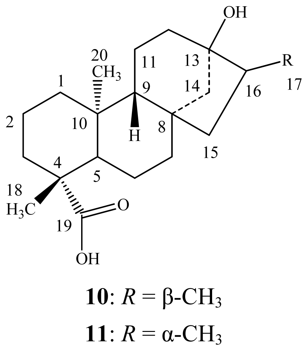
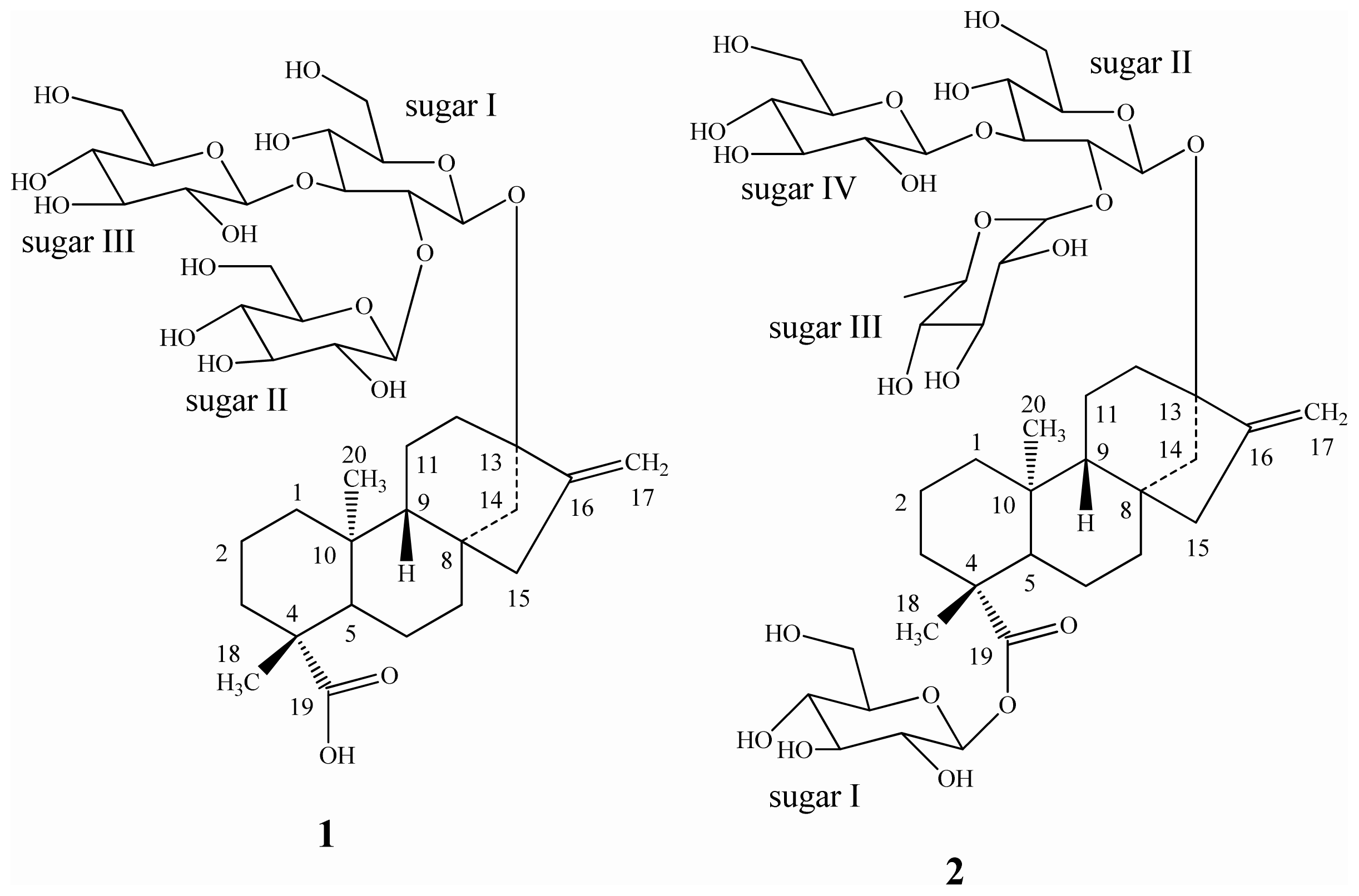
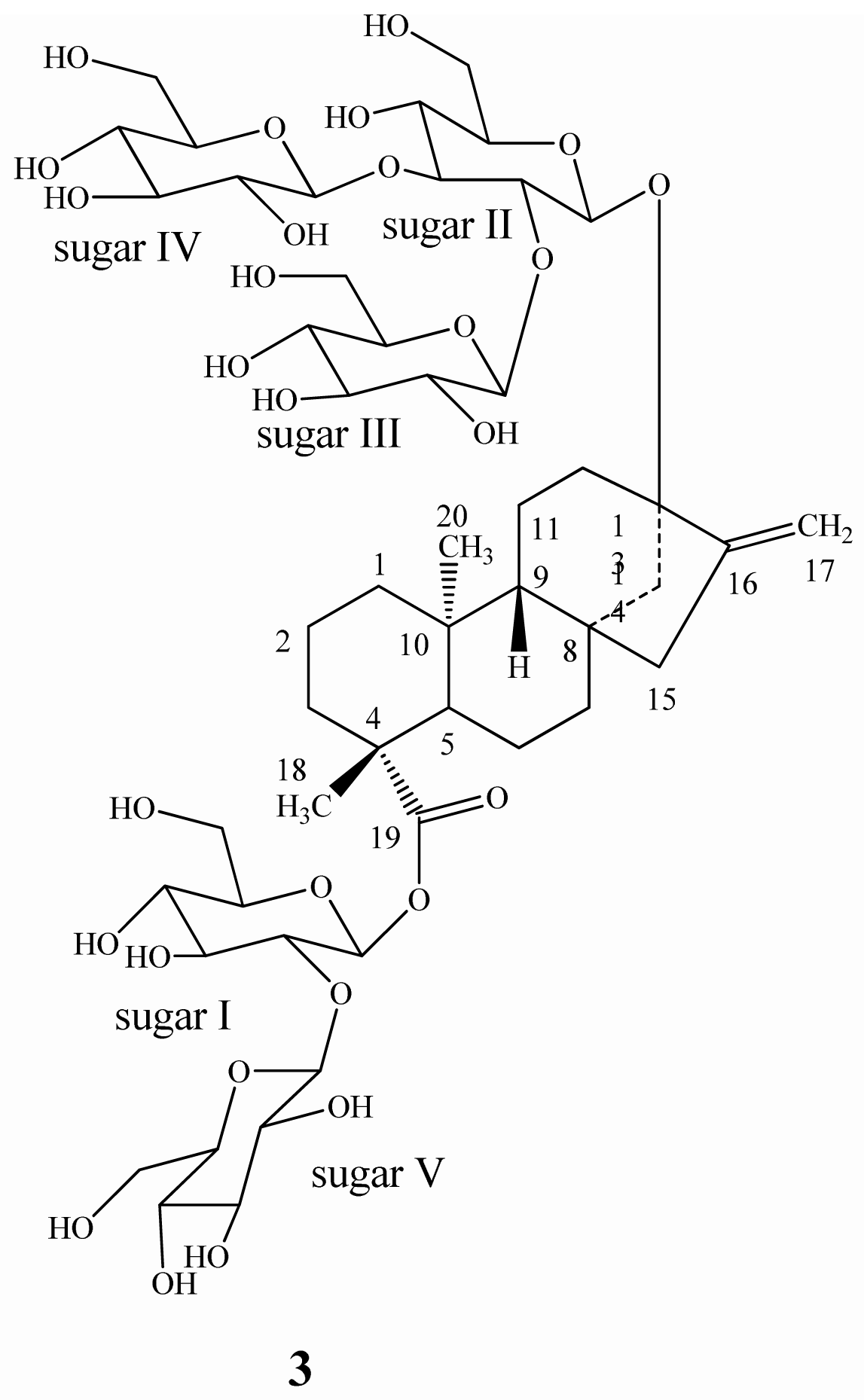
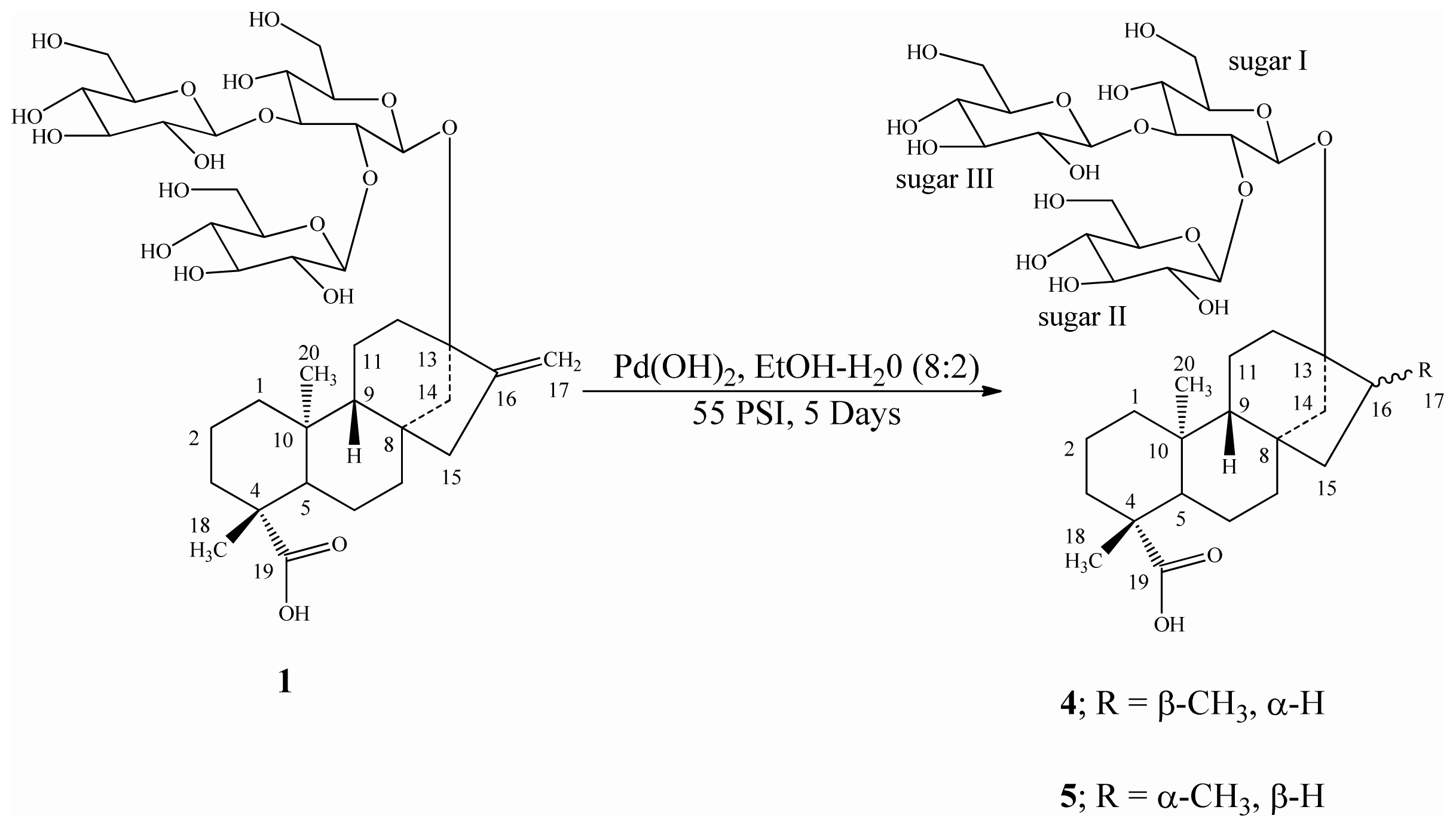
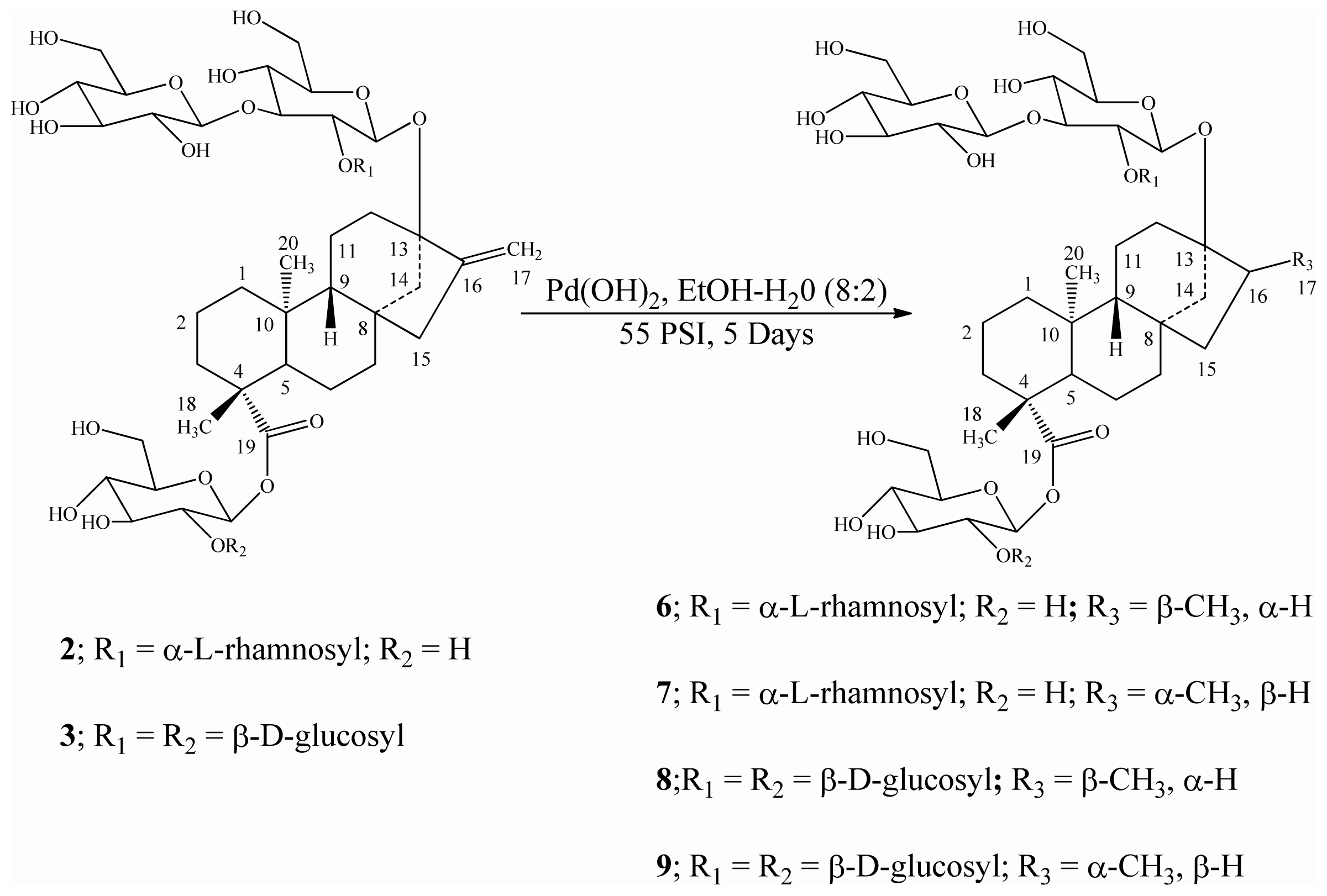
2.1. Catalytic Hydrogenation of Rebaudioside B (1), Rebaudioside C (2), and Rebaudioside D (3)

Reduction of the three ent-kaurane diterpene glycosides isolated from S. rebaudiana namely rebaudioside B (1), rebaudioside C (2), and rebaudioside D (3) was performed using catalytic hydrogenation with Pd(OH)2 in a solvent mixture of EtOH/H2O (8:2) at room temperature under 55 psi H2 as reported earlier [14], which furnished mixtures of dihydrorebaudioside-B 1/2 (4/5) (Scheme 1), dihydrorebaudioside-C 1/2 (6/7), and dihydrorebaudioside-D 1/2 (8/9) (Scheme 2). Further trials to separate the mixtures using various separation techniques failed; hence we are reporting the reduced compounds as is.
The 1H and 13C NMR spectral data analysis of the reaction products of rebaudioside B (1), rebaudioside C (2), and rebaudioside D (3) indicated the absence of the exocyclic double bond between C-16/C-17 position and the presence of their corresponding 17α and 17β methyl group isomers. The high resolution mass spectral (HRMS) data of the reduced products of rebaudioside B (1), rebaudioside C (2), and rebaudioside D (3) showed 2 amu more than their corresponding starting compounds which supported the hydrogenation.

2.2. Sensory Studies of Reduced Compounds 49

The sensory evaluations of the synthetically reduced steviol glycosides 49 at a concentration of 500 ppm were performed against several control samples of 0.75%, 2%, 4%, 6%, and 7.0% sucrose equivalence (SE) in carbon treated (CT) water at room temperature (rt) as reported earlier [14]. Also, the sensory comparison of the mixtures 4/5, 6/7, and 8/9 against their original steviol glycosides was studied at 500 ppm using the controlled, multi-sip and swallow taste methods as described below. Sweetness evaluation results indicated that the sweet taste of the hydrogenated rebaudioside B derivatives (4/5) was reduced by over 75%, and that of the rebaudioside D derivatives (8/9) was reduced by about 25%, whereas the rebaudioside C derivatives (6/7) completely lost their sweetness after catalytic reduction (Table 1). These results indicated that the C16–C17 methylene double bond in steviol glycosides could be regarded as a pharmacophore essential for the sweetness property of these molecules [15,16]. Similar data has been identified earlier during our study on the catalytic hydrogenation of rubusoside, stevioside, and rebaudioside A [14].

2.3. Spectroscopy and Structural Characterization of Reduced Compounds 49

Structural characterization of the reduced compounds 49 obtained by the catalytic hydrogenation of the three ent-kaurane diterpene glycosides isolated from S. rebaudiana; rebaudioside B (1), rebaudioside C (2), and rebaudioside D (3) was performed on the basis of one-dimensional (1H, 13C), two-dimensional [1H-1H correlation spectroscopy (COSY), 1H-13C heteronuclear multiple-quantum correlation (HMQC), 1H-13C heteronuclear multiple bond correlation spectroscopy (HMBC)] NMR and mass spectral data. The stereochemistry at the C-16 position was identified by comparison with their corresponding aglycone derivative NMR values reported in the literature [14,1719], as well as enzymatic hydrolysis studies. The 1H-NMR and 13C-NMR values for all the protons and carbons in dihydrorebaudioside-B1/dihydrorebaudioside-B2 (4/5), dihydrorebaudioside-C1/dihydrorebaudioside-C2 (6/7), and dihydrorebaudioside-D1/dihydrorebaudioside-D2 (8/9) were assigned on the basis of COSY, HMQC and HMBC correlations. Further it was found that the ratio of 17α/17β reduced compounds was observed to be 3:2 for rebaudioside B (1); and 2:1 for rebaudioside C (2), and rebaudioside D (3). The 1H-NMR spectral data for the sp3 methyl groups and anomeric protons for the mixture of reduced compounds 49 are given in Table 2, whereas the complete assignments of their carbon values are given in Table 3.

3. Experimental Section

3.1. General

Melting points were measured using a SRS Optimelt MPA 100 instrument and are uncorrected. Infrared (IR) spectral data was acquired using a Perkin Elmer 400 Fourier Transform Infrared (FT-IR) Spectrometer (Atlanta, USA) equipped with a Universal Attenuated Total Reflectance (UATR, Atlanta, USA) polarization accessory, whereas NMR spectra were acquired on Varian Unity Plus 600 MHz instrument (Atlanta, USA) in C5D5N using standard pulse sequences. Chemical shifts were given in δ (ppm), and coupling constants were reported in Hz. HRMS and MS/MS data were generated with a Waters Premier Quadrupole Time-of-Flight (Q-TOF, New Jersey, USA) mass spectrometer equipped with an electrospray ionization source operated in the positive-ion mode and ThermoFisher Discovery OrbiTrap (New Jersey, USA) in the positive mode of electrospray. All the samples were diluted with water: acetonitrile (1:1) containing 0.1% formic acid and introduced via infusion using the onboard syringe pump. CT water was prepared by passing water through granular or block carbon material to reduce toxic compounds as well as harmless taste- and odor-producing chemicals.

3.2. Isolation of Reduced Steviol Glycosides 49

3.2.1. General Procedure for the Catalytic Hydrogenation of Steviol Glycosides 13

Pd(OH)2 (50 mg) was added to a solution of each steviol glycoside 13 (2 g) in EtOH/H2O (8:2, 100 mL). The mixture was hydrogenated at ambient temperature for 5 days under hydrogen (H2) pressure at 55 psi. After each day, an aliquot of the reaction mixture was filtered through Celite and analyzed by high performance liquid chromatography (HPLC) for the absence of starting materials. At the end of hydrogenation (5 days), the reaction mixture was filtered through celite and concentrated under vacuum to afford a clear white product. The product was triturated with acetone, filtered and dried under vacuum at 50 °C for 48 h–72 h. The combined purity of each isomeric mixture 4/5, 6/7, and 8/9 was checked by HPLC and was found to be >97%.
Dihydrorebaudioside B1/Dihydrorebaudioside B2 (4/5). White powder; IR νmax: 3347 cm−1, 2928 cm−1, 2882 cm−1, 1726 cm−1, 1034 cm−1, 891 cm−1; 1H-NMR and 13C-NMR spectroscopic data see Tables 2,3, respectively; HRMS (M + NH4)+m/z 824.4282 (calcd. for C38H66NO18: 824.4280), (M + Na)+m/z 829.3838 (calcd. for C38H62O18Na: 829.3834).
Dihydrorebaudioside C1/Dihydrorebaudioside C2 (6/7). White powder; IR νmax: 3355 cm−1, 2932 cm−1, 2881 cm−1, 1722 cm−1, 1034 cm−1, 890 cm−1; 1H-NMR and 13C-NMR spectroscopic data see Tables 2,3, respectively; HRMS (M + NH4)+m/z 970.4864 (calcd. for C44H76NO22: 970.4859), (M + Na)+m/z 975.4418 (calcd. for C44H72NaO22: 975.4413).
Dihydrorebaudioside D1/Dihydrorebaudioside D2 (8/9). White powder; IR νmax: 3345 cm−1, 2920 cm−1, 2882 cm−1, 1724 cm−1, 1035 cm−1, 880 cm−1; 1H-NMR and 13C-NMR spectroscopic data see Tables 2,3, respectively; HRMS (M + H)+m/z 1131.5074 (calcd. for C50H83O28: 1131.5071), (M + NH4)+m/z 1148.5342 (calcd. for C50H86NO28: 1148.5336).

3.2.2. General Procedure for the Enzymatic Hydrolysis of Reduced Steviol Glycoside Mixtures

The mixture of each reduced steviol glycoside (250 mg) was dissolved in 0.1 M sodium acetate buffer, pH 4.5 (50 mL) and crude pectinase from Aspergillus niger (15 mL, Sigma-Aldrich, P2736) was added. The reaction mixture was stirred at 50 °C for 96 h. The product precipitated out during the reaction for all three mixtures 4/5, 6/7 and 8/9 was identified as the same based on spectral data and TLC. The filtered compound was purified over silica gel column chromatography; elution with n-hexane/acetone (9.5:0.5) yielded a pure Compound 10 (18 mg, mp: 186 °C –190 °C) whereas elution with n-hexane/acetone (9.0:1.0) yielded another pure Compound 11 (13 mg, mp: 212 °C –214 °C). The two Compounds 10 and 11 (Figure 2) were identified as dihydrosteviol A and dihydrosteviol B by comparison of their physical and 1H-NMR spectral data with the literature values [1719].

3.3. Sweetness Evaluation of the Reduced Steviol Glycoside Mixtures 49

Sweetness evaluation of the reduced steviol glycoside mixtures was performed using sucrose as a control along with their original compounds. Extra fine cane sucrose from Domino (lot#11:09 6843 1A10) was used for preparation of control samples. Carbon-treated (CT) water was used for all preparations of control, standard steviol glycosides and their reduced compounds. The reduced steviol glycoside mixtures were prepared at 500 ppm for sensory evaluation by adding the non-moisture compensated mass into a 100 mL sample of CT water. The mixtures were moderately stirred at room temperature (rt), and the reduced steviol glycoside samples were then evaluated against several control sucrose samples at 0.75%, 2%, 4%, 6% and 7.0% SE in water at RT by experienced Research and Technology panelists at The Coca-Cola Company, Atlanta, USA, for any tasting quality determinations using the controlled, multi-sip and swallow taste methods shown below.

3.4. Multi-Sip and Swallow Taste Method

  • Take first sip (~1.8 mL) of a full medicine cup and swallow the control, wait for 15 s–25 s, then take the second sip and lock it into memory and wait for 15 s–25 s.
  • Taste the first sip of the experimental sample; wait for 15 s–25 s, then use the second sip to compare to the second sip of the control.
  • Repeat steps #1 and #2 for the third and fourth sips of the same control and experimental samples to confirm the initial finding.

4. Conclusions

In conclusion, six ent-kaurane diterpene glycosides 49 were synthesized by catalytic hydrogenation of the three natural products rebaudioside B (1), rebaudioside C (2), and rebaudioside D (3) using Pd(OH)2. The structures of all synthesized compounds were characterized on the basis of NMR (1D and 2D) and mass spectral data, enzymatic hydrolysis as well as in comparison with the data reported in the literature. This is the first report of the complete spectral characterization of the reduced compounds of rebaudioside B, rebaudioside C, and rebaudioside D.

Acknowledgements

We wish to thank the Department of Chemistry, Emory University, Atlanta, GA, USA; Rafael Sa Miguel, Biosciences and Ingredient Department, The Coca-Cola Company, Atlanta, GA, USA for providing sensory data of reduced compounds; and Chris Mubarak, Analytical Sciences Department, The Coca-Cola Company, Atlanta, GA, USA for providing IR spectral data.
  • Conflict of InterestThe authors declare no conflict of interest.

References

  1. Brandle, J.E.; Starrratt, A.N.; Gijen, M. Stevia rebaudiana: Its agricultural, biological and chemical properties. Can. J. Plant Sci 1998, 78, 527–536. [Google Scholar]
  2. Chaturvedula, V.S.P.; Prakash, I. Diterpene glycosides from Stevia rebaudiana. J. Med. Plants Res 2011, 5, 4838–4842. [Google Scholar]
  3. Chaturvedula, V.S.P.; Mani, U.; Prakash, I. Diterpene glycosides from Stevia rebaudiana. Molecules 2011, 16, 3552–3562. [Google Scholar]
  4. Chaturvedula, V.S.P.; Prakash, I. A new diterpenoid glycoside from Stevia rebaudiana. Molecules 2011, 16, 2937–2943. [Google Scholar]
  5. Chaturvedula, V.S.P.; Prakash, I. Structures of the novel diterpene glycosides from Stevia rebaudiana. Carbohydr. Res 2011, 346, 1057–1060. [Google Scholar]
  6. Chaturvedula, V.S.P.; Rhea, J.; Milanowski, D.; Mocek, U.; Prakash, I. Two minor diterpene glycosides from the leaves of Stevia rebaudiana. Nat. Prod. Commun 2011, 6, 175–178. [Google Scholar]
  7. Chaturvedula, V.S.P.; Prakash, I. Additional minor diterpene glycosides from Stevia rebaudiana. Nat. Prod. Commun 2011, 6, 1059–1062. [Google Scholar]
  8. Chaturvedula, V.S.P.; Clos, J.F.; Rhea, J.; Milanowski, D.; Mocek, U.; DuBois, G.E.; Prakash, I. Minor diterpenoid glycosides from the leaves of Stevia rebaudiana. Phytochem. Lett 2011, 4, 209–212. [Google Scholar]
  9. Chaturvedula, V.S.P.; Mani, U.; Prakash, I. Structures of the novel α-glucosyl linked diterpene glycosides from Stevia rebaudiana. Carbohydr. Res 2011, 346, 2034–2038. [Google Scholar]
  10. Chaturvedula, V.S.P.; Klucik, J.; Mani, U.; Prakash, I. Synthesis of ent-kaurane diterpene glycosides. Molecules 2011, 16, 8402–8409. [Google Scholar]
  11. Chaturvedula, V.S.P.; Clos, J.F.; Prakash, I. Stability study of steviol glycosides in mock beverages using fluorescent light exposure under ICH guidelines. Int. J. Pharm. Pharm. Sci 2011, 3, 316–323. [Google Scholar]
  12. Chaturvedula, V.S.P.; Clos, J.F.; Prakash, I. Stability of steviol glycosides in mock beverages under acidic conditions. Int. J. Pharm. Pharm. Sci 2011, 3, 421–425. [Google Scholar]
  13. Prakash, I.; Clos, J.F.; Chaturvedula, V.S.P. Stability of rebaudioside A under acidic conditions and its degradation products. Food Res. Int 2012, 48, 65–75. [Google Scholar]
  14. Chaturvedula, V.S.P.; Campbell, M.; Miguel, R.I.S.; Prakash, I. Synthesis and sensory evaluation of ent-kaurane diterpene glycosides. Molecules 2012, 17, 8908–8916. [Google Scholar]
  15. Mani, U.; DuBois, G.; Prakash, I. Synthetic study on the relationship between structure and sweet taste properties of steviol glycosides. Molecules 2012, 17, 4186–4196. [Google Scholar]
  16. Kasai, R.; Kaneda, N.; Tanaka, O.; Yamasaki, K.; Sakamoto, I.; Morimoto, K.; Okada, S.; Kitahata, S.; Furukawa, H. Sweet diterpene glycosides of leaves of Stevia rebaudiana Bertoni: Synthesis and structure-sweetness relation of rebaudiosides A, D, and E and their related glycosides. Nippon Kagaku Kaishi 1981, 5, 726–735. [Google Scholar]
  17. Nanayakkara, N.P.D.; Klocke, J.A.; Compadre, C.M.; Hussain, R.A.; Pezzuto, J.M.; Kinghorn, A.D. Characteriztaion and feeding deterrent effects on the aphid, Schizaphis graminum, of some derivatives of the sweet compounds, stevioside and rebaudioside A. J. Nat. Prod 1987, 50, 434–441. [Google Scholar]
  18. Pezzuto, J.M.; Compadre, C.M.; Swanson, S.M.; Nanayakkara, N.P.D.; Kinghorn, A.D. Metabolically activated steviol, the aglycone of stevioside, is mutagenic. Proc. Nat. Acad. Sci. USA 1985, 82, 2478–2482. [Google Scholar]
  19. Kamiya, S.; Konishi, F.; Esaki, S. Synthesis and taste of some analogs of stevioside. Agric. Biol. Chem. 1979, 43, 1863–1867, Sample Availability: Samples of the catalytically reduced steviol glycosides 4–9 are available from the authors.. [Google Scholar]
Figure 1. Structures of rebaudioside B (1), rebaudioside C (2), and rebaudioside D (3).
Ijms 13 15126f1aIjms 13 15126f1b
Figure 2. Structures of dihydrosteviol A (10) and dihydrosteviol B (11).
Ijms 13 15126f2
Scheme 1. Hydrogenation of rebaudioside B (1) and its reduced compounds.
Ijms 13 15126f3
Scheme 2. Hydrogenation of rebaudioside C (2) and rebaudioside D (3) and their reduced compounds.
Ijms 13 15126f4
Table 1. Sensory evaluation of rebaudioside B (1), rebaudioside C (2) and rebaudioside D (3) versus catalytically hydrogenated steviol glycosides (49) at 500 ppm in CT water at RT.
Steviol Glycoside TypeSensory Evaluation of Original CompoundSensory Evaluation of Reduced Compound
Rebaudioside B (1)Slow onset of sweetness, sweet lingering aftertaste, about 5%–6% sucrose equivalenceWeak sweetness, about 1% sucrose equivalence
Rebaudioside C (2)Slow onset of sweetness, less sweet overall than sucrose, about 2%–3% sucrose equivalenceNo sweetness, moderate astringency
Rebaudioside D (3)Slow onset of sweetness, very clean, sweeter overall than sucrose, less sweet lingering aftertaste compared to sucrose, about 6%–7% sucrose equivalenceSlow onset of sweetness, no sweet lingering taste, about 5%–5.5% sucrose equivalence
Table 2. 1H-nuclear magnetic resonance (NMR) chemical shift values for reduced compounds 49 recorded in C5D5N ac.
Position456789
171.17 (d, 6.6, 1H)1.34 (d, 6.3, 1H)1.19 (d, 6.6, 1H)1.39 (d, 6.4, 1H)1.13 (d, 6.5, 1H)1.17 (d, 6.4, 1H)
181.16 (s, 3H)1.18 (s, 3H)1.24 (s, 3H)1.27 (s, 3H)1.14 (s, 3H)1.15 (s, 3H)
201.19 (s, 3H)1.32 (s, 3H)1.29 (s, 3H)1.28 (s, 3H)1.41 (s, 3H)1.42 (s, 3H)
Sugar I-1′5.04 (d, 6.6, 1H)5.03 (d, 6.4, 1H)6.15 (d, 6.8, 1H)6.16 (d, 6.5, 1H)6.88 (d, 6.4, 1H)6.84 (d, 6.5, 1H)
Sugar II-1″5.33 (d, 6.4, 1H)5.36 (d, 6.3, 1H)5.09 (d, 6.7, 1H)5.06 (d, 6.4, 1H)5.51 (d, 6.6, 1H)5.54 (d, 6.4, 1H)
Sugar III-1‴5.47 (d, 6.1, 1H)5.52 (d, 6.4, 1H)5.95 (d, 6.5, 1H)5.77 (d, 6.8, 1H)5.50 (d, 6.6, 1H)5.58 (d, 6.5, 1H)
Sugar IV-1″″6.53 (d, 1.8, 1H)6.86 (d, 1.6, 1H)5.38 (d, 6.4, 1H)5.42 (d, 6.6, 1H)
Sugar V-1‴″6.33 (d, 6.4, 1H)6.31 (d, 6.2, 1H)
Sugar III-6‴1.65 (d, 6.1, 3H)1.74 (d, 6.4, 3H)
aAssignments made on the basis of correlation spectroscopy (COSY), heteronuclear multiple-quantum correlation (HMQC) and heteronuclear multiple bond correlation spectroscopy (HMBC) correlations;
bChemical shift values are in δ (ppm);
cCoupling constants are in Hz.
Table 3. 13C-NMR chemical shift values for reduced compounds 49 recorded in C5D5N a, b.
Position456789
140.240.241.241.241.141.1
220.220.420.220.120.620.4
338.938.838.939.038.638.3
444.343.244.443.444.642.9
557.556.058.157.958.057.9
623.323.622.723.023.122.9
741.540.341.740.141.240.1
844.143.144.343.043.742.4
956.050.856.654.755.654.9
1040.240.340.340.340.140.3
1120.520.720.320.620.620.7
1235.444.035.344.235.944.0
1388.688.386.286.288.288.1
1447.850.847.350.247.750.9
1547.844.347.244.947.644.8
1641.538.741.239.041.138.9
1716.316.514.219.714.417.2
1829.729.828.628.629.429.8
19180.6180.5177.8177.7176.5176.4
2015.916.115.816.015.715.9
1′98.698.896.295.796.296.2
2′78.878.775.575.481.481.0
3′85.786.679.079.278.878.7
4′72.172.271.471.471.571.4
5′77.077.078.578.678.578.6
6′62.962.862.562.563.163.3
1″105.4105.098.396.894.294.3
2″74.674.678.478.679.179.2
3″77.877.887.186.186.086.7
4″72.172.370.670.571.271.1
5″79.079.275.675.477.277.0
6″62.962.862.762.862.862.9
1‴105.5105.3103.1102.1105.1104.8
2‴75.675.771.771.675.775.9
3‴81.981.372.972.878.778.6
4‴72.172.173.173.272.372.1
5‴79.179.070.370.079.079.1
6‴63.163.319.419.362.562.8
1″″105.0104.9105.3105.9
2″″74.974.774.374.4
3″″79.879.779.879.9
4″″72.272.172.272.1
5″″79.078.979.679.7
6″″63.663.163.263.1
1‴″106.1105.4
2‴″76.876.7
3‴″78.978.8
4‴″72.372.1
5‴″81.881.4
6‴″63.563.5
aAssignments made on the basis of COSY, HMQC and HMBC correlations;
bChemical shift values are in δ (ppm).

