Article

In Vitro Antioxidant Activity of Selected 4-Hydroxy-chromene-2-one Derivatives—SAR, QSAR and DFT Studies

1
Department of Chemistry, Faculty of Science, P.O. Box 60, University of Kragujevac, Radoja Domanovića 12, 34000 Kragujevac, Serbia
2
Department of Molecular Biology, Institute for Biological Research, University of Belgrade, Bulevar Despota Stefana 142, 11000 Beograd, Serbia
3
Department of Biology end Ecology, Faculty of Science, P.O. Box 60, University of Kragujevac, Radoja Domanovića 12, 34000 Kragujevac, Serbia
*
Author to whom correspondence should be addressed.
Int. J. Mol. Sci. 2011, 12(5), 2822-2841; https://doi.org/10.3390/ijms12052822
Submission received: 3 February 2011 / Revised: 15 March 2011 / Accepted: 13 April 2011 / Published: 29 April 2011
(This article belongs to the Special Issue Recent Advances in QSAR/QSPR Theory)

Abstract

:
The series of fifteen synthesized 4-hydroxycoumarin derivatives was subjected to antioxidant activity evaluation in vitro, through total antioxidant capacity, 1,1-diphenyl-2-picryl-hydrazyl (DPPH), hydroxyl radical, lipid peroxide scavenging and chelating activity. The highest activity was detected during the radicals scavenging, with 2b, 6b, 2c, and 4c noticed as the most active. The antioxidant activity was further quantified by the quantitative structure-activity relationships (QSAR) studies. For this purpose, the structures were optimized using Paramethric Method 6 (PM6) semi-empirical and Density Functional Theory (DFT) B3LYP methods. Bond dissociation enthalpies of coumarin 4-OH, Natural Bond Orbital (NBO) gained hybridization of the oxygen, acidity of the hydrogen atom and various molecular descriptors obtained, were correlated with biological activity, after which we designed 20 new antioxidant structures, using the most favorable structural motifs, with much improved predicted activity in vitro.

1. Introduction

Overall cell health depends on the balance between formation and elimination of free radicals [1]. Free radicals, which originate both in normal or pathological metabolic transformations, host-defense against undesirable invasion (chemical or biological), or host-response to a disturbance of the tissues’ integrity (due to trauma, cellular damage, etc.), may begin chain reactions initiated mostly by reactive oxygen species (ROS) [1]. They are, normally, continuously generated in the living cell, in low amounts by the transfer of one electron to an oxygen molecule during respiration chain and cellular immunization reactions [2] and are therefore needed for the normal redox-signaling and self-defense of the host [1]. Still, superoxide anion (O2) and hydroxyl radical (OH), in increased concentrations, can induce oxidative stress and cellular damage by altering the biological activities of lipids, proteins, DNA and carbohydrates [3], even to cellular death [1]. ROS are associated with incidence of heart diseases, thrombosis [4], hypertension [5], Alzheimer’s and Parkinson’s diseases [6] and cancer over the radical induced DNA double-strain breaks [7].
Some substances, like hydroxyl-coumarins [8], directly recombine free radicals and interrupt the initiation and/or propagation of the induced chain reactions. Due to the typical phenolic behavior [1] they act as potent metal chelators and free radical scavengers, resulting in a powerful antioxidant effect. Their antioxidant behavior could be applied in fat and oily foods to prevent oxidative deterioration, to replace known synthetic antioxidants. Deterioration of food quality occurs during processing and storage, and is related to presence of oxygen and oxidative processes [9]. To show antioxidant activity, a coumarin derivative has to possess at least one hydroxy1 group [10].
Due to their great importance, the structural aspects of both natural and synthetic coumarins as antioxidant compounds have been evaluated using the structure-activity relationships (SAR) and quantitative structure-activity relationships (QSAR) methodology.
Thus, the evaluation of coumarin isolates from Cortex Fraxini [11], Geranium wallichianum [12] and Korean medicinal plants [13] highlights the presence of catechol moiety and oxygen containing scaffold in C-6 and C-7 positions of the coumarin core for antioxidant activity. Also, the α-pyrone coumarin ring increases free radical scavenging activity, antilipid peroxidation ability and has a suppressive effect on enzymes [13]. Synthetic compounds, 4-methylcoumarins [14], substituted 7- or 8-hydroxybenzo[f]-coumarins, 6-hydroxybenzo[h]-coumarins and 7-azomethinecoumarins were tested for their antioxidant ability in vitro [15] and for their ability to interact with 1,1-diphenyl-2-picryl-hydrazyl (DPPH) stable free radical, scavenging of the superoxide anion and inhibition of lipid peroxidation, too [16].
Thereafter, this paper presents the antioxidant potential in vitro of various C-3 carbonyl/carboxyl (28b) [17] and N-thiazole (210c) [18] 4-hydroxy-2H-chromen-2-one derivatives introducing the 4-hydroxy group as highly potent in reducing chain reaction processes and coumarin C-3 scaffolds as good hydroxyl radical scavengers and metal chelators. The structure potential of our derivatives was confirmed by SAR, QSAR and Density Functional Theory (DFT) studies, which were used for the successful design of 20 novel improved coumarin antioxidant structures.

2. Results and Discussion

2.1. Determination of Total Antioxidant Capacity

Results of the total antioxidant capacity are expressed as μg equivalents of ascorbic acid per milliliter (Table 1), demonstrating level of coumarin activity as TAC = 26.76 – 742.67 μg/mL. The derivation of the compound 1 at C-3 position resulted in the increase of the total antioxidant potential within both tested groups.
The examined compounds 28b contain carboxyl or ester group trans-oriented towards the methyl group of the prop-1-en scaffold. The most active compound was 7b, a (E)-2-cyano-3-but-2-enoic acid derivative (TAC = 324.01 μg/mL). After the substitution of cyano group in 7b by carboxyethyl (2b; 278.24 μg/mL) and two acetyl (6b; 212.12 μg/mL) groups, activity was diminished. The decreasing order in the activity was 7b > 2b > 6b > 4b > 1 > 8b > 3b. Among the N-thiazole derivatives 210c, with the 4c > 2c > 7c > 8c > 6c > 3c > 9c > 10c decreasing activity, the N-thiazole p-sulfonic acid derivative, 4c, was pointed out as a very promising one, with the highest TAC value of 742.67 μg/mL. The O-hydroxybenzoic acid (2c), tolyl (5c, 8c) and N,N-diethyl (7c) derivatives also presented notable potential, in the range of 198.84 μg/mL to 514.24 μg/mL.
In accordance with the results presented for the other activities (Sections 2.2–2.5), the total antioxidant capacity is also presented as TAC50 values (Table 1) which are used in the QSAR study.
Since the total antioxidant capacity assay describes the possibility of the tested compound to reduce Mo(VI) to Mo(V), we considered HOMO-LUMO gap as a way to explain coumarin oxidation ability. The smaller the gap is, the more easily molecules will be excited [19], i.e., the test compound have higher potential to reduce Mo(VI). The range of ΔE HOMO-LUMO (Please see section 2.3.) was −8.53 eV (4b) to −7.05 eV (2c), suggesting potentially good activity. The high total antioxidant capacity had been obtained by compounds 7bE HOMO–LUMO = −8.35 eV), 2c (−7.05 eV) and 4c (−7.77 eV), presenting fine correlation between the activity and the phisico-chemical characterization (Equation 3) of the compounds.

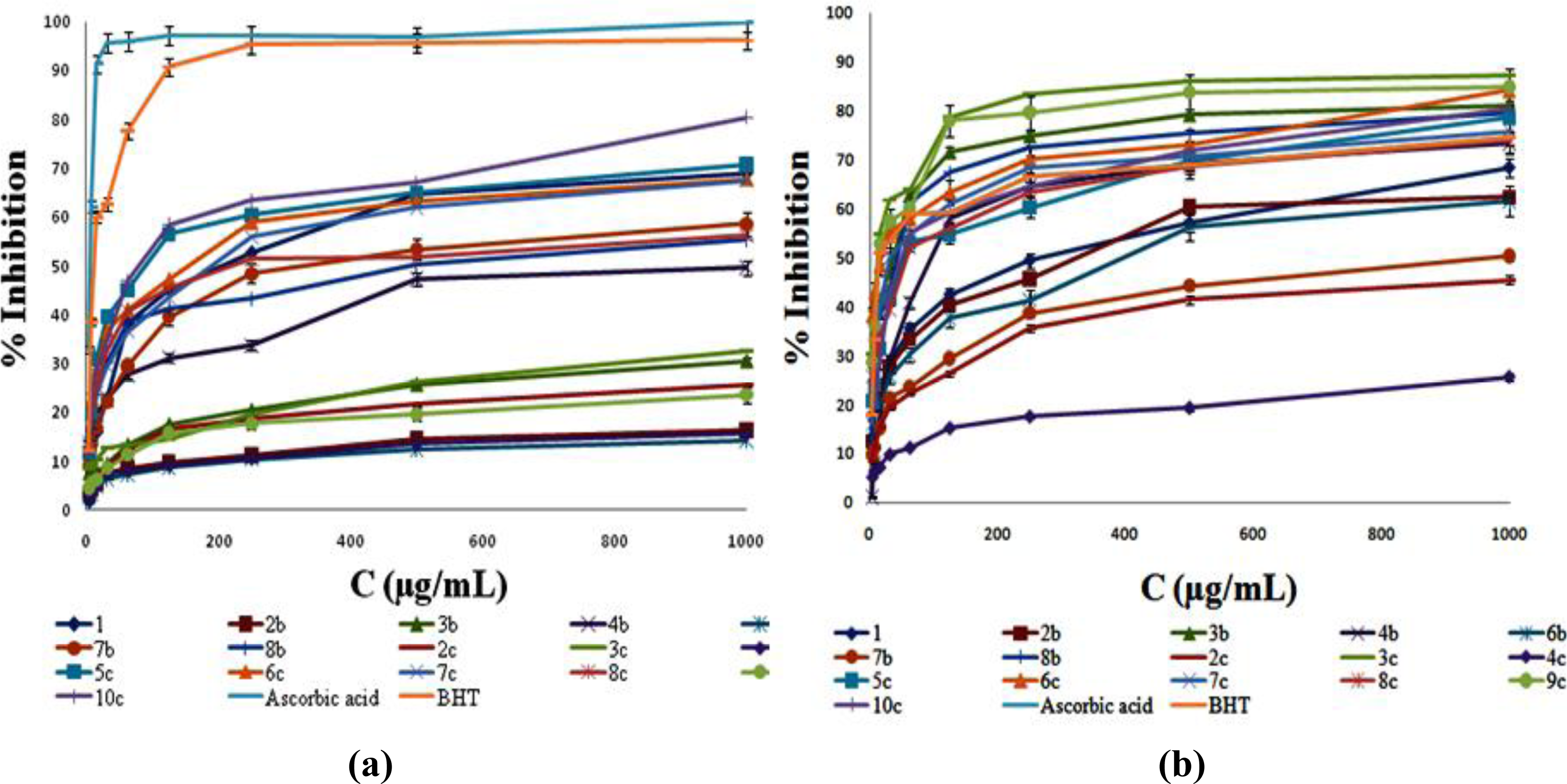
2.2. DPPH Radical Scavenging Activity

The in vitro activity of five coumarin derivatives 2b, 6b, 2c, 4c and 9c (Table 1) was comparable with the standard values of ascorbic acid (Asc) and butylated hydroxytoluene (BHT) (30 minutes: ascorbic acid IC50 = 24.17 μg/mL, BHT IC50 = 8.62 μg/mL; 60 minutes: ascorbic acid IC50 = 15.61 μg/mL, BHT IC50 = 6.05 μg/mL).
After the 30 min period, compounds 28b exhibited the following antioxidant potential: 6b > 2b > 8b > 4b > 3b > 1 > 7b, outlining acetyl groups of the prop-1-en moiety in 6b (IC50 equal to 5.14 μg/mL) as the most structurally favorable residues. The double carboxyethyl substitution of the prop-1-en moiety (2b) decreased the scavenging potential in a small manner (IC50 = 6.2 μg/mL). Still notable, but significantly lower activity (<50 μg/mL) was noticed for 3b, 4b and 8b containing carboxymethyl or carboxyl trans-substituents, with various cis-oriented scaffolds. It is worth mentioning that 4b was the only compound with cis-hydrogen. The presence of acetyl group (1) instead of prop-1-en scaffold has significantly reduced the activity. The lowest activity of the 7b can be attributed to the influence of cis-cyano group. The prolongation of the test time has magnified the potential of 28b, particularly within the triad 3b, 4b and 8b, which presented excellent activity (IC50 = 8.8 − 11.28 μg/mL). The higher scavenging potential of 3b with respect to 4b was caused by cis-acetyl 3b pharmacaphore. Once again, 6b was the most potent scavenger.
Within the second test participants, 210c, the strong hydrogen donor ability has distinguished the 4c as the most powerful DPPH radical scavenger, regarding both test periods (IC50 values of 4.72 μg/mL and 3.54 μg/mL). The activity decreased in the raw 4c > 2c > 9c > 3c > 5c (Figure 1, a). The N-thiazole motif linked with favorable p-SO3H group (4c) as well as the presence of the additional OH group (2c, IC50 equal to 4.9 μg/mL and 6.97 μg/mL) increased the antiradical activity. The high 30 min activity of the 2c was not a surprise due to more look-a-like phenolic structure of the compound, related to increased number of the hydroxyl groups. The m-NO2 N-thiazole derivative (9c), reached full scavenging potential after 60 minutes test (IC50 = 4.79 μg/mL) thus overpowering the decreasing activity of 2c in the same period. Despite the favorable structure, 2c was the only compound that presented weaker potential after 60 min testing. The thiazole derivatives containing only N-tolyl (5c and 8c), N,N-diethyl (7c) and N-naphtyl residues, presented a total lack of activity.
The scavenging potential of our compounds was: 4c > 2c > 6b > 2b > BHT > 9c > Asc after 30 min, while the prolonged reaction time changed the activity in the following manner: 6b > 4c > 2b > 9c > BHT > 2c > 3c > Asc.

2.3. Inhibition of Lipid Peroxidation in Linoleic Acid Emulsion

During the four days long inhibition of lipid peroxidation by coumarin derivatives, the absorbance of the control sample at 500 nm has been increasing up to the maximal value in 72nd hour, and then, on the 96th hour the absorbance decreased due to the decomposition of linoleic acid hydroperoxides generated during the peroxidatition [20]. Consequently, we present the results of coumarin induced inhibition of lipid peroxidation until the 72nd hour (Table 2). BHT was excellent standard for this measurement with the I50 = 7.81 μg/mL.
During the observed 24–72 h time interval, a group of five tested compounds, 2b, 6b, 2c, 4c and 9c, has presented significant lipid peroxide scavenging capacity, compared to 1 and BHT. Regarding the results, some patterns in the activity among derivatives could be obtained. Thus, the compounds 6b (I50 = < 3.90 μg/mL; 10.13 μg/mL; 10.76 μg/mL for 24 h, 48 h and 72 h, respectively) and 9c (I50 = 5.88 μg/mL; 10.06 μg/mL; 12.23 μg/mL) retained the level of activity after the second and third day. Compared to BHT, 6b expressed 50% higher potential in the first 24 hours. Within the following group, 2c, 4c and 2b, a small decrease in the activity on the 48th hour has been noticed, accompanied with amelioration on the third day of the test. The most active compound within the triad was 4c (I50 < 3.90 μg/mL; 26.31 μg/mL; 10.09 μg/mL). Furthermore, a group of structurally similar compounds, 3b, 4b and 8b, showed slightly lower radical scavenging intention than previously mentioned derivatives with I50 from 11.49 to 18.96 μg/mL on the 24th hour, 18.07 to 37.89 μg/mL on the 48th hour and from 10.46 μg/mL to 80.95 μg/mL on the final hour. The structural change of cis-hydrogen atom (4b) with acetyl (3b) or carboxymethyl (8b) group did not contribute to the activity.
The antioxidant capacity of an organic compound, whether natural or synthetic, is expressed as the ability of the compound to release hydrogen atom, i.e., to undergo keto-enol tautomerisation [21]. The molecular bonds with a length between a single and double bonds, also increases the radical scavenging potential. Furthermore, low Bond Dissociation Enthalpies (BDE) values are often attributed to the high antioxidant potential [21,22].
The proton transfer from the antioxidant towards radical is common for both DPPH and lipid peroxide scavenging. Therefore, we present the DFT study on the coumarin 4-OH group which is responsible for antioxidant activity. As has been obtained by the single-point DFT calculations and presented in Figure 2a and d, the 4-hydroxyl group of the tested derivatives is highly enolized and the hydrogen atom is almost released towards available free radical. In the methanol solution, the coumarin structure is transformed into hinon-like (Figure 2a and d) and the 4-OH hydrogen atom is attached to oxygen by weak electrostatic interactions. Therefore, the hydrogen is suitable for abstraction by the free radical. Moreover, by NBO analysis, we concluded that the acidity of the 4-OH hydrogen is very significant for the activity. Although the hybridization of the oxygen atom in the bond is sp2 by equations 0.7814(sp1.67)O + 0.3926(s100)H (6b) and 0.8947(sp1.62)O + 0.4837(s100)H (4c), it is still close to the sp one, regarding the (sp1.50) as the top border for sp hybridization, providing the relatively high acidity to the hydrogen atom. Furthermore, bond lengths of all our compounds show that the majority of the bonds are longer than double bonds and shorter than single bonds, indicating an extended conjugation with antioxidant properties [21]. The lengths of 4-OH bond in 6b (d = 1.03 Å) and 4c (d = 1.63 Å) are the longest ones (Table 3, D4), causing the low O-H bond dissociation enthalpy (Table 3, E1) [21,22], consequently facilitating the release of the proton towards radical. This influence of the bond length is also described by Equations (2) and (3). Beside the fact that the BDEs of 6b (205.84 kcal/mol) and 4c (239.26 kcal/mol) are higher compared to coumarin standards [22], the most decisive factor for the strong antioxidant activity is the hinon-like structure of coumarin compounds in the methanol solution and the great availability of the hydrogen. In agreement with these facts, the compounds 6b and 4c have shown excellent DPPH scavenging and lipid peroxidation activity. Compound 2c also presented notable potential due to long the 4-OH bond length (d = 1.55 Å) and considerable acidity of the hydrogen atom: 0.6826(sp1.73)O + 0.3726(s100)H. Still, this is the only compound possessing more than one OH group which possibly contributes to the activity.

2.4. Determination of Hydroxyl Radical Scavenging Activity

The hydroxyl radical scavenging activity of the BHT (OH50 equal to 33.92 μg/mL) has been impaired by ten compounds in the following order 4c > 2c > 7b > 6b > 2b > 1 > 3c > 9c > 6c > 3b > BHT, with 1.5–82.5% higher potential (Table 2) (Figure 1b). All of the tested compounds were better hydroxyl radical scavengers than ascorbic acid. The activities of 4c (OH50 = 5.94 μg/mL), 2c (OH50 = 9.81 μg/mL), 7b (OH50 = 9.89 μg/mL) and 6b (OH50 = 14.32 μg/mL) are worthy of note. Generally, the influence of the N-thiazole group linked to coumarin core, along with the functional groups that extended the conjugation (p-SO3H, p-, m-NO2 and Ph-o-OH-p-COOH), contributed to higher activity of 210c in comparison to 18b. On the other hand, the presence of cis-cyano group in 7b enhanced the hydroxyl radical scavenging activity for 40–89%, compared with 1, 2b and 6b. The hydroxyl radical scavenging was the only test during which 1 showed a certain potential. The OH radical abstraction is favored due to extended conjugation in our compounds (Please see section 3.1.).
All presented data are in strong correlation with observed radical scavenging activity.
The abstraction of the OH radical occurs in LUMO orbitals of the coumarin double bond system (Figure 2, c and f) which is confirmed by Equation (4). The LUMO orbitals of 6b (widespread trough molecule) and 4c (thiazole N = C) are presented as the OH radical abstraction centers.

2.5. Measurement of Ferrous Ion Chelating Ability

Starting with N-thiazole p-NO2 derivative 3c, (CE50 = 5.5 μg/mL), followed by m-NO2 derivative 9c (CE50 = 7.76 μg/mL), all tested compounds, except 1, had surpassed ferrous ion chelating ability (Table 2) of ascorbic acid (CE50 76.31 μg/mL) and BHT (CE50 85.48 μg/mL). The chelating effect in the first group of the compounds descended in following order 6b > 2b > 8b > 3b > 7b > 4b > 1. The decrease in number of oxygen atoms, starting with 3b, significantly reduced the chelating power. The influence of the carbonyl/carboxyl and N-thiazole residues on chelating activity was comparable.
The lactone part of coumarin derivatives can bind the iron atom, but that was not confirmed by Equation (5). Governed by the fact that the oxygen groups are crucial for the chelating ability [23], high activity of 6b, 2b and 8b is assigned to the presence of numerous oxygen atoms), Equation (5). Interestingly, 2b and 8b, containing carboxyl groups, had lower activity than 6b (two acetyl groups). The potential of the 3c and 9c depends on the binding of iron with nitro oxygen atoms.

2.6. QSAR Studies of the Antioxidant Activity

QSAR studies were administrated to the antioxidant activity as a tool to describe and explain the activity of the tested coumarin derivatives. The results of the in vitro testing and the concentrations of the test compounds that reduce 50% of the initial free radical concentration were used for the QSAR study. Logarithmic values of the biological activity were used for the generation of the QSAR equations with relevant molecular descriptors (Figure 3). From the equations, we ascertained the possible mechanisms by which our coumarins occur as antioxidants.

QSAR study on total antioxidant capacity

pTAC 50 = 8.46 ( ± 0.32 ) + 7.32 ( ± 0.99 ) HOMO 2.69 ( ± 0.47 ) LUMO 9.46 ( ± 1.48 ) Q 0 H + 0.6 ( ± 0.1 ) Q N 2.9 ( ± 0.4 ) Q S n = 15 ; r = 0.992 ; s = 0.032 ; F = 37.426 ; Q 2 = 0.993 s-PRESS = 0.004
According to the model that describes total antioxidant capacity, Equation (1), the potential of the compounds is highly dependent of HOMO electron energies and the partial atomic charge of the thiazole ring nitrogen. The equation describes the thiazole center as electrone rich and as participant in the Mo(VI) reduction, as well as the HOMO orbitals of the compounds. The HOMO orbital of 4c is the orbital of the coumarin lactone carbonyl double bond system (Figure 2), highlighting the importance of the coumarin residue in the mechanism.

QSAR study on in vitro 60 min DPPH scavenging activity

pIC 50 = + 6.72 ( ± 0.48 ) 1.23 ( ± 0.57 ) Q 0 H + 4.42 ( ± 0.35 ) 4 -OH bond length + 3.4 ( ± 0.94 ) Q N 7.6 ( ± 0.26 ) Q S
The high basicity of the thiazole nitrogen atom attenuates the strength of the 4-OH group C-O bond, making the hydrogen atom more acidic and more easily relaxed (Figure 2). Therefore, the basicity of the nitrogen facilitates DPPH radical scavenging, quantified by Equation (2). The positive sign [24] before the 6.72 in the Equation (2) suggests that the model is high specific towards DPPH scavenging.

QSAR study on inhibition of lipid peroxidation in linoleic acid emulsion (24 h):

pI 50 = + 8.46 ( ± 0.47 ) + 2.46 ( ± 0.28 ) log P 4.92 ( ± 0.36 ) HOMO + 1.96 ( ± 0.46 ) 4 -OH bond length 6.49 ( ± 0.76 ) Q S n = 15 ; r = 0.993 ; s = 0.046 ; F = 103.981 ; Q 2 = 0.991 ; s-PRESS = 0.007
Based on the similar behavior of an antioxidant towards DPPH scavenging activity and lipid peroxidation [25,26], which includes dissociation of the O-H bond and release of the hydrogen towards the radical, Equations (2) and (3) characterize the factor of the bond length as important for easy relieving of the hydrogen atom towards the free radical. As the bond is longer, the hydrogen atom is more easily relaxed due to the bond weakening. Furthermore, the ability of the compounds to inhibit peroxidation chain reaction is highly dependent on the coumarin solubility.

QSAR study on in vitro hydroxyl radical scavenging activity

pOH 50 = + 11.46 ( 0.94 ) 4.94 ( 0.32 ) HOMO + 7.46 ( 0.24 ) LUMO + 5.96 ( 0.32 ) Q 0 H n = 15 ; r = 0.997 ; s = 0.028 ; F = 174.273 ; Q 2 = 0.994 ; s-PRESS = 0.013
Free radicals can be easily scavenged on the double bond system. Continued conjugation through the 110c coumarin system, consisted of benzene, 4-hydroxy-enol and the C-3 scaffold double bonds (Figure 5) provides the ability for possible hydroxyl radical addition on LUMO coumarin orbital level. The strong participation of LUMO energy and the partial atomic 4-hydroxyl group oxygen charge in the highly specific Equation (4) correlates with high activity of our derivatives.

QSAR study on iron chelating ability

pCE 50 = + 1.29 ( 0.52 ) + 9.01 ( 0.003 ) CSEV + 0.95 ( 0.33 ) Q N n = 15 ; r = 0.993 ; s = 0.054 ; F = 43.383 ; Q 2 = 0.894 ; s-PRESS = 0.034
In the structure of tested coumarin derivatives there are high voluminous scaffolds which define the compound’s stereochemistry during the chelating of the iron atom. Although the model (Equation 5) does not specify the carbonyl/carboxyl pharmacophores influence, it does specify the great importance of the spatial arrangement of the C-3 residues on the activity. This particular stereochemistry is characterized with the steric molecular descriptor CSEV by Equation (5).
The QSAR statistics are outlined in Table 4 and Table 5. Table 4 presents calculated antioxidant activity values, used for the validation of the models. Excellent validation was further used for the calculation of the activity of the designed compounds (Please see Section 2.7).
Used molecular descriptors were cross-correlated with the experimental activity (Table 5), specifying the part of each descriptor in the related activity.

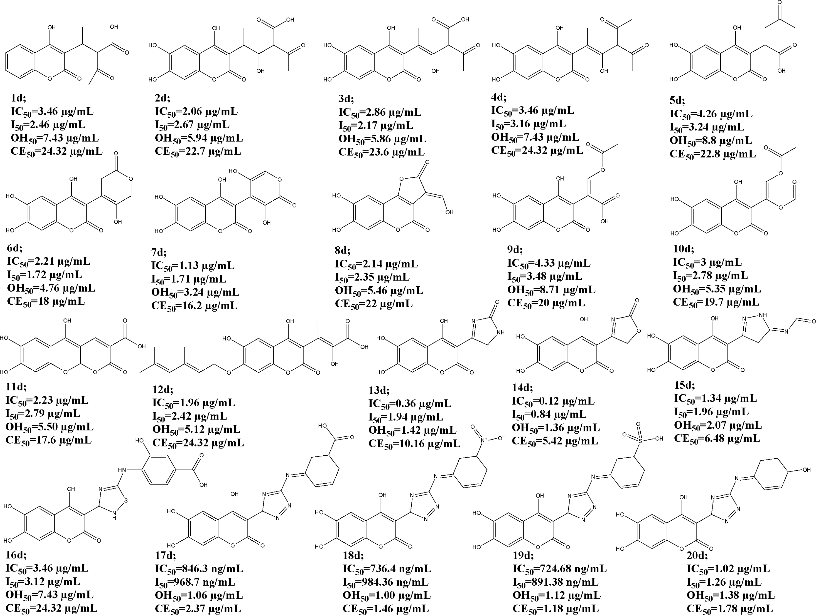
2.7. Structure-Based Design of Novel 4-Hydroxy Coumarin Antioxidants

We have learned how carbonyl/carboxyl and substituted N-thiazole moieties contribute to our compounds antioxidant activity and, by the use of QSAR, we designed twenty new improved coumarin structures and calculated their values of potential scavenging and chelating activity (Figure 4).
Therefore, the catehol moiety [1113] is an important structural part in the design, completing a new backbone with 1,4-pyrone system. The structures were considered in a way that modified C-3 scaffolds, extend the double bond conjugation in many, providing the large number of existing tautomeric forms obtained by the ChemSketch 12 software [27], yet retaining favorable structural properties of the test compounds.
Hence, the number of structural tautomers rises from 3 of compounds 4d and 8d, up to even 10 tautomers of 17d. As the result shows, extended conjugation had improved the overall predicted antioxidant ability. The unfavorable prop-1-ene part of the b condensates had been modified by the total removal of the double bond (1d, 5d), addition of one hydrogen group (12d) followed by the extension of the carbone chain (3d, 4d), or exploiting carbonyl/carboxyl scaffold which led to various lactone and ether structures (611d). The sulfur atom in the thiazole ring did not contribute to the scavenging activity (Equations (2), (3)), and so is replaced by the construction of oxazole, pyrazole, imidazole and triazole rings (1320d). All of the designed compounds had been subjected to the same molecular modeling methods as the original ones and the obtained molecular descriptors were used for the prediction of scavenging and chelating activity using existing QSAR equations. The residue of the triazole ring continued by the Schiff base moiety (17d, 18d and 19d) is presented as the most successful structural modification, which, along with the appropriate –COOH, –SO3H and –NO2 ring substituents, reduced the values of coumarin active concentrations to below 1 μg/mL for the IC50 and I50, and about the same value for the OH50 and CE50. The most promising structure is, therefore, 19d with a predicted DPPH scavenging activity of IC50 = 724.68 ng/mL. The 3,5-dihydroxy-2H-pyran-2-one C-3 structural part (7d) contributed the most to the predicted activity of the compounds that ensued from the carbonyl/carboxyl scaffold modifications. By further synthesis and an antioxidant assessment, the design will be vindicated.

3. Experimental Section
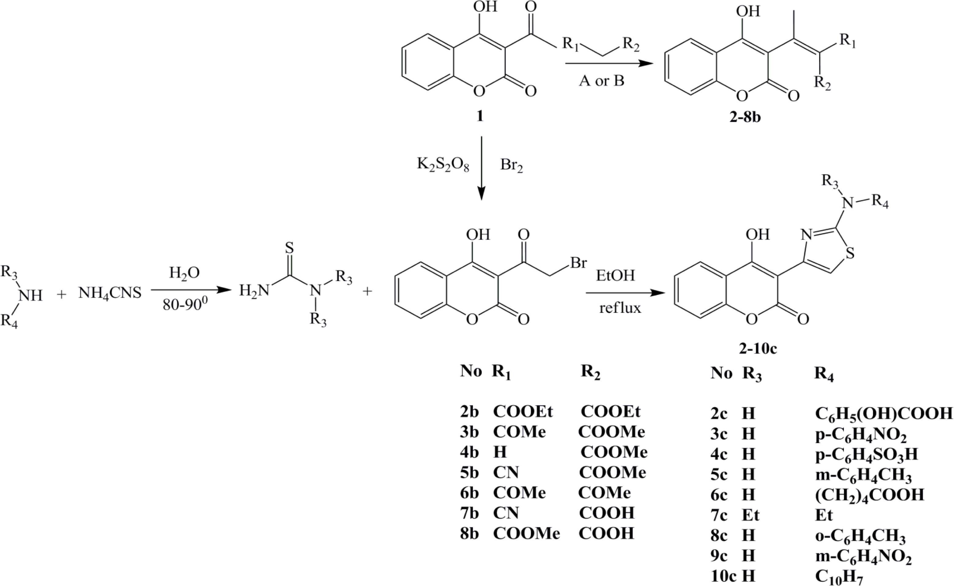
3.1. Chemistry

In previous papers we had reported synthesis and characterization of the group of 15 substituted chromene-2H-one derivatives (110c) (Figure 5) [17,18]. Synthesized compounds were characterized by elemental analysis (C, H, N, O and S) and determination of molecular weights by Mass Spectroscopy (MS). Structural characterization was performed by IR, H1 NMR and MS spectra. The purity of synthesized compounds over 98% was confirmed by the HPLC and TLC analysis.

3.2. Chemicals

All applied chemicals and reagents were of the highest purity available and purchased from the Sigma-Aldrich Chemical Company (St. Louis, MO, USA), Difco (Sparks, MD, USA) and Merck Laboratory Supplies (Darmstadt, Germany).

3.3. The Antioxidant Evaluation in Vitro

3.3.1. Determination of Total Antioxidant Capacity by Phosphomolibdenium Assay

The antioxidant activity of the tested compounds was evaluated by the phosphomolibdenium method according to the procedure of Prieto [28]. The assay is based on the reduction of Mo(VI)–Mo(V) by the test compounds and subsequent formation of the green phosphate/Mo(V) complex at acid pH. An aliquot of 100 μL of the methanol solution of the tested compounds (3.901–1000 μg/mL) was combined with 1 mL of reagent solution (0.6 M sulfuric acid, 28 mM sodium phosphate and 4 mM ammonium molybdate). The tubes containing the reaction solutions were incubated at 95 °C for 90 min. Then the absorbance of the solution was measured at 695 nm, using a Perkin-Elmer Lambda 25 UV/Vis spectrophotometer, against blank probe after cooling to room temperature. Methanol (100 μL) in the place of solution of the tested compound was used as the blank. The total antioxidant capacity of the tested samples was calculated according to the Equation (6):
TAC ( % ) = [ ( A 0 A t ) / A 0 ] × 100
where At is the absorbance value of the tested sample and A0 is the absorbance of the blank sample, in particular time. Ascorbic acid was used as reference standard. The results (TAC) are presented as the μg equivalents of the ascorbic acid per milliliter, obtained from the linear regression analysis. All the experiments were performed in triplicate and the average absorbance was noted for each concentration. As the TAC value is higher, the better is the antioxidant activity. Also, the results of all tests are expressed as EC50 values (i.e., TAC50, IC50, I50, OH50 or CE50) presenting the concentration of the test compound that reduces 50% of the initial free reactive species concentration, calculated as μg/mL, for various concentrations of coumarin derivatives (3.901–1000 μg/mL), with OriginPro 8 statistical software [29] using Nonlinear Curve Fit Growth/Sigmoidal Dose-response function. Percent of inhibition was plotted against concentration, and the equation was the line used to obtain TAC50 value. A lower TAC50 value indicates greater antioxidant activity.

3.3.2. DPPH Radical Scavenging Assay

The method used by Takao et al. [30] was adopted with suitable modifications. DPPH (8 mg) was dissolved in MeOH (100 mL) to obtain a concentration of 80 μg/mL. Serial dilutions (3.901–1000 μg/mL) were carried out with the stock solutions of the compounds 110c in methanol. Diluted solutions (2 mL each) were mixed with DPPH (2 mL) and allowed to stand for 30 min and 60 min for any reaction to occur. The absorbance was recorded at 517 nm. Control sample was prepared containing the same volume without test compounds and reference compounds. The radical-scavenging activity of the tested samples, expressed as percentage inhibition of DPPH, was calculated according to the Equation (7).
IC ( % ) = [ ( A 0 A t ) / A 0 ] × 100
Percent of inhibition after 30 min and 60 min was plotted against concentration, and the equation was the line used to obtain IC50 value. A lower IC50 value indicates greater antioxidant activity.

3.3.3. Inhibition of Lipid Peroxidation in a Linoleic Acid Emulsion Assay

The ability of coumarin derivatives to inhibit lipid chain peroxidation process was tested in a linoleic acid system [31]. Dilutions (3.901–1000 μg/mL) of the test compounds were prepared in methanol, to add to the linoleic acid emulsion. The linoleic acid emulsion was prepared by mixing 0.2804 g of linoleic acid, 0.2804 g of Tween 20 as emulsifier and 50 mL of phosphate buffer (0.2 M, pH 7) and the mixture was then homogenized. A 0.5 mL of coumarin methanol solution in different concentrations was mixed then in linoleic acid emulsion (2.5 mL, 0.02 M, pH 7) and phosphate buffer (0.2 M, pH 7). The reaction mixture was incubated at 37 °C in the dark to accelerate the peroxidation. Aliquots of 100 μL were taken at different intervals (24–96 h) during incubation. The degree of oxidation was measured by sequentially adding ethanol (4.7 mL, 75%), ammonium thiocyanate sample solution (100 μL, 30%) and FeCl2 (100 μL, 0.02 M in 3.5% HCl). After 3 min, the peroxide values were determined by reading the absorbance at 500 nm. Control was performed with linoleic acid but without the tested compounds. Percent inhibition of lipid peroxide generation was calculated using Equation (8).
I ( % ) = [ ( A 0 A t ) / A 0 ] × 100
Percent inhibition was plotted against concentration, and the equation for the line was used to obtain I50. All determinations were carried out in triplicate. The lower I50 value indicates greater antioxidant activity.

3.3.4. Hydroxyl Radical Scavenging Activity Assay

The evaluation of coumarins as inhibitors of hydroxyl radical-mediated oxidation was performed with method described by Halliwell et al. [32]. The reaction mixture contained 100 μL of tested compounds (with 3.901–1000 μg/mL dilutions) dissolved in ethanol, 500 μL of 5.6 mm 2-deoxy-d-ribose in KH2PO4/NaOH buffer (50 mM, PH 7.4), 200 μL of premixed FeCl3 (104 μM) and EDTA (104 μM) (1:1 v/v) solution, 100 μL of H2O2 (1.0 mM) and 100 μL of aqueous ascorbic acid (1.0 mM). Tubes were vortexed and heated in water bath at 50 °C for 30 min. Thereafter, 1 mL of 2.8% TCA and 1 mL of 1% TBA were added to each tube with repeat of the 30 min long water heat. The extent of oxidation was estimated from the absorption of solution at 532 nm. The percentage inhibition values were calculated from the Equation (9).
OH ( % ) = [ ( A 0 A t ) / A 0 ] × 100
Percent inhibition was plotted against concentration, and the equation for the line was used to obtain OH50 value. The lower OH50 value indicates greater antioxidant activity.

3.3.5. Ferrous Ion Chelating Ability Assay

The ferrous ion chelating activity of methanol coumarin solutions was measured by decrease in absorbance at 562 nm of the iron(II)-ferrozine complex [33]. One milliliter of 0.125 mM FeSO4 was added to 1.0 mL of sample (with 1000–3.901 μg/mL dilutions), followed by 1.0 mL of 0.3125 mM ferrozine. The mixture was allowed to equilibrate for 10 min before measuring the absorbance. The ability of the sample to chelate ferrous ion was calculated relative to the control (consisting of iron and ferrozine only) using the Equation (10).
CE ( % ) = [ ( A 0 A t ) / A 0 ] × 100
Percent inhibition was plotted against concentration, and the equation for the line was used to obtain CE50 value. The lower CE50 value indicates greater chelating power ability.

3.4. Statistical Analysis

All results were expressed as means ±standard deviation (SD). The significance of difference was calculated by one-way ANOVA test and values <0.05 were considered to be significant.

3.5. QSAR Study

The QSAR analysis was performed correlating the antioxidant activity presented in Tables 1 and 2, with various molecular descriptors (Table 3) to reveal predictions for the lead optimization in the training set of compounds of synthesized coumarins [17,18]. Although this set is small, it provides QSAR equations that are statistically significant. The results of regression analysis are shown in Equations (1), (2), (3), (4) and (5) and by Figure 3, where n is number of molecules, r is correlation, F is Fisher’s significance factor and s is standard deviation. Cross-validation resulted with Q2 as the square of predictive power of coefficient and s-PRESS as predictive residual sum of squares. The generation of the QSAR models was taken out with OriginPro 8, by the Multiple Linear Regression (MLR) analysis, using cross validation leave-one-out method. The ratios of observed vs. calculated antioxidant activity values and the correlation between the molecular descriptors and the activity are presented in the Tables 4 and 5, respectively.

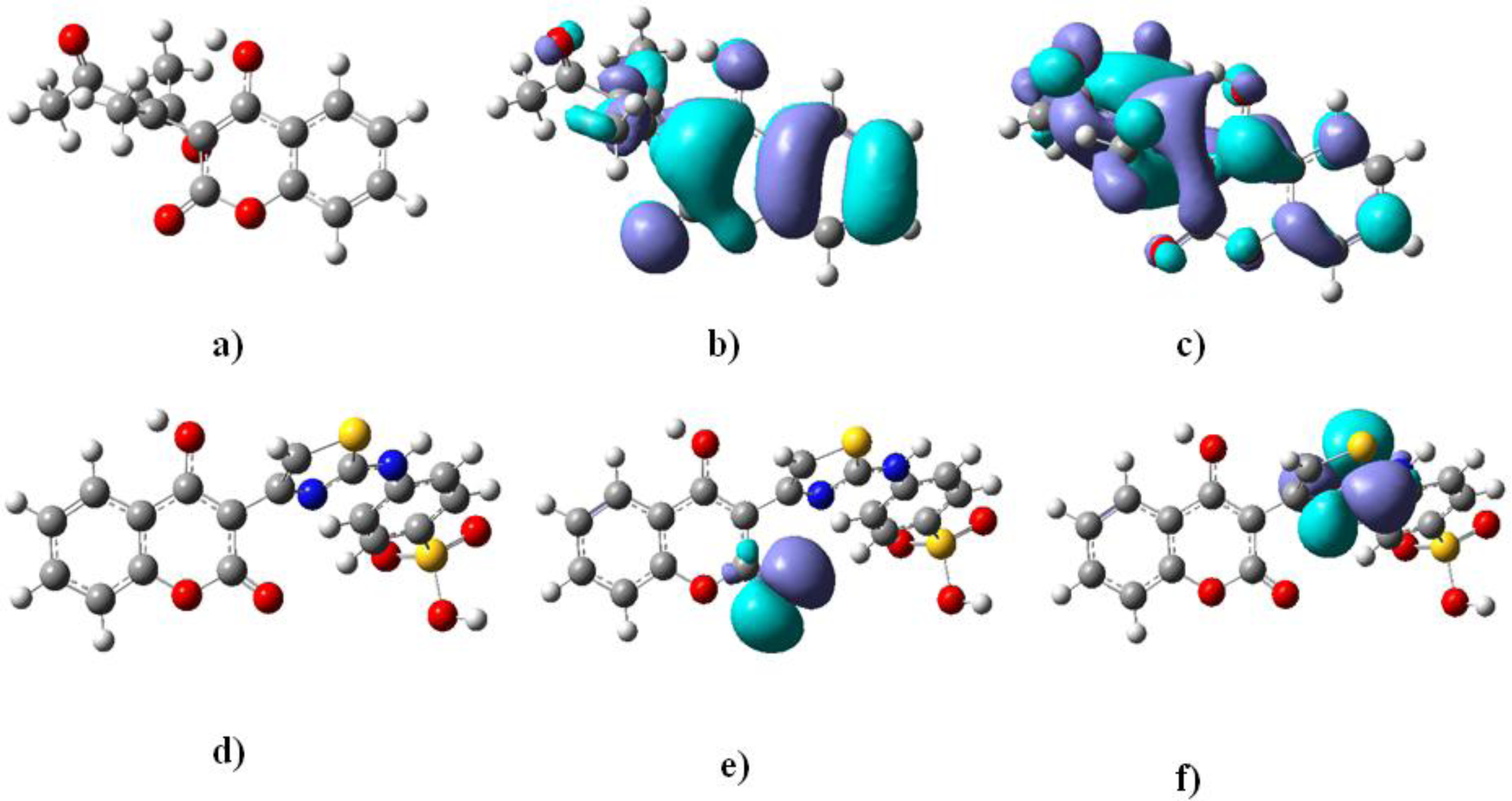
3.5.1. Molecular Modeling

The initial structures were built in Spartan 2006 for Windows [34] and imported in VegaZZ 2.3.1 [35] molecular modeling package. The Gasteiger charges are assigned with OPLS 2005 force field. Structures have been optimized firstly with AMMP incorporated in VegaZZ 2.3.1. The minimization was performed with 3000 steps Conjugate gradients optimizer model, 0.01 Toler and 0 steepest steps. Further, a 1000 steps Boltzman jump conformation search method was applied searching the flexible torsions only. Temperature had been set to 375.15 K, covering all of the experiment conditions. Torsion root square difference had been set to 60°. Dielectric constant of 33.6 simulated coumarin solution environment, using long range cutoff of 20 and short range of 6 Å. After the cluster analysis, on the selected lowest energy structure full optimization was performed by MOPAC 2009/PM6 [36] Hamiltonian semi empirical method imported in Vega ZZ, by fixing gradient norm as 0.01, with ɛ = 33.6 in order to include solubility of compounds 110c in MeOH, according to the experiment conditions. The most stable structures, with determined final heat of formation, were selected as representative conformations in calculation was used for calculation of the electronic descriptors, presented as dominant ones in the antioxidant activity QSAR study of flavonoids, chromones, coumarins and similar plant natural products [21].
Final optimizations were performed by density functional theory (DFT) with the CS Gaussian 03 program [37]. In order to calculate bond dissociation enthalpies of 4-OH groups, optimizations of parent molecules, appropriate radicals and hydrogen radicals were considered. Therefore, parent molecules were optimized by (RO)B3LYP functional and 6-31G(d) basis set, whereas (U)B3LYP/6-31G(d) was applied for radical structures. On the optimized structures full NBO analysis was performed. The geometrical parameters of all stationary points were optimized in methanol solution by CPCM routine. All calculated structures were verified to be local minima (all positive eigenvalues) for ground state structures, by frequency calculation. The zero point vibrational energy and the vibrational contribution to the enthalpy were scaled by a factor of 0.9805 [22]. In order to obtain more reliable relative energy on each stationary point, on the potential energy surface (PES), single-point energy calculations were performed with 6-311G++(2d,2p) diffuse function. The particularly basis set was selected since the optimized molecules are conjugated ones.

3.5.2. Molecular Descriptors

Molecular descriptors generated for this study were electronic and steric. Solubility factor was considered for the lipid peroxidation environment. Electronic molecular descriptors obtained after optimization were: HOMO and LUMO energies, ΔE HOMO-LUMO calculated as the difference of the energies, dipole, total energy, electronic energy, O–H bond length, with Mulliken population analysis applied for the calculation of the partial atomic charges (Q). The molecular descriptors values are presented in the Table 3.
During the DPPH and lipid peroxidation assays, the hydroxyl group proton from the antioxidant is transferred towards the free radical. The process is, taking peroxyl radical (ROO.) as a sample, presented by Equation (11) [22].
ROO + ArOH ROOH + ArO
Covering the fact that process is governed by the O-H bond dissociation enthalpy (BDE) of the antioxidant, we calculated BDE via the formula: BDEs = HKOHHKOHH, where HKOH is the enthalpy for the parent coumarin molecule, HKO is the enthalpy for radical generated after H-abstraction and HH is the enthalpy for hydrogen radical.
Coumarin antioxidant potential is increased with the extended conjugation, including bond orders [21] and some steric parametes like Connolly Accessible Area (CAA), Connolly Molecular Area (CMA), Connolly Solvent-Excluded Volume (CSEV) and principal moment of inertia, calculated by VEGA ZZ, as useful molecular descriptors.

4. Conclusions

Within the series of the examined coumarin derivatives in accordance with the presented experimental results for the total antioxidant capacity and the affinity towards DPPH, lipid peroxide and hydroxyl radicals, it was concluded that compounds 2b, 6b, 2c and 4c are notable antioxidants. Therefore, these compounds could be practically applied as antioxidant agents and as the starting compounds for future selective modifications of the coumarin molecule according to performed SAR, QSAR and experimental conditions based theoretical studies on their activity.

Abbreviations:

NBO
Natural Bond Orbital;
OPLS
Optimized Potentials for Liquid Simulations;
AMMP
Another Molecular Mechanics Program;
PM6
Parametric Method 6;
B3LYP
Becke, three-parameter, Lee-Yang-Parr.

Acknowledgments

This work was financially supported by Serbian Ministry of Science, Grants No. III 43004, III 41010 and OI 173020. The authors would like to thank Svetlana Marković, Faculty of Science, Kragujevac, for useful suggestions during Gasussian DFT calculations.

References

  1. Traykova, M; Kostova, I. Coumarin derivatives and antioxidative stress. Int. J. Pharmacol 2005, 1, 29–32. [Google Scholar]
  2. Droge, W. Free radicals in the physiological control of the cell function. Physiol. Rev 2002, 82, 47–95. [Google Scholar]
  3. Halliwell, B. How to characterize a biological antioxidant. Free Rad. Res 1990, 9, 1–32. [Google Scholar]
  4. Juliano, L; Colavita, AR; Leo, R; Pratico, D; Violi, F. Oxygen free radicals and platelet activation. Free Rad. Biol. Med 1997, 22, 999–1006. [Google Scholar]
  5. Lassegue, B; Griendling, KK. Reactive oxygen species in hypertension. Am. J. Hyper 2004, 17, 852–860. [Google Scholar]
  6. McIntosh, LJ; Trush, MA; Tronsoco, JC. Increased susceptibility of Alzheimer’s disease temporal cortex to oxygen-free radical-mediated processes. Free Rad. Biol. Med 1997, 23, 183–190. [Google Scholar]
  7. Festa, G; Aglitti, T; Duranti, G; Ricordi, R; Perticone, P; Cozzi, R. Strong antioxidant activity of ellagic acid in mammalian cells in vitro revealed by comet assay. Anticancer Res 2001, 21, 3903–3908. [Google Scholar]
  8. Hoult, J; Paya, M. Pharmacological and biochemical actions of simple coumarins: Natural products with therapeutic potential. Gen. Pharmacol 1996, 27, 713–722. [Google Scholar]
  9. Nićiforović, N; Mihailović, V; Mašković, P; Solujić, S; Stojković, A; Pavlović Muratspahić, D. Antioxidant activity of selected plant species; potential new sources. Food Chem. Toxicol 2010, 48, 3125–3130. [Google Scholar]
  10. Sinhg, R; Singh, B; Singh, S; Kumar, N; Kumar, N; Arora, S. Umbeliferone—An antioxidant isolated from Acacia nilotica (L.) Willd. Ex. Del. Food Chem 2010, 120, 825–830. [Google Scholar]
  11. Wu, C; Huang, M; Lin, Y; Ju, H; Ching, H. Antioxidant properties of Cortex Fraxini and its simple coumarins. Food Chem 2007, 104, 1464–1471. [Google Scholar]
  12. Ismail, M; Ibrar, M; Iqbal, Z; Hussain, J; Hussain, H; Ahmed, M; Ejaz, A; Choudhary, MI. Chemical constituents and antioxidant activity of Geranium wallichianum. Rec. Nat. Prod 2009, 3, 193–197. [Google Scholar]
  13. Thuong, PT; Hung, MT; Ngoc, TM; Ha do, T; Min, SB; Kwack, JS; Kang, ST; Choi, SJ; Bae, HK. Antioxidant activities of coumarins from Korean medicinal plants and their structure-activity relationships. Phytother. Res 2010, 24, 101–106. [Google Scholar]
  14. Ćavar, S; Kovač, F; Maksimović, M. Synthesis and antioxidant activity of selected 4-methylcoumarins. Food Chem 2009, 117, 135–142. [Google Scholar]
  15. Kontogiorgis, AC; Hadjipavlou-Litina, JD. Synthesis and biological evaluation of novel coumarin derivatives with a 7-azomethine linkage. Bioorg. Med. Chem. Lett 2004, 14, 611–614. [Google Scholar]
  16. Mladenović, M; Vuković, N; Nićiforović, N; Sukdolak, S; Solujić, S. Synthesis and molecular descriptor characterization of novel 4-hydroxy-chromene-2-one derivatives as antimicrobial agents. Molecules 2009, 14, 1495–1512. [Google Scholar]
  17. Vuković, N; Sukdolak, S; Solujić, S; Milošević, T. Synthesis and antimicrobial evaluation of some novel 2-aminothiazole derivatives of 4-hydroxy-chromene-2-one. Arch. der Pharm 2008, 341, 491–496. [Google Scholar]
  18. Pope, M; Swenberg, EC. Electronic Processes in Organic Crystals and Polymers, 2nd ed; Oxford Science Publications; Oxford University Press: New York, NY, USA, 1999; pp. 143–197. [Google Scholar]
  19. Vuković, N; Sukdolak, S; Solujić, S; Nićiforović, N. Substituted imino and amino derivatives of 4-hydroxycoumarins as novel antioxidant, antibacterial and antifungal agents: Synthesis and in vitro assessments. Food Chem 2010, 120, 1011–1018. [Google Scholar]
  20. Marković, SZ; Manojlović, TN. DFT study on the reactivity of OH groups in emodin: Structural and electronic features of emodin radicals. Monats. Chem 2009, 140, 1311–1318. [Google Scholar]
  21. Zhang, YH; Wang, FL. Theoretical elucidation of structure–activity relationship for coumarins to scavenge peroxyl radical. J. Mol. Struc. (TEOCHEM) 2004, 673, 199–202. [Google Scholar]
  22. Duh, PD; Tu, YY; Yen, GC. Antioxidant activity of aqueous extract of Harnjyur (Chrysanthemum morifolium Ramat). Lebensmwiss Technol 1999, 32, 269–277. [Google Scholar]
  23. Selassie, CD. History of quantitative structure-activity relationships. In Burger’s Medicinal Chemistry & Drug Discovery, 6th ed; Abraham, DJ, Ed.; John Wiley and Sons, Inc: New York, NY, USA, 2003; Volume 1; pp. 1–48. [Google Scholar]
  24. Liu, JK; Hu, L; Dong, ZJ; Hu, Q. DPPH radical scavenging activity of ten natural p-terphenyl derivatives obtained from three edible mushrooms indigenous to China. Chem. Biodiver 2004, 1, 601–605. [Google Scholar]
  25. Marx, JL. Oxygen free radicals linked to many diseases. Science 1985, 235, 529–531. [Google Scholar]
  26. Spessard, OG. ACD Labs/LogP dB 3.5 and ChemSketch 3.5. J. Chem. Inform. Comp. Sci 1998, 38, 1250–1253. [Google Scholar]
  27. Prieto, P; Pineda, M; Aguilar, M. Spectrophotometric quantification of antioxidant capacity trough the formation of a phosphomolybdenium complex: Specific application to the determination of vitamin E. Anal. Biochem 1999, 269, 337–341. [Google Scholar]
  28. Origin Pro 8. OriginLab Corporation, One Roundhouse Plaza: Northampton, MA, USA. 2009.
  29. Takao, T; Watanabe, N; Yagi, I; Sakata, K. A simple screening method for antioxidants and isolation of several antioxidants produced by marine bacteria from fish and shellfish. Biosci. Biotech. Biochem 1994, 58, 1780–1783. [Google Scholar]
  30. Masude, T; Isibe, D; Jitoe, A; Naramati, N. Antioxidant curcuminoids from rhizomes of Curcuma zanthorrhiza. Phytochemistry 1992, 33, 3645–3647. [Google Scholar]
  31. Halliwell, B; Gutteridge, JMC; Aruoma, O. The deoxyribose method: A simple “test-tube” assay for determination of rate constants for reactions of hydroxyl radicals. Anal. Biochem 1987, 165, 215–219. [Google Scholar]
  32. Carter, P. Spectrophotometric determination of serum iron at the submicrogram level with a new reagent (ferrozine). Anal. Biochem 1971, 40, 450–458. [Google Scholar]
  33. Software Spartan for Windows, Wavefunction, Inc.: Irvine, CA, USA, 2006.
  34. Pedretti, A; Villa, A; Vistoli, G. VEGA: A versatile program to convert, handle and visualize molecular structure on Windows-based PCs. J. Mol. Graph. Model 2002, 21, 47–49. [Google Scholar]
  35. MOPAC 2009. Stewart Computational Chemistry: Colorado Springs, CO, USA, 2009. Available online: http://www.openMOPAC.net (accessed on 13 April 2011).
  36. Frisch, MJ; Trucks, GW; Schlegel, HB; Scuseria, GE; Robb, MA; Cheeseman, JR; Montgomery, JA, Jr; Vreven, T; Kudin, KN; Burant, JC; Millam, JM; Iyengar, SS; Tomasi, J; Barone, V; Mennucci, B; Cossi, M; Scalmani, G; Rega, N; Petersson, GA; Nakatsuji, H; Hada, M; Ehara, M; Toyota, K; Fukuda, R; Hasegawa, J; Ishida, M; Nakajima, T; Honda, Y; Kitao, O; Nakai, H; Klene, M; Li, X; Knox, JE; Hratchian, HP; Cross, JB; Bakken, V; Adamo, C; Jaramillo, J; Gomperts, R; Stratmann, RE; Yazyev, O; Austin, AJ; Cammi, R; Pomelli, C; Ochterski, JW; Ayala, PY; Morokuma, K; Voth, GA; Salvador, P; Dannenberg, JJ; Zakrzewski, VG; Dapprich, S; Daniels, AD; Strain, MC; Farkas, O; Malick, DK; Rabuck, AD; Raghavachari, K; Foresman, JB; Ortiz, JV; Cui, Q; Baboul, AG; Clifford, S; Cioslowski, J; Stefanov, BB; Liu, G; Liashenko, A; Piskorz, P; Komaromi, I; Martin, RL; Fox, DJ; Keith, T; Al-Laham, MA; Peng, CY; Nanayakkara, A; Challacombe, M; Gill, PMW; Johnson, B; Chen, W; Wong, MW; Gonzalez, C; Pople, JA. Gaussian 03; Revision C02; Gaussian, Inc: Wallingford, CT, USA, 2003. [Google Scholar]
Figure 1. (a) DPPH radical scavenging activity in the 60th minute; and (b) determination of hydroxyl radical scavenging activity, in vitro.
Ijms 12 02822f1
Figure 2. (a) and (d) 4-OH bonds of 6b and 4c with (b) and (f) HOMO; (c) and (f) LUMO orbitals of 6b and 4c, respectively.
Ijms 12 02822f2
Figure 3. Plot of observed vs. calculated biological values of the training set compounds obtained from (a) Equation (2); (b) Equation (3); (c) Equation (4); (d) Equation (5).
Ijms 12 02822f3
Figure 4. Designed 4-hydroxy-chromene-2H-one structures with predicted biological activity in vitro.
Ijms 12 02822f4
Figure 5. Synthesized coumarin derivatives 110c.
Ijms 12 02822f5
Table 1. Total antioxidant capacity and DPPH radical scavenging activity of synthesized coumarin derivatives in vitro.
Comp.aTAC (μg/mL)bTAC50 (μg/mL)cIC50 (μg/mL)
30 min60 min
1121.46 ± 0.28b97.45 ± 0.31d133.70 ± 0.2487.47 ± 0.24
2b278.24 ± 0.3647.65 ± 0.246.2 ± 0.114.6 ± 0.26
3b54.08 ± 0.76197.62 ± 0.2144.93 ± 0.168.80 ± 0.14
4b138.32 ± 0.8784.57 ± 0.6541.64 ± 0.149.93 ± 0.22
6b212.12 ± 0.2650.59 ± 0.125.14 ± 0.062.45 ± 0.17
7b324.01 ± 0.2835.69 ± 0.17246.63 ± 0.31135.01 ± 0.31
8b106.64 ± 0.1599.71 ± 0.2837.76 ± 0.2111.28 ± 0.19
2c514.24 ± 0.6433.35 ± 0.244.94 ± 0.086.97 ± 0.25
3c86.14 ± 0.95132.66 ± 0.1629.07 ± 0.049.22 ± 0.17
4c742.67 ± 0.2817.25 ± 0.154.72 ± 0.033.54 ± 0.32
5c198.84 ±0.2453.16 ± 0.0968.56 ± 0.0766.54 ± 0.26
6c164.61 ± 0.3272.35 ± 0.15115.42 ± 0.1594.30 ± 0.24
7c224.26 ± 0.3146.67 ± 0.11161.73 ± 0.4693.58 ± 0.17
8c219.94 ± 0.5647.18 ± 0.28140.48 ± 0.2660.31 ± 0.06
9c82.22 ± 0.96136.94 ± 0.5313.72 ± 0.254.79 ± 0.03
10c26.76 ± 0.48219.43 ± 0.8978.25 ± 0.1176.41 ± 0.05
Asc//24.17 ± 0.0715.61 ± 0.04
BHT//8.62 ± 0.026.05 ± 0.01
aTotal antioxidant capacity of the coumarin derivatives expressed as μg equivalents of ascorbic acid per milliliter;
bTotal antioxidant capacity of the coumarin derivatives; TAC50, the concentration of coumarin required to inhibit 50% of Mo(VI) reduction;
cDPPH radical scavenging activity;
dResults are mean values ± SD from at least three experiments.
Table 2. Lipid peroxide, hydroxyl radical scavenging and chelating effect of coumarin derivatives in vitro.
Comp.aI50 (μg/mL)
bOH50 (μg/mL)cCE50 (μg/mL)
24 h48 h72 h
126.31 ± 0.3155.23 ± 0.2255.23 ± 0.3217.77 ± 0.15475.24 ± 0.21
2b7.77 ± 0.1216.85 ± 0.1513.01 ± 0.1417.19 ± 0.0645.0 ± 0.54
3b12.08 ± 0.1228.07 ± 0.4110.46 ± 0.2832.21 ± 0.4157.35 ± 0.34
4b18.96 ± 0.0437.89 ± 0.2880.95 ± 0.2484.04 ± 0.0262.5 ± 0.11
6b<3.90110.13 ± 0.1610.76 ± 0.1614.32 ± 0.1528.64 ± 0.28
7b76.17 ± 0.25216.85 ± 0.22167.66 ± 0.469.89 ± 0.0360.74 ± 0.41
8b11.49 ± 0.2418.07 ± 0.3116.02 ± 0.2437.82 ± 0.0855.18 ± 0.13
2c6.72 ± 0.3717.89 ± 0.347.07 ± 0.349.81 ± 0.0334.64 ± 0.11
3c24.94 ± 0.3412.02 ± 0.0951.53 ± 0.1319.83 ± 0.245.5 ± 0.08
4c<3.90126.31 ± 0.1610.09 ± 0.275.94 ± 0.0443.19 ± 0.02
5c24.49 ± 0.37100.94 ± 0.1751.33 ± 0.2770.51 ± 0.6352.04 ± 0.45
6c33.46 ± 0.4828.66 ± 0.1783.29 ± 0.1930.32 ± 0.3462.25 ± 0.27
7c53.13 ± 0.1940.28 ± 0.36119.34 ± 0.2436.88 ± 0.7254.77 ± 0.16
8c33.01 ± 0.16731.60 ± 0.2880.98 ± 0.3169.76 ±0.2852.81 ± 0.25
9c5.88 ± 0.3410.06 ± 0.3412.23 ± 0.1724.32 ± 0.117.76 ± 0.05
10c28.20 ± 0.2662.34 ± 0.1957.06 ± 0.2954.82 ± 0.0351.64 ± 0.26
Asc246.14 ± 0.3514.36 ± 0.16>1000160.55 ± 0.1976.31 ± 0.25
BHT<7.81<7.81<7.8133.92 ± 0.3485.48 ± 0.17
aLipid peroxidation scavenging capacity of coumarin derivatives;
bHydroxy radical scavenging capacity of coumarin derivatives;
cChelating capacity of coumarin derivatives.
Table 3. Bond dissociation enthalpies and the relevant molecular descriptors for the QSAR studies.
Comp.E1D1D2D3D4D5D6D7D8D9
1114.32−9.99−1.49−8.500.99−0.6200124.41−0.529
2b199.74−10.01−1.56−8.450.99−0.6600203.33−1.561
3b227.25−9.96−1.54−8.421.04−0.6300165.18−1.318
4b224.96−9.98−1.45−8.531.02−0.6100192.32−0.035
6b205.84−10.04−1.65−8.391.03−0.6700171.66−1.679
7b211.94−10.04−1.69−8.351.06−0.6900173.41−1.765
8b216.27−9.04−1.45−7.590.96−0.6600246.34−1.709
2c242.37−9.00−1.95−7.051.55−0.61−0.590.25233.721.216
3c255.69−8.99−1.62−7.371.53−0.46−0.620.27249.553.129
4c239.26−8.90−1.13−7.771.63−0.65−0.310.29225.940.702
5c244.56−8.92−0.94−7.781.36−0.70−0.660.47230.132.904
6c249.78−8.89−0.95−7.941.59−0.67−0.680.27208.371.921
7c269.29−8.91−1.07−7.841.69−0.63−0.690.26225.741.856
8c251.48−8.97−1.76−7.211.61−0.68−0.620.24237.203.380
9c281.35−8.87−1.23−7.640.99−0.65−0.640.25248.003.129
10c271.52−8.79−1.16−7.630.99−0.65−0.650.26222.343.603
aE-energy: E1 BDEs (kcal/mol);
bD-descriptor: D1: HOMO (eV); D2: LUMO (eV); D3: H-L gap (eV); D4: 4-OH bond length (Å); D5: Q0H; D6: QN; D7: QS; D8: CSEV2); D9: log P.
Table 4. Observed vs. calculated values of coumarin derivatives antioxidant activity from QSAR studies.
Comp.pTAC50pIC50pI50pOH50pCE50
Obser.Calc.Obser.Calc.Obser.Calc.Obser.Calc.Obser.Calc.
14.013.894.064.264.574.784.714.803.323.42
2b4.324.715.345.435.125.184.764.724.344.37
3b3.703.695.065.114.925.144.494.624.244.36
4b4.074.885.005.094.724.824.064.134.204.44
6b4.294.285.615.655.415.414.845.024.544.44
7b4.454.763.873.754.124.185.005.134.224.52
8b4.003.894.954.964.945.084.424.474.264.36
2c4.474.915.165.255.175.485.005.234.464.52
3c3.883.925.035.134.604.924.704.865.265.31
4c4.765.015.455.695.415.615.235.554.364.26
5c4.274.144.124.064.664.634.154.263.283.34
6c4.144.084.634.804.474.734.524.724.214.21
7c4.334.364.224.424.274.384.434.444.264.26
8c4.324.4455.325.684.484.564.164.224.284.45
9c3.863.154.124.125.235.534.614.715.115.34
10c3.663.494.064.264.554.724.124.324.294.26
Table 5. Correlation matrix of molecular descriptors values and antioxidant activity.
D1aD2D3D4D5D6D7D8D9A1bA2A3A4A5
D11.00
D20.481.00
D30.960.911.00
D40.480.510.371.00
D50.360.290.910.961.00
D60.230.790.610.290.511.00
D70.220.240.410.340.550.841.00
D80.110.130.240.090.380.310.411.00
D90.260.420.350.140.180.310.290.191.00
A10.970.740.000.000.870.640.790.000.121.00
A20.000.000.000.980.000.780.980.000.260.001.00
A30.640.000.000.930.000.000.680.000.940.000.681.00
A40.760.950.000.000.980.000.000.000.090.000.340.171.00
A50.000.000.000.000.000.910.000.980.140.000.000.000.001.00
aD- Please see Table 3;
bA1: pTAC50; A2: pIC50; A3: pI50; A4: pOH50; A5: pCE50.

