Article

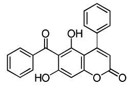
Anti-UVC Irradiation and Metal Chelation Properties of 6-Benzoyl-5,7-dihydroxy-4-phenyl-chromen-2-one: An Implications for Anti-Cataract Agent

1
Institute of Biological Chemistry, Academia Sinica, 128 Academia Road, Section 2, Nankang, Taipei 115, Taiwan
2
School of Pharmacy, College of Pharmacy, Taipei Medical University, 250 Wuxing Street, Taipei 11031, Taiwan
3
Institute of Biochemical Sciences, National Taiwan University, No. 1, Section 4, Roosevelt Road, Taipei 10617, Taiwan
*
Authors to whom correspondence should be addressed.
Int. J. Mol. Sci. 2011, 12(10), 7059-7076; https://doi.org/10.3390/ijms12107059
Submission received: 29 July 2011 / Revised: 23 September 2011 / Accepted: 11 October 2011 / Published: 21 October 2011
(This article belongs to the Special Issue Antioxidants)

Abstract

:
Coumarin derivative 1, 5,7-dihydroxy-6-(3-methyl-1-butyryl)-4-phenyl-chromen- 2-one, has been reported to possess radical scavenging activity and DNA protection. We have synthesized a series of coumarins with structural modifications at positions C4, C5, C6 and C7 and evaluated them for their anti-UVC properties. Coumarin 7, 6-benzoyl-5,6-dihydroxy-4-phenyl-chromen-2-one, was found to have the most potent activity in protecting porcine γ-crystallin against UVC insults. Results of fluorescence assays indicated that compound 7 was capable of decreasing the loss of intensity while lens crystallins and DNA PUC19 were irradiated with UVC. Presence of compound 7 decreased hydroxyl radical levels determined by probe 1b and the free iron concentrations determined by Ferrozine reagent. The chelation assay showed that compound 7 was chelated to metal via 6-CO and 5-OH on the benzopyrone ring. The observed protective effects of compound 7 towards crystallins from insults of UVC and free radicals may be due to its iron-chelating activity and its peak absorption at 254 nm.

Graphical Abstract

1. Introduction

Solar radiation can be divided into five regions in increasing order of wavelengths: ultraviolet UVC, UVB, UVA, visible range, and infrared range. Among these radiations, UVC and UVB are responsible for photochemical reactions. UVA, visible-range, and infrared-range radiation were traditionally thought to be less damaging. UV-induced oxidative damage is highly related to cataractogenesis [1,2]. Compounds with anti-UV properties can be used as anti-cataract agents. Coumarin derivatives were reported to protect rabbit corneal-derived cells (SIRCs) from UVB-induced oxidative damage [3].
Coumarins are a class of phenolic substances commonly found in plants, fungi, and bacteria [4]. Structures of these compounds consist of a benzene moiety fused to an α-pyrone. The structural diversity found in this family of compounds can be classified into different categories, from coumarins to polycyclic coumarins, such as furocoumarins and pyranocoumarins. This diversity leads to multiple pharmacological activities; natural and synthetic coumarins have anti-inflammatory [5,6], antimicrobial [6,7], antiviral [6,8], anticarcinogenic [6,9], anticoagulant [6,10], and antioxidant activities [6,10]. Generally speaking, their mechanisms of action are not well understood.
Among activities of coumarin derivatives, their antioxidant properties have garnered attention due to their potential applications. Compounds containing the moiety of coumarin (1,2-benzopyrone, 2H-1-benzopyran-2-one, 2) were shown to possess antioxidant activity [1120]. Many different types of antioxidant activity of coumarins were investigated in various biological systems, but it was difficult to determine a structure-activity relationship [5,6,11,21]. The scavenging capacities of coumarins against various free-radical types were investigated. Those results implied that compounds with phenolic hydroxyl groups are thought to possess antioxidant activities. A hydroxyl group on an aromatic ring structure can carry out the single-electron reduction of a free radical; therefore, compounds with phenolic hydroxyl groups are good radical scavengers.
The eyes are constantly exposed to reactive oxygen species (ROS) induced by UV light [22]. A previous report showed that corneal and tear fluids possess an elaborate antioxidant defense system [23]. However, UV light can pass through the cornea and enter the lens. UV irradiation is one of the significant risk factors in cataractogenesis. Chemical modifications or oxidation of lens proteins may also contribute to the emergence of opacities. Eye drops containing antioxidants such as tropicamide, phenylephrine, flurbiprofen, and tobramycin are also given preoperatively [24]. In recent years, the protective effects of compounds against UV-induced oxidative damage of the eye were investigated [3,25,26]. It was shown that UV irradiation can cause turbidity of γ-crystallin [27,28]. Although the detailed mechanism of this phenomenon remains vague, it is believed to be related to ROS induced by UV light. The focus on the antioxidant activities of coumarin derivatives brought our attention to cataracts, which are thought to be correlated with oxidative stress [2934].
Previously, we showed that coumarin 1 (Chart 1) possess anti-inflammatory, antioxidant activities, and the pUC-19 plasmids co-incubated in the presence of coumarin 1 exhibited less DNA strand damage and most of the plasmids were in the supercoiled form [20]. Those results demonstrated that our 6-acyl-4-aryl-alkyl-5,7-dihydroxycoumarins have the ability to scavenge the hydroxyl radical under Fenton reaction conditions. Since phenolic compounds are well-known antioxidants, we are interested in examining the antioxidative effects of the compounds where (i) a phenolic moiety is attached to the 6-acyl position of 6-acyl-4-phenyl-5,7-dihydroxycoumarin; (ii) the hydroxyl group of 6-benzoyl is replaced with methyl or hydrogen; and (iii) 5- and 7-hyroxyl groups are present on the coumarin ring. Therefore, here we aimed to established an in vitro anti-UVC assay and use it to screen for the most potent anti-UVC radiation compound from the library of synthetic coumarin derivatives 311 (Chart 1). This compound was further investigated towards crystallins and DNA protection, and the involvement of iron-chelation activity was then further examined by Ferrozine assay and NMR spectroscopy.

2. Results and Discussion

2.1. Chemistry

Coumarin is a flavonoid, which is a natural constituent of many plants and essential oils. Coumarins from plants are derivatives of cinnamic acid with a benzo-α-pyrone skeleton [35]. They are classically synthesized by the Perkin, Pechmann, or Knoevenagel reactions [6]. Pechmann condensation utilizes a phenol and carbozylic acid or ester for the coumarin synthesis. We synthesized several coumarin derivatives using this method [20,36]. Herein, we followed a similar strategy to synthesize most of coumarin derivatives (Scheme 1).

2.2. Biological Results

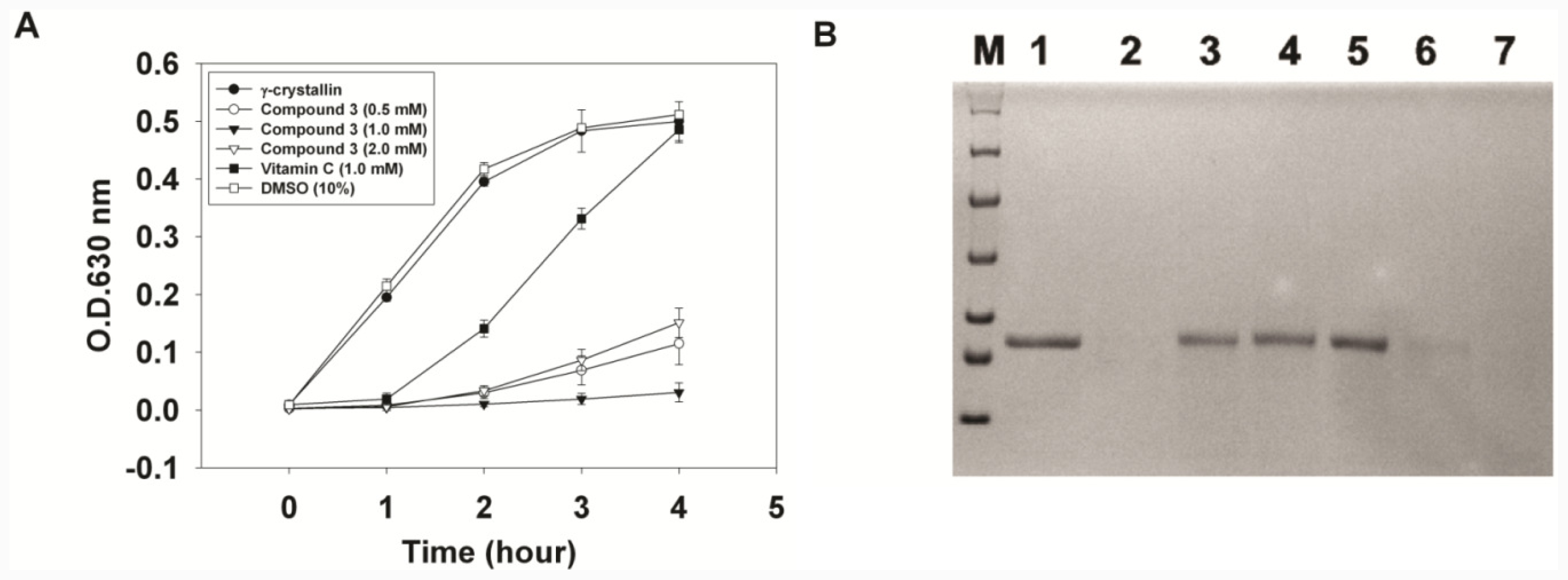
2.2.1. Turbidity Assay of γ-Crystallin and Comparison of Anti-UVC Activity

Previously, UVC irradiation to induce turbidity of γ-crystallin was applied to measure the chaperone activity of lens γ-crystallin [24,25]. Lens γ-crystallin was correlated to cataracts and demonstrated in a mouse model [37]. Formation of amyloid fibrils in vitro from partially unfolded intermediates of human γ-crystallin was investigated [38]. Amongst the lens crystallins, γ-crystallins are particularly sensitive to oxidation, because of their high amounts of Cys and Met residues [39]. Therefore, we chose γ-crystallins as the reported proteins for our assays. Compounds that protect γ-crystallins from damage by UVC irradiation can be anti-cataract agents. Hydroxycoumarins were previously shown to have antioxidant activities [11,20,21]. They are believed to act like classic phenol- or quinol-based antioxidants which can carry out the single-electron reduction of a free radical. Compound 3 (5,6-dihydroxy-6-(4-hydroxybenzoyl)-4-phenyl-2H-chromen-2-one) was dissolved in DMSO to create a stock solution which was then added to the sample solution to achieve a maximum final DMSO concentration of 10% (v/v). The sample solutions of 200 μL each were exposed to a UV lamp (254 nm), and turbidity was measured before and after UV irradiation. For each condition, four sample solutions were used in the experiment. The results indicated that compound 3 can effectively suppress the turbidity induced by UV irradiation (Figure 1A). After UV irradiation, the sample with 10% DMSO showed a slight increase in turbidity, whereas the sample with vitamin C (1 mM) showed a delayed increase of turbidity. It was obvious that vitamin C possesses statistically significant less anti-UV activity than compound 3 at 4 h observation (P < 0.05). SDS-PAGE analysis showed that γ-crystallin was effectively protected by compound 3 (Figure 1B), whereas the samples with vitamin C and DMSO only (10%) were photo-degraded after 4 h of UV irradiation.
In order to study the structural activity relationship, various compounds were compared in the same activity assay (Table 1). Due to differences in the solubilities and colors of these compounds, the change in turbidity was used in this assay. Porcine γ-crystallins with or without compounds were irradiated by UVC (254 nm), and the turbidity was measured at 630 nm on an ELISA reader. The changes in turbidity after 4 h of UVC irradiation were recorded, and a sample of only porcine γ-crystallin, without other compounds, was used as a blank. The results indicated that potency ranking of anti-UVC activity was as follows: 7 > 4 = 6 > 3 > 9 > 10 > 11 > 8 > 2 > 5 (Table 1). Coumarin (2) showed 20% suppression of turbidity, and SDS-PAGE showed it had poor protective activity. Compounds 3–7 respectively showed 66%, 103%, 12.5%, 99.9%, and 116% suppression of turbidity. SDS-PAGE also indicated that compounds 3, 4, 6, and 7 showed good protective activities. Compound 3 possesses three phenolic hydroxyl groups, but it showed only 66% suppression of turbidity. Compound 4 was more potent than compound 3 indicating that the 6-p-methylbenzoyl group is more potent than the 6-p-hydroxybenzoyl group. Compound 7 showed the best protective activity among them. The 6-p-hydroxybenzoyl group was less potent than the 6-benzoyl group when comparing compounds 3 and 7. Compound 5 showed only 12.5% suppression of turbidity, and SDS-PAGE showed that it had less protective activity, which indicated that the 5-hydroxyl group may play an important role in protective activity. Substitution of the 5,7-dihydroxyl groups in compound 7 with O-acetyl groups (compound 6) did not show significant effects in augmenting protective activity. However, compound 6 showed less solubility in the buffer. The result indicated that compound 7 was the most potent compound in this study.
Previous reports have indicated that water irradiated by UVC forms free radicals [40,41]. It was reported that ascorbic acid exhibits potent free radical-scavenging activity and was used as the control [42]. Ascorbyl radicals are long-lived free radicals which make vitamin C a good radical scavenger [43]. The sample with vitamin C (1 mM) showed a delayed increment in turbidity after UVC irradiation. A high concentration of vitamin C can protect γ-crystallin from photodegradation by UVC. Hence, photodegradation of γ-crystallin can be correlated to free radical reactions.

2.2.2. Evaluation of the Substitution Groups

An additional interesting aim of the investigation was to determine the structure-activity relationship of coumarin derivatives. From the above study, it was shown that substitution of a 6-benzoyl group in compound 7 affected its activity. Therefore, by means of the Pechmann reaction, we synthesized compound 8 which does not contain a 6-benzoyl group (Scheme 1). The anti-UVC activity assay showed that compound 7 was more potent than compound 8. Compound 7 showed 94% suppression of turbidity, whereas compound 8 showed only 36% suppression of turbidity. SDS-PAGE showed that both compounds could protect against γ-crystallin degradation (data not shown). Hence, we concluded that the 6-benzoyl group promoted anti-UVC activity. This result may have been due to the addition of the 6-benzoyl group helping to stabilize free radicals at position 6 when phenolic hydroxyl groups carry out the single-electron reduction.
To evaluate the function of the 4-phenyl group of compound 7, compound 9 was synthesized in a similar way as compound 8 (Scheme 1). The 4-phenyl group was changed to a 4-ethyl group in compound 9. The results of the activity assay revealed that compound 7 showed 89.3% suppression of turbidity, whereas compound 9 showed only 54% suppression of turbidity. It was demonstrated that compound 7 was more potent than compound 9. SDS-PAGE showed that both compounds can protect against γ-crystallin degradation (data not shown). The 4-phenyl group also played a role in promoting the anti-UVC activity of compound 7. It was proposed that the addition of a 4-phenyl group would enhance the resonance effects of coumarin derivatives.
Table 1 shows the comparisons of all tested coumarin derivatives. Results show that coumarin possesses poor anti-UVC activity. The addition of hydroxyl groups to coumarin derivatives greatly improved the anti-UVC activities of these compounds. Compound 5 showed poor anti-UVC activity, indicating that the 5-hydroxyl group may play an important role in the activity of compound 7. Therefore, we synthesized compounds 10 and 11 (Scheme 1) to investigate the influence of the phenolic hydroxyl group. The anti-UVC activity assay of compounds 8, 10, and 11 demonstrated that both compound 10 and 11 possessed poor activity. Compound 8 showed 77.8% suppression of turbidity, while compounds 10 and 11 respectively showed only 68.7% and 35.8% suppression. The SDS-PAGE analysis also revealed that compounds 10 and 11 possessed poorer protective activities (data not shown). Compound 11 exhibited slightly higher anti-UVC activity than coumarin, which indicates that the 5,7-dihydroxyl groups are important in this activity assay. However, compared to compound 11, the activity of compound 10 was lower by about 10%, but SDS-PAGE showed that γ-crystallin was severely degraded. Comparing compounds 10 and 5 showed that the 5-p-cyanobenzoyl group of compound 5 led to the poor activity. The phenolic hydroxyl group improved the anti-UVC activity of the compound. It should be noted that hydroxyl groups also improved the solubility of the coumarin derivatives in the water-based buffer.

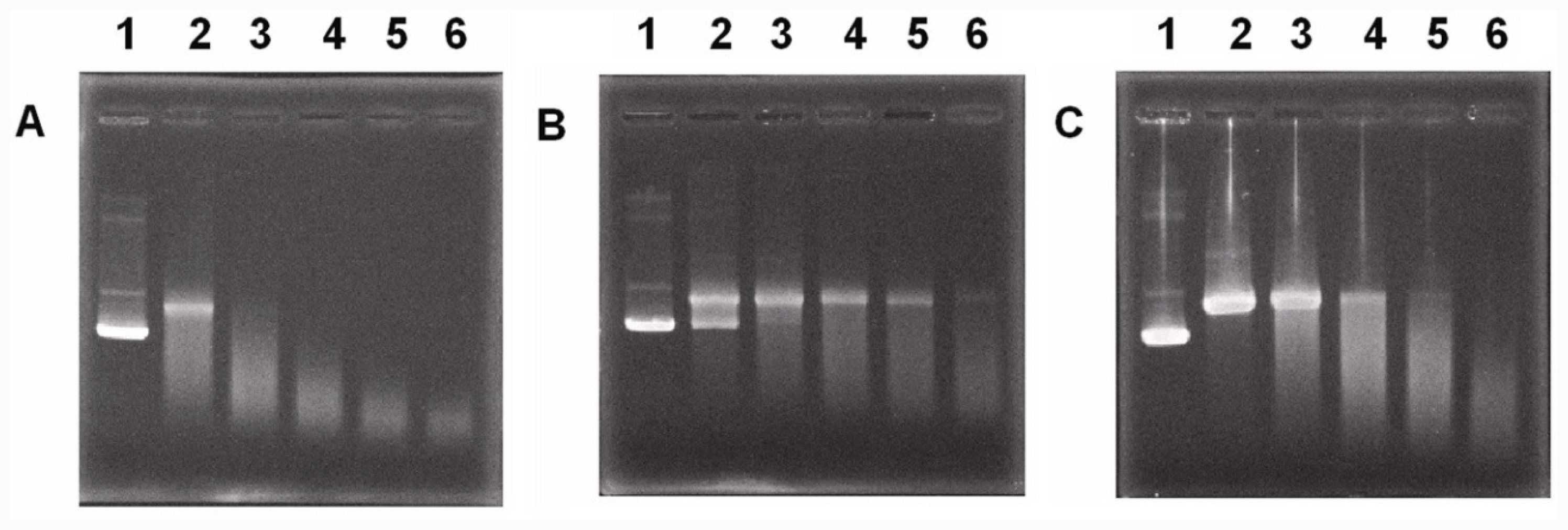
2.2.3. Protection of DNA

It was demonstrated that compound 7 possessed the best anti-UVC activity among these coumarin derivatives. However, UVC irradiation damages both proteins and DNA. UVC irradiation may damage the DNA of cells. The outside surface of the lens is a layer of lens epithelial cells. UVC damage to lens epithelial cells may lead to acute UVC cataractogenesis [44]. Therefore, the ability of compound 7 to protect DNA from UVC damage was tested (Figure 2). Supercoiled DNA (PUC19 plasmid) irradiated by UVC (254 nm) became linear DNA and then photodegraded. The addition of compound 7 prevented the supercoiled DNA from photodegradation. After 30 min of irradiation, a portion of the supercoiled DNA was preserved in the presence of compound 7, while all DNA had become linear in the presence of vitamin C. It was shown that the UVC-protective property of compound 7 is more potent than that of vitamin C.

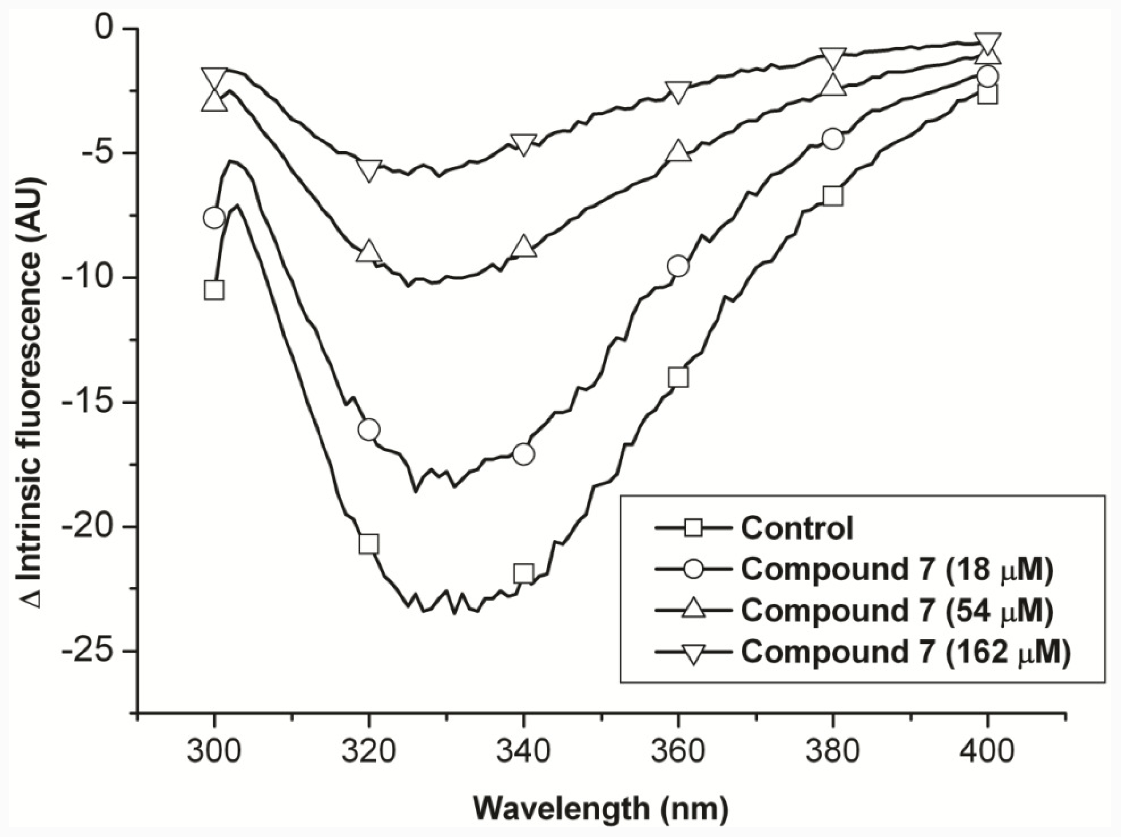
2.2.4. Compound 7 Protects Porcine Lens Proteins from Oxidative Damage by Hydroxyl Radicals

In the abovementioned discussion on the mechanism of compound 7’s anti-UVC activity, we have assumed that compound 7 possesses free radical-scavenging properties. Therefore, we tested if compound 7 can protect lens crystallins from the Fenton reaction (Figure 2). Although all amino acid residues in the protein chain are susceptible to modification by the hydroxyl radical, tryptophan is the most vulnerable amino acid. In our previous report, the intrinsic fluorescence of lens crystallins was used as an index of the effectiveness of the tested compound [45]. Herein, we showed that compound 7 protected lens crystallins in a dose-dependent manner (Figure 3). The solvent, DMSO, had no protective function. It was demonstrated that compound 7 is a free radical scavenger and can protect crystallins. However, the commonality between UV irradiation and the Fenton reaction was not provided. It is known that hydroxyl radicals are produced during UV-light dissociation of H2O2. Hence, we tested if UV irradiation increased H2O2 in solution. A sensitive probe for H2O2 was used to evaluate H2O2 produced by UV irradiation [46]. These results indicated that UV irradiation produced H2O2 and showed some commonality with the Fenton reaction (data not shown).

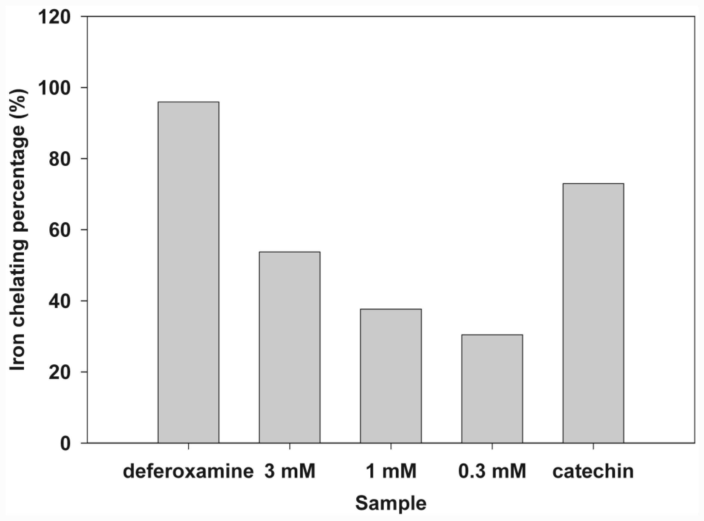
2.3. Iron-Chelating Activity

Although compound 7 with phenolic hydroxyl groups showed free radical-scavenging properties, there are other possibilities in preventing the Fenton reaction. A previous report indicated that the chelator is capable of binding ferrous and ferric iron, reducing concentrations of Fe(II) and Fe(III) in solution, and thus alleviating the Fenton reaction [47]. In view of its chemical structure, results suggested that compound 7 can chelate Fe(II). The chelation assay showed that compound 7 was capable of binding ferrous iron (Figure 4). To further confirm this property, 1H NMR titration studies were performed to identify the likely site of iron binding on compound 7. A diamagnetic metal ion, Zn2+, was used as the probe for Fe2+ [48]. Figure 5 shows the 1D-NMR spectrum of compound 7 in the presence and absence of ZnAc. As Zn2+ was added, little change was observed in most of the compound 7 peaks; however, the H8 proton peak showed a slight upfield shift (of ca. 0.03 ppm). H8 resonance (shifted and broadened) was observed by 1:2 stoichiometric Zn complexation indicating that Zn is chelated to 6-CO and 5-OH on the benzopyrone ring. Since the H8 proton experienced a small change of an upfield shift, it is unlikely to have been chelated to 6-CO and 7-OH on the benzopyrone ring. Due to the similar coordination properties of Fe2+ and Zn2+, it is reasonable to suggest that Fe2+ was chelated at the same site as Zn2+.

3. Experimental Section

3.1. General Experimental Conditions

Melting points were taken in a capillary tube using a MEL-TEMP II melting-point apparatus by Laboratory Devices (CA, USA). Nuclear magnetic resonance (NMR) spectra were recorded on a Bruker DMX-500 FT-NMR spectrometer (Madison, WI, USA); chemical shifts were recorded in parts per million downfield from Me4Si. IR spectra were determined with a Perkin-Elmer 1760-X FT-IR spectrometer (Massachusetts, USA). Mass spectra were recorded on Jeol JMS-D300 (Tokyo, Japan) and FINNIGAN TSQ-46C mass spectrometers (Thermo scientific, USA); HRMS was obtained with a Jeol JMS-HX110 spectrometer. TLC was performed on Merck (Art. 5715) silica gel plates (Darmstadt, Germany) and visualized under UVC light (254 nm). Flash column chromatography was performed with Merck (Art. 9385) 40–63 μm silica gel 60.

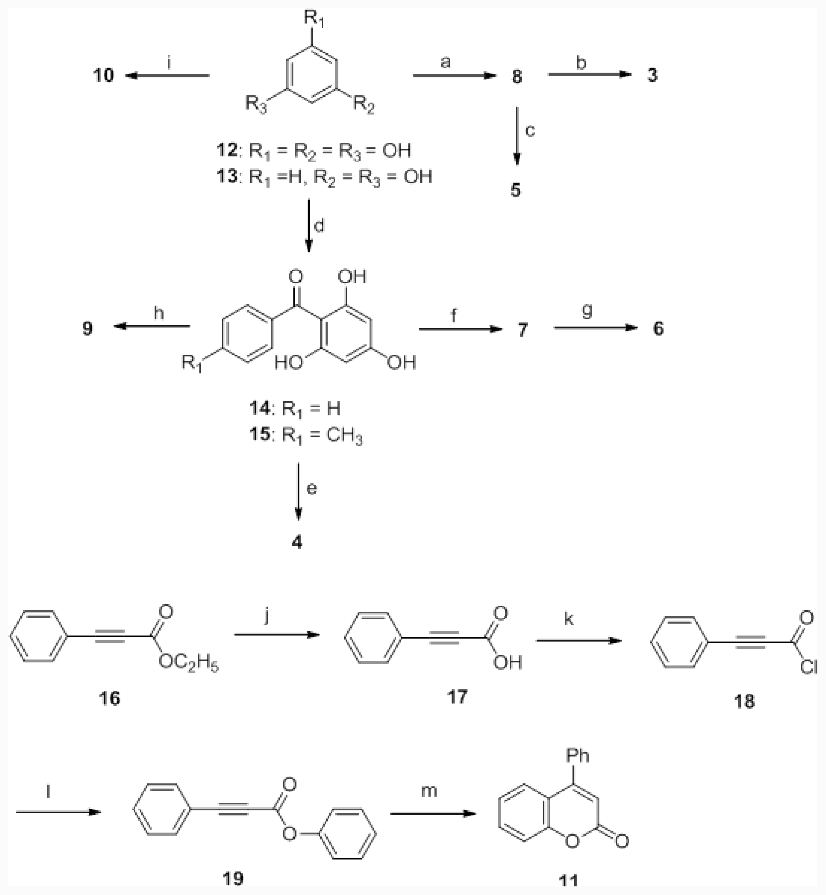
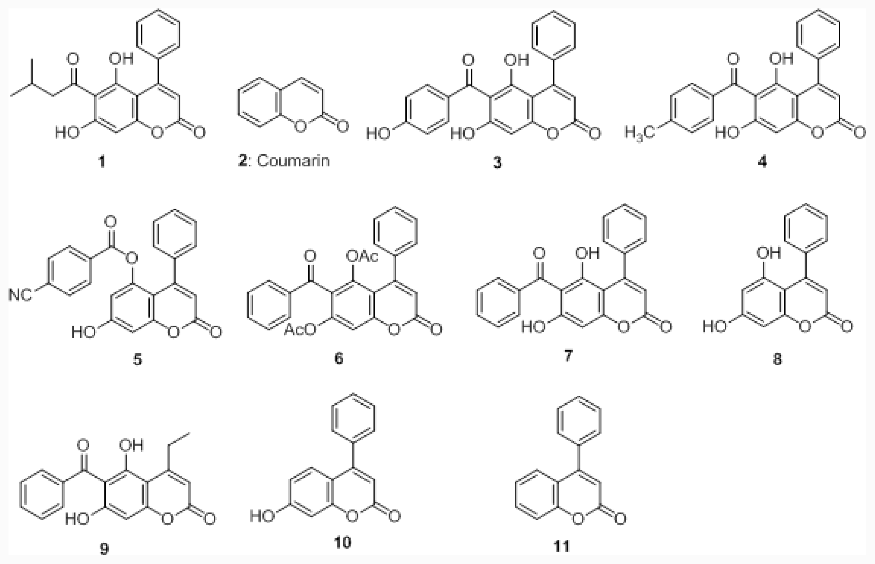
3.2. Preparation of Coumarin Derivatives 3–11

Synthesis of coumarins 3–5, 7, and 9 were reported by our laboratory [20,36]. Acetylation of the phenolic hydroxyl group of coumarin derivative 7 was yielded coumarin derivative 6. Resorcinol treated with ethyl benzoylacetate resulted in coumarin derivative 10. Compounds 8 and 11 were prepared by a method reported in the literature [49,50].

3.3. Iron-Chelating Activity

The chelation of ferrous ions by compound 7 was estimated using a modified method of Lopes et al. [51,52]. Ferrous ammonium sulfate was added to a solution containing 5% ammonium buffer and different concentrations of compound 7 (0.3, 1, and 3 mM). The final concentration of ferrous ammonium sulfate was 0.5 mM. Ferrozine was added to initiate the reaction, and the final concentration was 0.25 mM. The reaction mixture was incubated at room temperature for 15 min after being shaken. The absorbance of the solution was measured at 562 nm in a Helius Alpha spectrophotometer (Unicam Instruments, Cambridge, UK). Deferoxamine was used as a control at a final concentration 30 μM. The higher the iron-chelating activity of a tested sample was, the lower the resulting ultraviolet absorbance. Therefore, the relative iron-chelating capacity was calculated as the percent decrease in absorbance of the test samples compared to the controls that lacked the test drugs.

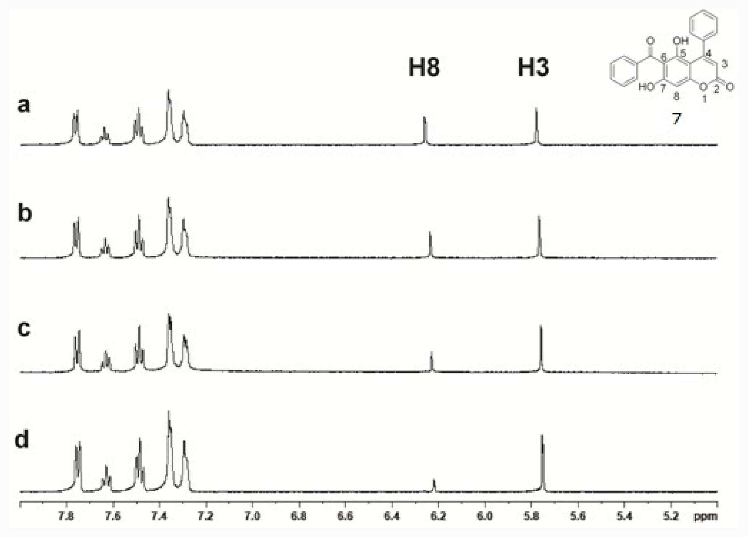
3.4. 1H-NMR Studies of Metal Chelation

1H NMR studies were carried out on a Bruker AC 400 spectrometer. A stock solution of 7 (150 mM) was freshly prepared in DMSO-d6. Zinc acetate (300 mM) was prepared in D2O. Titrations were done in a 50/50 (v/v) DMSO-d6/D2O solvent buffered with 50 mM Tris-HCl at pH 7.20. The pH of the solutions was determined with a Corning pH meter (Corning, NY, USA) using a Sigma-Aldrich micro combination electrode (St. Louis, MO, USA). The pH meter readings for D2O were recorded as pH values, i.e., uncorrected for the effect of deuterium.

3.5. Absorption Spectra of Coumarin Derivatives

Coumarin derivatives 2–7 (0.1 mM) were prepared in methanol, and absorption spectra were measured on a UV/Vis Scanning Spectrophotometer (DU series 700, Beckman Coulter, CA, USA).

3.6. Biological Assay

3.6.1. Purification of Porcine γ-Crystallin

Porcine lenses were decapsulated and homogenized in buffer containing 50 mM Tris-HCl, 0.1 M NaCl, 5 mM EDTA, 0.01% β-mercaptoethanol, and 0.02% sodium azide (pH 8.0). After centrifugation at 16,060× g for 30 min, the supernatant was collected. The supernatant was applied to a TSK HW-55(F) column (Tosoh Bioscience, Japan) and eluted at 25 mL/h. Five well-resolved peaks were obtained and identified as HMα-, α-, βH-, βL-, and γ-crystallins (Data not shown) based on their subunit compositions as revealed by sodium dodecylsulfate polyacrylamide gel electrophoresis (SDS-PAGE) [28,53,54]. Fractions of porcine γ-crystallin were collected, and the protein concentration was determined according to the Bradford method (BioRad Laboratories, USA).

3.6.2. Turbidity Assays of γ-Crystallin under UVC Irradiation

Photoaggregation of γ-crystallin (20–25 μM) was monitored in a buffer of 50 mM Tris-HCl and 0.1 M NaCl at pH 8.0, in the absence or presence of coumarin derivatives. Sample solutions of 200 μL each were exposed to a UV lamp (254 nm) of Ultraviolet Crosslinker CL-1000 (Bulletin UVP-AB-114, USA). Turbidity changes represented by relative light-scattering intensities after irradiation for four hours were measured at 630 nm by an enzyme-linked immunosorbent assay (ELISA) reader. Four sample solutions were used in the experiment for each condition. The percentage of suppression, (Iγ–Ic)/Iγ × 100%, was used to evaluate the anti-UVC activity, where Iγ is the intensity of scattered light for γ-crystallin without a compound, and Ic is the intensity of scattered light for γ-crystallin with a compound. SDS-PAGE (with 5% stacking and 12% resolving gels) was performed as previously reported [55]. Each well contained approximately 4 μg of porcine γ-crystallin.

3.6.3. Protection of Supercoiled DNA from Strand Breakage by UVC Irradiation

The activity of preventing photodegradation was evaluated by incubating compounds with DNA and irradiating them under UV light. Plasmid DNA (PUC 19) was incubated at 25 °C in water, with or without compound 6 or vitamin C. Sample solutions of 200 μL (0.0225 μg/μL PUC19) were each exposed to a UV lamp (254 nm) of Ultraviolet Crosslinker CL-1000 (Bulletin UVP-AB-114, USA), and samples were collected every 30 min. DNA samples were analyzed on 0.8% agarose gels. Each well contained approximately 450 μg of PUC19.

3.6.4. Fenton Reaction

Crude porcine lens proteins were incubated with various Fenton solutions modified based on the method of Huang et al. [56]. The final concentrations of Fe2+ and H2O2 in Fenton solutions of experiments were 0.02 and 0.2 mM, respectively. The fluorescence emission spectra were immediately taken at room temperature and recorded with a Hitachi F-4500 FL fluorescence spectrophotometer excited with 295-nm light. The emission spectra were recorded at 300–400 nm using a light slit of 5 nm for both the excitation and emission modes. Spectra of normal lens proteins were used as baselines for calculating changes in the tryptophan fluorescence intensity.

4. Conclusions

We established an in vitro assay to screen compounds with anti-UV properties. Compound 7 was the most potent compound among these coumarin derivatives that suppressed turbidity induced by UV irradiation. The 5,7-dihydroxyl groups play important roles in maintaining the anti-UV activity of this compound. The phenyl group at position 4 and benzoyl group at position 6 of compound 7 are partially responsible for the anti-UV activity. It is reasonable that compounds with a phenolic hydroxyl group possess free radical-scavenging properties. Therefore, coumarin derivatives can protect γ-crystallin and DNA from free-radical damage produced by UVC irradiation. In addition to the free-radical-scavenging properties, we also noted the UV-filtering property of coumarin derivatives. The human lens absorbs most of the UV radiation at 300–400 nm. This absorption is largely performed by a group of low-molecular-weight fluorescent compounds, known as UV-filter compounds [5759]. In order to evaluate the UV-filtering properties of coumarin derivatives, we measured the UV spectra of these compounds (Figure 6). The optical density at 254 nm of each compound was plotted in this figure. It is interesting that compounds with higher anti-UVC activities possess higher absorption at 254 nm. The only exception was compound 5, which showed low activity but high absorption. The low activity may have been due to the phenolic hydroxyl group’s inability to carry out the single-electron reduction of the p-cyanobenzoyl group, leading to an unstable free radical at position 6. Compound 7 was shown to possess the highest anti-UVC activity and best UV-filtering property at 254 nm. In addition, the iron-chelating activity gives compound 7 an additional potential application in iron-related cataracts. Increasing evidence relating iron to cataractogenesis includes reports of cataract formation following ocular siderosis from an iron foreign body in the lens [6064]. The benefits of iron chelators are currently being investigated for limiting iron-induced oxidative damage. In conclusion, we have developed a set of in vitro assays and 6-benzoyl-5,7-dihydroxy-4-phenyl-chromen-2-one (7) protected lens γ-crystallins against UVC insults and chelated metal via its structure of 6-CO and 5-OH on the benzopyrone ring. Further in vivo anti-cataractogenesis assay is warranted.

Acknowledgments

We greatly appreciate Shu-Chuan Jao (Biophysics Core Facility, Academia Sinica) for her technical support in NMR and Lee-Chiang, Lo for providing probe 1b. This study was supported by grants from the National Science Council (NSC98-2313-B-038-002-MY3) and Academia Sinica, Taipei, Taiwan.

References

  1. Davies, MJ; Truscott, RJ. Photo-oxidation of proteins and its role in cataractogenesis. J. Photochem. Photobiol. B 2001, 63, 114–125. [Google Scholar]
  2. Brian, G; Taylor, H. Cataract blindness—Challenges for the 21st century. Bull. World Health Organ 2001, 79, 249–256. [Google Scholar]
  3. Lodovici, M; Caldini, S; Morbidelli, L; Akpan, V; Ziche, M; Dolara, P. Protective effect of 4-coumaric acid from UVB ray damage in the rabbit eye. Toxicology 2009, 255, 1–5. [Google Scholar]
  4. Hoult, JR; Paya, M. Pharmacological and biochemical actions of simple coumarins: Natural products with therapeutic potential. Gen. Pharmacol 1996, 27, 713–722. [Google Scholar]
  5. Fylaktakidou, KC; Hadjipavlou-Litina, DJ; Litinas, KE; Nicolaides, DN. Natural and synthetic coumarin derivatives with anti-inflammatory/antioxidant activities. Curr. Pharm. Des 2004, 10, 3813–3833. [Google Scholar]
  6. Borges, F; Roleira, F; Milhazes, N; Santana, L; Uriarte, E. Simple coumarins and analogues in medicinal chemistry: Occurrence, synthesis and biological activity. Curr. Med. Chem 2005, 12, 887–916. [Google Scholar]
  7. Musicki, B; Periers, AM; Laurin, P; Ferroud, D; Benedetti, Y; Lachaud, S; Chatreaux, F; Haesslein, JL; Iltis, A; Pierre, C; et al. Improved antibacterial activities of coumarin antibiotics bearing 5,5′-dialkylnoviose: Biological activity of RU79115. Bioorg. Med. Chem. Lett 2000, 10, 1695–1699. [Google Scholar]
  8. Thaisrivongs, S; Watenpaugh, KD; Howe, WJ; Tomich, PK; Dolak, LA; Chong, KT; Tomich, CC; Tomasselli, AG; Turner, SR; Strohbach, JW; et al. Structure-based design of novel HIV protease inhibitors: Carboxamide-containing 4-hydroxycoumarins and 4-hydroxy-2-pyrones as potent nonpeptidic inhibitors. J. Med. Chem 1995, 38, 3624–3637. [Google Scholar]
  9. Valenti, P; Fabbri, G; Rampa, A; Bisi, A; Gobbi, S; Da Re, P; Carrara, M; Sgevano, A; Cima, L. Synthesis and antitumor activity of some analogues of flavone acetic acid. Anticancer Drug Des 1996, 11, 243–252. [Google Scholar]
  10. Stirling, Y. Warfarin-induced changes in procoagulant and anticoagulant proteins. Blood Coagul. Fibrinolysis 1995, 6, 361–373. [Google Scholar]
  11. Pedersen, JZ; Oliveira, C; Incerpi, S; Kumar, V; Fiore, AM; De Vito, P; Prasad, AK; Malhotra, SV; Parmar, VS; Saso, L. Antioxidant activity of 4-methylcoumarins. J. Pharm. Pharmacol 2007, 59, 1721–1728. [Google Scholar]
  12. Manojkumar, P; Ravi, TK; Gopalakrishnan, S. Antioxidant and antibacterial studies of arylazopyrazoles and arylhydrazonopyrazolones containing coumarin moiety. Eur. J. Med. Chem 2009, 44, 4690–4694. [Google Scholar]
  13. Thuong, PT; Hung, TM; Ngoc, TM; Ha do, T; Min, BS; Kwack, SJ; Kang, TS; Choi, JS; Bae, K. Antioxidant activities of coumarins from Korean medicinal plants and their structure-activity relationships. Phytother. Res 2009, 24, 101–106. [Google Scholar]
  14. Manojkumar, P; Ravi, TK; Subbuchettiar, G. Synthesis of coumarin heterocyclic derivatives with antioxidant activity and in vitro cytotoxic activity against tumour cells. Acta Pharm 2009, 59, 159–170. [Google Scholar]
  15. Gacche, RN; Gond, DS; Dhole, NA; Dawane, BS. Coumarin Schiff-bases: As antioxidant and possibly anti-inflammatory agents. J. Enzym. Inhib. Med. Chem 2006, 21, 157–161. [Google Scholar]
  16. Torres, R; Faini, F; Modak, B; Urbina, F; Labbe, C; Guerrero, J. Antioxidant activity of coumarins and flavonols from the resinous exudate of Haplopappus multifolius. Phytochemistry 2006, 67, 984–987. [Google Scholar]
  17. Kontogiorgis, CA; Savvoglou, K; Hadjipavlou-Litina, DJ. Antiinflammatory and antioxidant evaluation of novel coumarin derivatives. J. Enzym. Inhib. Med. Chem 2006, 21, 21–29. [Google Scholar]
  18. Yu, J; Wang, L; Walzem, RL; Miller, EG; Pike, LM; Patil, BS. Antioxidant activity of citrus limonoids, flavonoids, and coumarins. J. Agric. Food. Chem 2005, 53, 2009–2014. [Google Scholar]
  19. Lee, BC; Lee, SY; Lee, HJ; Sim, GS; Kim, JH; Kim, JH; Cho, YH; Lee, DH; Pyo, HB; Choe, TB; Moon, DC; Yun, YP; Hong, JT. Anti-oxidative and photo-protective effects of coumarins isolated from Fraxinus chinensis. Arch. Pharm. Res 2007, 30, 1293–1301. [Google Scholar]
  20. Lin, CM; Huang, ST; Lee, FW; Kuo, HS; Lin, MH. 6-Acyl-4-aryl/alkyl-5,7-dihydroxycoumarins as anti-inflammatory agents. Bioorg. Med. Chem 2006, 14, 4402–4409. [Google Scholar]
  21. Kostova, I. Synthetic and natural coumarins as antioxidants. Mini Rev. Med. Chem 2006, 6, 365–374. [Google Scholar]
  22. Kennedy, M; Kim, KH; Harten, B; Brown, J; Planck, S; Meshul, C; Edelhauser, H; Rosenbaum, JT; Armstrong, CA; Ansel, JC. Ultraviolet irradiation induces the production of multiple cytokines by human corneal cells. Invest. Ophthalmol. Vis. Sci 1997, 38, 2483–2491. [Google Scholar]
  23. Choy, CK; Cho, P; Chung, WY; Benzie, IF. Water-soluble antioxidants in human tears: Effect of the collection method. Invest. Ophthalmol. Vis. Sci 2001, 42, 3130–3134. [Google Scholar]
  24. Chandra, P; Hegde, KR; Varma, SD. Possibility of topical antioxidant treatment of cataracts: Corneal penetration of pyruvate in humans. Ophthalmologica 2009, 223, 136–138. [Google Scholar]
  25. Kawada, H; Kojima, M; Kimura, T; Natori, S; Sasaki, K; Sasaki, H. Effect of 5-S-GAD on UV-B-induced cataracts in rats. Jpn. J. Ophthalmol 2009, 53, 531–535. [Google Scholar]
  26. Varma, SD; Hegde, KR; Kovtun, S. UV-B-induced damage to the lens in vitro: Prevention by caffeine. J. Ocul. Pharmacol. Ther 2008, 24, 439–444. [Google Scholar]
  27. Lee, JS; Samejima, T; Liao, JH; Wu, SH; Chiou, SH. Physiological role of the association complexes of alpha-crystallin and its substrates on the chaperone activity. Biochem. Biophys. Res. Commun 1998, 244, 379–383. [Google Scholar]
  28. Liao, JH; Lee, JS; Chiou, SH. Distinct roles of alphaA- and alphaB-crystallins under thermal and UV stresses. Biochem. Biophys. Res. Commun 2002, 295, 854–861. [Google Scholar]
  29. Sawada, H; Fukuchi, T; Abe, H. Oxidative stress markers in aqueous humor of patients with senile cataracts. Curr. Eye Res 2009, 34, 36–41. [Google Scholar]
  30. Zoric, L; Kosanovic-Jakovic, N; Colak, E; Radosavljevic, A; Jaksic, V; Stevic, S. Oxidative stress in association with risk factors for the occurrence and development of age-related macular degeneration. Vojnosanit. Pregl 2008, 65, 313–318. [Google Scholar]
  31. Berthoud, VM; Beyer, EC. Oxidative stress, lens gap junctions, and cataracts. Antioxid. Redox. Signal 2009, 11, 339–353. [Google Scholar]
  32. Li, L; Duker, JS; Yoshida, Y; Niki, E; Rasmussen, H; Russell, RM; Yeum, K-J. Oxidative stress and antioxidant status in older adults with early cataract. Eye (Lond. ) 2009, 23, 1464–1468. [Google Scholar]
  33. Gul, A; Rahman, MA; Hasnain, SN; Salim, A; Simjee, SU. Could oxidative stress associate with age products in cataractogenesis? Curr. Eye Res 2008, 33, 669–675. [Google Scholar]
  34. Williams, DL. Oxidative stress and the eye. Vet. Clin. North Am. Small Anim. Pract 2008, 38, 179–192. [Google Scholar]
  35. Murray, RD. Coumarins. Nat. Prod. Rep 1989, 6, 591–624. [Google Scholar]
  36. Chin, YP; Huang, WJ; Hsu, FL; Lin, YL; Lin, MH. Synthesis and evaluation of antibacterial activities of 5,7-Dihydroxycoumarin derivatives. Arch. Pharm 2011, 344, 386–393. [Google Scholar]
  37. Graw, J. Mouse models of cataract. J. Genet 2009, 88, 469–486. [Google Scholar]
  38. Wang, Y; Petty, S; Trojanowski, A; Knee, K; Goulet, D; Mukerji, I; King, J. Formation of amyloid fibrils in vitro from partially unfolded intermediates of human gammaC-crystallin. Invest. Ophthalmol. Vis. Sci 2010, 51, 672–678. [Google Scholar]
  39. Skouri-Panet, F; Bonnete, F; Prat, K; Bateman, OA; Lubsen, NH; Tardieu, A. Lens crystallins and oxidation: The special case of gammas. Biophys. Chem 2001, 89, 65–76. [Google Scholar]
  40. Azrague, K; Bonnefille, E; Pradines, V; Pimienta, V; Oliveros, E; Maurette, MT; Benoit-Marquié, F. Hydrogen peroxide evolution during V-UV photolysis of water. Photochem. Photobiol. Sci 2005, 4, 406–408. [Google Scholar]
  41. Zhan, M. Determination of photochemically-generated reactive oxygen species in natural water. J. Environ. Sci. (China) 2009, 21, 303–306. [Google Scholar]
  42. Wagner, AE; Huebbe, P; Konishi, T; Rahman, MM; Nakahara, M; Matsugo, S; Rimbach, G. Free radical scavenging and antioxidant activity of ascorbigen versus ascorbic acid: Studies in vitro and in cultured human keratinocytes. J. Agric. Food. Chem 2008, 56, 11694–11699. [Google Scholar]
  43. Warren, JJ; Mayer, JM. Surprisingly long-lived ascorbyl radicals in acetonitrile: Concerted proton-electron transfer reactions and thermochemistry. J. Am. Chem. Soc 2008, 130, 7546–7547. [Google Scholar]
  44. Oriowo, OM; Cullen, AP; Chou, BR; Sivak, JG. Action spectrum and recovery for in vitro UV-induced cataract using whole lenses. Invest. Ophthalmol. Vis. Sci 2001, 42, 2596–2602. [Google Scholar]
  45. Wu, TH; Liao, JH; Hou, WC; Huang, FY; Maher, TJ; Hu, CC. Astaxanthin protects against oxidative stress and calcium-induced porcine lens protein degradation. J. Agric. Food. Chem 2006, 54, 2418–2423. [Google Scholar]
  46. Lo, LC; Chu, CY. Development of highly selective and sensitive probes for hydrogen peroxide. Chem. Commun 2003, 2003, 2728–2729. [Google Scholar]
  47. Lewis, S; Lynch, A; Bachas, L; Hampson, S; Ormsbee, L; Bhattacharyya, D. Chelate-modified fenton reaction for the degradation of trichloroethylene in aqueous and two-phase systems. Environ. Eng. Sci 2009, 26, 849–859. [Google Scholar]
  48. Perez, CA; Wei, Y; Guo, M. Iron-binding and anti-Fenton properties of baicalein and baicalin. J. Inorg. Biochem 2009, 103, 326–332. [Google Scholar]
  49. Jurd, L; Corse, J; King, AD, Jr; Bayne, H; Mihara, K. Phytochemistry Antimicrobial properties of 6,7-dihydroxy-7,8-dihydroxy-,6-hydroxy- and 8-hydroxycoumarins. Phytochemistry 1971, 10, 2971–2974. [Google Scholar]
  50. Shi, Z; He, C. Efficient functionalization of aromatic C–H bonds catalyzed by gold(III) under mild and solvent-free conditions. J. Org. Chem 2004, 69, 3669–3671. [Google Scholar]
  51. Lopes, GK; Schulman, HM; Hermes-Lima, M. Polyphenol tannic acid inhibits hydroxyl radical formation from Fenton reaction by complexing ferrous ions. Biochim. Biophys. Acta 1999, 1472, 142–152. [Google Scholar]
  52. Wu, TH; Liao, JH; Hsu, FL; Wu, HR; Shen, CK; Yuann, JMP; Chen, ST. Grape Seed Proanthocyanidin Extract Chelates Iron and Attenuates the Toxic Effects of 6-Hydroxydopamine: Implications for Parkinsons’s Disease. J. Food Biochem 2010, 34, 244–262. [Google Scholar]
  53. Liao, JH; Hung, CC; Lee, JS; Wu, SH; Chiou, SH. Characterization, cloning, and expression of porcine alpha B crystallin. Biochem. Biophys. Res. Commun 1998, 244, 131–137. [Google Scholar]
  54. Keenan, J; Orr, DF; Pierscionek, BK. Patterns of crystallin distribution in porcine eye lenses. Mol. Vis 2008, 14, 1245–1253. [Google Scholar]
  55. Laemmli, UK. Cleavage of structural proteins during the assembly of the head of bacteriophage T4. Nature 1970, 227, 680–685. [Google Scholar]
  56. Huang, FY; Chia, CM; Ho, Y. The formation of oxidatively induced high-molecular-weight aggregate of alpha-/gamma-crystallins. Biochem. Biophys. Res. Commun 1999, 260, 60–65. [Google Scholar]
  57. Wood, AM; Truscott, RJ. UV filters in human lenses: Tryptophan catabolism. Exp. Eye Res 1993, 56, 317–325. [Google Scholar]
  58. Wood, AM; Truscott, RJ. Ultraviolet filter compounds in human lenses: 3-hydroxykynurenine glucoside formation. Vis. Res 1994, 34, 1369–1374. [Google Scholar]
  59. Truscott, RJ; Wood, AM; Carver, JA; Sheil, MM; Stutchbury, GM; Zhu, J; Kilby, GW. A new UV-filter compound in human lenses. FEBS Lett 1994, 348, 173–176. [Google Scholar]
  60. Loh, A; Hadziahmetovic, M; Dunaief, JL. Iron homeostasis and eye disease. Biochim. Biophys. Acta 2009, 1790, 637–649. [Google Scholar]
  61. Goralska, M; Nagar, S; Colitz, CM; Fleisher, LN; McGahan, MC. Changes in ferritin H- and L-chains in canine lenses with age-related nuclear cataract. Invest. Ophthalmol. Vis. Sci 2009, 50, 305–310. [Google Scholar]
  62. Sneed, SR; Weingeist, TA. Management of siderosis bulbi due to a retained iron-containing intraocular foreign body. Ophthalmology 1990, 97, 375–379. [Google Scholar]
  63. Hope-Ross, M; Mahon, GJ; Johnston, PB. Ocular siderosis. Eye (Lond. ) 1993, 7, 419–425. [Google Scholar]
  64. Girelli, D; Corrocher, R; Bisceglia, L; Olivieri, O; de Franceschi, L; Zelante, L; Gasparini, P. Molecular basis for the recently described hereditary hyperferritinemia-cataract syndrome: A mutation in the iron-responsive element of ferritin l-subunit gene (the “Verona mutation”). Blood 1995, 86, 4050–4053. [Google Scholar]
Figure 1. Turbidity assay of compound 3 under UV irradiation. (A) Porcine γ-crystallins, with or without compound 3, were irradiated by UV (254 nm), and the turbidity was measured at 630 nm by ELISA reader. Solvent, DMSO, and ascorbic acid were used as controls. The bars denote ranges of variation from four measurements; (B) SDS-PAGE of porcine γ-crystallin before and after UV irradiation (4 h) in various conditions. Lane 1: γ-crystallin before UV irradiation. Lane 2: γ-crystallin after UV irradiation. Lanes 3, 4, and 5: γ-crystallin with compound 3 (0.5, 1.0, and 2.0 mM, respectively) after UV irradiation. Lane 6: γ-crystallin with ascorbic acid (1.0 mM) after UV irradiation. Lane 7: γ-crystallin with DMSO (10%) after UV irradiation. M: markers. The masses of markers are 116.0 kDa, 66.2 kDa, 45.0 kDa, 35.0 kDa, 25.0 kDa, 18.4 kDa and 14.4 kDa from the top to bottom of the gel.
Ijms 12 07059f1
Figure 2. Activities of compound 7 and vitamin C in protecting DNA from photodegradation by UV. (A) Plasmid DNA (PUC19) was broken and then degraded under UV irradiation. Lane 1, supercoiled DNA without UV irradiation; lanes 2–6, DNA after UV irradiation for 30, 60, 90, 120, and 150 min, respectively; (B) Plasmid DNA incubated with compound 7 (1 mM) showed that some fraction of DNA was preserved under UV irradiation; (C) Plasmid DNA incubated with vitamin C (1 mM) showed that most DNA became the linear form and then degraded under UV irradiation.
Ijms 12 07059f2
Figure 3. Free radical-scavenging activity. Porcine crystallins were subjected to oxidative stress by Fenton solutions with/without the protection of compound 7. Negative changes indicate the loss of fluorescence intensity of tryptophan of lens crystallins. The protective activity of compound 7 was concentration-dependent.
Ijms 12 07059f3
Figure 4. Iron-chelating activity of compound 7. Comparative analyses of ferrous ion-chelating potency with various concentrations of compound 7, and positive controls (30 mM of catechin or deferoxamine) were performed to determine their ability to complex with ions. The percentage of the chelating effect was calculated using the following equation: [1 – (absorbance of a sample at 562 nm)/(absorbance of the control, without tested a sample or positive control, at 562 nm)]/100.
Ijms 12 07059f4
Figure 5. 1D-nuclear magnetic resistance (NMR) spectrum of 5 mM of compound 7 without and in the presence of ZnAc in 50 mM Tris-HCl at pH 7.2 (50/50, v/v, DMSO-d6/D2O-Tris-HCl): (a) at a 1: 0; (b) at a 1: 0.5; (c) at a 1: 1; and (d) at a 1: 2 Zn equivalent.
Ijms 12 07059f5
Figure 6. The relationship of UV absorbance of compounds and their anti-UV activity. (A) The absorption spectra of compounds 2–7; (B) The UV absorbance (254 nm) of compounds 2–7; (C) The anti-UV activities of compounds 2–7 are shown in percentages of the suppression of turbidity.
Ijms 12 07059f6
Scheme 1. Synthesis of coumarin derivatives. Conditions: a: 12, ethyl benzoylacetate, trifluoroacetic acid, reflux, 40%; b: p-anisoyl chloride, AlCl3, nitrobenzene, 60 °C, 3 h, 16%; c: p-cyanobenzoyl chloride, AlCl3, nitrobenzene, 60 °C, 3 h, 15%; d: 12, benzoyl chloride, AlCl3, nitrobenzene, 60 °C, 3 h, 15%; e: 15, p-toluoyl chloride, AlCl3, nitrobenzene, 60 °C, 3 h, 36%; f: 14, ethyl benzoylacetate, glacial HOAc, conc. H2SO4, 15%; g: sodium acetate, acetic anhydride, 160 °C, 1 h, 86%; h: 14, ethyl propionylacetate, glacial HOAc, conc. H2SO4, 27%; i: 13, ethyl benzoylacetate, glacial HOAc, conc. H2SO4, 60%; j: 1.5 N NaOH, THF, reflux 1 h, 75%; k: oxalyl chloride, THF, DMF; l: phenol, NaH, THF; m: 5 mol% AuCl3, AgOTf, 70%.
Ijms 12 07059f7
Chart 1. Structures of coumarin derivatives.
Ijms 12 07059f8
Table 1. Optical density (OD) at 4 h observations. Turbidity assay of various coumarin derivatives under UV irradiation at a concentration of 0.5 mM. Porcine γ-crystallins, with or without co-incubation with test compounds, were irradiated by UV (254 nm) for four hours, and the turbidity was measured at 630 nm with an ELISA reader and expressed as mean ± standard derivation.
cpd no.23456
OD0.636 ± 0.0410.193 ± 0.043−0.031 ± 0.0080.698 ± 0.1500.0003 ± 0.074

cpd no.7891011
OD−0.041 ± 0.0120.502 ± 0.0120.284 ± 0.0350.364 ± 0.0180.406 ± 0.032

